Note: Descriptions are shown in the official language in which they were submitted.
CA 02375034 2002-01-11
WO 01/07915 PCTIUSOO/20354
-1-
METHOD AND DEVICE FOR ANALYSIS OF BIOLOGICAL SPECIMENS
FIELD OF THE INVENTION
The present invention is related to the separation and identification of
components of cellular specimens. In particular, the present invention
involves
expression scanning, and in particular examples a method of identifying
specimen
components while maintaining the spatial relationship between the location of
the
specimen component of interest and the remainder of the specimen.
BACKGROUND OF THE INVENTION
The Human Genome Project and other gene discovery initiatives are
dramatically increasing the information available regarding the number,
genomic
location, and sequences of human genes. Accompanying the expanding base of
genetic knowledge are several new technologies geared toward high-throughput
mRNA and proteomic analysis of biological samples, allowing a global view of
the genes and gene products that reflect normal physiology and pathological
states.
Utilized together, the expanding genetic database and newly developing
analysis
technologies hold tremendous potential to increase the understanding of normal
cellular physiology and the molecular alterations that underlie disease
states.
However, many biological specimens, such as whole cell tissue samples, remain
uniquely difficult to analyze due to their complex cellular heterogeneity.
The first report of the application of tissue sections directly onto paper
strips and subsequent electrophoresis was made by Lindner et al. (1956).
Later,
Saravis et al. (1979) utilized agarose gels and Bonte (1978) utilized
polyacrylamide gels to achieve better separation of the analyzed proteins. As
reported in a review by Neuhoff (1980), routine application of these
procedures to
whole cell tissues was not widespread because of technical difficulties, so
methods
using extraction of the proteins from the sample through cell lysis before
separation predominated.
CA 02375034 2002-01-11
WO 01/07915 PCT/US00/20354
-2-
More recently, Inczedy-Marcsek et al. (1988) described the use of
electrophoresis and isoelectric focusing of cryostat samples placed directly
upon
ultra thin polyacrylamide gels. The use of ultra thin gels allowed for
extraction of
the proteins from the tissue sample without lysis of the cells of the sample,
and did
overcome some of the technical difficulties experienced by early workers in
this
field. Schumacher et al. (1990) also described the use of isoelectric focusing
to
identify enzymes, glycoproteins, and neuropeptides present in cryostat
sections.
This process involved the direct placement of the sample upon ultra thin gels,
followed by isoelectric focusing. The processes of both Inczedy-Marcsek et al.
and Schumacher et al. produce gels in which the proteins or other molecules of
interest move through the gel medium according to physical characteristics
related
to charge and molecular weight. However, these approaches provide information
only on the total molecular content of the sample being analyzed, representing
the
aggregate proteins and nucleic acids present in all of the various cell types
present
in the specimen.
Isofocusing and electrophoresis processes have been disclosed for
cryostat tissue samples, followed by immunochemical analysis. Specifically,
Schumacher and Trudrung (1991) and van der Sluis et al. (1988) describe the
identification of alkaline phosphatases and peptides such as vassopressin,
respectively, through direct tissue isoelectric focusing followed by Western
blotting. This immunochemical analysis technique involves the movement of the
protein or molecules of interest, through capillary action, from the focusing
gel to
nitrocellulose membranes. The membrane-bound protein is then detected using
immunostaining procedures. Van der Sluis et al. (1988) did attempt to
generally
localize the proteins within the tissue sample by applying this procedure to a
series
of sliced tissue sections. However, the immunodetection process was preceded
by
an isofocusing step, so the results only indicated presence of the protein
within a
particular tissue sample.
Molecular analysis of cell populations in tissue sections have been
performed using immunohistochemistry (IHC) and in-situ hybridization (ISH).
The ISH technique is reviewed by Jin and Lloyd (1997), and the IHC technique
is
CA 02375034 2002-01-11
WO 01/07915 PCTIUSOO/20354
-3-
reviewed by Grogan (1992). While these techniques have been valuable tools to
investigate the cellular localization of a particular protein or mRNA in a
complex
tissue section, they both suffer from three major drawbacks. First, IHC and
ISH
are limited to analysis of a single molecular species per sample. Second,
artifact
staining based on cross-hybridization severely affects the accuracy of the
test
results. Finally, these methods have limited ability to visualize proteins and
mRNAs expressed at moderate or low levels of abundance.
Techniques have been disclosed for separating particular subsets of
cells from a whole tissue sample. For example, Emmert-Buck et al. (1996)
describe the use of laser-based microdissection techniques to rapidly procure
microscopic, histopathologically defined cell populations. Alternatively,
tissue
arrays, such as those described by Kononen et al. (1998) permit individual
molecules to be studied simultaneously in hundreds of separate tissue samples.
However, there remains a need in the art for an improved method of analyzing
proteins or other molecules of interest present in cellular specimens where
the
method is capable in some embodiments of providing information concerning the
location of the proteins or molecules of interest in the initial tissue
sample, and/or
provide a method that avoids some of the problems encountered with IHC and
ISH.
SUMMARY OF THE DISCLOSURE
The present disclosure describes methods, systems, and devices for
analyzing a biological specimen, such as a cellular specimen. The method
includes placing the specimen on a substrate with capture regions, such as
matrices
or layers, wherein the different regions of the substrate contain different
identification molecules, and transferring components of the specimen through
the
regions under conditions that allow the components to interact with different
identification molecules in the different regions (such as contiguous layers)
of the
substrate. In one embodiment, components of the cellular specimen are
transferred through the substrate (such as different matrices or layers of a
substrate) by electrophoresis, or by capillary action of a transfer buffer
moving
CA 02375034 2002-01-11
WO 01/07915 PCT/US00/20354
-4-
through the cellular specimen. In specific examples, the components are
transferred sequentially through a plurality of substantially parallel layers.
The transfer of components of a cellular specimen through the substrate
can occur while maintaining the cellular architecture of the specimen, if
desired.
Because the cellular architecture of the specimen may be maintained in some
embodiments, a correlation can be established between the location of the
different
identification molecules interacting with the cellular components, and the
original
location of the cellular components within the cellular specimens. The
analysis
can be performed with one or more different discrete cellular specimens on a
surface of the substrate. Examples of cellular specimens include, but are not
limited to, tissue sections (particularly tumor tissue sections), a cytology
sample,
microdissected cells and cultured cells. Cytostat tissue sections cut slightly
thicker
than usual, that is about 25 to about 50 m, improve the detection of
molecules of
moderate and low level abundance.
The regions (such as matrices or layers) of the substrate can range from
about 1 to more than a hundred, for example several hundred, several thousand,
or several tens of thousands in number, with each region (such as a layer)
having a
thickness (for example) of at least about 25 nm. In particular embodiments,
the
regions may extend across the substrate (as in layers), and components of the
specimen are transferred generally transverse to the layers, but they may be
transferred substantially parallel or at other angles to the layers.
Identification
molecules present in the substrate layers may, for example, be antibodies that
interact with the components of the cellular specimen, and can be used to
identify
particular molecules of interest present in the specimen. Other
representative,
non-limiting examples of identification molecules include nucleic acids,
peptides,
receptors, and ligands. The identification molecule can, for example, comprise
a
capture molecule that retains a component of the specimen in the layer. If
this is
done, the analysis can be completed by exposing the identification molecule to
a
detection molecule that associates with a combination of the capture molecule
and
the component of the sample, or associates with a region of the component
different than the region that was recognized by the identification molecule.
For
CA 02375034 2002-01-11
WO 01/07915 PCT/US00/20354
-5-
example, the molecule of interest can be a protein, and the identification
molecule
can recognize a first domain of the protein, and the detection molecule
recognizes
a second domain of the protein.
Another particular embodiment is a method of analyzing a specimen by
providing a substrate that includes different regions (such as layers) having
contiguous faces, each layer including a corresponding capture molecule
capable
of interacting with and capturing a component of the specimen; applying the
specimen to a face of the substrate, and transferring components (such as
intact
components) of the specimen through the contiguous faces of the different
layers
of the matrix. The components of the specimen react with the capture molecule
and the pattern of capture in the different layers can be correlated with
information
about the specimen. For example, interaction with a specific antibody in a
particular layer indicates the presence of the antigen in the specimen. The
location
of the interaction in a layer can be correlated with a position of the
specimen. In
the instance of cellular specimens, the cellular architecture of a tissue
specimen
from which the specimen was taken may be preserved, to permit a correlation
between the pattern of capture and a cellular or sub-cellular component of the
specimen.
The capture molecule used in some embodiments of the present
invention has the ability to inhibit the transfer of at least some of one or
more
molecules of interest present in the specimen to a downstream region (such as
a
layer) of the substrate. In some embodiments the method results in a pattern
of
capture that can be viewed as a plurality of two-dimensional patterns that,
when
stacked, forms a three-dimensional matrix. The two-dimensional patterns may,
in
specific embodiments, be cytocoherent, in that the patterns reflect the
pattern of
expression or presence of the molecule of interest within the specimen. When
the
specimen is a cellular specimen, and the two dimensional patterns are
cytocoherent, the third dimensional matrix of capture can be correlated to
specific
cellular architecture in a cellular specimen. Since the presence of proteins
or
mRNA are associated with expression of certain gene products, the scan can in
some embodiments be referred to as an expression scan.
CA 02375034 2002-01-11
WO 01/07915 PCT/USOO/20354
-6-
Another embodiment of the invention includes a device for analyzing a
specimen, where that device includes a substrate containing different regions
(such
as matrices or layers) having a surface to which the specimen may be applied
and
maintained in a spatial coherence, such as cytocoherence. In such examples,
successive regions (such as layers of the substrate) contain different
identification
molecules, each of which is capable of interacting with and retaining a
corresponding intact component of the specimen, even when the cellular
specimen
has not undergone previous proteolytic, nucleolytic or other degradation prior
to
transfer through the substrate layers. The device can have substrate layers
that are
contiguous and conductive, and are capable of transferring intact components
of
the cellular specimen through the layers, while maintaining a correspondence
between a position on a surface of the substrate and a position in the
substrate to
which the component is transferred. Alternatively, the layers may be separated
(particularly when the components are transferred by electrophoresis).
In particular examples, the substrate is structured to be capable of
exerting capillary pressure on the specimen to transfer the component through
the
substrate, where an example of such a structure is a stack of nitrocellulose
membranes. If movement by electrophoresis is desired, the device includes
electrodes positioned in relationship to the substrate to introduce an
electrical
current through the substrate, for example through the different layers of a
substrate. In such an embodiment, the electrical current moves the components
of
interest from the specimen through one or more layers of the substrate. If
movement by means of a fluid pressure differential is utilized, the device
includes
a means for establishing and maintaining a fluid pressure differential across
the
substrate layers.
In another aspect, certain embodiments also include a system for the
molecular analysis of a biological sample, such as a cellular specimen. The
system may, for example, contain a sample support, multiple contiguous
separation regions (such as matrices or layers), a transport means, and at
least two
housings. The sample support is capable of holding the sample during the
movement of a component of the sample from the sample through separation
CA 02375034 2002-01-11
WO 01/07915 PCT/US00/20354
-7-
regions. The separation regions may, for example, be aligned (for example
stacked) face to face and each region (e.g. matrix or layer) includes capture
molecules that are capable of hybridizing to one or more components of the
sample. The transport means of the present system can move at least one
component of the sample from the sample support, through the faces, and into
the
separation matrices. The transport means can include, for example, capillary
action, a fluid pressure differential, or a pair of electrodes that create an
electrical
current through the matrices.
An example of a specific housing of the present system holds multiple
separation matrices in face to face alignment during the movement of the
sample
components, but allows for separation of the multiple separation matrices from
each other so further analysis can be performed. The second housing is the
location for the further analysis of the hybridization between the capture
molecule
and the component of interest of the cellular specimen.
The foregoing and other objects, features, and advantages of the
invention will become more apparent from the following detailed description of
several embodiments which proceeds with reference to the accompanying figures.
The inclusions of particular embodiment examples in this Summary does not
imply that there are essential to the invention.
BRIEF DESCRIPTION OF THE FIGURES
FIG. 1 is an illustration of a prostate section, showing how different
areas of the prostate, and different cell populations, can be targeted for
analysis,
using the present invention. In this particular embodiment, the method is
called
Layered Expression Scanning (LES).
FIG. 2A is a schematic drawing of the method of the present invention.
Three different types of starting specimens are shown: a whole mount tissue
specimen; dissected, intact cells; and dissected, lysed cells. This FIG. 2A
also
includes an enlarged, perspective view of an example of a substrate of the
present
invention having multiple contiguous porous layers, each layer having a
different
identification molecule bound within it.
CA 02375034 2002-01-11
WO 01/07915 PCT/US00/20354
-8-
FIG. 2B shows an embodiment of the substrate, similar to that shown
in FIG. 2A, but wherein the individual layers are separated.
FIG. 3 presents a set of photomicrographs that illustrate retention of the
two-dimensional architecture of a whole mount tissue sample during transfer
through multiple capture layers. The figures show an intact section of
prostate
tissue (FIG. 3B) and an image after (FIG. 3A) capillary transfer through
capture
membrane layers, each layer having a different type of antibody bound
throughout
it. The whole mount section of human prostate represents a cross section of
the
entire organ, which was placed on a top layer of ten capture layers, then
transferred through the layers and on to a nitrocellulose membrane. The
membrane was subsequently processed similar to a standard immunoblot, using an
antibody against cytokeratin, which selectively stains epithelium (FIG. 3A).
Retention of the basic organization of the tissue section throughout the
transfer
process is demonstrated by comparing FIG. 3A with FIG. 3B, which is a
hematoxylin and eosin stained slide of an adjacent recut from the same tissue
block. Retention of tissue section architecture after transfer through 100
capture
layers is also demonstrated by comparing FIG. 3C with FIG. 3D, which show,
respectively, the anti-cytokeratin antibody stained nitrocellulose layer
obtained
after transfer of a whole mount tissue section through 100 layers and a
hematoxylin and eosin stained slide of an adjacent recut from the same tissue
block.
FIG. 4A is a diagram illustrating the staining pattern obtained in a
capture layer linked to anti-PSA antibody after five cell lysate samples (only
one
of which contains PSA) and a positive control of purified PSA were passed as
discrete 4 mm spots through ten capture membranes, each capture membrane
being linked to a different antibody.
FIG 4B is a diagram illustrating the staining pattern obtained when each
individual layer of a stack of ten LES layers was analyzed separately by
electrophoresis for PSA.
CA 02375034 2002-01-11
WO 01/07915 PCT/US00/20354
-9-
FIG. 4C is a diagram illustrating the staining pattern obtained when
each individual layer of a stack of 100 LES layers was analyzed separately by
electrophoresis for PSA.
FIG 4D is a diagram illustrating the gel zymography results obtained
after mmp-2 was transferred through 100 LES layers.
FIG. 5 shows the autoradiograms obtained for ten LES layers and a
nitrocellulose membrane after radiolabeled PCR products from povl and fl-actin
transcripts were transferred as discrete spots through ten capture layers.
Layer 5
was linked to a plasmid containing the entire povl cDNA. A non-blocked
nitrocellulose membrane (shown at the top) was used to bind the noncaptured
transcripts after they traversed the set of layers.
FIG. 6 is a schematic drawing which shows an initial gel with twenty
different samples which are passed through ten layers (A through J), and the
PSA
staining pattern on the tenth layer.
DETAILED DESCRIPTION OF SEVERAL EMBODIMENTS
This detailed description discloses a method of placing a cellular
specimen on a substrate with capture regions, which are identifiable sub-
divisions
of the substrate, wherein the different regions of the substrate contain
different
identification molecules, and transferring components of the cellular specimen
through the regions under conditions that allow the components to interact
with
different identification molecules in the different regions of the substrate.
The
different regions can take a variety of forms, such as separately identifiable
substrate sub-units, including a matrix in which the identification molecules
are
suspended or attached. A matrix is not necessarily a regular array, but
instead
refers to a unit having a relatively shallow depth, and a face with width and
length. The face of the matrix can be parallel, transverse, or at some other
angle
to a direction of movement of the sample through the substrate. The matrix may
extend completely or partially across the substrate, and the different
matrices may
be of substantially uniform or different dimensions (such as width and length
and
CA 02375034 2002-01-11
WO 01/07915 PCT/US00/20354
-10-
depth). An example of a particular matrix is a layer, which is one of a series
of
discrete thin strata which may or may not be separable from one another.
Although it should be clear that the substrate can take many different forms,
for
purposes of illustration, the substrate will be described in association with
a
layered substrate in which the layers may be physically separated from one
another.
In this particularly discussed embodiment, biological specimens (such
as tissue sections or other cell populations, which are referred to herein as
cellular
specimens) are separated into multiple layered substrates, such that each of
the
layers can be subjected to a separate analysis that can be correlated with the
cytological architecture of the original specimen. The prostate tissue section
of
FIG. 1 illustrates how intact tissue sections may have different microscopic
variations, which can be usefully correlated with the results of the different
analyses. FIG. 1 shows a section of prostate tissue, having an area 1 of
lymphocytes not associated with tumor; area 2 of normal epithelium, adjacent
to
tumor; area 3 of low grade tumor; area 4 of stroma; area 5 of high grade
tumor;
area 6 of hyperplasia; area 7 of low grade prostatic intraepithelial neoplasia
(PIN);
area 8 of normal epithelium, not adjacent to tumor; and area 9 of lymphocytes,
associated with tumor. It is of interest to be able to determine different
molecular
characteristics of the intact tissue specimen, and correlate those molecular
characteristics with particular regions of the tissue. Particular embodiments
of the
layered expression scans (LES) of the present invention make this possible.
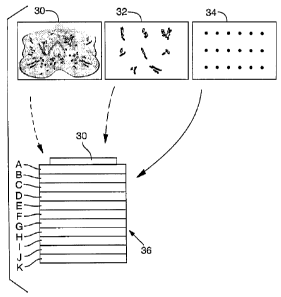
One example of a layered expression scan is shown in schematic form
in FIG. 2. One or more biological samples, such as an intact tissue section
(for
example prostate section 30), dissected intact cell lysates 32, or dissected
cell
lysates 34, are prepared and placed within or upon an ultra thin gel, called a
sample gel, which is applied to a multilayered gel, for example to a surface
(such
as a top surface) of a multilayered substrate 36.
The sample gel can utilize any known gel matrix including agarose,
polyacrylamide and gelatin based matrices. If the sample gel is agarose, its
concentration is, for example, in the range of about 0.1 % to about 5 %, and
it
CA 02375034 2002-01-11
WO 01/07915 PCTIUSOO/20354
-11-
may be cast to be "ultrathin," that is, in the range of about 0.10 m to about
1
mm thick. Alternatively, the biological samples can be placed directly in the
substrate or on a surface, such as the top surface, of the multilayered
substrate 36.
For purposes of simplified illustration in FIG. 2, the intact prostate section
30 is
placed directly on a top surface of the multi-layered substrate 36.
The specimen 30 is placed on the top surface of the substrate layer A,
which surface is substantially parallel to separations between the layers. For
purposes of illustration, eleven layers are shown (although many more can be
used, for example at least hundreds or thousands of layers), and the layers
are
labeled A through K. Each of the layers may be a membrane or film, each of
which may contain one (or more) identification molecules, such as an antibody
that recognizes a particular antigen, or a DNA sequence that functions as a
probe
by hybridizing to complementary DNA sequences in the specimen. The
identification molecule can be different in each of the layers A-K or the
same.
After application of the specimen 30 to the flat top surface of layer A,
the soluble contents of the specimen are transferred (for example by capillary
action or electrophoresis) through the series of layers A-K, while maintaining
the
overall two-dimensional architecture within the sample. As the specimen
components, such as proteins and nucleic acids, pass through the membranes,
the
identification molecules of the substrate layers interact with the proteins or
molecules of interest. After this interaction occurs, the membranes are
separated
(FIG. 2B) and subjected to further analysis, such as exposure to a second
antibody
or DNA sequence, producing a highly sensitive and specific molecular profile,
or
"expression scan" of the cellular specimen. If the analysis is applied to a
whole
tissue specimen, the final step of the method can involve examination of a
reference specimen cut from a location immediately adjacent to the first
tissue
specimen, so that areas of interest in the intact specimen (such as areas of
cellular
atypia) can be correlated with findings in the expression scan. In this
manner,
molecular characteristics of the specimen (such as the expression of
particular
proteins) can be correlated with areas of histological interest (such as
invasion of
the prostate capsule). In the context of this example, expression of
particular
CA 02375034 2002-01-11
WO 01/07915 PCTIUSOO/20354
-12-
proteins associated with capsular invasion (or metastasis in general) can be
located.
The present example of analyzing a cellular specimen includes placing
the cellular specimen on a layered substrate, where the different layers of
the
substrate contain different identification molecules, and transferring
components of
the cellular specimen through the layers under conditions that allow the
components to interact with different identification molecules in the
different
contiguous layers of the substrate. Cellular specimens include, but are not
limited
to, tissue sections, cultured cells, or a cytology sample. Tumor tissue
sections
produced by the cryostat method are particularly suited for use in the present
method. Standard methods of preparing tissue sections are taught in Lefkovits
et
al. (1996). If the molecule of interest is present at moderate or low level
abundance, such as those present in the range of one to 10,000 copies per cell
or
even one to 100 copies per cell, the thickness of the tissue section to be
analyzed
can be increased to intensify the expression scan produced. The thickness of
such
samples are about 25 m to about 50 m. Since an adjacent reference specimen
may be used to view the tissue microscopically, and the sections are thin, the
histological detail of the analysis is not compromised by utilizing the
thicker tissue
section for the present method.
The cellular specimen to be analyzed by the method of the present
invention may also be obtained by dissecting a cell population of interest
from a
larger cell population, for example, through laser capture microdissection, or
the
cellular specimen can be lysates of a dissected cell population. Methods of
preparing tissue samples for microdissection are disclosed in Emmert-Buck et
al.
(1996) and Bonner et al. (1997). The laser capture microdissection procedure,
described by Emmert-Buck et al. (1996) and Bonner et al. (1997) allows
dissection
of particular cell populations of interest from a tissue sample, providing
individual
samples for experiments that compare the contents of various tissue types
within
one specimen. FIG. 1 illustrates a tissue sample containing nine populations
of
interest, where each could be separately isolated using the laser capture
microdissection process. Alternatively, comparisons of the same tissue over
time,
CA 02375034 2002-01-11
WO 01/07915 PCT/US00/20354
- 13-
such as changes in protein expression or mRNA during tumor development, can
be obtained. If an investigator wishes to study a protein or mRNA of very low
abundance, such as menin, the gene responsible for Multiple Endocrine
Neoplasia
Type 1, then preparation of a highly concentrated lysate derived from
microdissected cells can be utilized. Very low abundance mRNA would be
present in the cell in a range of one to 10,000 copies. It is also possible to
amplify low abundance mRNAs by reverse transcription/polymerase chain reaction
(RT/PCR) and then analyze for their corresponding cDNAs.
As previously discussed, the prepared cellular specimen is optionally
placed in a gel, to allow ease of handling prior to analysis. In some
embodiments,
the sample gel may be an ultra thin gel made of agarose or polyacrylamide. The
sample gel could be made using standard 2 % agarose dissolved in tris-borate
EDTA buffer. Two hundred l of this preparation is pipetted onto a standard
glass histology slide and coverslipped, thus creating an ultrathin gel on the
order
of 0.5-1 mm thick. The sample gel can be selected to participate in separating
the
different components of the cellular specimen. This separation function is
accomplished by providing the sample gel with a particular structure that
alters or
aids the migration of certain components into the layers of substrate 36,
and/or
retards the migration of components that should remain in the sample gel.
Structural changes that aid the separation function include varying the gel
concentration to alter the gel pore size, or varying gel composition, such as
using
an acidic or basic formulation to aid or retard the migration of certain
components. If no separation function by the sample gel is desired, a gel with
neutral characteristics can be chosen, such as 2 % agarose in TBE with a pH of
7.4.
If no gel separation function is desired and the physical form of the
sample is appropriate (for example a tissue section), the specimen 30 is
placed
directly on a planar top face of the first layer A (FIG. 2A) of the substrate
36.
Even if a gel is not used, the analyzed cellular specimen can be treated
before
transfer to allow selective transfer of certain target molecules into the
substrate
layers. An example of such a treatment is the use of a transfer buffer that
contains
CA 02375034 2002-01-11
WO 01/07915 PCTIUSOO/20354
-14-
detergents, which would tend to increase the transfer of components of a
cellular
specimen that are present in the cellular membrane (such as the plasma
membrane).
If the samples are solubilized cellular lysates, purified proteins, or
nucleic acids, it is possible to prepare a sample gel as follows. A 2 mm thick
2%
agarose gel is "punched" to generate a series of holes (4 mm in diameter, for
example) that serve as sample "wells." The samples may then be added to 1 %
liquid agarose, placed into the wells, and then allowed to solidify to form a
sample
gel 34. The sample gel created by this process may then be placed on top of
the
layered substrate 36.
The layered substrate 36 of the embodiment disclosed in FIG. 2A
includes separable layers of a material (such as layers A-K of nitrocellulose,
which
can be obtained from Schleicher and Schuell, Keene, NH, product #BA-85) which
is capable of placement in multiple contiguous layers, as shown in FIG. 2A,
and
subsequent separation into multiple separate (non-contiguous) layers, as shown
in
FIG. 2B. The nitrocellulose layers may be treated with a blocking agent, to
inhibit binding of proteins to the nitrocellulose of the layers, which allows
proteins
to pass through the layer unless it interacts with and is captured by the
identification molecule. Once the components of the specimen have migrated
through the contiguous layers, the layers are separated to permit
individualized
analysis of the components of the cellular specimen retained in each separated
layer.
Other examples of the substrate layers include, but are not limited to
high concentration agarose gels, low concentration agarose gels, high
concentration polyacrylamide gels, a low concentration polyacrylamide gel, and
membranes, such as porous membranes like nitrocellulose paper. Low
concentration agarose is from about 0.1 to about 3 %, while high concentration
is
above about 3 %. Low concentration acrylamide is about 2 % to about 20 %,
while
high concentration is above about 20%. Such gels or membranes may optionally
be backed with a polyester membrane or the like to provide mechanical strength
and to provide a "contact substance" that permits efficient transfer of the
CA 02375034 2002-01-11
WO 01/07915 PCT/US00/20354
- 15-
components of the cellular specimen between the layers of the substrate and
reduces loss of the two-dimensional architecture of the sample (such as sample
30)
as the components migrate through the substrate 36.
Nitrocellulose layers are examples of porous layers, that exert capillary
pressure on the specimens (such as specimen 34) on the top surface of layer A
(FIG. 2A), and conduct components of the specimens through the layers. Such
porous layers or membranes allow the movement of liquid from one face to an
opposite face of the membrane, and exert capillary action on the specimen to
move
soluble components of the specimen through the multiple layers. The pore size
of
the porous layers may be any that are available, particularly the about 0.45
m
pore-size nitrocellulose membrane. The number of layers in the substrate can
vary
widely, for example from about 1 to at least 2, 5, 10 or even 1000 layers,
although for purposes of illustration eleven layers A through K are shown in
FIGS. 2A and 2B. The number of layers can be varied, depending in part on the
number of different binding or other identification molecules being used, and
is
ultimately limited only by the ability to promote migration of the cellular
components through the substrate levels. The substrate layers can be of
identical
structure, or the layers can be mixtures of different substrate types.
In a disclosed embodiment, each layer (or other type of region) of the
substrate is impregnated with multiple copies of at least one identification
molecule that can interact with one or more molecules of interest. Similarly,
different layers of the substrate can contain multiple different
identification
molecules, for example each layer (or other type of region) can have one or
more
identification molecules present. In an alternative embodiment of the
substrate, all
the layers (or other type of region) would contain the same identification
molecule
and differential migration through the various substrate layers would allow
separation. The differential migration can be promoted by differing physical
characteristics of the substrate layers, such as different pore diameters or
pH, or
porosity or pH gradients, in the direction of layers A to K. Likewise, in
other
embodiments, some of the substrate layers do not contain identification
molecules
CA 02375034 2002-01-11
WO 01/07915 PCT/US00/20354
-16-
and may serve to promote differential migration of sample components through
the
layers.
Representative examples of identification molecules include, but are not
limited to antibodies, nucleic acids, peptides, receptors, ligands, dyes,
stains, or
colorimetric enzymes. Specific examples of identification molecules include
anti-
prostate specific antigen antibodies (Scripps, San Diego, CA; anti-cytokeratin
antibodies, anti-alpha-actin antibodies (Sigma, St. Louis, MO); anti-PB39
antibodies, and anti-menin antibodies (National Cancer Institute Core Antibody
Lab, Fredrick, MD). Identification molecules can interact specifically with
the
molecule of interest, such as the binding of an antibody or complementary
interaction with a single stranded DNA sequence, or more generally, such as
the
interaction between a dye and a molecule colored by that dye. If the
identification
molecule prevents the migration of the molecule of interest into subsequent
layers
of the substrate, the identification molecule is referred to as a capture
molecule.
When the transfer of the components of the cellular specimen occurs
through capillary movement of liquid present in the sample through the
substrate,
it is desirable to have the multiple layers (or other regions) of the
substrate in
physical contact with each other. The use of contiguous substrate layers A-K
(as
in FIG. 2A) reduces the effects of diffusion on the accurate migration of the
proteins or molecules of interest through the substrate and enhances the
capillary
movement of the components. Alternatively, the components can be moved
through the substrate layers (or other regions) using electrophoresis, a
variation of
isoelectric focusing, or other similar methods of moving charged molecules. If
electrophoresis or another method using electricity is used, the different
layers of
the substrate are ideally conductive, such as an agarose or polyacrylamide
gel.
Methods based on electrophoresis would be limited generally to separation of
charged species from the cellular specimen. However, the use of
electrophoresis
can avoid the use of contiguous substrate layers. For example, the layers
could be
separated from one another, as long there is an electrically conductive medium
(such as a liquid, particularly a liquid comprising ions, such as may be
formed by
CA 02375034 2002-01-11
WO 01/07915 PCTIUSOO/20354
- 17-
dissolving a salt in a liquid) between the layers through which the specimen
is
electrophoresed.
Another means of transferring sample components through the substrate
layers (or other regions) is by way of liquid movement in response to a fluid
pressure differential. For example, pressure, such as provided by a compressed
gas, may be applied to the sample to force the liquid present in the sample
into
and through the substrate 36. Alternatively, another liquid under pressure may
be
used to carry sample constituents into and through the substrate layers to an
area
of lower pressure. Liquid present in a sample or provided to carry sample
constituents into the substrate layers may also be induced to move through the
substrate 36 by a vacuum applied to the substrate 36 opposite the surface
where
the sample (such as sample 30) is applied. Since a continuous fluid medium can
be established with such an approach, the layers can be either contiguous or
non-
contiguous.
After the molecules of interest have been transferred through the
substrate layers in the disclosed example, the various layers can be separated
from
each other to allow analysis using a second identification molecule, separate
from
that used for initial capture, such as a second antibody or DNA sequence. For
example, the second antibody can be a specific binding agent such as an
antibody
that recognizes the original antibody bound to its antigen in the substrate
layer.
The use of the second identification agent ensures high specificity of the
staining
signal present in the expression scan.
Separate analysis of different substrate layers is illustrated in FIGS. 2A-
B. In this example, a whole mount section of human prostate tissue,
representing
a cross section of the entire organ, was placed on top of the substrate and
transferred through ten capture layers, and onto a nitrocellulose membrane.
The
membrane was subsequently processed similar to a standard immunoblot using an
antibody against cytokeratin, which selectively stains epithelium.
Retention of the basic organization of the tissue section throughout the
transfer process is demonstrated by comparing FIG. 3A (cytokeratin antibody
CA 02375034 2002-01-11
WO 01/07915 PCTIUS00/20354
- 18-
transfer layer) with FIG. 3B (hematoxylin and eosin stained slide of an
adjacent
recut from the same tissue block).
The specificity of molecular capture using this technique was also
illustrated by transferring a whole mount section of prostate tissue through
ten
capture membranes, each having a different antibody linked throughout the
membrane. After transfer of the tissue section, each membrane was placed into
denaturing buffer to remove captured molecules, and subsequently analyzed by
immunoblot using anti-PSA (prostate specific antigen). Specific capture of PSA
was demonstrated by isolation of a single PSA band of 30 kDa following
electrophoresis.
To demonstrate the potential of the method for very high throughput
analysis, a repeat of the PSA capture experiment was performed, except the
tissue
was transferred through 100 capture layers, with anti-PSA placed on layer
#100.
Successful capture of PSA in layer #100 was achieved. There does not appear to
be a limit to the number of capture membranes which can be utilized, hence the
method can include expression scanning using hundreds or even thousands of
layers, to allow for simultaneous measurement of thousands of molecular
species.
To demonstrate the use of the scanning technique with microdissected
samples, nine separate cell populations from three different subjects were
procured
from tissue sections by laser capture microdissection, solubilized, and
transferred
as nine separate, 5 mm spots, through ten capture layers, in which polyclonal
anti-
PSA was present on layer #10. A dissected cell population of prostate
epithelial
cells was placed in the upper left corner of the top layer of the substrate.
After
tissue transfer, layer #10 was probed with monoclonal antibody against PSA,
and
visualized by enhanced chemiluminesence (ECL). Specific PSA staining was
visualized only for the tissue sample containing prostate epithelium,
consistent
with the known epithelial localization of PSA. Samples 2-9 were appropriately
negative for PSA staining.
The maintenance of cellular architecture helps determine associations
between cellular findings and molecular characteristics determined by the
expression scan. For example, the presence of the lymphocytes can be
correlated
CA 02375034 2002-01-11
WO 01/07915 PCT/US00/20354
- 19-
with findings associated with other of the layers. Also, expression of a
particular
receptor may be correlated or mapped to epithelium. Alternatively, another
molecular marker can be associated with areas of metaplasia or capsular
invasion.
Separate analysis of the substrate layers allows one to investigate
multiple regions of the molecule of interest, i.e., domains of a protein or
exons of
a RNA transcript, as described more fully in the Examples. The present method
can provide a quantitative indication of the relative abundance of the
components
in the cellular specimen when the identification molecules interact in
relative
abundance to the quantity of the component of interest in the cellular
specimen.
Mass spectroscopy sequencing can also be performed after separation to
characterize a captured amino acid sequence.
The foregoing explanation will be better illustrated by the following
additional specific examples.
Example 1
Identification of PSA, Tubulin, Actin, and Cytokeratin in Prostate Tumor
The LES procedure was performed on prostate tumor sections. The
preliminary experiment used cytokeratin as the protein of interest. A whole
mount
cryostat section of human prostate tissue was prepared by making a thin frozen
section of prostate, the section having a thickness of about 10 m. As shown
in
Figure 1, the section includes multiple cell populations of biological
interest
including normal epithelium, pre-malignant lesions, high and low grade tumor
foci, and significant tumor-host interactions such as lymphocytes interacting
with
cancer cells. This section was placed on an ultrathin 2% agarose gel that had
been
cast on a glass histology slide. The section was covered with 2% agarose
solution. A cover slip was applied on top of the section and the agarose was
allowed to polymerize, thus creating a two-layered sample gel with the tissue
section in between. The agarose sample gel containing the tissue sample was
applied to the surface of a single layer substrate made of a 1.75" X 1.75"
0.45
pore size nitrocellulose membrane (Schlieicher and Schuell, Keen, NH). The
membrane was then probed with an antibody against cytokeratin (Sigma, 1:1000
CA 02375034 2002-01-11
WO 01/07915 PCTIUSOO/20354
-20-
dilution) overnight at 41 C. This membrane was then probed a second time with
a
biotinylated secondary antibody (Sigma, 1:5000 titer) for 30 minutes at room
temperature. The membranes were visualized by autoradiography using enhanced
chemiluminescence (ECL) as recommended by the manufacturer (Pierce,
Rockford, IL).
A second experiment to test the specificity of "capture molecules" in
the membrane layers was then performed. A 20 gm cryostat section of prostate
tissue was prepared within an ultrathin 2 % agarose gel as described above.
Components of this tissue section as transferred overnight at room temperature
through ten contiguous nitrocellulose membranes (0.5" X 0.5," 0.45 pore size,
Schliecher and Schuell) by capillary action. Prior to use, each membrane was
linked to a different identification molecule, in this case, antibodies, for 1
hour at
room temperature. The membranes were washed 3 times for 10 minutes in 1X
PBS, and treated with a commercial blocking agent (Pierce) for 1 hour at room
temperature, followed by a repeat wash. The nitrocellulose/antibody membranes
(illustrated as A-J in Figure 2) were prepared as follows:
Layer Identification Molecule Source
A Anti-PB39, 644 NCI
B Anti-actin Sigma
C Anti-tubulin Sigma
D Anti-PB39, 655 NCI
E Polyclonal anti-PSA Scirpps, San Diego, CA
F Anti-CAIR 1 NCI
G Anti-PB-39, 656 NCI
H Anti-cytokeratin Sigma
I Anti-CD-3 NCI
J Anti-PB-39, 645 NCI
Antibodies were linked to the nitrocellulose membranes according well
known procedures such as those disclosed in U.S. Pat. No. 4,774,177, issued to
CA 02375034 2010-08-09
-21-
Marks on 9/27/88 or U.S. Pat. No. 4,727,037, issued to Ring on February 23,
1988.
Nitrocellulose layers are examples of porous layers that exert capillary
pressure on the specimens on the top surface of the substrate, and conduct
components of the specimens through the layers. Such porous layers or
membranes allow the movement of liquid from one face to an opposite face of
the
membrane, and exert capillary action on the specimen to move soluble
components
of the specimen through the multiple layers. Although nitrocellulose avidly
binds
biomolecules such as proteins, the nitrocellulose can be altered with well
known
blocking agents to inhibit e.g. protein binding, and promote movement of the
protein or other biomolecule through the nitrocellulose layers.
Blocking agents serve to prevent non-specific interactions between the
substrate and the components of the sample as they are transferred through the
substrate. "Blocking agent" is a collective term for various additives that
prevent
non-specific binding, but that have no active part in the specific reaction,
such as
an immunochemical reaction, between a particular identification molecule and
its
target. Blocking agents are most commonly concentrated protein solutions.
Examples of such solutions include 10-20% fetal calf serum and 5 % non-fat dry
milk powder dissolved in a buffer such as PBS, TBS, or TBST. Commercially
available blocking agents include SuperBlocktm, Blocker' BLOTTO, Blockert
BSA, and SeaBlockC" (Pierce Chemical, Rockford Ill) as well as NAP-
SureBlockerTM, a non-animal protein blocking agent (Deno Technology,
Maplewood, MO).
After transfer, each membrane was separately placed into 30 l of SDS
sample buffer (Novex, San Diego, CA) to remove any captured molecules. The
removed, solubilized molecules were separated by electrophoresis on a 4-20%
tris-
glycine acrylamide gel (Novex) for 1.5 hr at 110V. The proteins were
transferred
to a 0.2 m pore size PVDF membrane for 2 hours at 40V and analyzed by a
standard immunoblotting procedure using a 1:1000 titer of monoclonal anti-PSA
molecules (Scripps). In each case, the signal obtained was restricted to. the
CA 02375034 2002-01-11
WO 01/07915 PCTIUSOO/20354
-22-
appropriately sized molecular weight band for the molecule captured by the
antibody.
The feasibility of transfer through 100 membrane layers was shown by
repeating the experiment above with 99 layers treated only with blocking agent
and a final layer 100, that had polyclonal anti-PSA antibody linked to its
surface.
The Western blot showed capture of PSA only in layer 100. Nonspecific capture
of PSA in layers 1-99 is avoided by the blocking agent pre-treatment. This
experiment was repeated using an antibody against matrix metalloproeinase-2 in
layer 100. Instead of Western immunoblotting, the isolated protein was
analyzed
by gel zymography, as disclosed in Zucker et al. (1994). Thus, it is possible
for
to allow simultaneous measurement of thousands of molecular species present in
the tissue samples or isolated cell populations, through the use of thousands
of
substrate layers.
A further experiment was done to detect the presence of PSA in a
dissected cell population. Different cell populations, distinguished by tissue
type,
are separately collected using laser microdissection techniques as described
by
Emmert-Buck et al. (1997). Ten epithelium samples 1-10 were placed in a row on
a sample gel, as shown in FIG. 6, and ten non-epithelium samples 11-20 were
placed in a second row immediately below the epithelial samples. All twenty
samples were transferred through a substrate containing ten nitrocellulose
membranes (A through J), in which only membrane J had anti-PSA antibodies
linked to its surface. After transfer, each of the ten membranes was probed
with a
monoclonal antibody against PSA and visualized by enhanced chemiluminescence
(ECL) as described above. The first nine membranes A through I did not produce
an ECL signal, indicating no capture of PSA had occurred. However, positive
staining for PSA was visualized on membrane J in all of the samples containing
epithelium (sample numbers 1-10). This result is consistent with the known
epithelial localization of PSA. Samples 11-20 did not contain epithelial cells
and
were appropriately negative for PSA staining.
CA 02375034 2002-01-11
WO 01/07915 PCT/US00/20354
-23-
Example 2
Selective Capture of Prostate Specific Antigen (PSA)
To demonstrate selective molecular capture within substrate layers, cell
samples from five separate patients were procured from tissue specimens and
solubilized in standard protein extraction buffer. The samples included
lysates of
normal lung, lung cancer, esophageal cancer, normal prostate, and breast
cancer
tissue. Each of the cell lysates was placed within a discrete 4 mm diameter
spot
on the top layer of a capture membrane set. This was accomplished by punching
4
mm diameter holes ("wells") in a 2 mm thick agarose gel, adding the lysates to
1 % liquid agarose, filling the 4 mm wells with the lysate/agarose solution,
and
allowing them to solidify. The sample gel thus created was placed on the top
layer of a capture membrane set. Additionally, purified PSA was used as a
positive control sample. In this experiment, the capture membranes consisted
of
ten nitrocellulose layers, each coupled to a different antibody. Polyclonal
anti-
PSA was linked to layer number ten (the tenth successive capture membrane).
The six tissue samples were placed on the surface of the substrate and
transferred
through the capture membranes by capillary action, and each membrane was
subsequently analyzed. FIG. 4A shows capture layer number 10 after probing
with a monoclonal antibody against PSA and visualization by enhanced
chemiluminescence (ECL). Samples 1 (purified PSA) and 5 (normal prostate
tissue) show a positive signal, which indicates that PSA has been successfully
captured. Samples 2 (normal lung), 3 (lung tumor), 4 (esophageal tumor), and 6
(breast cancer) do not contain PSA and are appropriately negative.
A location of each of the samples that was placed on the top layer was
substantially preserved and reproduced on the membranes through which the
samples were transferred. Their substantial retention of spatial relationship
conveniently allows the resulting patterns to be correlated with the original
specimens.
CA 02375034 2002-01-11
WO 01/07915 PCTIUSOO/20354
-24-
Example 3
Specificity of PSA Capture
To show the specificity of the capture process, a single sample of
prostate tissue was solubilized and transferred through a set of capture
layers as
described in Example 2 above, except that polyclonal anti-PSA was placed on
membrane 5. After the transfer of the prostate tissue through the layers, each
membrane was placed in denaturing buffer to remove captured molecules. The
proteins recovered from every membrane were subsequently separated by gel
electrophoresis (the proteins recovered from layer 1 were run in Lane 1, the
proteins recovered from layer 2 were run in lane 2, and so forth) and analyzed
by
immunoblot using a monoclonal anti-PSA antibody. FIG 4B shows the results
from each of capture layers one through nine. Lane 5 (representing layer 5,
linked to anti-PSA) shows a single, distinct PSA band at Mr = 30,000 (30 kDa).
The remaining capture membranes are negative for PSA. This result demonstrates
that PSA was captured only on the membrane containing its antibody. Moreover,
the single band on the immunoblot indicates that the ECL signal derived from
the
capture membrane in Example 2 was specific for PSA.
To illustrate the potential of the method for high-throughput analysis,
a repeat of the experiment was performed except the tissue was transferred
through 101 capture layers with anti-PSA placed on layer number 100.
Successful
and specific capture of PSA is shown in Fig. 4C. Only lane 100 (representing
layer 100, linked to anti-PSA) shows a single, distinct PSA band at M, =
30,000
(30 kDa). The remaining capture membranes are negative for PSA. The specific
and selective capture observed after transfer through this large number of
layers
indicates that it is possible to utilize layered expression scanning for the
simultaneous measurement of hundreds, thousands, or even tens of thousands of
molecular species, by providing different capture agents in different layers.
CA 02375034 2002-01-11
WO 01/07915 PCT/US00/20354
-25-
Example 4
Capture of Active Enzymes
To demonstrate the ability of layered expression scanning to capture
and analyze active enzymes a repeat of the ten layer experiment described
above in
Example 3 was performed, except the anti-PSA antibody that was linked to
capture layer 5 was replaced by an antibody against matrix metalloproteinase-2
(MMP-2). Purified MMP-2 protein was transferred through the capture layers,
and each membrane was subsequently analyzed by gelatin zymography. FIG. 4D
shows successful capture of MMP-2 represented by a single band at M, = 72,000
(72 kDa) in lane 5 that corresponds to capture layer 5. All other lanes,
corresponding to layers not containing anti-MMP-2 antibodies, were negative
for
MMP-2.
Example 5
Selective and Specific Capture of Nucleic Acids
This example demonstrates the ability of layered expression scanning to
analyze nucleic acids. 32P-labeled PCR products (200 bp) were amplified from
plasmids containing cDNAs of the POV1 (PB39, NCI) and J3-actin (Clonetech,
Palo Alto, CA) genes, respectively. The radiolabeled PCR products were excised
from an agarose gel, and 5 % of each product was placed in discrete 4 mm spots
as
described for the tissue samples in Example 2. The PCR products were
transferred through 10 capture layers overnight by capillary transfer using 6X
SSC. In this experiment, the capture layers consisted of ultrathin (<50 m) 2%
agarose gels. Capture layer five contained a plasmid containing the entire
cDNA
for the POV1 gene. During preparation of layer 5, the POV1 cDNA-containing
plasmid was added to the agarose prior to gel polymerizationat a final
concentration of 30 ng/.iL. A nonblocked nitrocellulose membrane was used to
bind the noncaptured POV1 and fl-actin PCR products after they traversed the
membrane set. After transfer, the layers were separated and visualized by X-
OMAT radiography. FIG. 5 shows successful and selective capture of POVI
CA 02375034 2002-01-11
WO 01/07915 PCTIUSOO/20354
-26-
cDNA in layer 5, while the actin PCR product moved through the entire set of
layers and was not captured until it reacted the nonblocked nitrocellulose
layer.
Example 6
Transfer of Intact Tissue Sections
The Examples above show the feasibility of layered expression
scanning to analyze tissue samples after they have been appropriately procured
and
solubilized. Layered expression scanning may also be utilized to analyze
intact
tissue sections. If an intact tissue section is used as the sample, it is
possible to
correlate the two-dimensional architecture of the tissue section with the two-
dimensional pattern of cellular components localized in particular capture
layers
following transfer.
To demonstrate the retention of the two-dimensional architecture of a
tissue section, 10 m thick whole-mount cryostat sections of human prostate
from
radical prostatectomy specimens were placed on top of either a ten-layer or a
one
hundred-layer agarose gel set. The intact tissue section was transferred
through
the layers by capillary fluid movement overnight at room temperature to a 1.75-
square inch, 0.45 m pore size nitrocellulose membrane (Schleicher and
Schuell).
After transfer of the tissue sections, the nitrocellulose membranes were
probed
with an antibody against cytokeratin (Sigma 1:1000 dilution) to selectively
identify
epithelial elements and were visualized by ECL according to the
recommendations
of the manufacturer (Pierce).
Retention of the basic organization of the tissue section throughout the
transfer process is demonstrated in FIG 3 A-D by comparing the transferred
sections (FIG. 3A and FIG 3C) with a hematoxylin and eosin (H&E) stained slide
of an adjacent recut section. The overall architecture of the transferred
sections is
highly similar to the corresponding H&E stained slides, and the location of
individual glandular epithelial elements within the tissue sections can be
determined. Thus, layered expression scanning can be used for analyzing intact
CA 02375034 2002-01-11
WO 01/07915 PCTIUSOO/20354
-27-
tissue sections while retaining a correspondence between the two-dimensional
architecture of the tissue section and the two-dimensional position of
components
transferred to the capture layers. Single cell-level of resolution will permit
individual cells to be analyzed for the presence of particular molecules. For
example, in prostate cancer, all of the individual normal glands premalignant
foci,
and high- and low- grade tumor glands could be simultaneously analyzed, as
well
as important sub-populations, such as tumor glands, that are invading through
the
prostate capsule. Alternatively, microscopic structure level resolution could
allow
localization of particular proteins to individual subcellular organelles.
Example 7
Layered Expression Scanning Membranes
Membranes and gels useful for creating identification and capture
layers as utilized in the Examples may have one or more of the following
properties. First, the membranes or gels are able to immobilize individual
identification or capture molecules (e.g. antibodies, nucleic acids, and
dyes).
Second, the membranes or gels permit cellular components transferred from a
sample to efficiently traverse the set of layers and accumulate or react in
the
appropriate layer. Third, the membranes or gels facilitate transfer with
minimal
loss of the two-dimensional relationship of the biological sample(s).
Particular examples of materials appropriate for constructing a set of
layers for layered expression scanning include nitrocellulose membranes,
derivatized nitrocellulose membranes, high concentration agarose gels, low
concentration agarose gels, high concentration polyacrylamide gels, a low
concentration polyacrylamide gel, and membranes, such as porous membranes like
nitrocellulose paper. Low concentration agarose is from about 0.1 to about 3
%,
while high concentration is above about 3 %. Low concentration acrylamide is
about 2 % to about 20 %, while high concentration is above about 20 %.
Individual layers may also be composites of two or more membranes or
gels. For example, thin polymer membranes, such as polar polymer membranes,
for instance polyester membranes, may be combined with nitrocellulose
CA 02375034 2002-01-11
WO 01/07915 PCTIUSOO/20354
-28-
membranes or agarose or polyacrylamide gels to form composite layers for
layered expression scanning.
In a particular embodiment, the composite membrane is formed as
follows. A thin (10 m) polyester membrane is used as a backbone layer. The
polyester membrane is then coated with a soluble polymer material, such as 2%
agarose, to form an ultrathin (< 1 m) layer covering the polyester backbone.
A
capture molecule (e.g., an antibody or nucleic acid) is added to the polymer
material prior to its addition to the polyester backbone. After the polymer is
coated on the backbone, it forms a gel and irreversibly traps the capture
molecule
within the gel structure. The polyester backbone/polymer gel composite
containing the capture molecule may then be used as a layered expression
scanning
capture membrane. Experiments have demonstrated that such composite
membranes are highly efficient at meeting the criteria described above. A
particular advantage of the composite membranes is that the polymer gel that
is
coated on the polyester backbone serves as a "contact substance" between each
of
the layers, thereby permitting efficient transfer of biomolecules with minimal
loss
of correspondence with the two-dimensional architecture in the sample.
Example 8
Determination of the Binding Status or Binding Partner of a Molecule of
Interest during Tumor Progression
Different tumor cell populations, distinguished by the stage of tumor
progression, are separately collected using laser microdissection techniques
as
described by Emmert-Buck et al. (1997). Each different cell population is
placed
in its own location within a sample gel, as described above in Example 1. The
sample gel is placed on a multi-layer substrate, containing at least one layer
cross-
linked with antibodies against one or more known binding partners of the
molecule
of interest. The molecules could be treated with a cross-linking agent, thus
binding partners will remain in the state they are in at the time of the
preparation
of the cryostat during transfer. After transfer of the components of the cell
CA 02375034 2002-01-11
WO 01/07915 PCT/USOO/20354
-29-
populations through the substrate layers as described above, the layers are
separated and the molecules of interest are run on a gel and probed by the
capture
antibody. Thus, this experiment shows whether or not a molecule of interest is
bound or free at various stages of tumor development by determining the
molecular weight of the species when the tissue sample is prepared.
In order to search for new binding partners, the experiment is
performed as described above for binding status without the pre-transfer cross-
linking. After transfer of the cellular specimen, mass spectrometry can be
used to
determine the identity of proteins that are captured along with the protein of
interest. After separation from the capture molecule and isolation in a gel,
MS-
MS (mass spectrometry-mass spectrometry) sequencing can identify the proteins
recovered from relatively few numbers of microdissected cells as described in
Huang et al. (1999).
Example 9
Comparative Expression Between Normal and Diseased Cell Populations
LES can be used as an "open system" to search for disease associated
molecular alterations in tissue samples. In this example, normal and diseased
cell
samples are placed within the sample gel as described in Example 1. The
information molecules cross-linked on the membrane layers can be antibodies,
peptides, or DNA sequences for either known proteins, or libraries of ssDNA or
mRNA. Large numbers of capture molecules are simultaneously used to analyze
the comparative expression between normal and diseased cell populations of the
targets of the capture molecules. The samples tested can be derived from one
or
multiple patients. Once a protein or nucleic acid is shown to be expressed
differently in normal and diseased cells, its identity can be determined by
the
capture molecule to which it binds. This identity can be confirmed using
standard
sequencing techniques, or such sequencing techniques can be used initially to
determine whether the target of the capture molecule is unknown.
CA 02375034 2002-01-11
WO 01/07915 PCT/USOO/20354
-30-
Example 10
Determination of the Structure of a Protein of Interest
During Tumor Progression
Different cell populations, distinguished by the stage of tumor
progression, are separately collected using laser microdissection techniques
as
described by Emmert-Buck et al. (1996). Each cell population is placed in its
own
location within a sample gel, as described above in Example 1. The sample gel
is
placed on a substrate, containing at least one membrane cross-linked with
polyclonal antibody against tumor suppressor protein. After transfer of the
components of the cell populations through the substrate layers, the membranes
are separated and the anti-tumor suppressor protein membrane, with its
captured
molecules, is probed with two differentially labeled monoclonal antibodies
that
recognize different regions of the tumor suppressor protein. One antibody is
specific for the N-terminus of the protein, and the other is specific for the
C-
terminus of the protein. By comparing the presence or absence of the N- or C-
terminus of the protein at various stages of tumor progression, this
investigation
can detect if the tumor suppressor protein has been truncated at some point
during
tumor development. Mutation is one example of an event that could lead to
protein truncation. Such alterations in proteins during the transition between
normal and tumor cells is known to occur, for example in the adenomatous
polyposis coli (APC) tumor suppressor gene product, as reported by Smith et
al.
(1993).
Example 11
Use of Differential Transfer from the Sample Gel
Initial placement of the tissue specimen into a high concentration gel
limits migration to relatively small proteins. Alternatively, low
concentration gels
allow larger molecules to be transferred and analyzed. In the normal prostate,
PSA is localized exclusively within epithelial cells, whereas in tumors PSA is
able
to enter the stroma and is bound by alpha-1 anti-chymotrypsin (ACT) as
described
CA 02375034 2002-01-11
WO 01/07915 PCT/US00/20354
-31-
by Chen et al. (1995). PSA and ACT form an enzyme-inhibitor complex with a
significantly larger aggregate molecular weight than PSA alone. By altering
the
characteristics of the gel into which the tissue sample is placed, it is
possible to
separately analyze PSA and PSA-ACT complex in tumors. There is selective
membrane capture of PSA after placing a prostate tumor section into a 2%
agarose
gel. However, when the concentration of the gel is reduced to 0.5 %, both PSA
and PSA-ACT migrate through the membranes and are captured. Alteration of
experimental conditions to effect molecular migration can allow investigators
to
customize experiments as needed for particular objectives. For example, study
of
subcellular molecular profiles may be performed by utilizing transfer buffers
with
and without detergents to selectively mobilize soluble or membrane-bound
proteins.
Example 12
Automated Expression Scanning
The layered expression scanning of the present invention can also be used
in association with an automated laboratory instrument capable of multiple
applications. For example, the capture layers in the present prototype system
are
replaced by thin transparent membranes such that several thousand stacked
layers
will cumulatively be only a few millimeters in thickness. Thus, the total
migration
distance of the tissue sample during transfer and detection or immobilization
is
minimal, thereby optimizing the cellular resolution of the system. In this
application the tissue sample, wash buffers, and fluorescently labeled
secondary
detection molecules are transferred through the intact membrane set, thus
obviating the need to separate and individually process each capture layer.
The
sample, wash buffers and fluorescently labeled secondary detection molecules
may
be transferred into the stacked layers either in the same direction as the
sample
components are conducted through the stacked layers or in another direction,
such
as in the reverse direction or along the direction of the layers themselves.
The
intact membrane set is then analyzed by confocal fluorescence microscopy, and
the
expression data of each individual layer is determined and overlayed with the
high
CA 02375034 2002-01-11
WO 01/07915 PCTIUSOO/20354
-32-
quality histological image of the tissue section. The approach was
demonstrated in
an experiment similar to that shown in FIG 4, in which each of the detection
reagents were transferred through the capture membranes while the membranes
remained as an intact set. Successful capture and analysis occurred.
In yet another embodiment, the set of capture layers may be utilized
repeatedly to produce expression scans by washing the stacked layers with a
denaturing buffer between scans to remove captured molecules. Suitable buffers
for this purpose include buffers containing denaturants, such as detergents or
urea,
and salts, such as sodium chloride, at concentrations that are sufficient to
remove
captured molecules from the stacked layers. A particular example of a suitable
denaturing buffer is a buffer containing 1 % sodium dodecyl sulfate (SDS) and
500
mM sodium chloride. Other denaturing buffer systems are known in the art and
their suitability for use with automated expression scanning can be determined
by
analyzing the layers for the continued presence of bound molecules after they
are
washed with a particular denaturing buffer system.
In another approach, the capture membranes will be separable and
processed individually after tissue transfer. The separated membranes may then
be studied beyond measurement of expression levels of individual molecules.
For
example, mass spectrometry can be used to identify binding partners which are
"co-captured" along with targeted proteins.
Example 13
Analysis of Individual Cloned Biomolecules
The layered expression scanning (LES) methods can be used to analyze
for individual cloned biomolecules, such as messenger RNAs recovered from a
cell population and cloned into bacteria using standard methods.
In a particular embodiment, the bacteria are plated on media and
individual colonies are grown in the presence of a labeled nucleotide.
Individual
colonies are then placed on top of an LES device and the nucleic acids from
each
colony are transferred through a set of LES layers such as those described in
Example 5 above and where each LES layer contains an individual cDNA clone.
CA 02375034 2002-01-11
WO 01/07915 PCT/US00/20354
-33-
The identity of the cDNA in all bacterial colonies is simultaneously
determined by
analyzing for the presence or absence of hybridization on each capture
membrane
after the cloned DNA has traversed the LES layer set. One application of this
particular method is to perform high-throughput gene expression analysis of a
given cell population by determining the identity of a large number of
bacterial
clones derived from a particular cells messenger RNA population.
Example 14
Analysis of the Genomic Content of Cells
The layered expression scanning method may be used to analyze the
genomic DNA content of individual cells or cells within a tissue section. One
example of this application is as follows.
DNA from a series of cell lines is purified, labeled with a "tagged"
(radiolabeled or fluorescently labeled) nucleotide and placed in a grid on a
membrane on top of the LES device, such as described above in Example 2. In
this particular embodiment, each of the LES layers contains a specific genomic
DNA clone. The DNA samples are transferred through the LES layers such that
the DNA gragments from the cell samples specifically hybridize to the LES
layer
that contains the corresponding genomic clone. The LES layers are then
analyzed
(by radiography or fluorescence) to provide a quantitative measure of the
amount
of DNA in each cell sample at each genomic locus included in the LES layer
set.
This application would be useful in determining the specific regions of DNA
(and
associated genes) that are amplified or deleted in a series of cell lines.
Although many of the foregoing examples have been described in
association with a layered substrate, in which discrete or separable layers
extend
successively transverse to the path of movement of the material being
analyzed,
these same principles can be applied to other configurations of the substrate.
For
example, layers can be arranged substantially parallel, or at some other
angular
relationship, to the path of movement. In other embodiments, each layer may be
subdivided into multiple regions, each with a different capture molecule,
which
are capable of producing more complex patterns that can be recognized by the
user
CA 02375034 2010-08-09
-34-
or image processing software. Each of the regions can extend in any desired
shape throughout the layer, which can extend in any direction relative to the
direction of movement of the sample through the substrate. However, in
particularly useful embodiments, the different regions are transverse to the
direction of movement to maintain a spatial correspondence between a surface
of
the substrate to which the specimen is applied, and the region which captures
a
molecule of interest.
Although disclosed embodiments examine a pattern of interaction in
successive layers which correspond to positions on a surface of the substrate,
any
pattern that conveys information about the molecular content of the specimen
may
be used. With particularly complex patterns (of the type that may be generated
by
multiple different types of capture molecules in each layer, in regular or
irregular
patterns, that may extend to different depths of the substrate), pattern
recognition
software is particularly effective to store and compare patterns.
In view of the many possible embodiments to which the principles of
this invention may be applied, it should be recognized that the illustrated
embodiments are only particular examples of the invention.
CA 02375034 2002-01-11
WO 01/07915 PCT/US00/20354
-35-
References
W. Bonte, Acta histochem. 62: 68-77 (1978).
Z. Chen et al., Clin. Chem. 41:1273-82 (1995).
M. Emmert-Buck et al., Science 274: 998-1001 (1996).
M. Inczedy-Marcsek et al., Acta Histochem. Supp. 36: S377-94 (1988).
T. Grogan, Am. J. Clin. Pathol. 4(Supp. 1): S35-8 (1992).
Z. Huang et al., Anal Biochem 268:305-17 (1999).
L. Jin and R. Lloyd, J. Clin. Lab. Anal. 11:2-9 (1997).
J. Kononen et al., Nat. Med. 4: 844-847 (1998).
I. Lefkovits et al. (eds.), Immunology Methods Manual (1996).
J. Lindner et al., Naturwissenschaften 43: 201 (1956).
V. Neuhoff, Electrophoresis '79 (1980).
C. Saravis et al., J. Immun. Meth. 29:97-100 (1979).
U. Schumacher et al. (1990), Histochem. J. 22:433-438 (1990).
U. Schumacher and D. Trudrung, Anal. Biochem. 194: 256-58 (1991).
M. Schena et al., Science 270: 467-469 (1995).
K. Smith et al., Proc. Natl. Acad. Sci. 90: 2846-2850 (1993).
P. van der Sluis et al., Electrophoresis 9: 654-66 (1988).
L. Zhang et al., Science 276: 1268-1272 (1997).
S. Zucker et al., Clin, Exp. Metastasis 12:13-23 (1994).