Note: Descriptions are shown in the official language in which they were submitted.
WO 00/79255 CA 02375249 2001-12-18 PCT/AUOO/00702
1
TITLE
"A METHOD AND DEVICE FOR MEASURING TISSUE OEDEMA"
FIELD OF THE INVENTION
THIS INVENTION relates to the measurement of tissue
oedema and, in particular, lymphoedema using bioelectrical impedance
analysis.
BACKGROUND
Measurement of extracellular tissue fluid may be of
importance in a range of situations. This is particularly so in the case of
lymphoedema which is a condition characterised by excess protein and
oedema in the tissues as a result of reduced lymphatic transport capacity
and/or reduced tissue proteolytic capacity in the presence of a normal
lymphatic load. Lymphoedema normally occurs in a limb and may cause
pain, scarring and reduced limb function. The condition is incurable,
progressive, often disfiguring and physically disabling. Its course,
however, can be arrested or slowed by intervention using physical
therapy, compression bandaging, massage and other physical
techniques.
Acquired or secondary lymphoedema is caused by damaged
or blocked lymphatic vessels. The commonest inciting events are surgery
and/or radiotherapy. However, onset of lymphoedema is unpredictable
and may develop within days of its cause or at any time during a period of
many years after that cause.
There is a need for an accurate and effective technique to
detect the onset of lymphoedema, assess its severity and monitor its
response to treatment. The simplest known technique involves
measurement of limb circumferences and comparison with a paired
unaffected limb. A further technique is available by way of immersion of
the affected part and measurement of displaced liquid with subsequent
comparison against the result of the same measurement performed on an
unaffected limb.
WO 00/79255 CA 02375249 2001-12-18 PCT/AUOO/00702
2
It is also known to use multiple frequency bioelectrical
impedance analysis (MFBIA) to assess lymphoedema (Watanabe et al.,
1989, Lymphology 22:85). The authors noted that when a low frequency
voltage is applied to tissue, the impedance of the cell membrane is
substantial. With increased frequency, the impedance of the cell
membrane decreases and current is able to flow through both
extracellular and intracellular fluids. The results obtained by Watanabe et
al were subject to analysis of equivalent resistivity of extracellular and
intracellular fluid calculated after measurement of electrical bioimpedance
at multiple frequencies. Further development of the technique was
subsequently disclosed (Ward et al., 1992, European Journal of Clinical
Investigation 22:751) in which MFBIA was used and the impedance at
zero frequency was estimated by extrapolation. Differences were then
calculated between left-hand and right-hand sides of patients for the
impedance calculations 50kHz and OkHz frequency. The bilateral
differences in impedance between a group of controls and a group of
affected patients were significant. This test relies on the use of a
multifrequency bioimpedance meter and relatively complex analysis of the
results to provide an indication of lymphoedema.
US Patent No 5,372,141 describes a body composition
analyser that provides information in relation to body fat and ideal body
weight. The analyser compares the bioimpedance of the body "network"
against a reference network of known impedance. It is, however, of little
or no use in assessing tissue oedema.
US Patent No 4,947,862 discloses an analyser to determine
the amount of body fat on a patient. The analyser uses a high frequency
low-voltage signal in the body and measures magnitudes and phase shift
of the induced signal but again is of little use in measuring tissue
oedema.
It would be of advantage to provide a method for
determining the presence of oedema and, in particular, lymphoedema by
WO 00/79255 CA 02375249 2001-12-18 PCT/AUOO/00702
3
measurements taken at a single frequency. It would further be
advantageous to produce a device for measuring bioelectrical impedance
at a single frequency and analysing that measurement to produce an
indicator of the presence of oedema.
OBJECT OF THE INVENTION
It is an object of the present invention to overcome or
ameliorate one or more of the difficulties of known methods used to
assess tissue oedema and, in particular, lymphoedema.
DISCLOSURE OF THE INVENTION
In one form, although it need not be the only or broadest
form, the invention resides in a method of assessing tissue oedema
comprising the steps of:
performing a first measurement of bioelectrical impedance
of a first anatomical region in a subject at a single low frequency
alternating current;
performing a second measurement of bioelectrical
impedance of a second anatomical region in the same subject at the
same low frequency alternating current; and
analysing the two measurements to obtain an indication of
the presence of tissue oedema.
The first anatomical region and second anatomical region
may be paired with at least one of the anatomical regions unaffected by
tissue oedema.
Alternatively, the first and second anatomical regions may
be dissimilar with at least one of the anatomical regions unaffected by
tissue oedema.
The first anatomical region and the second anatomical
region may be the same region with the first and second measurements
separated in time. The anatomical regions may be limbs or parts of limbs.
The low frequency is preferably in the range of 5 to 20kHz.
More suitably, the range is 10 to 15kHz. Most preferably, the
WO 00/79255 CA 02375249 2001-12-18 PCT/AUOO/00702
4
measurements are made at 10kHz.
The analysis may include the step of dividing the lesser
result of the two measurements into the greater result of the two
measurements to obtain a product or quotient. The results of the two
measurements may further include the steps of applying a correcting
factor or term to the product and deriving an indication of tissue oedema.
The step of analysing the two measurements may be
conducted according to the algorithm
Zh
F=Z~-cf
where:
F is an indication of the presence of tissue oedema;
Zh is the greater bioelectrical measurement;
Z, is the lesser bioelectrical measurement; and
cf is a correcting factor.
The method may include the step of establishing "cf'.
Establishing "cf' may include the step of establishing a ratio of the
bioelectrical impedance of a first anatomical region of at least one subject
unaffected by tissue oedema compared to the bioelectrical impedance of
a second anatomical region of that subject wherein the first and second
anatomical regions of the at least one unaffected subject are paired with
the first and second anatomical regions of the subject being assessed for
tissue oedema.
When analysing the results of two measurements obtained
on paired limbs, the correcting factor may suitably be 1.066.
Alternatively, the step of analysing the two measurements
may be conducted according to the algorithm
Zz
F= cf1- Zh
where:
WO 00/79255 CA 02375249 2001-12-18 PCT/AUOO/00702
F is an indication of the presence of tissue oedema;
cf, = a correcting factor;
ZQ is the lesser bioelectrical impedance measurement; and
Zh is the greater bioelectrical impedance measurement.
5 When analysing the results of two paired limbs cf, may be
0.862.
The indication of tissue oedema may be displayed by the
step of representing the indication as a position on a scale.
In an alternate form, the invention resides in an apparatus
for determining the presence of tissue oedema, including:
current means for applying an alternating current to an
anatomical region at a single frequency;
monitoring means to monitor the bioelectrical impedance of
said region and produce signals characteristic of bioimpedance; and
analysis means to analyse the signals indicative of
bioimpedance to provide an indication of tissue oedema.
The current means may suitably be a proximal electrode
and distal electrode in electrical connection with a power source. The
monitoring means is suitably a first connection and second connection for
location on or near the anatomical region. Preferably, the monitoring
means includes display means to display the signals indicative of
bioimpedance.
Suitably, the analysis means is at least one processing
means programmed to perform analysis of data to provide an indication of
the presence of tissue oedema.
The analysis means may be programmed to analyse data
according to the algorithm
Zh
F= ZQ- cf
where:
F is an indication of the presence of tissue oedema;
WO 00/79255 CA 02375249 2001-12-18 PCT/AUOO/00702
6
Zh is a greater bioelectrical impedance measurement
obtained from a first anatomical region;
ZQ is a lower bioelectrical impedance measurement obtained
from a second anatomical region; and
cf is a correcting factor.
Suitably, cf may equal 1.066 when the first and second
anatomical regions of a subject undergoing assessment for tissue
oedema are paired limbs.
The apparatus preferably includes means for recording
bioimpedance in anatomical regions of the same subject simultaneously.
Preferably, said means includes duplicated electrodes and
connections.
BRIEF DESCRIPTION OF THE DRAWINGS
FIG. 1 represents the results of Limits of Agreement
Analysis between MFBIA analysis and the method of the invention.
FIG. 2 represents the results of Correlation Analysis
between MFBIA analysis and the method of the present invention.
FIG. 3 is a schematic drawing of the apparatus of the
invention.
FIG. 4 is a diagram of the apparatus of the invention applied
to the arm of a subject.
FIG. 5 is a diagram of the apparatus of the invention applied
simultaneously to both arms of a subject.
FIG. 6 represents the results of correlation analysis between
circumferential changes in a limb and results from the apparatus of the
invention.
DETAILED DESCRIPTION OF THE DRAWINGS
In the following discussion, like numbers apply to like parts.
The inventors have discovered a method of assessing
tissue oedema based on measuring bioelectrical impedance at a single
low alternating voltage frequency and, hence, alternating current
WO 00/79255 CA 02375249 2001-12-18 PCT/AUOO/00702
7
frequency. "Low" in this specification means up to 30kHz. In order to
interpret readings taken at the single frequency, it is necessary to
compare a reading taken at an anatomical region of interest against a
second reading.
The second reading may be taken in a paired unaffected
anatomical region. For example, a first measurement may be made at a
location on the left leg and a second measurement made at the same
location on the right leg of the same patient where the right leg is
unaffected by tissue oedema. It is clear to a skilled addressee that other
paired anatomical regions may be similarly used when performing the
invention. For example, paired areas of the thorax may be assessed.
It is, however, possible to take the second reading at a
dissimilar anatomical region. For example, the first reading may be taken
on a leg and a second reading may be taken on an arm. The analysis of
these readings will necessarily involve some different considerations,
such as a different correcting factor. Again, it is clear to a skilled
addressee that a wide range of dissimilar anatomical structures may be
used for these measurements, such as a leg and the chest wall. This
form of the method is of particular use where two paired anatomical sites
are both affected by tissue oedema. The comparison of readings taken in
two such affected sites will be distorted and will not produce a reliable
indicator of tissue oedema.
As a further alternative, the method of the invention may be
applied to two or more readings on the same anatomical region of a
subject where those readings are separated in time. For exampie, a
series of readings may be taken on a single limb subsequent to surgery
with a known risk of lymphoedema as a side effect. Analysis of any two
or more readings may indicate the early stage of developing
lymphoedema and thereby provide a distinct advantage in that the
prognosis may be greatly improved by early and aggressive therapeutic
intervention. This technique may also be used to monitor the progress of
WO 00/79255 CA 02375249 2001-12-18 PCT/AUOO/00702
8
lymphoedema with comparison made between measurements of an
affected site.
The single frequency is suitably in a range such as 5 to
20kHz as at this level, the impedance of cell walls is high and current
flows mainly through extracellular fluid. Information obtained from
readings at a low frequency therefore relates essentially to the
extracellular fluid. The preferred range is in the order of 10 to 15kHz and
preferably measurements are made at 10kHz.
Comparison of the results of measuring the bioelectrical
impedance may be compared by dividing a lesser result into a greater
result to provide a product greater than 1. For example, when comparing
bioimpedance readings in paired limbs of unaffected subjects, there is
typically a variation between sides due to the effect of left- or right-
handedness or dominance. The results of surveying a population have
established that when the lesser measurement is divided into the greater,
over 99% of the clinically unaffected population will have a result less
than 1.066. This figure may be used as a correcting factor when
comparing paired limbs.
With increasing tissue oedema, the bioimpedance reading
will decrease, thereby resulting in a greater product as a smaller reading
is divided into the relatively constant reading of an unaffected limb or
other anatomical region. As the difference between the product and the
correcting factor increase, the likelihood of tissue oedema being present
also increases, as discussed further below.
In the case of comparison of any two dissimilar regions, a
correcting factor may be established by surveying a population of
clinically unaffected subjects.
The inventors have found that a comparison of impedance
of two anatomical regions at a single low level frequency of current will
produce a reliable indicator of the presence or possible presence of
lymphoedema. This overcomes the need to use multifrequency
WO 00/79255 CA 02375249 2001-12-18 PCT/AUOO/00702
9
bioelectrical impedance analysis. The present testing method is quicker
and simpler and the apparatus is substantially cheaper to produce. In
addition, the complex analysis of MFBIA is avoided.
As there is some overlap between the results of unaffected
subjects and those affected by tissue oedema, the determination of its
presence is more accurate when the disparity between the quotient and
the correcting factor is large.
A suitable classification of results when comparing paired
sites on limbs is as follows.
RANGE EXAMPLE RESULT
<1.066 1.02 -
1.066 - 1.10 1.08 +
1.1 -1.2 1.15 ++
>1.2 1.3 +++
In the above results, the example is the actual result of
analysing readings taken from a subject. The presence of lymphoedema
of increasing severity is represented by increasing numbers of "+" signs.
The inventors' preferred method of analysis is to divide the
lesser bioimpedance reading into the greater to thereby produce a
quotient greater than 1 and to then subject that quotient to subtraction of
a correcting factor.
However, it is clear to a skilled addressee that the higher
reading could be divided into the lesser to provide a fractional ratio less
than one. That ratio could then be subtracted from a correcting factor
determined from an unaffected control group. The algorithm for this
process could be:
Ze
F= cfi- Zh
cf, is a correcting factor which may be established by
WO 00/79255 CA 02375249 2001-12-18 PCT/AUOO/00702
surveying subjects unaffected by lymphoedema and dividing a lower
impedance reading of one region by a larger impedance reading for a
paired region. It has been found that over 99% of the unaffected
population will have a ratio of 0.862 or greater when comparing paired
5 limbs on the same subject.
The inventors have arranged trials of the method of their
invention on approximately 70 subjects in which contemporaneous
assessment was made using the known MFBIA technique to assess
tissue oedema. FIG. 1 is a graph of the results of limits of agreement of
10 analysis between MFBIA analysis and the present method. The graph
has an X axis 11 recording the mean of the methods and a Y axis 12
recording difference between the methods around a zero line 13. The
limits of plus and minus two standard deviation variations are set by lines
14, 15, respectively. The mean agreement between the methods is very
high with only a 3.6% difference. The majority of the results also fall well
within the two standard deviation agreement limits which also indicates a
high level of agreement between the methods.
Referring to FIG. 2, there is shown a graph representing the
results of correlation analysis between MFBIA analysis and the method of
the present invention. An index of lymph accumulation as measured by
MFBIA analysis is shown on the X axis 16 and an index of lymph
accumulation as measured by the present method is shown on the Y axis
17. A line of identity 18 is plotted, as is a line of best fit 19. The
correlation between the methods is high at 0.87.
The result of these comparative methods is to establish that
the present method works as well as an MFBIA approach but it is clearly
much simpler, since it avoids the technical complexity required for MFBIA
measurement and also the difficulty of an analysis which requires
complex mathematical modelling.
Referring to FIG. 3, there is shown an apparatus for
performing the method of the invention. It includes an oscillator 20,
WO 00/79255 CA 02375249 2001-12-18 PCT/AUOO/00702
11
divider 21 and filter 22 connected in series to produce alternating current
of a desired frequency when connected to a power source (not shown).
The alternating current passes through cable 23 to electrode 24 through
intervening tissue (not shown) to electrode 25 which is connected to earth
7 via cable 27. Monitoring electrodes 28, 29 are in connection with
bioimpedance measuring meter 30 via cables 31, 32. Signals from
bioimpedance measuring meter 30 are passed to analogue/digital
convertor 33 which is in signal connection with data storing unit 34 which
retains the digitised reading of bioimpedance.
A first reading of bioelectrical impedance is taken from a
first anatomical region of a subject and stored in data storing unit 34.
A second reading is taken from a second anatomical region
of the same subject. On receipt of the second reading, the processor 35
analyses the two readings according to the algorithm
F= ZQ-cf
where F is an indicator of the presence of tissue oedema, Zh
is the greater impedance measurement, ZQ is the lesser bioelectrical
impedance measurement and cf is the correcting factor. The greater the
difference between the product of Z~ and the correcting factor, the
greater is the chance of the presence of tissue oedema.
The processor 35 transfers the result to second data storing
unit 36, and the result is also presented on display 37. The display may
be a scale with a movable indicator. It may also be a simple series of
lights which, when illuminated, indicate any one of "unaffected", "possibly
affected" or "affected". The display may be any other suitable form of
indicator.
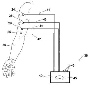
FIG. 4 shows an apparatus 38 of the invention attached to a
subject's arm 39. Electrodes 24, 25 are applied at spaced positions on
the subject's skin. A module 40 is in connection with the electrodes 24,
WO 00/79255 CA 02375249 2001-12-18 PCT/AUOO/00702
12
25 through electrical leads 41, 42. Module 40 includes the power source
which provides alternating current between the electrodes 24, 25 at a set
frequency. The inventors have found that the alternating current is
preferably at a frequency between 5 to 20kHz but, most preferably, at
10kHz.
Monitoring electrodes 28, 29 are applied to the skin. They
are separated from each other but located between electrodes 24, 25 and
connected via electrical leads 43, 44 to a bioeletrical impedance
measuring meter (not shown) in module 40.
A reading of bioelectrical impedance is taken on one limb
and stored in first data storing unit (see FIG. 1).
The electrodes 24, 25 and monitoring electrodes 28, 29 may
then be located in similar positions on the contra-lateral limb and a
reading of bioelectrical impedance taken in a similar manner. A similar
step may be conducted on dissimilar anatomical regions, such as an arm
and a leg or on the same anatomical site at different times. For example,
in the latter case, a regular reading may be taken every month to monitor
changes in an anatomical region.
Module 40 further includes a processor programmed to
divide the lesser of the bioelectrical impedance reading into the greater to
produce a quotient. A correcting factor is then applied to the quotient to
provide an indication of the presence of lymphoedema.
A correcting factor may be estabiished by surveying a
population of clinically unaffected subjects.
If a limb is affected by lymphoedema, its bioelectrical
impedance will decrease due to the presence of extracellular fluid.
Therefore, the variation between the impedance of the two limbs is such
as to move the quotient of the two measurements outside the expected
range for an unaffected population.
As shown in FIG. 4 the result may be displayed in display
window 45 and by illumination of one of the three LEDs 46 which
WO 00/79255 CA 02375249 2001-12-18 PCT/AUOO/00702
13
individually represent indications of "unaffected", "possibly affected" and
"positively affected".
As shown in FIG. 5, it is within the scope of the invention to
include a two channel bioimpedance meter with duplication of peripheral
accessories so that measurements of both sides of a subject can occur
simultaneously. In this case, current is simultaneously passed between
electrodes 24, 25 on one arm 47 and electrodes 24A, 25A on the opposite
arm 48. Monitoring electrodes 28, 29 on the first arm 47 measure
bioelectrical impedance while monitoring electrodes 28A, 29A measure.
bioelectrical impedance on the opposite arm 48. A measuring meter 30
has two channels for simultaneously monitoring signals provided from the
monitoring electrodes 28, 29, 28A, 29A. The signals are passed through
an analogue/digital converter 33 and then analysed by processor 35. The
results are stored in storing unit 36 and shown on display 37. '
FIG. 6 is correlation of results obtained from the present
invention when compared to volume assessed from circumferential
changes of a limb. The X axis 49 represents volume increase in a limb
during a one year period. The Y axis 50 is an indicator of lymph
accumulation as measured by the apparatus of the invention. The line 51
is the line of identity and the broken line 52 is the line of best fit. The
correlation between the two methods is high at 0.75. Measurement of
circumference is currently the most commonly used method of assessing
lymphoedema.
The discussion has referred, to both oedema and
lymphoedema, as it is clear to a skilled addressee that the above method
and apparatus may be utilised on any form of tissue oedema. However, it
is also likely that the predominant use of the method and apparatus will
be directed mainly to lymphoedema due to its clinical relevance.
However, this may change in a specific situation or with time. The
method may also be used in comparing a reading from one anatomical
region with a separate unpaired region. For example, a reading taken on
WO 00/79255 CA 02375249 2001-12-18 PCT/AUOO/00702
14
central localised oedema (eg. ascites) may be referenced against a
nonoedematous structure such as a limb.
Throughout the specification, the aim has been to describe
the preferred embodiments of the invention without limiting the invention
to any one embodiment or specific collection of features. Various
changes and modifications may be made to the embodiments described
and illustrated without departing from the present invention.