Note: Descriptions are shown in the official language in which they were submitted.
CA 02375606 2001-11-23
WO 01/07653 PCT/US00/19502
SPATIALLY ENCODED ANALYTE DETECTION
CROSS-REFERENCE TO RELATED APPLICATIONS
This application claims priority to USSN 09/358,204, filed on July 21, 1999
which is incorporated herein by reference in its entirety for all purposes.
STATEMENT AS TO RIGHTS TO INVENTIONS MADE UNDER FEDERALLY
SPONSORED RESEARCH AND DEVELOPMENT
This work was supported by the National Institutes of Health (GM44112-
OlAl) and the UC BioSTAR project. The Government of the United States of
America
may have certain rights in this invention.
FIELD OF THE INVENTION
This invention relates to the field of diagnostics. In particular this
invention
provides devices and methods that allow rapid detection and/or quantitation of
multiple
1 S analytes and yet does not require the use of labels or labeling steps.
BACKGROUND OF THE INVENTION
Immunoassays and nucleic acid hybridization chemistries are rapidly being
developed towards the goal of detecting genetic defects, performing disease
diagnostics,
and performing prognostic evaluations (Sosnowski et al. (1997) Proc. Natl.
Acacl. Sci.
USA, 94: 1119-1123). Antibodies, nucleic acid binding proteins, receptor
ligands, and
nucleic acids are known to bind very specifically and with high efficiency to
their
congnate "binding partner" under suitable conditions. This phenomenon is
frequently used
for the recognition and diagnosis of disease-causing organisms (e.g., HIV),
pathological
conditions (e.g. cancer, liver disease, kidney disease, degenerative joint
disease, etc.),
substance abuse (e.g. detection of products such as cotinine, etc.), and the
like.
Numerous disease markers, and pathogen markers (e.g. proteins and/or
nucleic acids) are well known and have been thoroughly characterized. Thus,
binding
partners (e.g. nucleic acids, antibodies, and the like), that specifically
bind such markers
can be synthesized and/or isolated and used as markers for recognition of the
disease state,
or disease-causing agent (Landegren et al. (1988) Science, 242: 229, Mikkelson
(1996)
-1-
CA 02375606 2001-11-23
WO 01/07653 PCT/US00/19502
Electroanalysis, 8: 15-19). Various assays are earned out routinely in
microbiology
laboratories or pathology laboratories using such methodologies.
Nucleic acid hybridization, antibody binding reactions, protein binding
reactions, and lectin binding reactions are generally detected through the use
of labels that
either intercalate into the molecule (e.g. into the double helix of a DNA) or
are covalently
attached to either the target or the probe molecule (see, e.g., Sosnowski et
al. (1997) Proc.
Natl. Acad. Sci. USA, 94: 1119-1123, LePecq and Paoletti (1966) Anal.
Biochem., 17: 100-
107, Kapuscinski and Skoczylas (1977) Anal. Biochem., 83: 252-257). In some
cases,
electrogenerated chemiluminescence has also been utilized to detect an
intercalated
electroactive luminescent marker (Pollard-Knight et al. (1990) Anal. Biochem.,
185: 84-89,
Pollard-Knight et a1.(1990) Anal. Biochem., 185: 353-358, Tizard et al. (1990)
Proc. Natl.
Acad. Sci. USA, 12: 4514-4518). All of these detection strategies require the
derivatization
of the target or probe molecule, either before (e.g. for covalent labeling) or
after (e.g. for
intercalation or indirect labeling) the binding reaction between the probe and
target
molecule. This introduces contamination problems. In addition, where multiple
analytes
are analyzed simultaneously multiple labels must be employed. In addition,
tedious
sample handling is required which further enhances the risk of contamination
and/or leads
to false analysis. These and other problems are overcome by the present
invention.
SUMMARY OF THE INVENTION
This invention provides novel devices and methods for detecting and/or
quantifying a plurality of analytes in a sample. This invention provides a
flow-through
microfluidic (e.g., capillary) biosensor for detecting different target
analytes (e.g. nucleic
acids) in a sample after binding to their cognate "binding partners" (e.g.
nucleic acids,
antibodies, lectins, etc.). In general, binding partner "probes", specific to
various analytes
are immobilized in different sections of a capillary channel, e.g., using
photolabile
biotin/avidin technology. The sample is then flushed through the capillary, so
that the
target analytes are bound to the binding partners (capture agents) immobilized
on the
capillary wall and the rest of the sample is eluted from the capillary.
Finally, the
complexed (bound) analyte is released along the entire length of the channel
and flushed
past a detector. In a preferred embodiment, the desorbed, target-analytes are
detected at a
copper electrode poised downstream using sinusoidal voltammetry (Singhal and
Kuhr
(1997) Anal. Chem., 69: 3552-3557, Singhal et al. (1997) Anal. Chem., 69: 1662-
1668).
-2-
CA 02375606 2001-11-23
WO 01/07653 PCT/US00/19502
The time from the elution of the target analyte(s) to detection is used to
determine the
identity of each analyte. Multiple analytes, of the same species of molecule
(e.g., all
nucleic acids), or of different species (e.g. proteins and nucleic acids), can
be diagnosed by
using a single biosensor in this manner. The sensor is highly specific due to
the use of
specific binding partners, and extremely sensitive due to electrochemical
detection.
Thus, in one embodiment, this invention provides devices for detecting a
two or more analytes in a sample. The devices comprise a channel having
affixed therein a
binding partner for each of the two or more analytes, where the binding
partners for each
of the two or more analytes are located in different regions of the channel
and the channel
has a cross-sectional area small enough such that when analytes are released
from the two
or more binding partners into a fluid flowing through the channel, the
analytes remain
spatially segregated until they reach a detection point along, or at the end
of, the channel
downstream from the binding partners; and a detector that detects the analytes
at the
detection point.
The channel can be any convenient channel, e.g. a capillary tube, a capillary
electrophoresis tube, a channel etched in a surface, a channel formed by
hydrophobic
agents printed onto a surface, etc. The channel can have essentially any
dimensions) as
long as the analytes remain sufficiently segregated to be distinguished when
they reach a
detection region in the channel or at the channel end. Preferred channels have
a cross-
sectional area that provides a Renold's number (Re) of less than about 1.
Preferred
channels have a cross-sectional diameter or width less than or equal to about
500 Vim, more
preferably less than or equal to about 100 Vim, and most preferably less than
or equal to
about 50 pm. In particularly preferred devices the two or more target analytes
comprise at
least three, preferably at least 4, more preferably at least 5, and most
preferably at least 10,
at least 50, at least 100, or at least 500 different analytes (and hence that
many different
binding partners). A wide variety of binding partners are suitable including,
but not
limited to antibodies, binding proteins, and nucleic acids. Similarly many
detectors are
suitable and include spectrophotometers (e.g. absorbance spectrophotometers),
and
electroanalytic detectors (including essentially any amperometric and/or
voltammetric
and/or potentiometric and/or coulometric detectors). Preferred detectors
include
voltammeters, especially a sinusoidal voltammeters.
-3-
CA 02375606 2001-11-23
WO 01/07653 PCT/US00/19502
In another embodiment, this invention provides methods of detecting two or
more target analytes in a sample. The methods involve providing a detection
device as
described herein; ii) passing a fluid comprising a sample through the channel
under
conditions where the target analytes present in the fluid bind to their
respective binding
partners thereby spatially encoding the analytes along the channel; iii)
releasing the
analytes from the binding partners into fluid flow passing along the channel;
and iv)
detecting the analytes at a position along the channel downstream from the
binding
partners. In preferred methods, the analytes are not labeled. In particularly
preferred
embodiments the analytes are not labeled. In particularly preferred devices
the two or
more target analytes comprise at least three, preferably at least 4, more
preferably at least
5, and most preferably at least 10, at least 50, at least 100, or at least 500
different analytes
(and hence that many different binding partners are present in the channels)
comprising
the detection device). In some preferred embodiments, the fluid flow induced
by a
pressure difference and/or by electroosmotic flow. fluid flow. Preferred
"sample" fluids
for the detection of analytes include blood, plasma, serum, urine, oral fluid,
cerebrospinal
fluid, and lymph. Detecting can be by a variety of means including
spectrophotometers
(e.g. absorbance spectrophotometry), and electroanalytic methods (including
essentially
any amperometric and/or voltammetric and/or potentiometric and/or coulometric
method).
Preferred detection methods voltammetry, especially sinusoidal voltammetry. In
particularly preferred methods, the analytes are nucleic acids and the
detecting detects
target analytes at a concentration of less than 1 x 10⁻⁹ M.
DEFINITIONS
The terms "polypeptide", "peptide" and "protein" are used interchangeably
herein to refer to a polymer of amino acid residues. The terms apply to amino
acid
polymers in which one or more amino acid residue is an artificial chemical
analogue of a
corresponding naturally occurnng amino acid, as well as to naturally occurnng
amino acid
polymers.
The term "antibody", as used herein, includes various forms of modified or
altered antibodies, such as an intact immunoglobulin, an Fv fragment
containing only the
light and heavy chain variable regions, an Fv fragment linked by a disulfide
bond
(Brinkmann et al. (1993) Proc. Natl. Acad. Sci. USA, 90: 547-551), an Fab or
(Fab)'2
fragment containing the variable regions and parts of the constant regions, a
single-chain
-4-
CA 02375606 2001-11-23
WO 01/07653 PCT/US00/19502
antibody and the like (Bird et al. (1988) Science 242: 424 426; Huston et al.
(1988) Proc.
Nat. Acad. Sci. USA 85: 5879 5883). The antibody may be of animal (especially
mouse
or rat) or human origin or may be chimeric (Morrison et al. (1984) Proc Nat.
Acad. Sci.
USA 81: 6851-6855) or humanized (Jones et al. (1986) Nature 321: 522-525, and
published UK patent application #8707252).
The terms "binding partner", or "capture agent", or a member of a "binding
pair" refers to molecules that specifically bind other molecules to form a
binding complex
such as antibody-antigen, lectin-carbohydrate, nucleic acid-nucleic acid,
biotin-avidin, etc.
In particularly preferred embodiments, the binding is predominantly mediated
by non-
covalent (e.g. ionic, hydrophobic, etc.) interactions.
The term "specifically binds", as used herein, when referring to a
biomolecule (e.g., protein, nucleic acid, antibody, etc.), refers to a binding
reaction which
is determinative of the presence biomolecule in heterogeneous population of
molecules
(e.g., proteins and other biologics). Thus, under designated conditions (e.g.
immunoassay
conditions in the case of an antibody or stringent hybridization conditions in
the case of a
nucleic acid), the specified ligand or antibody binds to its particular
"target" molecule and
does not bind in a significant amount to other molecules present in the
sample.
The term channel refers to a path that directs fluid flow in a particular
direction. The channel can be formed as a groove or trench having a bottom and
sides, or
as a fully enclosed "tube". In some embodiments, the channel need not even
have "sides".
For example, a hydrophobic polymer can be applied to a flat surface and
thereby confine
and/or direct fluid flow on that surface in a narrow (e.g. hydrophilic)
domain. The channel
preferably includes at least one surface to which a binding partner (capture)
agent can be
affixed.
A "target analyte" is any molecule or molecules that are to be detected
and/or quantified in a sample. Preferred target analytes include biomolecules
such as
nucleic acids, antibodies, proteins, sugars, and the like.
The term "microchannel" is used herein for a channel having dimensions
which provide low Reynolds number operation (Re <_ l, preferably Re <_ 0.1,
more
preferably Re <_ 0.01, and most preferably Re S 0.001). Generally low Reynolds
number
operation, fluid dynamics are dominated by viscous forces rather than inertial
forces.
-5-
CA 02375606 2001-11-23
WO 01/07653 PCT/US00/19502
The term capillary tube refers to a tube of narrow dimension (e.g. typically
providing low Re flow). Open-ended capillary tubes, when contacted with water
will
typically uptake the water by capillary action. Capillary tubes can be
fabricated of a
number of materials including, but not limited to, glass, plastic, quartz,
ceramic, and
various silicates.
A "capillary electrophoresis tube" refers to a "capillary tube" designed for
and/or typically used or intended to be used in a capillary electrophoresis
device.
The terms "nucleic acid" or "oligonucleotide" or grammatical equivalents
herein refer to at least two nucleotides covalently linked together. A nucleic
acid of the
present invention is preferably single-stranded or double stranded and will
generally
contain phosphodiester bonds, although in some cases, as outlined below,
nucleic acid
analogs are included that may have alternate backbones, comprising, for
example,
phosphoramide (Beaucage et al. (1993) Tetrahedron 49(10):1925) and references
therein;
Letsinger (1970) J. Org. Chem. 35:3800; Sprinzl et al. (1977) Eur. J. Biochem.
81: 579;
Letsinger et al. (1986) Nucl. Acids Res. 14: 3487; Sawai et al. (1984) Chem.
Lett. 805,
Letsinger et al. (1988) J. Am. Chem. Soc. 110: 4470; and Pauwels et al. (1986)
Chemica
Scripta 26: 141 9), phosphorothioate (Mag et al. (1991) Nucleic Acids Res.
19:1437; and
U.S. Patent No. 5,644,048), phosphorodithioate (Briu et al. (1989) J. Am.
Chem. Soc. 111
:2321, O-methylphophoroamidite linkages (see Eckstein, Oligonucleotides and
Analogues:
A Practical Approach, Oxford University Press), and peptide nucleic acid
backbones and
linkages (see Egholm (1992) J. Am. Chem. Soc. 114:1895; Meier et al. (1992)
Chem. Int.
Ed. Engl. 31: 1008; Nielsen (1993) Nature, 365: 566; Carlsson et al. (1996)
Nature 380:
207). Other analog nucleic acids include those with positive backbones (Denpcy
et al.
(1995) Proc. Natl. Acad. Sci. USA 92: 6097; non-ionic backbones (U.S. Patent
Nos.
5,386,023, 5,637,684, 5,602,240, 5,216,141 and 4,469,863; Angew. (1991) Chem.
Intl. Ed.
English 30: 423; Letsinger et al. (1988) J. Am. Chem. Soc. 110:4470; Letsinger
et al.
(1994) Nucleoside & Nucleotide 13:1597; Chapters 2 and 3, ASC Symposium Series
580,
"Carbohydrate Modifications in Antisense Research", Ed. Y.S. Sanghui and P.
Dan Cook;
Mesmaeker et al. (1994), Bioorganic & Medicinal Chem. Lett. 4: 395; Jeffs et
al. (1994) J.
Biomolecular NMR 34:17; Tetrahedron Lett. 37:743 (1996)) and non-ribose
backbones,
including those described in U.S. Patent Nos. 5,235,033 and 5,034,506, and
Chapters 6 and
7, ASC Symposium Series 580, Carbohydrate Modifications in Antisense Research,
Ed.
-6-
CA 02375606 2001-11-23
WO 01/07653 PCT/US00/19502
Y.S. Sanghui and P. Dan Cook. Nucleic acids containing one or more carbocyclic
sugars
are also included within the definition of nucleic acids (see Jerkins et al.
(1995), Chem.
Soc. Rev. pp169-176). Several nucleic acid analogs are described in Rawls, C &
E News
June 2, 1997 page 35. These modifications of the ribose-phosphate backbone may
be done
to facilitate the addition of additional moieties such as labels, or to
increase the stability
and half life of such molecules in physiological environments.
The terms "hybridizing specifically to" and "specific hybridization" and
"selectively hybridize to," as used herein refer to the binding, duplexing, or
hybridizing of
a nucleic acid molecule preferentially to a particular nucleotide sequence
under stringent
conditions. The term "stringent conditions" refers to conditions under which a
probe will
hybridize preferentially to its target subsequence, and to a lesser extent to,
or not at all to,
other sequences. Stringent hybridization and stringent hybridization wash
conditions in
the context of nucleic acid hybridization are sequence dependent, and are
different under
different environmental parameters. An extensive guide to the hybridization of
nucleic
acids is found in, e.g., Tijssen (1993) Laboratory Techniques in Biochemistry
and
Molecular Biology--Hybridization with Nucleic Acid Probes part l, chapt 2,
Overview of
principles of hybridization and the strategy of nucleic acid probe assays,
Elsevier, NY
Tijssen ). Generally, highly stringent hybridization and wash conditions are
selected to be
about 5°C lower than the thermal melting point (Tm) for the specific
sequence at a defined
ionic strength and pH. The Tm is the temperature (under defined ionic strength
and pH) at
which 50% of the target sequence hybridizes to a perfectly matched probe. Very
stringent
conditions are selected to be equal to the Tm for a particular probe. An
example of
stringent hybridization conditions for hybridization of complementary nucleic
acids which
have more than 100 complementary residues on an array or on a filter in a
Southern or
northern blot is 42°C using standard hybridization solutions (see,
e.g., Sambrook (1989)
Molecular Cloning. A Laboratory Manual (2nd ed.) Vol. 1-3, Cold Spring Harbor
Laboratory, Cold Spring Harbor Press, NY, and detailed discussion, below),
with the
hybridization being carried out overnight. An example of highly stringent wash
conditions
is 0.15 M NaCI at 72°C for about 15 minutes. An example of stringent
wash conditions is
a 0.2x SSC wash at 65°C for 15 minutes (see, e.g., Sambrook supra.) for
a description of
SSC buffer). Often, a high stringency wash is preceded by a low stringency
wash to
remove background probe signal. An example medium stringency wash for a duplex
of,
_7_
CA 02375606 2001-11-23
WO 01/07653 PCT/US00/19502
e.g., more than 100 nucleotides, is lx SSC at 45°C for 15 minutes. An
example of a low
stringency wash for a duplex of, e.g., more than 100 nucleotides, is 4x to 6x
SSC at 40°C
for 15 minutes.
"Spatial segregation" refers to the differences in the localization of a the
concentration distributions of two or more species of molecule (e.g. analytes)
in a fluid
stream. Where the analytes are spatially segregated (i.e., flow encoded) it is
possible to
detect distinct signals for each analyte of interest even though the type of
signal for all of
the analyte may be identical. Thus, the analyte identity can be determined by
position
along the "flow path" or time of detection, and differences in labels
associated with each
analyte are not required.
Electroanalytic methods refer to methods that exploit the "electrical"
properties (e.g., resistance, conductance, capacitance, impedance, etc.) of a
system or
analyte to extract information regarding that system. Electroanalytic methods
include,
essentially any amperometric and/or voltammetric and/or potentiometric and/or
coulometric method. Preferred electroanalytic methods include cyclic
voltammetry, ac, dc,
or rotating ring-disc voltammetry, sinusoidal voltammetry, impedance
spectroscopy, and
the like.
The terms "cyclic voltammetry" or "time-varying voltammetry" are used
interchangeably to refer to cyclic voltammetry. The term "sinusoidal
voltammetry" is used
to refer generally to cyclic voltammetry (e.g. with any time-varying voltage
including, but
not limited to a square wave, a triangle wave, etc.), or to the use of a large
amplitude
sinusoidal potential waveform which is used in analogous fashion to cyclic
voltammetry,
e.g., as described in U.S. Patent, 5,650,061.
BRIEF DESCRIPTION OF THE DRAWINGS
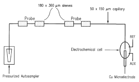
Figure 1 shows a schematic of a capillary based DNA-biosensor with
electrochemical detection. Two different probe sections are present in the
capillary; probe
1, a TB-specific probe, and probe 2, an HIV-specific probe. An HPCE
autosampler is used
for various stringent washes and rinses required for specific-hybridization of
complementary DNA-targets to these immobilized probes. A copper electrode is
positioned at outlet of the capillary biosensor by using a machined two-part
system.
Figure 2 shows protocols for performing stringent hybridization and alkali
denaturation of DNA-targets inside the capillary biosensor. (1) Hybridize the
various
_g_
CA 02375606 2001-11-23
WO 01/07653 PCT/US00/19502
DNA-targets to the probes immobilized on the capillary surface. (2) Stringent
washes are
then performed to remove any non-specific adsorbed or hybridized DNA. (3)
Finally,
alkali denaturation is performed to elute the previously hybridized DNA-
targets from the
capillary biosensor.
Figure 3 provides a schematic of the elution of alkali denatured DNA-
targets from a capillary biosensor, and consequent electrochemical detection.
The
electrode is fabricated inside a capillary piece with the same diameter as the
biosensor
capillary to facilitate auto-alignment. The electrode lies very close (< 5 pm)
to the outlet
of the biosensor capillary. The lower trace shows a schematic of the detection
of the
DNA-targets as they elute from the biosensor capillary.
Figure 4 illustrates detection of HIV-specific target using capillary
biosensor and sinusoidal voltammetric detection. 10 p.g/ml HIV-specific target
flushed
inside a capillary biosensor with HIV-specific probe immobilized only. All
hybridization
conditions as described herein. The sinusoidal voltammetric excitation
waveform was 2
Hz, at 0-700 mVp-p. The signal shown was obtained at the 5'h harmonic.
Figure 5 shows the detection of multiple DNA-targets simultaneously using
flow-encoded hybridization assay. Sample used contained a 1:1 mixture of HN,
and TB-
specific targets, each at a concentration of 10 ~g/ml. All hybridization and
elution
conditions are the same as in Ffigure 4 and as described in Example 1. The
signal shown
was obtained at the 5'h harmonic, as it was found to have the best sensitivity
for detection.
Figure 6 shows the background subtracted frequency spectrum for Arginine
at a copper microelectrode. The three dimensional graph consists of the
frequency (x-
axis), magnitude (z-axis) and phase angle (y-axis) information out to the 10'h
harmonic.
Figure 7 shows the sinusoidal time domain response from 1 p,M arginine at
the fifth harmonic (10 Hz).
Figure 8 demonstrates the linear dynamic range of various arginine
concentrations.
Figure 9 shows the subtracted frequency spectra for asparagine and
glutamine at a copper microelectrode. The squares represent 10~M asparagines,
while the
circles represent 10~M glutamine.
_9_
CA 02375606 2001-11-23
WO 01/07653 PCT/US00/19502
Figures 10A and lOB show the sinusoidal time domain response of
asparagine and glutamine at the sixth harmonic (12 Hz). Figure 10A shows IOpM
asparagines, while Figure lOB shows l OpM glutamine.
Figure 11 shows the background subtracted frequency domain spectrum for
10 pM Insulin B-chain.
Figure 12 shows the sinusoidal time domain component of insulin B-chain
at the fourth harmonic (8 Hz).
Figure 13 shows the subtracted frequency spectra for Luteinizing Hormone-
Releasing Hormone (circles) and Bradykinin (squares) at a copper
microelectrode.
Figures 14A and 14B show the time domain response of Bradykinin and
Luteinizing Hormone- Releasing Hormone at the second harmonic (4 Hz),
respectively.
Figure 15 shows the background subtracted frequency domain response for
Neurotensin (squares) and Substance P (circles), respectively.
Figure 16A and 16B show the time domain response of Neurotensin and
Substance P, respectively, at the first harmonic (2 Hz).
DETAILED DESCRIPTION
I. Methods for efficient detection of multiple analytes.
This invention provides novel methods and instruments for the rapid
detection and/or quantification of multiple analytes in a sample. In one
preferred
embodiment, this invention comprises a channel having attached therein binding
partners)
specific for the analytes that it is desired to detect. Different binding
partners are located
in different regions of the channel so that when the analyte(s) are bound they
are "spatially
encoded" by their location along the channel. The bound analytes are
subsequently
released from the binding partner, or the binding partner/analyte complex is
released from
the wall of the channel, into a fluid flowing through the channel. The channel
dimensions
are such that the analytes remain spatially segregated until they reach a
detection point in
the channel downstream from said binding partners.
When the analytes or analyte/binding partner complexes are released into
the flow, they are spatially encoded; their position in the stream relative to
each other
being dependent on the position of the binding partners when they were affixed
to the
channel wall(s). The time difference between the release and the detection can
therefore
-10-
CA 02375606 2001-11-23
WO 01/07653 PCT/US00/19502
be used to specifically identify the particular analyte generating (or not
generating) an
output signal.
Because the analytes can each be specifically identified without the use of a
label to distinguish them from other analytes numerous and tedious sample-
handling and
labeling steps are eliminated. This eliminates multiple labeling and
contamination
problems. Also, the risk of sample contamination, that could lead to an
elevated incidence
of false positives is reduced or eliminated.
It is noted that the channels can be prepared well before use and different
microfluidic structures (e.g. channels) can be swapped into and out of the
device that
provides sample handling, fluid flow and analyte detection. Different channels
can be
provided for different collections of analytes and multiple channels, either
the same or
different, can be run simultaneously.
The methods and devices of this invention are therefore well suited to the
detection of analytes in a clinical setting. The ability to detect
underivatized analytes (e.g.
DNA, mIRNA, etc.) greatly simplifies the procedure and helps in preventing
sample
contamination and false identification problems.
In one particularly preferred embodiment, the use of copper electrodes with
cyclic (e.g. sinusoidal) voltammetry overcomes many of the problems
encountered by
traditional electrochemical measurements, and thereby allows the detection of
the analyte.
The sensitivity of the detection strategy is due to the effective decoupling
of the faradic
signal from the capacitive background currents in the frequency domain. Thus,
for
example, ssDNA and dsDNA can be detected in the picomolar concentration range,
and
the electrochemical signal is due to the oxidation of easily accessible sugars
on the outer
perimeter of the DNA double helix compared to a ssDNA of the same size.
A sensor that can detect multiple targets by using only one detector
provides a cheaper and more compact detection system that is also easier to
fabricate.
II. System Components.
A) Channel.
1) Channel types and dimensions.
Virtually any type of channel is suitable for the practice of the present
invention so long as the channel allows the passage of materials through it
without
-11-
CA 02375606 2001-11-23
WO 01/07653 PCT/US00/19502
substantial mixing between components in a solution at different positions
along the
channel. In other words, in a preferred capillary, analytes (or other
detectable reagents)
initially released at distinct locations along the channel remain spatially
segregated at a
detection point "downstream" from the initial release point. Spatial
segregation refers to
the ability to detect distinct signals for each analyte of interest even
though the type of
signal for all of the analyte may be identical. Thus, the analyte identity can
be determined
by position along the "flow path" or time of detection, and differences in
labels associated
with each analyte are not required.
Spatial segregation, however, does not require complete segregation of the
analytes away from each other. To the contrary, there can exist significant
overlap and
peak concentrations can be detected and, associated concentration profiles and
be
measured and/or calculated to provide positive/negative detection and/or full
analyte
quantification.
Particularly preferred channels for use in this invention are "
microchannels". The term microchannel is used herein for a channel having
dimensions
that provide low Reynolds number operation, i.e., for which fluid dynamics are
dominated
by viscous forces rather than inertial forces. Reynolds number, sometimes
referred to the
ratio of inertial forces to viscous forces is given as:
Re = pdz/rh + pud/rl
where a is the velocity vector, p is the fluid density, r1 is the viscosity of
the fluid, d is the
characteristic dimension of the channel, and T is the time scale over which
the velocity is
changing (where u/i = 8u/dt). The term "characteristic dimension" is used
herein for the
dimension that determines Reynolds number, as is known in the art. For a
cylindrical
channel it is the diameter. For a rectangular channel, it depends primarily on
the smaller of
the width and depth. For a V-shaped channel it depends on the width of the top
of the "V",
and so forth. Calculation of Re for channels of various morphologies can be
found in
standard texts on fluid mechanics (e.g. Granger (1995) Fluid Mechanics, Dover,
N.Y.;
Meyer (1982) Introduction to Mathematical Fluid Dynamics, Dover, N.Y.).
Fluid flow behavior in the steady state (i -~ infinity)is characterized by the
Reynolds number, Re = pud/r) . Because of the small sizes and slow velocities,
microfabricated fluid systems are often in the low Reynolds number regime (Re
less than
-12-
CA 02375606 2001-11-23
WO 01/07653 PCT/US00/19502
about 1). In this regime, inertial effects, that cause turbulence and
secondary flows, and
therefore mixing within the flow, are negligible and viscous effects dominate
the
dynamics. Under these conditions, flow through the channel is generally
laminar.
Since the Reynolds number depends not only on channel dimension, but on
fluid density, fluid viscosity, fluid velocity and the timescale on which the
velocity is
changing, the absolute upper limit to the channel diameter is not sharply
defined. In fact,
with well designed channel geometries, turbulence can be avoided for R < 100
and
possibly for R < 1000, so that high throughput systems with relatively large
channel sizes
are possible. The preferred channel characteristic dimension range is between
about 0.5
pm and 100 mm. Particularly preferred channel range from a characteristic
dimension of
about 1 pm to about 100 ~,m, most preferably from about 5 p,m to about 100 pm.
A more
preferred range is between about 5 p.m and SO pm.
The devices of this invention need not be confined to low Reynolds number
operation. Where the binding probes are widely separated and hence the
released analytes
1 S are widely separated in the flow considerable connective mixing can occur
in the channel
without the different analytes "overlapping" and masking each other's signal.
In addition,
it will be appreciated that considerable mixing of the two analytes can occur
and as long as
there is a significant (e.g., statistically significant) spatial separation
between the peak
concentrations of the two analytes, the signals will be distinguishable and
detection of each
analyte can be effected. However, as analytes co-mix, quantification of each
individual
analyte may become progressively more difficult. Nevertheless, even in this
context
quantification can be obtained by estimating or modeling the spatial
distribution of the
analyte based on the location of the concentration peaks) and the rate of fall-
off to provide
an approximation of the integrated signal for each analyte.
As indicated above, any channel configuration is suitable so long as the
mixing requirements described above are met. Thus, appropriate channels
include, but are
not limited, to channels formed by opposed barriers, open-topped grooves,
closed
channels, and the like. The channels can have virtually any cross-section,
e.g. circular,
square, rectangular, triangular v-shaped, u-shaped, hexagonal, octagonal,
irregular, and so
forth. The channels) used in this invention also need not be continuous. Thus,
for
example, channels can be formed by an aggregation of porous particles, by
mixed or cross-
linked polymers, and so forth.
-13-
CA 02375606 2001-11-23
WO 01/07653 PCT/US00/19502
Any channel material is suitable for practice of this invention so long as the
material is essentially stable to the solutions passed through it. Preferred
materials are
capable of binding, or being derivatized to bind, to the binding partner or a
linker to the
binding partner. In addition, in a preferred embodiment, the material is
selected and/or
modified so that it does not substantially bind to the analyte. Preferred
materials also do
not bind, or otherwise interact with the probes in regions other than where it
is desired to
affix the probes.
Particularly preferred materials include, but are not limited to glass,
silicon,
quartz or other minerals, plastic(s), ceramics, metals, paper, metalloids,
semiconductive
materials, cements, and the like. In addition, substances that form gels, such
as proteins
(e.g., gelatins), lipopolysaccharides, silicates, agarose and polyacrylamides
can be used. A
wide variety of organic and inorganic polymers, both natural and synthetic may
be
employed as the material for the solid surface. Illustrative polymers include
polyethylene,
polypropylene, poly(4-methylbutene), polystyrene, polymethacrylate,
polyethylene
terephthalate), rayon, nylon, polyvinyl butyrate), polyvinylidene difluoride
(PVDF),
silicones, polyformaldehyde, cellulose, cellulose acetate, nitrocellulose, and
the like.
In the case of conductive or semiconductive substrates ,there is preferably
an insulating layer on the substrate. This is particularly important where the
device
incorporates electrical elements (e.g. electrical fluid direction systems,
sensor, and the like
or uses electroosmotic forces to move materials about the system). In the case
of
polymeric substrates, the substrate materials may be rigid, semi-rigid, or non-
rigid,
opaque, semi-opaque, or transparent depending upon the use for which they are
intended.
For example, devices that include an optical or visual detection element are
generally
fabricated, at lease in part, from transparent materials to allow or at least
facilitate that
detection. Alternatively, transparent windows of e.g. glass or quartz can be
incorporated
into the device for these types of detection elements. Additionally , the
polymeric
materials may have linear or branched backbones and may be crosslinked or
noncrosslinked. Example of particularly preferred polymeric materials include
e.g.
polydimethylsiloxanes (PDMS), polyurethane, polyvinylchloride (VPC),
polystyrene,
polysulfone, polycarbonate, and the like.
The channel can be a component of a larger object. Thus, the channel can
be assembled with one or more other channels to provide a multiplicity of
channels
-14-
CA 02375606 2001-11-23
WO 01/07653 PCT/US00/19502
whereby a number of different assays can be run simultaneously. The channel
can be a
component of an instrument that includes appropriate liquid handling, and/or
detection,
and/or sample processing/application functions.
The channels) can also be fabricated as a as a reusable or disposable unit
that can be conveniently "plugged" into an instrument for running the assays
of this
invention. It will be appreciated that the channels) can be provided on any
one or more of
a wide variety of objects including, but not limited to a microtiter dish
(e.g., PVC,
polypropylene, or polystyrene), a test tube (glass or plastic), a dipstick
(e.g. glass, PVC,
polypropylene, polystyrene, latex, and the like), a microcentrifuge tube, or a
glass, silica,
plastic, metallic or polymer bead.
In particularly preferred embodiments, one or more channels are provided
as a capillary tube (e.g. a capillary electrophoresis tube), on a glass or
silicon slide, as a
capillary charmel, or fabricated as an element of an "integrated circuit"
having on board
circuit elements for control of liquid flow, sample application, and/or signal
detection. In a
most preferred embodiment, the channel is provided as a capillary tube, e.g. a
capillary
electrophoresis tube as illustrated herein in the Examples.
2) Channel fabrication.
Methods of fabricating the channels of this invention are well known to
those of skill in the art. For example, where the channel is formed of one or
more capillary
tubes, the capillaries can be purchased from commercial vendors (e.g.
Polymicron
Technologies, Tucson, Az) or pulled or extruded by conventional capillary
"pulling"
machines.
Where the channels are fabricated on a surface, they can be formed using
standard techniques, e.g. they can be machined, molded, carved, etched,
laminated,
extruded, or deposited, etc.
In one preferred embodiment, the channels) are fabricated using
micromachining processes (e.g. photolithography) well known in the solid state
electronics
industry. Commonly, microdevices, e.g. microchannels, are constructed from
semiconductor material substrates such as crystalline silicon, widely
available in the form
of a semiconductor wafer used to produce integrated circuits, or from glass.
Because of
the commonality of material(s), fabrication of microdevices from a
semiconductor wafer
substrate can take advantage of the extensive experience in both surface and
bulk etching
-15-
CA 02375606 2001-11-23
WO 01/07653 PCT/US00/19502
techniques developed by the semiconductor processing industry for integrated
circuit (IC)
production.
Surface etching, used in IC production for defining thin surface patterns in a
semiconductor wafer, can be modified to allow for sacrificial undercut etching
of thin
layers of semiconductor materials to create movable elements. Bulk etching,
typically
used in IC production when deep trenches are formed in a wafer using
anisotropic etch
processes, can be used to precisely machine edges or trenches in microdevices.
Both
surface and bulk etching of wafers can proceed with "wet processing", using
chemicals
such as potassium hydroxide in solution to remove non-masked material from a
wafer. For
microdevice construction, it is even possible to employ anisotropic wet
processing
techniques that rely on differential crystallographic orientations of
materials, or the use of
electrochemical etch stops, to define various channel elements.
Another etch processing technique that allows great microdevice design
freedom is commonly known as "dry etch processing". This processing technique
is
particularly suitable for anistropic etching of fine structures. Dry etch
processing
encompasses many gas or plasma phase etching techniques ranging from highly
anisotropic sputtering processes that bombard a wafer with high energy atoms
or ions to
displace wafer atoms into vapor phase (e.g. ion beam milling), to somewhat
isotropic low
energy plasma techniques that direct a plasma stream containing chemically
reactive ions
against a wafer to induce formation of volatile reaction products.
Intermediate between high energy sputtering techniques and low energy
plasma techniques is a particularly useful dry etch process known as reactive
ion etching.
Reactive ion etching involves directing an ion containing plasma stream
against a
semiconductor, or other, wafer for simultaneous sputtering and plasma etching.
Reactive
ion etching retains some of the advantages of anisotropy associated with
sputtering, while
still providing reactive plasma ions for formation of vapor phase reaction
products in
response to contacting the reactive plasma ions with the wafer. In practice,
the rate of
wafer material removal is greatly enhanced relative to either sputtering
techniques or low
energy plasma techniques taken alone. Reactive ion etching therefore has the
potential to
be a superior etching process for construction of microdevices, with
relatively high
anistropic etching rates being sustainable. The micromachining techniques
described
above, as well as many others, are well known to those of skill in the art
(see, e.g.,
-16-
CA 02375606 2001-11-23
WO 01/07653 PCT/US00/19502
Choudhury (1997) The Handbook of Microlithography, Micromachining, and
Microfabrication, Soc. Photo-Optical Instru. Engineer, Bard & Faulkner (1997)
Fundamentals of Microfabrication). In addition, examples of the use
of.micromachining
techniques on silicon or borosilicate glass chips can be found in U.S. Patents
5,194,133,
5,132,012, 4,908,112, and 4,891,120.
In one embodiment, the channel is micromachined in a silicon (100) wafer
using standard photolithography techniques to pattern the channels and
connection ports.
Ethylene-diamine, pyrocatechol (EDP) is used for a two-step etch and a Pyrex
7740
coverplate can be anodically bonded to the face of the silicon to provide a
closed liquid
system. In this instance, liquid connections can be made on the backside of
the silicon.
As indicated above, in a preferred embodiment, the channel is fabricated
from a glass, quartz, or other capillary tube, such as a capillary
electrophoresis tube.
In other embodiments, the channel can be built up by depositing material on
a substrate to form channel walls (e.g. using sputtering or other deposition
technology) or
the channel can be cast/molded in a material. Cast/molded channels are easily
fabricated
from a wide variety of materials including but not limited to various metals,
plastics, or
glasses. In certain preferred embodiments, the channels) are cast in various
elastomers.
(e.g. alkylated chlorosulfonated polyethylene (Acsium~), polyolefin elastomers
(e.g.
Engage~), chlorosulfonated polyethylene (e.g. Hypalon~), perfluoroelastomer
(e.g.,
Kalrez~), neoprene-polychloroprene, ethylene-propylene-dime terpolymers
(EPDM),
chlorinated polyethylene (e.g. Tyrin~), various siloxane polymers (e.g.
polydimethylsiloxane), etc).
B) Binding Partners.
In a preferred embodiment, the channels) utilized in this invention bear,
affixed to one or more surfaces one or more biological "binding partner(s)." A
biological
"binding partner" or a member of a "binding pair" refers to a molecule or
composition that
specifically binds other molecules to form a binding complex such as antibody-
antigen,
lectin-carbohydrate, nucleic acid-nucleic acid, biotin-avidin, etc.
The term "specifically binds", as used herein, when refernng to a
biomolecule (e.g., protein, nucleic acid, antibody, etc.), refers to a binding
reaction which
is determinative of the presence biomolecule heterogeneous population of
proteins and
other biologics. Thus, under designated conditions (e.g. immunoassay
conditions in the
-17-
CA 02375606 2001-11-23
WO 01/07653 PCT/US00/19502
case of an antibody, or stringent hybridization conditions in the case of a
nucleic acid), the
specified ligand or antibody binds to its particular "target" (e.g. a protein
or nucleic acid)
and does not bind in a significant amount to other molecules.
The binding partners) used in this invention are selected based upon the
targets that are to be identified/quantified. Thus, for example, where the
target is a nucleic
acid the binding partner is preferably a nucleic acid or a nucleic acid
binding protein.
Where the target is a protein, the binding partner is preferably a receptor, a
ligand, or an
antibody that specifically binds that protein. Where the target is a sugar or
glycoprotein,
the binding partner is preferably a lectin, and so forth.
Suitable binding partners (capture agents) include, but are not limited to
nucleic acids, proteins, receptor binding proteins, nucleic acid binding
proteins, lectins,
sugars, glycoproteins, antibodies, lipids, and the like. Methods of
synthesizing or isolating
such binding partners are well known to those of skill in the art.
1) Preparation of binding partners (capture agents).
a) Nucleic acids
Nucleic acids for use as binding partners in this invention can be produced
or isolated according to any of a number of methods well known to those of
skill in the art.
In one embodiment, the nucleic acid can be an isolated naturally occurnng
nucleic acid
(e.g., genomic DNA, cDNA, mRNA, etc.). Methods of isolating naturally occurnng
nucleic acids are well known to those of skill in the art (see, e.g., Sambrook
et al. (1989)
Molecular Cloning -A Laboratory Manual (2nd Ed.), Vol. 1-3, Cold Spring Harbor
Laboratory, Cold Spring Harbor, New York).
However, in a preferred embodiment, the nucleic acid is created de novo,
e.g. through chemical synthesis. In a preferred embodiment, nucleic acids
(e.g.,
oligonucleotides) are chemically synthesized according to the solid phase
phosphoramidite
triester method described by Beaucage and Caruthers ( 1981 ), Tetrahedron
Letts., 22(20):
1859-1862, e.g., using an automated synthesizer, as described in Needham-
VanDevanter et
al. (1984) Nucleic Acids Res., 12: 6159-6168. Purification of
oligonucleotides, where
necessary, is typically performed by either native acrylamide gel
electrophoresis or by
anion-exchange HPLC as described in Pearson and Regnier (1983) J. Chrom. 255:
137-
149. The sequence of the synthetic oligonucleotides can be verified using the
chemical
-18-
CA 02375606 2001-11-23
WO 01/07653 PCT/US00/19502
degradation method of Maxam and Gilbert (1980) in Grossman and Moldave (eds.)
Academic Press, New York, Meth. Enzymol. 65: 499-560.
b) Antibodies/antibody fragments.
Antibodies or antibody fragments for use as binding partners (capture
agents) can be produces by a number of methods well known to those of skill in
the art
(see, e.g., Harlow & Lane (1988) Antibodies: A Laboratory Manual, Cold Spring
Harbor
Laboratory, and Asai (1993) Methods in Cell Biology Yol. 37: Antibodies in
Cell Biology,
Academic Press, Inc. N.Y.). In one approach, the antibodies are produced by
immunizing
an animal (e.g. a rabbit) with an immunogen containing the epitope it is
desired to
recognize/capture. A number of immunogens may be used to produce specifically
reactive
antibodies. Recombinant protein is the preferred immunogen for the production
of
monoclonal or polyclonal antibodies. Naturally occurring protein may also be
used either
in pure or impure form. Synthetic peptides made as well using standard peptide
synthesis
chemistry (see, e.g., Barany and Merrifield, Solid-Phase Peptide Synthesis;
pp. 3-284 in
The Peptides: Analysis, Synthesis, Biology. Vol. 2: Special Methods in Peptide
Synthesis,
Part A., Merrifield et al. (1963) J. Am. Chem. Soc., 85: 2149-2156, and
Stewart et al.
(1984) Solid Phase Peptide Synthesis, 2nd ed. Pierce Chem. Co., Rockford,
Ill.)
Methods of production of polyclonal antibodies are known to those of skill
in the art. In brief, an immunogen, preferably a purified cytoskeletal
component, is mixed
with an adjuvant and animals are immunized. The animal's immune response to
the
immunogen preparation is monitored by taking test bleeds and determining the
titer of
reactivity to the cytoskeletal components and test compositions. When
appropriately high
titers of antibody to the immunogen are obtained, blood is collected from the
animal and
antisera are prepared. Further fractionation of the antisera to enrich for
antibodies reactive
to the cytoskeletal component can be done if desired. (See Harlow and Lane,
supra).
Monoclonal antibodies may be obtained by various techniques familiar to
those skilled in the art. Briefly, spleen cells from an animal immunized with
a desired
antigen are immortalized, commonly by fusion with a myeloma cell (See, Kohler
and
Milstein (1976) Eur. J. Immunol. 6: 511-519). Alternative methods of
immortalization
include transformation with Epstein Barr Virus, oncogenes, or retroviruses, or
other
methods well known in the art. Colonies arising from single immortalized cells
are
screened for production of antibodies of the desired specificity and affinity
for the antigen,
-19-
CA 02375606 2001-11-23
WO 01/07653 PCT/US00/19502
and yield of the monoclonal antibodies produced by such cells may be enhanced
by
various techniques, including injection into the peritoneal cavity of a
vertebrate host.
Alternatively, one may isolate DNA sequences which encode a monoclonal
antibody or a
binding fragment thereof by screening a DNA library from human B cells
according to the
general protocol outlined by Huse et al. (1989) Science, 246:1275-1281.
Antibodies fragments, e.g. single chain antibodies (scFv or others), can also
be produced/selected using phage display technology. The ability to express
antibody
fragments on the surface of viruses that infect bacteria (bacteriophage or
phage) makes it
possible to isolate a single binding antibody fragment from a library of
greater than 10'°
nonbinding clones. To express antibody fragments on the surface of phage
(phage
display), an antibody fragment gene is inserted into the gene encoding a phage
surface
protein (pIII) and the antibody fragment-pIII fusion protein is displayed on
the phage
surface (McCafferty et al. (1990) Nature, 348: 552-554; Hoogenboom et al.
(1991)
Nucleic Acids Res. 19: 4133-4137).
Since the antibody fragments on the surface of the phage are functional,
phage bearing antigen binding antibody fragments can be separated from non-
binding
phage by antigen affinity chromatography (McCafferty et al. (1990) Nature,
348: 552-
554). Depending on the affinity of the antibody fragment, enrichment factors
of 20 fold -
1,000,000 fold are obtained for a single round of affinity selection. By
infecting bacteria
with the eluted phage, however, more phage can be grown and subjected to
another round
of selection. In this way, an enrichment of 1000 fold in one round can become
1,000,000
fold in two rounds of selection (McCafferty et al. (1990) Nature, 348: 552-
554). Thus
even when enrichments are low (Marks et al. (1991) J. Mol. Biol. 222: 581-
597), multiple
rounds of affinity selection can lead to the isolation of rare phage. Since
selection of the
phage antibody library on antigen results in enrichment, the majority of
clones bind
antigen after as few as three to four rounds of selection. Thus only a
relatively small
number of clones (several hundred) need to be analyzed for binding to antigen.
Human antibodies can be produced without prior immunization by
displaying very large and diverse V-gene repertoires on phage (Marks et al.
(1991) J. Mol.
Biol. 222: 581-597). In one embodiment natural VH and VL repertoires present
in human
peripheral blood lymphocytes are were isolated from unimmunized donors by PCR.
The
V-gene repertoires were spliced together at random using PCR to create a scFv
gene
-20-
CA 02375606 2001-11-23
WO 01/07653 PCT/US00/19502
repertoire which is was cloned into a phage vector to create a library of 30
million phage
antibodies (Id.). From this single "naive" phage antibody library, binding
antibody
fragments have been isolated against more than 17 different antigens,
including haptens,
polysaccharides and proteins (Marks et al. (1991) J. Mol. Biol. 222: 581-597;
Marks et al.
S (1993). BiolTechnology. 10: 779-783; Griffiths et al. (1993) EMBO J. 12: 725-
734;
Clackson et al. (1991) Nature. 352: 624-628). Antibodies have been produced
against self
proteins, including human thyroglobulin, immunoglobulin, tumor necrosis factor
and CEA
(Griffiths et al. (1993) EMBO J. 12: 725-734). It is also possible to isolate
antibodies
against cell surface antigens by selecting directly on intact cells. The
antibody fragments
are highly specific for the antigen used for selection and have affinities in
the 1 :M to 100
nM range (Marks et al. (1991) J. Mol. Biol. 222: 581-597; Griffiths et al.
(1993) EMBO J.
12: 725-734). Larger phage antibody libraries result in the isolation of more
antibodies of
higher binding affinity to a greater proportion of antigens.
c) Binding proteins.
In one embodiment, the binding partner (capture agent) can be a binding
protein. Suitable binding proteins include, but are not limited to receptors
(e.g. cell surface
receptors), receptor ligands, cytokines, transcription factors and other
nucleic acid binding
proteins, growth factors, etc.
The protein can be isolated from natural sources, mutagenized from isolated
proteins or synthesized de novo. Means of isolating naturally occurring
proteins are well
known to those of skill in the art. Such methods include but are not limited
to well known
protein purification methods including ammonium sulfate precipitation,
affinity columns,
column chromatography, gel electrophoresis and the like (see, generally, R.
Scopes,
(1982) Protein Purification, Springer-Verlag, N.Y.; Deutscher (1990) Methods
in
Enzymology Yol. 182: Guide to Protein Purification, Academic Press, Inc.
N.Y.).
Where the protein binds a target reversibly, affinity columns bearing the
target can be used to affinity purify the protein. Alternatively the protein
can be
recombinantly expressed with a HIS-Tag and purified using Ni2+/NTA
chromatography.
In another embodiment, the protein can be chemically synthesized using
standard chemical peptide synthesis techniques. Where the desired subsequences
are
relatively short the molecule may be synthesized as a single contiguous
polypeptide.
Where larger molecules are desired, subsequences can be synthesized separately
(in one or
-21-
CA 02375606 2001-11-23
WO 01/07653 PCT/US00/19502
more units) and then fused by condensation of the amino terminus of one
molecule with
the carboxyl terminus of the other molecule thereby forming a peptide bond.
This is
typically accomplished using the same chemistry (e.g., Fmoc, Tboc) used to
couple single
amino acids in commercial peptide synthesizers.
Solid phase synthesis in which the C-terminal amino acid of the sequence is
attached to an insoluble support followed by sequential addition of the
remaining amino
acids in the sequence is the preferred method for the chemical synthesis of
the
polypeptides of this invention. Techniques for solid phase synthesis are
described by
Barany and Merrifield (1962) Solid-Phase Peptide Synthesis; pp. 3-284 in The
Peptides:
Analysis, Synthesis, Biology. Vol. 2: Special Methods in Peptide Synthesis,
Part A.,
Merrifield et al. (1963) J. Am. Chem. Soc., 85: 2149-2156, and Stewart et al.
(1984) Solid
Phase Peptide Synthesis, 2nd ed. Pierce Chem. Co., Rockford, Ill.
In a preferred embodiment, the can also be synthesized using recombinant
DNA methodology. Generally this involves creating a DNA sequence that encodes
the
binding protein, placing the DNA in an expression cassette under the control
of a particular
promoter, expressing the protein in a host, isolating the expressed protein
and, if required,
renaturing the protein.
DNA encoding binding proteins or subsequences of this invention can be
prepared by any suitable method as described above, including, for example,
cloning and
restriction of appropriate sequences or direct chemical synthesis by methods
such as the
phosphotriester method of Narang et al. (1979) Meth. Enzymol. 68: 90-99; the
phosphodiester method of Brown et al. (1979) Meth. Enzymol. 68: 109-151; the
diethylphosphoramidite method of Beaucage et al. (1981) Tetra. Lett., 22: 1859-
1862; and
the solid support method of U.S. Patent No. 4,458,066.
The nucleic acid sequences encoding the desired binding proteins) may be
expressed in a variety of host cells, including E. coli, other bacterial
hosts, yeast, and
various higher eukaryotic cells such as the COS, CHO and HeLa cells lines and
myeloma
cell lines. The recombinant protein gene will be operably linked to
appropriate expression
control sequences for each host. For E. coli this includes a promoter such as
the T7, trp, or
lambda promoters, a ribosome binding site and preferably a transcription
termination
signal. For eukaryotic cells, the control sequences will include a promoter
and preferably
-22-
CA 02375606 2001-11-23
WO 01/07653 PCT/US00/19502
an enhancer derived from immunoglobulin genes, SV40, cytomegalovirus, etc.,
and a
polyadenylation sequence, and may include splice donor and acceptor sequences.
The plasmids can be transferred into the chosen host cell by well-known
methods such as calcium chloride transformation for E. coli and calcium
phosphate
treatment or electroporation for mammalian cells. Cells transformed by the
plasmids can
be selected by resistance to antibiotics conferred by genes contained on the
plasmids, such
as the amp, gpt, neo and hyg genes.
Once expressed, the recombinant binding proteins can be purified according
to standard procedures of the art as described above.
d) Sugars and carbohydrates.
Other binding partners include sugars and carbohydrates. Sugars and
carbohydrates can be isolated from natural sources, enzymatically synthesized
or
chemically synthesized. A route to production of specific oligosaccharide
structures is
through the use of the enzymes which make them in vivo; the
glycosyltransferases. Such
enzymes can be used as regio- and stereoselective catalysts for the in vitro
synthesis of
oligosaccharides (Ichikawa et al. (1992) Anal. Biochem. 202: 215-238).
Sialyltransferase
can be used in combination with additional glycosyltransferases. For example,
one can use
a combination of sialyltransferase and galactosyltransferases. A number of
methods of
using glycosyltransferases to synthesize desired oligosaccharide structures
are known.
Exemplary methods are described, for instance, WO 96/32491, Ito et al. (1993)
Pure Appl.
Chem. 65:753, and U.S. Patents 5,352,670, 5,374,541, and 5,545,553. The
enzymes and
substrates can be combined in an initial reaction mixture, or alternatively,
the enzymes and
reagents for a second glycosyltransferase cycle can be added to the reaction
medium once
the first glycosyltransferase cycle has neared completion. By conducting two
glycosyltransferase cycles in sequence in a single vessel, overall yields are
improved over
procedures in which an intermediate species is isolated.
Methods of chemical synthesis are described by Zhang et al. (1999) J. Am.
Chem. Soc., 121 (4): 734-753. Briefly, in this approach, a set of sugar-based
building
blocks is created with each block preloaded with different protecting groups.
The building
blocks are ranked by reactivity of each protecting group. A computer program
then
determines exactly which building blocks must be added to the reaction so that
the
sequences of reactions from fastest to slowest produces the desired compound.
-23-
CA 02375606 2001-11-23
WO 01/07653 PCT/US00/19502
2) Attachment of binding partners to the channel.
Many methods for immobilizing biomolecules to a variety of solid surfaces
are known in the art. The desired component may be covalently bound, or
noncovalently
attached through specific or nonspecific bonding.
If covalent bonding between a compound and the surface is desired, the
surface will usually be polyfunctional or be capable of being
polyfunctionalized.
Functional groups which may be present on the surface and used for linking can
include
carboxylic acids, aldehydes, amino groups, cyano groups, ethylenic groups,
hydroxyl
groups, mercapto groups and the like. The manner of linking a wide variety of
compounds
to various surfaces is well known and is amply illustrated in the literature.
See, for
example, Ichiro Chibata (1978) Immobilized Enzymes, , Halsted Press, New York,
and
Cuatrecasas, (1970) J.-Biol. Chem. 245: 3059.
In addition to covalent bonding, various methods for noncovalently binding
an assay component can be used. Noncovalent binding is typically nonspecific
absorption
1 S of a compound to the surface. Typically, the surface is blocked with a
second compound to
prevent nonspecific binding of labeled assay components. Alternatively, the
surface is
designed such that it nonspecifically binds one component but does not
significantly bind
another. For example, a surface bearing a lectin such as concanavalin A will
bind a
carbohydrate containing compound but not a labeled protein that lacks
glycosylation.
Various solid surfaces for use in noncovalent attachment of assay components
are
reviewed in U.S. Patent Nos. 4,447,576 and 4,254,082.
Where the binding partner is a nucleic acid or a polypeptide, the molecule
can be chemically synthesized in situ. This involves essentially standard
chemical
synthesis methods substituting photo-labile protecting groups for the usual
protecting
groups (e.g. dimethoxy trityl group (DMT) used in nucleic acid synthesis).
Irradiation of
the microchannel at discrete locations results in selective coupling of the
monomer (e.g.
amino acid or nucleotide) to the growing polypeptide(s) or nucleic acids) at
the irradiated
site. Methods of light-directed polymer synthesis are well known to those of
skill in the art
(see, e.g., U.S. Patent No. 5,143,854; PCT Publication Nos. WO 90/15070, WO
92/10092
and WO 93/09668; and Fodor et al. (1991) Science; 251, 767-77).
In preferred embodiments, the binding partner is immobilized by the use of
a linker (e.g. a homo- or heterobifunctional linker). Linkers suitable for
joining biological
-24-
CA 02375606 2001-11-23
WO 01/07653 PCT/US00/19502
binding partners are well known to those of skill in the art. For example, a
protein or
nucleic acid molecule may be linked by any of a variety of linkers including,
but not
limited to a peptide linker, a straight or branched chain carbon chain linker,
or by a
heterocyclic carbon linker. Ileterobifunctional cross linking reagents such as
active esters
of N-ethylmaleimide have been widely used (see, for example, Lerner et al.
(1981) Proc.
Nat. Acad. Sci. USA, 78: 3403-3407 and Kitagawa et al. (1976) J. Biochem., 79:
233-236,
and Birch and Lennox (1995) Chapter 4 in Monoclonal Antibodies: Principles and
Applications, Wiley-Liss, N.Y.).
In one preferred embodiment, the binding partner is immobilized utilizing a
biotin/avidin interaction. In this embodiment, biotin or avidin with a
photolabile
protecting group can be placed in the channel. Irradiation of the channel at a
distinct
location results in coupling of the biotin or avidin to the channel at that
location. Then, the
binding agent bearing a respective biotin or avidin is placed into the channel
whereby it
couples to the respective binding partner and is localized in the irradiated
site. The process
1 S can be repeated at each distinct location it is desired to attach a
binding partner.
Another suitable photochemical binding approach is described by Sigrist et
al. (1992) BiolTechnology, 10: 1026-1028. In this approach, interaction of
ligands with
organic or inorganic surfaces is mediated by photoactivatable polymers with
carbene
generating trifluoromethyl-aryl-diazirines that serve as linker molecules.
Light activation
of aryl-diazirino functions at 350 nm yields highly reactive carbenes and
covalent coupling
is achieved by simultaneous carbene insertion into both the ligand and the
inert surface.
Thus, reactive functional groups are not required on either the ligand or
supporting
material.
In a most preferred embodiment, fused silica capillaries (50 pm i.d. are
coated with a thin layer of epoxy (Epotek 350) in order to cover the fused
silica surface
with an organic coating. The organic coating of the surface not only minimizes
DNA
adsorption on the walls of the capillary, but also provides a polymerized
surface to which
DNA-probes can be immobilized directly. A protocol for coating the capillary
surface
with the epoxy is described by Liu et al. (1996) J. Chromatogr. 723: 157-167.
Briefly, the
capillary was rinsed first with acetone for 15 min., then dried in an oven at
100°C for 1 h
under a nitrogen pressure of 20 psi. Epoxy 314 ND (Epo-Tek, Billerica, MA) was
dynamically coated onto the capillary surface by aspirating a solution of an
epoxy mixture
-25-
CA 02375606 2001-11-23
WO 01/07653 PCT/US00/19502
in acetone. The residual solvent was removed from the epoxy-coated capillaries
by
flushing with nitrogen at room temperature for 30 min. The epoxy coating is
cross-linked
at 80°C for 30 min, then at 150°C for 2 h under a nitrogen
pressure of 20 psi. The coated
capillaries are washed with buffer for 30 min prior to use.
A 1 cm section of the epoxy-coated capillary is then flushed with a specific
DNA-probe solution. The DNA-probe solution is allowed to react with the
capillary piece
overnight to bind the DNA-probe to the capillary walls via hydrophobic and
electrostatic
interactions. Other DNA-probes are attached to similar one cm long pieces of
coated
capillaries in a similar manner. Once the are immobilized onto the capillary
walls, these
hybridization regions are rinsed with deionized water, and are then ready to
assemble into
a capillary biosensor having different binding partners at different
locations.
C) Analyte detection methods.
Virtually any method of biological molecule detection can be used in
accordance with the methods of this invention. Since the identity of the
various analytes is
determined by their spatial position in the flow moving through the channel,
there is not
need for different labeling systems on each analyte. To the contrary, one
advantage of the
present assay system is that there is no need to label the analyte at all.
Methods of detecting analytes are well known to those of skill in the art.
Were the analyte is labeled (e.g. with a radioactive, fluorescent, magnetic,
or mass label),
the analyte is detected by detecting the label. However, in a preferred
embodiment, the
analyte is not labeled and preferred detection methods do not rely on the use
of labels
attached to the analyte. Such detection means include, but are not limited to
detection of
optical signals (e.g. emission and/or absorption spectroscopy), detection of
electrical and
magnetic signals, detection of changes of the electrical properties (e.g.
conductance/resistance, capacitance, impedance, etc.) of the medium containing
the
analyte.
In one simple embodiment, the optical absorption of the fluid containing the
analyte is monitored (e.g. with a standard ultra-violet) detector. However, in
a preferred
embodiment, an electroanalytic detector is utilized. In a most preferred
embodiment, the
electroanalytic detector utilizes time-varying (e.g. sinusoidal) voltammetry.
In a particularly preferred embodiment, sinusoidal voltammetry involves
providing a small amount of the analyte of interest to a voltammetric
electrode. A
-26-
CA 02375606 2001-11-23
WO 01/07653 PCT/US00/19502
sinusoidal (or other time-varying) voltage is applied to the electrode. The
time-varying
(e.g., sinusoidal) voltage has an amplitude large enough to sweep through the
formal
potential of the redox species of interest in a single cycle at a given
frequency. The
response of the analyte to the sinusoidal voltage is selectively detected at a
harmonic of the
fundamental frequency of the time-varying voltage. Methods of performing time-
varying
voltammetry are provided in U.S. Patent 5,650,061 and the references cited
therein.
While a particularly preferred embodiment utilizes sinusoidal voltammetry,
other voltammetric methods are well suited to this invention. As indicated
above, time-
varying voltammetric methods are particularly preferred and such voltammetric
methods
are not limited to the use of sinusoidal time-varying voltages. Other wave
forms are also
suitable. Such methods include, but are not limited to, the use of square
waves and
triangle waves. Such time-varying voltammetric methods are well known to those
of skill
in the art (see, e.g., Cullison and Kuhr (1996) Electroanalysis, 7(1): 1-6).
It was a discovery of this invention that combination of sinusoidal
voltammetry detection with spatially encoded analyte separation provides
highly specific
analyte detection/quantitation at extremely low levels in a complex sample
(e.g. serum).
III. Integrated assay device.
State-of the-art chemical analysis systems for use in chemical production,
environmental analysis, medical diagnostics and basic laboratory analysis are
preferably
capable of complete automation. Such total analysis systems (TAS) (Fillipini
et al. (1991)
J. Biotechnol. 18: 153; Garn et al (1989) Biotechnol. Bioeng. 34: 423;
Tshulena (1988)
Phys. Scr. T23: 293; Edmonds (1985) Trends Anal. Chem. 4: 220; Stinshoff et
al. (1985)
Anal. Chem. 57:1148; Guibault (1983) Anal. Chem Symp. Ser. 17: 637; Widmer
(1983)
Trends Anal. Chem. 2: 8) automatically perform functions ranging from
introduction of
sample into the system, transport of the sample through the system, sample
preparation,
separation, purification and detection, including data acquisition and
evaluation.
Recently, sample preparation technologies have been successfully reduced
to miniaturized formats. Thus, for example, gas chromatography (Widmer et al.
(1984)
Int. J. Environ. Anal. Chem. 18: 1), high pressure liquid chromatography
(Muller et al.
(1991) J. High Resolut. Chromatogr. 14: 174; Manz et al.. (1990) Sensors &
Actuators
B1:249; Novotny et al., eds. (1985) Microcolumn Separations: Columns,
Instrumentation
and Ancillary Techniques J. Chromatogr. Library, Vol. 30; Kucera, ed. (1984)
Micro-
-27-
CA 02375606 2001-11-23
WO 01/07653 PCT/US00/19502
Column High Performance Liquid Chromatography, Elsevier, Amsterdam; Scott, ed.
(1984) Small Bore Liguid Chromatography Columns: Their Properties and Uses,
Wiley,
N.Y.; Jorgenson et al. (1983) J. Chromatogr. 255: 335; Knox et al. (1979) J.
Chromatogr.
186:405; Tsuda et al. (1978) Anal. Chem. 50: 632) and capillary
electrophoresis (Manz et
S al. ( 1992) J. Chromatogr. 593: 253; Olefirowicz et al. ( 1990) Anal. Chem.
62: 1872;
Second Int'l Symp. High-Perf. Capillary Electrophoresis (1990) J. Chromatogr.
516;
Ghowsi et al. (1990) Anal. Chem. 62:2714) have been reduced to miniaturized
formats.
Similarly, in another embodiment, this invention provides an integrated
assay device (e.g., a TAS) for detecting and/or quantifying a multiplicity of
analytes. The
assay device comprises the channels) with attached binding partners as
described above.
In addition, preferred integrated assay devices also include one or more of
the following: a
detection system (e.g. voltammetry system including electrodes and/or
associated
electronics), one or more reservoirs to provide buffers and/or flushing
fluids, sample
application wells) and/or injection port(s), a computer controller (for
control of pumps,
reservoir flow switching, detector, and signal analysis system, and the like.
In a particularly preferred embodiment, the integrated assay device contains
the channels in a "removable" unit. Thus, for example, where the capillaries
can be
provided as channels in a module that can be easily inserted and removed from
the
ancillary equipment thereby readily allowing the device to be run with assay
for different
sets of analytes.
Where the channel used in the device is a tube (e.g. a capillary
electrophoresis tube), a conventional capillary electrophoresis device
contains much of the
ancillary plumbing, sample handling and delivery components, and computer
controllers)
for an "integrated" assay device according to the present invention. Little
more is required
than fairly straightforward introduction/addition of a detector (e.g. a
sinusoidal
voltammetry detector) and associated electronics in accordance with this
invention to
provide an integrated assay device well suited to detection and/or
quantitation of a wide
variety of analytes.
IV. Running assays.
In general, assays are run by introducing the sample into the channel having
affixed binding partners. The sample is preferably held under conditions that
allow the
respective binding partners to specifically bind to the target analytes that
may be present in
-28-
CA 02375606 2001-11-23
WO 01/07653 PCT/US00/19502
the sample. The sample is then flushed out of the channel, typically by
introduction of a
buffer that facilitates release of the bound analyte. The released arialyte is
then detected at
a downstream detection point and the identity of the analyte is determined by
the time
from release to detection.
A) Sample preparation.
Virtually any sample can be analyzed using the devices and methods of this
advantage. However, in a preferred embodiment, the sample is a biological
sample. The
term "biological sample", as used herein, refers to a sample obtained from an
organism or
from components (e.g., cells) of an organism. The sample may be of any
biological tissue
or fluid. Frequently the sample will be a "clinical sample" which is a sample
derived from
a patient. Such samples include, but are not limited to, sputum, cerebrospinal
fluid, blood,
blood fractions (e.g. serum, plasma), blood cells (e.g., white cells), tissue
or fine needle
biopsy samples, urine, peritoneal fluid, and pleural fluid, or cells
therefrom. Biological
samples may also include sections of tissues such as frozen sections taken for
histological
purposes.
Biological samples, (e.g. serum) may be analyzed directly or they may be
subject to some preparation prior to use in the assays of this invention. Such
preparation
can include, but is not limited to, suspension/dilution of the sample in water
or an
appropriate buffer or removal of cellular debris, e.g. by centrifugation, or
selection of
particular fractions of the sample before analysis.
B) Sample delivery into system
The sample can be introduced into the devices of this invention according
to standard methods well known to those of skill in the art. Thus, for
example, the sample
can be introduced into the channel through an injection port such as those
used in high
pressure liquid chromatography systems. In another embodiment the sample can
be
applied to a sample well that communicates to the channel. In still another
embodiment
the sample can be pumped into the channel. Means of introducing samples into
channels
are well known and standard in the capillary electrophoresis and
chromatography arts.
C) Binding conditions
Once in the channel, the sample is held under conditions that promote
specific binding between the sample and the binding partner. Conditions
compatible with
specific binding between a binding partner and an analyte are well known to
those of skill
-29-
CA 02375606 2001-11-23
WO 01/07653 PCT/US00/19502
in the art. For example, buffers suitable for promoting binding between an
antibody and a
target protein are well known in the immunoassay art (see, e.g., U.S. Patents
4,366,241;
4,376,110; 4,517,288; and 4,837,168, Asai (1993) Methods in Cell Biology
Volume 37.~
Antibodies in Cell Biology, Academic Press, Inc. New York; Stites & Terr
(1991) Basic
and Clinical Immunology 7th Edition). Similarly conditions under which a
nucleic acids
specifically hybridize to each other are also well know to those of skill in
the art (see,
Tijssen (1993) supra.). The particular binding conditions are optimized for
particular sets
of binding partners and target analytes according to standard methods well
known to those
of skill in the art (see, e.g., Tijssen (1993) supra., U.S. Patents 4,366,241;
4,376,110;
4,517,288; and 4,837,168, Asai (1993) Methods in Cell Biology Volume 37.~
Antibodies in
Cell Biology, Academic Press, Inc. New York; Stites & Terr (1991) Basic and
Clinical
Immunology 7th Edition).
D) Release conditions.
After the analyte(s) in the sample are specifically bound to the binding
partner attached to the channel, they are released. Release is preferably
accomplished by
contacting the binding partner/analyte complex with a buffer or with
temperature
conditions that disrupt the binding partner/analyte interaction. Depending on
the particular
analyte/binding partner pair such associations can be disrupted by the use of
high
temperature, denaturants (e.g. urea, formamide, etc.), high or low pH, high or
low salt, and
various chaotropic agents (e.g., guanidine HCl).
E) Analyte/flow through the channel.
Samples and/or carrier/buffer fluids can be introduced into and/or moved
through the channel according to standard methods. For example, fluid can be
introduced
and moved through the cannel by a simple gravity feed from a "reservoir".
Alternatively,
fluids can be moved through the channel by gas pressure, or by fluid pressure
produced by
any of a variety of suitable pumps (e.g. peristaltic pumps, metering pumps,
etc.), pressure
on a deformable chamber/diaphragm, etc.. Analytes can also be driven through
the
channel by electroosmotic methods.
F) Detection.
As indicated above, analyte detection can be by any of a number of
methods well known to those of skill in the art as indicated above. In a
preferred
-3 0-
CA 02375606 2001-11-23
WO 01/07653 PCT/US00/19502
embodiment, electrochemical detection methods are utilized and in a most
preferred
embodiment, detection is by sinusoidal voltammetry.
The protocol for performing sinusoidal voltammetry has been described
previously (Singhal et al. (1997) Anal. Chem. 69: 4828-4832; and U.S. Patent
5,650,061).
Briefly, a sine wave at 2 Hz, 0.7 Vp-p, +0.35 V D.C. offset is generated
digitally using a
software program. This sine wave serves as the applied potential for a copper
electrode.
The current response from the electrode is collected by the software in real
time for the
entire length of a single elution run. This time domain current response is
then converted
into the frequency domain with fast Fourier transforms. The protocol for
analyzing
frequency spectra has been explained previously (Singhal et al. (1997) Anal.
Chem. 69:
1662-1668). The spectrum corresponding to the analyte is obtained after
background
subtraction and digital phase locking as described previously (Singhal et al.
(1997) supra.).
V. Kits for multiple analyte detection.
In one embodiment this invention provides kits for screening for identifying
the presence or absence, or quantifying a multiplicity of analytes in a
sample. The kits
include of the channels of this invention bearing affixed to their surfaces
various binding
partners as described herein. The channels can be designed for simple and
rapid
incorporation into an integrated assay device, e.g. a device comprising
electrochemical
detector (e.g. sinusoidal voltammetry) circuitry, appropriate plumbing for
administration
of a sample and maintenance of a fluid flow through the channel, and computer
control
systems) for control of sample application, fluid flow, and analysis of signal
output as
described herein. The kit can additionally include appropriate buffers and
other solutions
and standards for use in the assay methods described herein.
In addition, the kits may include instructional materials containing
directions (i.e., protocols) for the practice of the methods of this
invention. While the
instructional materials typically comprise written or printed materials they
are not limited
to such. Any medium capable of storing such instructions and communicating
them to an
end user is contemplated by this invention. Such media include, but are not
limited to
electronic storage media (e.g., magnetic discs, tapes, cartridges, chips),
optical media (e.g.,
CD ROM), and the like. Such media may include addresses to Internet sites that
provide
such instructional materials.
-31-
CA 02375606 2001-11-23
WO 01/07653 PCT/US00/19502
EXAMPLES
The following examples are offered to illustrate, but not to limit the claimed
W vention.
Example 1
Nanoliter Volume Electrochemical Sensing of DNA-Hybridization
Materials and methods.
Reagents.
The water used was deionized and then passed through a Milli-Q Water
Purification System (Millipore Corp., Bedford, MA). The biotinylated DNA-probe
and the
complementary DNA-target specific to Tuberculosis (TB) and Human
Immunodeficiency
Virus (HIV) identification were custom synthesized through Genemed Synthesis,
Inc., San
Francisco, CA (Table 1). DNA-probe solutions were made by diluting a 100 pg/ml
solution of DNA-probe dissolved in deionized water into a 1:1 mixture with DNA-
binding
solution (Pierce Chemicals, CA). This binding solution facilitates binding DNA
to
polymerized surfaces via hydrophobic and electrostatic interactions. Fused
silica
capillaries (Polymicron Technologies, Inc., AZ) were used for making the
capillary
biosensors. These capillaries were not flushed with acetone and dried before
any
derivatization was done to the capillary surface.
Capillary Derivatization and Immobilization of DNA-probes.
Fused silica capillaries (SO p,m i.d. x 150 ~m o.d., one meter in length)
were used for the biosensor. The capillary was coated with a thin layer of
epoxy (Epotek
350) in order to cover the fused silica surface with an organic coating.
Organic coating of
the surface not only minimizes DNA adsorption on the walls of the capillary,
but also
provides a polymerized surface to which DNA-probes can be immobilized
directly. The
protocol for coating the capillary surface with the epoxy was exactly as
described by Liu et
al. (1996) J. Chromatogr. 723: 157-167. Briefly, the capillary was rinsed
first with
acetone for 15 min., then dried in an oven at 100°C for 1 h under a
nitrogen pressure of 20
psi. Epoxy 314 ND (Epo-Tek, Billerica, MA) was dynamically coated onto the
capillary
surface by aspirating a solution of an epoxy mixture in acetone. The residual
solvent was
removed from the epoxy-coated capillaries by flushing with nitrogen at room
temperature
for 30 min. The epoxy coating was cross-linked at 80°C for 30 min, then
at 150°C for 2 h
-32-
CA 02375606 2001-11-23
WO 01/07653 PCT/US00/19502
under a nitrogen pressure of 20 psi. The coated capillaries were washed with
buffer for 30
min prior to use.
A 1 cm section of the epoxy-coated capillary was then flushed with a
specific DNA-probe solution. The DNA-probe solution was allowed to react with
the
capillary piece overnight to bind the DNA-probe to the capillary walls via
hydrophobic
and electrostatic interactions. Other DNA-probes was attached to similar one
cm long
pieces of coated capillaries in a similar manner. Once the probes were
immobilized onto
the capillary walls, these hybridization regions were rinsed with deionized
water, and then
were ready to assemble into the capillary biosensor. These hybridization
regions were
epoxied into a "separation column" at two different locations, where the
distance from the
inlet to the first probe (TB probe) was about 25 cm, and the two probes were
spaced 15 cm
apart. This left a distance of approximately 60 cm from the second probe (HIV
probe) to
the detector. The different segments of capillary were linked together by
epoxying the
capillaries into sleeves (180 x 360 ~m capillary sections) which were also
approximately
one cm in length each. The total length of the capillary biosensor was
approximately one
meter.
Hybridization, Elution and Detection of DNA-Target.
The capillary was mounted in a commercial capillary electrophoresis
instrument (Biorad Instruments Inc, Hercules, CA), which was used for its
pressure flow
and autosampler capabilities. The protocols used for hybridizing complementary
targets to
these DNA-probes with high stringency have been described extensively in the
literature.
The specific procedures used for this experiment are as follows:
The capillary was initially flushed with prehybridization buffer (0.75 M
NaCI, 75 mM sodium citrate, pH=7.0, 0.1% N-lactoyl sarcosine, 0.02% SDS, in
50%
formamide, 40°C) in order to selectively bind complementary DNA-targets
to the probe.
DNA-target solutions for both TB and HIV targets were dissolved in
Prehybridization
Buffer, and flushed and incubated in the capillary for nearly 30 minutes in
order to achieve
complete hybridization and saturation of the surface immobilized probes.
The excess target solution was then rinsed out with Hybridization Buffer
(0.3 M NaCI, 30 mM sodium citrate, pH=7.0, 0.1% SDS). A stringent wash was
subsequently done with Stringent Wash Buffer (75 mM NaCI, 7.5 mM sodium
citrate,
pH=7.0, 0.1 % SDS, at 40°C) in order to remove any non-specifically
bound DNA-targets).
-33-
CA 02375606 2001-11-23
WO 01/07653 PCT/US00/19502
This stringent wash ensured that only perfectly complementary DNA-targets
remained
behind inside the capillary biosensor, as everything else is washed out under
these
stringent conditions.
The capillary was then filled up with Electrochemical Wash Buffer (89 mM
TRIS, 89 mM boric acid, and 1 mM EDTA, pH = 10), in order to rinse out the
high
stringency wash buffer which was not compatible with the copper electrode (due
to the
presence of surfactants).
Once the capillary was filled with Electrochemical Wash Buffer, the copper
electrode was placed at the biosensor capillary outlet. The electrode was
automatically
aligned with the capillary outlet due to a two-part machined design (Kuhr
(1993) U.S.
Patent 5,650,061). The capillary was then filled rapidly (at 100 psi) with
Elution Buffer
(89 mM Tris, 89 mM boric acid, and 1 mM EDTA, pH = 11), and incubated for 30
minutes at room temperature. The Elution Buffer promoted denaturation of the
hybridized
DNA-targets, thereby releasing the oligomers into solution inside the
capillary at specific
locations.
The Elution Buffer, containing the dehybridized target DNA was then
pumped at a constant flow rate using pressure induced flow at nearly 5 psi,
thereby eluting
the released DNA-targets as they moved with the buffer. As the DNA-target
oligomers
flowed past the detector, the DNA was electrocatalytically oxidized at the
copper
electrode, and thereby generated a signal which could be detected using
sinusoidal
voltammetry as described previously (see U.S. Patent 5650,061). Each separate
zone of
DNA was then detected at the copper electrode at the outlet at it moves past
the detector.
Electrochemical Detection.
Forty-micron diameter copper microelectrodes were fabricated inside a 5
cm, 50 x 360 ~m fused silica capillary. The capillary was filled with gallium
using a
syringe. Next, a small length of copper wire was inserted into the capillary
at one end, and
then sealed in place using S-minute epoxy. Another wire was inserted in from
the back
end of the capillary to provide an electrical connection to the copper wire.
The gallium
inside the capillary provided an electrical connection between the two wires.
These
capillary microelectrodes were very rugged, and reusable after polishing.
These electrodes
were not pretreated in any form except for a manual polish using 600-grit sand
paper.
-34-
CA 02375606 2001-11-23
WO 01/07653 PCT/US00/19502
Sinusoidal voltammetry was used to detect the dehybridized DNA target at
the copper microelectrode as it eluted from the capillary. The protocol for
performing
sinusoidal voltammetry has been described previously (Singhal et al. (1997)
Anal. Chem.
69: 4828-4832; and U.S. Patent 5,650,061). Briefly, a sine wave at 2 Hz, 0.7
Vp-p, +0.35
V D.C. offset was generated digitally using a in-house software program. This
sine wave
served as the applied potential for the copper electrode. The current response
from the
electrode was collected by the software in real time for the entire length of
a single elution
run. This time domain current response was then converted into the frequency
domain
with fast Fourier transforms. The protocol for analyzing frequency spectra has
been
explained previously (Singhal et al. (1997) Anal. Chem. 69: 1662-1668). The
spectrum
corresponding to the analyte was obtained after background subtraction and
digital phase
locking as described previously (Singhal et al. (1997) supra.).
Results and Discussion
Low volume, direct detection of DNA hybridization is desirable due to the
clinical importance of DNA as an indicator of disease. Once a specific
nucleotide
sequence has been shown to be uniquely or distinguishably associated with a
given marker
(e.g. an infectious agent, genetic trait, tumor type), that sequence can be
synthesized in
large quantity and used as a probe for nucleic acid from other sources to
determine if the
specific sequence is present. DNA assays based on hybridization have been
developed for
a number of different applications, and in many cases, multiple tests need to
be performed
on every sample to completely fingerprint and identify the DNA present.
Sinusoidal Voltammetry, a frequency domain voltammetric detection
technique, can be used to detect nucleic acids under experimental conditions
similar to
those used for the detection of sugars. Since nucleotides also contain amine
moieties on
the nucleobases, and these are also electroactive at a copper surface, it was
possible that
some signal on the nucleotides could be contributed by these bases apart from
that due to
the sugar backbone.
Detection of underivatized DNA is highly desirable in order to avoid any
sample handling losses and contamination problems. Electrochemical detection
is
particularly suited for the generally sample-limited case of DNA analysis, as
it can be
miniaturized with ease (capable of working in nanoliter to picoliter volumes)
without
sacrificing its capabilities as a sensitive detector.
-35-
CA 02375606 2001-11-23
WO 01/07653 PCT/US00/19502
In the development of this capillary biosensor, specific sequences of DNA
have been immobilized in different regions inside a continuous microfluidic
channel (i.e., a
fused silica capillary). A 1-cm section of a 50 ~.m i.d. capillary,
corresponding to a sample
volume of 20 nL, was used to provide the recognition region of the sensor. The
sample
was pumped through each region sequentially, where the appropriate DNA targets
(if
present) could bind to each immobilized DNA probe independently. Once the
sample had
a chance to interact with each immobilized target, it was eluted from the
capillary and the
entire capillary was washed with a series of stringent washes, thereby
removing any
potential contaminating materials. The target DNA that remained bound to each
region of
immobilized probe was then eluted in a spatially encoded manner.
Figure 1 shows the fundamental approach used in this design to enable the
possibility of observing multiple hybridization events in a single experiment.
Zones 1 and
2 are immobilization zones to which DNA-probes for TB and HIV were attached,
respectively. These zones were subsequently combined to make a single
capillary system,
so as to use only one injection of a sample containing the DNA targets. The
reagents
needed to wash a more complex sample with very high stringency (i.e., a
clinical sample
which contains many other biomolecules like proteins, other cell lines etc.)
can be
introduced via pressure-induced flow from reservoirs at the head of the
capillary. The
copper microelectrode is poised at the outlet end of the capillary; it is
positioned using a
machined two-part system that allows automatic alignment of the capillary with
the
electrode (Kuhr et al. U.S. Patent 5,650,061). Thus the system is very easy to
put together,
and robust once in operation.
The sequence of steps used to achieve specific hybridization, washing, and
elution of the denatured targets oligonucleotides is shown in Figure 2.
Similar steps can be
utilized for any kind of stringent hybridization of DNA-targets to their
complementary
probes. In this scheme:
1) Hybridization is carried out under stringent conditions to avoid any non-
specific binding of the targets to the capillary walls or to probes that are
not
perfect complements to the target analyte. Consequently, the TB-target (the
oligomer which has a sequence characteristic of DNA coding for TB, Zone
1) only hybridizes to the immobilized TB-probe (the complementary
sequence), and the HIV-target only hybridizes to the immobilized HIV-
-36-
CA 02375606 2001-11-23
WO 01/07653 PCT/US00/19502
probe (Zone 2) under stringent conditions. These zones are spatially
segregated and stringent washes remove all interfering components form
each zone, as well as from the capillary separating the zones.
2) The final wash with Elution Buffer (TBE, pH = 11 ) denatures the
hybridized complementary nucleic acids simultaneously, thereby releasing
the bound DNA targets into the solution immediately adjacent to the
immobilized probes) in the capillary. The spatial selectivity for these two
targets is conserved since this buffer is rapidly moved into place (on a time
scale much faster than dehybridization can take place), then flow within the
capillary is halted, and the denaturation process is complete after 30
minutes of incubation.
3) Finally, the solution containing the "free" spatially-resolved target-DNA
oligomers was eluted. Since the zones containing the two targets are
spatially distinct, they flow past the copper electrode poised at the outlet
at
different times. The scheme shown in Figure 3 illustrates this aspect of
detecting the eluting DNA-targets. The elution time of each target at the
detector indicate its identity, thereby encoding the site of DNA-
hybridization.
The detection of HIV-target DNA using a capillary biosensor with a 1-cm
zone of immobilized DNA-probe is shown in Figure 4. A sample containing 100 pL
of 10
pg/ml of a synthetic HIV-target was flushed through the capillary biosensor,
where the
HIV-probe was immobilized. The sequence of steps described in Figure 2 was
followed to
allow the detection of HIV-oligonucleotide target in the sample. Originally,
the sequence
did not include the electrochemical wash buffer (89 mM TRIS, 89 mM boric acid,
and 1
mM EDTA, pH = 10). This was added to minimize the artifact observed when the
Elution
Buffer hits the copper electrode. The pH of this buffer is critical, since too
high a pH will
lead to dehybridization of the target DNA and result in loss of signal, while
too low a pH
will result in a large artifact when the Elution Buffer reaches the detector.
As shown in Figure 4, the signal obtained with sinusoidal voltammetry
demonstrates the elution of the DNA-target after dehybridization in Elution
Buffer. The
elution of a blank solution shows that the signal is very stable, but it is
difficult to assess
-37-
CA 02375606 2001-11-23
WO 01/07653 PCT/US00/19502
the specificity of binding of the HIV-target with a single probe system. Thus,
this kind of
detection could lead to false positives in DNA testing.
A multiple probe system can not only address the issues of parallel
processing of nucleic acid samples, but also provides an internal standard
against non-
specific hybridization in its inherent design. If non-specific hybridization
occurs in a
given sample, it would give more than one peak in a multiple probe system.
This would
immediately indicate the need for an even more stringent hybridization
protocols, until a
single peak is detected for a single injected target. The specificity of
hybridization for the
current system is demonstrated in Figure 5(A), with the detection of
hybridization of TB
and HIV-specific targets simultaneously present in the same sample. The sample
was
allowed to interact with each DNA-probe only once, but the two targets can be
detected
simultaneously in one run. The migration times for the two zones match with
internal
standards for TB and HIV targets shown in Figures 5(B) and 5(C) respectively.
Thus, not
only does this illustrate that the two targets can be detected simultaneously,
but also that
there was no non-specific hybridization occurnng under the hybridization
conditions being
used. Otherwise, the internal standard runs would have shown not one, but two
peaks (i.e.,
the TB-specific target would have hybridized to its perfect complement probe,
and to the
HIV-specific probe, and similarly for the HIV-specific target). Thus, the
detection of two
peaks in figure S(A) definitely indicates the detection of synthetic TB and
HIV-specific
targets simultaneously and demonstrates the absence of non-specific
hybridization,
reducing the likelihood of the generation of any false positive results.
DNA sequencing by hybridization relies on the molecular recognition
imparted via the hybridization of a sample (e.g. target) DNA molecule to an
immobilized
probe DNA. Preferred probe oligonucleotides are at least about 7 nucleotides
in length
more preferably at least about 10 nucleotides in length, more preferably at
least 15 or 20
nucleotides in length and most preferably at least 30, 40 or 50 nucleotides in
length. This
probe has a known sequence that is complementary to at least one region of the
target.
While there are many different assay formats, the probe is typically
immobilized to
nitrocellulose, agarose, plastic or other inert substrate that can be placed
in contact with the
sample, washed clean of non-recognized DNA, then assayed for content. The
assay of
hybridized DNA can be accomplished in the system described here following
denaturation
-38-
CA 02375606 2001-11-23
WO 01/07653 PCT/US00/19502
of the DNA, elution from the capillary or channel and detection with SV at a
copper
microelectrode.
('.onclusions
A new capillary-based DNA-biosensor has been developed utilizing direct
electrochemical detection which is capable of detecting multiple DNA oligomers
simultaneously. This detection scheme utilized the flow-encoded hybridization
assay of
DNA-targets in a sample with various DNA-probes immobilized at various
positions on a
capillary surface. The simultaneous hybridization of various types of DNA-
targets is
complemented by the direct detection of these targets once they are eluted, at
a copper
electrode by using sinusoidal voltammetry. Such parallel, and native detection
of disease-
specific oligonucleotide sequences can pave the way for a multi-disease DNA-
sensor
which is robust, rugged, and cheap. Thereby, it avoids the problems with
existing DNA-
sensors which are based on various optical detection schemes that are operator
intensive
and expensive to perform.
Example 2
Sensitive and Selective Detection of Amino Acids and Peptides with Sinusoidal
Voltammetry
Experimental Parameters
Reagents.
The water used was deionized and then passed through a Milli-Q water
purification system (Millipore Corp., Bedford, MA). The amino acids and
Insulin (98-
99%, Sigma Chemical Corp., St. Louis, MO), and the remaining peptides
(Peninsula
Laboratories, Inc., San Carlos, CA) were used as received. All experiments
were done
with 0.10 M sodium hydroxide (A.C.S. Grade, Fisher Scientific, Fair Lawn NJ)
as the
running electrolyte. Stock solutions of 0.10 M were prepared in deionized
water.
Subsequent dilutions were made using the running electrolyte.
Copper Microelectrodes.
Copper microelectrodes were prepared by first pulling glass capillaries with
a microelectrode pulley (Model PE-2, Narishige, Tokyo Japan). Then, under a
microscope
the end of the capillary was clipped with a scalpel. A 20~m diameter copper
wire
-39-
CA 02375606 2001-11-23
WO 01/07653 PCT/US00/19502
(99.99%, Goodfellow, Cambridge,~England) was then inserted into the freshly
clipped end
and sealed with epoxy (Epoxy Technology, Billerica, Massachusetts). The
electrode was
polished on a diamond-polishing wheel and cleaned by sonication in deionized
water. To
make electrical connection with the copper wire, the back end of the capillary
was filled
with gallium (Sigma Chemical Co.) and a 150pm diameter copper wire inserted
into the
gallium. Alternatively, the back end of the capillary was filled with epoxy
and the larger
diameter copper wire was placed in the epoxy filled capillary until it
physically made
contact with the 20p.m wire. No electrochemical pretreatment was performed and
the
electrode was allowed to stabilize under experimental conditions for about an
hour or until
a stable response was observed.
Electrochemical instrumentation and experimental conditions.
The flow cell was constructed of Plexiglass and the tubing was matched so
that diffusional broadening was avoided. The introduction of the sample plug
was
controlled via a pneumatic actuator, which is controlled by a solenoid valve.
The flow rate
was maintained by gravity flow by keeping the buffer reservoir 19 cm above the
flow cell.
The flow rate was determined to be 0.5 ml/min and the volume of the sample was
determined from the flow rate and length of the injection. The injection time
was
determined so that the electrode saw the full concentration of the analyte.
The conditions for the experiments reported are described here. For the
amino acids and peptides a 2 Hz sine wave (0 to 690 mV vs Ag/AgCI) was applied
with
software written by the author in Labview (National Instruments, Austin, TX).
The
waveform was filtered with a 4-pole low pass filter using a cyberamp (Model
380, Axon
Instruments Inc., Foster City, CA) with a 3-db point of three times the
fundamental
frequency (6 Hz). The output current was filtered with a 4-pole low pass
filter. The filter
was set at 40 Hz (4 times the maximum frequency observed, 10'h harmonic or 20
Hz). The
current was converted from digital to analog with a 16 bit analog to digital
converter (PCI-
4451, National Instruments) using a 300 MHz Pentium II personal computer. A
single
scan comprised 4 sinusoidal cycles.
The time domain collected was converted into the frequency domain by
Labview software (National Instruments) and further processed by using Matlab
programming (The Mathworks, Inc., Englewood Cliffs NJ). The signal only
spectra were
obtained by subtracting the background vector obtained prior to injection from
the
-40-
CA 02375606 2001-11-23
WO 01/07653 PCT/US00/19502
instantaneous signal current vector. A digital lock-in amplification method
was used to
acquire the time domain spectra. The time spectra were Fourier transformed at
a rate of
512 points to generate the magnitude and phase angle of each frequency
harmonic (up to
the 10'h harmonic). The phase information at each harmonic was obtained by
using the
S signal only vector and projecting it onto the background-subtracted signal
vector. Lastly,
the phase resolved vectors were low-passed filtered using a moving average
smooth
(boxcar integration).
Results.
Figure 6 shows the background subtracted frequency spectrum for Arginine
at a copper microelectrode. The experiment was performed using 1 ~.M Arginine.
The
excitation signal was a sine wave: 2 Hz, 0-690 mV vs. Ag/AgCI. Current from
four
sinusoidal periods which consisted of 512 points ( total time =1 sec) was used
to generate
each frequency spectrum. The three dimensional graph consists of the frequency
(x-axis),
magnitude (z-axis) and phase angle (y-axis) information out to the 10'h
harmonic.
Figure 7 shows the sinusoidal time domain response from 1pM arginine at
the fifth harmonic (10 Hz). This harmonic gave the highest signal/noise and a
limit of
detection (S/N = 3) of 39nM.
Figure 8 demonstrates the linear dynamic range of various arginine
concentrations. Arginine concentrations of 1, 10, 100, and 1000 p,M were
injected into the
flow injection analysis system. The magnitudes at the fifth harmonic (10 Hz)
are plotted
against the four different concentrations injected. This plot shows excellent
linearity
(R=0.9997) over three orders of magnitude at the fifth harmonic.
Figure 9 shows the subtracted frequency spectra for asparagine and
glutamine at a copper microelectrode. The squares represent l Op,M
asparagines, while the
circles represent IOpM glutamine. The experimental conditions are the same as
those used
to generate Figure 1.
Figures 10A and lOB show the sinusoidal time domain response of
asparagine and glutamine at the sixth harmonic (12 Hz). Figure 10A shows 10~M
asparagines, while Figure lOB shows l OpM glutamine. The sixth harmonic is
where these
two amino acids optimized phase angles are closest to 90 degrees apart. This
harmonic
-41-
CA 02375606 2001-11-23
WO 01/07653 PCT/US00/19502
gives the greatest selectivity between these two analytes. The limit of
detection at this
harmonic (S/N = 3) for asparagine is 400nM and SOOnM for glutamine.
Figure 11 shows the background subtracted frequency domain spectrum for
p.M Insulin B-chain. The same conditions as figure 1 were used.
5 Figure 12 shows the sinusoidal time domain component of insulin B-chain
at the fourth harmonic (8 Hz). The fourth harmonic gave the greatest
signal/noise and a
limit of detection (S/N =3) of SOOnM.
Figure 13 shows the subtracted frequency spectra for Luteinizing Hormone-
Releasing Hormone (circles) and Bradykinin (squares) at a copper
microelectrode.
10 Figures 14A and 14B show the time domain response of Bradykinin and
Luteinizing Hormone- Releasing Hormone at the second harmonic (4 Hz),
respectively.
Figure 15 shows the background subtracted frequency domain response for
Neurotensin (squares) and Substance P (circles), respectively.
Figure 16A and 16B show the time domain response of Neurotensin and
Substance P, respectively, at the first harmonic (2 Hz).
It is understood that the examples and embodiments described herein are for
illustrative purposes only and that various modifications or changes in light
thereof will be
suggested to persons skilled in the art and are to be included within the
spirit and purview
of this application and scope of the appended claims. All publications,
patents, and patent
applications cited herein are hereby incorporated by reference in their
entirety for all
purposes.
-42-