Note: Descriptions are shown in the official language in which they were submitted.
CA 02375689 2002-O1-09
WO 01/03616 PCT/US00/40328
COLLAPSIBLE AND EXPANDABLE INTERBODY FUSION DEVICE
TECHNICAL FIELD
This invention relates to bone fusion devices. More specifically, it
relates to devices that fuse spinal vertebrae together.
BACKGROUND ART
Fusion cages provide a space for inserting a bone graft between
adj scent portions of bone. In time, the bone and bone graft grow together
through or
around the fusion cage to fuse the graft and the bone solidly together. One
current use
of fusion cages is to treat a variety of spinal disorders, including
degenerative disc
diseases such as Grade I or II spondylolistheses of the lumbar spine. Spinal
fusion
cages (included in the general term, "fusion cages") are inserted into the
intervertebral
disc space between two vertebrae for fusing them together. They distract (or
expand)
a collapsed disc space between two vertebrae to stabilize the vertebrae by
preventing
them from moving relative to each other.
The typical fusion cage is cylindrical, hollow, and threaded.
Alternatively, some known fusion cages are unthreaded or made in tapered,
elliptical,
or rectangular shapes. Known fusion cages are constructed from a variety of
materials including titanium alloys, porous tantalum, other metals, allograft
bone, or
ceramic material.
Fusion cages may be used to connect any adjacent portions of bone,
however one primary use is in the lumbar spine. Fusion cages can also be used
in the
cervical or thoracic spine. Fusion cages can be inserted in the lumbar spine
using an
anterior, posterior, or lateral approach. Insertion is usually accomplished
through a
traditional open operation, but a laparoscopic or percutaneous insertion
technique can
also be used.
With any of the approaches, threaded fusion cages are inserted by first
opening the disc space between two vertebrae of the lumbar spine using a wedge
or
other device on a first side of the vertebrae. Next, a tapered plug is
hammered in to
hold the disc space open. A threaded opening is then drilled and tapped on a
second
side opposite the first side of the vertebrae for producing the equivalent of
a "split"
CA 02375689 2002-O1-09
WO 01/03616 PCT/US00/40328
2
threaded bore defined by the walls of the vertebrae above and below the bore.
The
threaded fusion cage is then threaded into the bore and the wedge is removed.
The
first side is then drilled and tapped before inserting a second threaded
fusion cage.
Typically, two threaded fusion cages are used at each invertebral disc level.
There are problems with all of the standard approaches. With a
posterior approach, neural structures in the spinal canal and foramen need to
be
properly retracted before the plug is hammered into the disc space. Proper
neural
retraction is critical to the insertion process. If the retraction is not done
properly, the
procedure could cause neural injury, i.e., nerve damage and potential
neurologic
deficit. With either the anterior or lateral approach, blood vessels or other
vital
structures need to be retracted and protected to reduce or eliminate internal
bleeding.
The general technique for inserting fusion cages is well known.
Insertion techniques and additional details on the design of fusion cages is
described
in Internal Fixation and Fusion of the Lumbar Spine Using Threaded Interbody
Cages, by Curtis A. Dickman, M.D., published in BNI Quarterly, Volume 13,
Number
3, 1997, which is hereby incorporated by reference.
U.S. Patent No. 5,782,832 to Larsen et al. (the "Larsen reference")
discloses an alternate type of spinal fusion implant. Fig. 1 of the Larsen
reference
shows an implant apparatus with two separable support components which are
adapted for adjusting sliding movement relative to each other to selectively
vary the
overall width of the implant to accommodate vertebral columns of various sizes
or to
vary the supporting capacity of the implant during healing. Each of the
support
components include upper and lower plate portions that are operatively
connected by
respective linkage mechanisms. The linkage mechanisms allow relative movement
of
the upper and lower plate portions between an extended position and a
collapsed
position. The device disclosed in the Larsen reference has several problems.
One
problem is that, because the width of the implant is adjusted prior to
insertion, a wide
insertion slot is necessary despite the reduced profile presented by the
collapsed
implant. Another problem is that at least part of the linkage mechanism
extends
beyond the upper and lower plate portions, thus requiring more invasion into
the body
cavity to position the implant. Yet another problem is that the linkage
mechanisms
CA 02375689 2002-O1-09
WO 01/03616 PCT/US00/40328
3
must be locked into the expanded position by conventional arrangements such as
locking screws.
The problems discussed above in regard to known fusion cages are
substantially solved by the present invention.
DISCLOSURE OF INVENTION
The present invention is directed to a fusion cage that can be inserted
less intrusively and requires a reduced size opening for insertion than known
fusion
cages. Reducing the size of the opening reduces and perhaps eliminates the
need for
retraction of neural structures, vascular structures, or other vital
structures.
Consequently, compared to known fusion cages, the fusion cage of the present
reduces operating time, reduces blood loss, and reduces the risk of injury. It
is
believed that the present invention provides these and other advantages.
One preferred embodiment of the interbody fusion cage of the present
invention includes an upper body and a lower body connected by articulated
supports.
The articulated supports enable the fusion cage to be collapsed prior to its
insertion
between adjacent vertebrae. Once inserted, the articulated supports allow the
fusion
cage to be expanded to a fully expanded position.
In another preferred embodiment, the fusion cage includes protrusions
on the articulated supports, or ridges or other surface irregularities along
the fusion
cage's upper and lower surfaces, to secure the fusion cage in position.
In an alternate preferred embodiment, an overcenter latch mechanism
may be incorporated to maintain the fusion cage in the fully expanded
position.
Buttressing, or stops, located where the articulated supports attach to the
upper or
lower body, prevents the articulated supports from continuing out past the
desired
maximum height. Once at the maximum height, the forces exerted on the fusion
cage
by the bone surfaces above and below it continue to force the articulated
supports
outward against the stops.
Bone, or other material intended to promote bone growth, can be
inserted into the cavity formed by the upper and lower body and the fully
extended
articulated supports. Eventually, adjacent vertebrae will grow through and
around the
fusion cage, effectively fusing the two vertebrae together.
CA 02375689 2002-O1-09
WO 01/03616 PCT/US00/40328
4
BRIEF DESCRIPTION OF DRAWINGS
Fig. 1 is an end view of a spinal fusion cage in accordance with the
present invention in a fully expanded position.
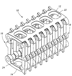
Fig. 2 is a perspective view of a spinal fusion cage in accordance with
the present invention in a fully expanded position.
Fig. 3 is an end view of a spinal fusion cage in accordance with the
present invention in a partially collapsed position.
Fig. 4 is a perspective view of a spinal fusion cage in accordance with
the present invention in a partially collapsed position.
Fig. 5 is an end view of a spinal fusion cage in accordance with the
present invention in a fully collapsed position.
Fig. 6 is a perspective view of a spinal fusion case in accordance with
the present invention in a fully collapsed position.
Fig. 7 is an end view of an alternative embodiment of the fusion cage
in a fully collapsed position.
Fig. 8 is a perspective view of an alternative embodiment of the fusion
cage in a fully collapsed position.
Fig. 9 is an exploded view of a spinal fusion cage in accordance with
the present invention.
Fig. 10 is an exploded view of one embodiment of an articulated
support utilizing a pin in accordance with the present invention.
Fig. 11 is a perspective view of the assembled articulated support of
Fig. 10.
Fig. 12 is an exploded view of an alternative embodiment of an
articulated support utilizing a post in accordance with the present invention.
Fig. 13 is a perspective view of the assembled articulated support of
Fig. 12.
Fig. 14 is a perspective view of an alternative preferred embodiment of
the articulated supports.
Fig. 15 is an end view of an alternate embodiment of a spinal fusion
cage with an exaggerated raised edge.
CA 02375689 2002-O1-09
WO 01/03616 PCT/LJS00/40328
S
Fig 16 is a perspective view of an alternate embodiment of a spinal
fusion cage with an exaggerated raised edge.
Fig. 17 is an end view of an alternative embodiment of the fusion cage
in an exaggerated fully open position.
$ Fig. 18 is a perspective view of an alternative embodiment of the
fusion cage in an exaggerated fully open position.
BEST MODES FOR CARRYING OUT THE INVENTION
The several embodiments of the fusion cage according to this invention
are shown in the figures. Like numbers correspond to identical or
corresponding
parts. References to "upper," "lower," "left," "right," or other terms
denoting relative
position refer to the drawing as shown and are for convenience only as the
fusion cage
can assume any orientation when in use.
A fusion cage 10 of the present invention includes an upper body 12
connected to a lower body 14 by side articulated supports 16, 18. The fusion
cage 10
expands and contracts between a fully open or expanded position (Figs. 1 and
2),
through a midway, partially collapsed position (Figs. 3 and 4), to a fully
closed or
collapsed position (Figs. 5 and 6). This ability to expand and collapse allows
the
fusion cage to be inserted through a much smaller opening than is possible
with rigid
fusion cages, while providing the same final spacing between the vertebrae as
provided by rigid fusion cages.
When in the open position, as shown in Figs. 1 and 2, the upper body
12, the lower body 14, and the articulated supports 16 and 18 form a hollow
cavity 20
adapted to contain autologous bone, artificial bone, bone matrix, bone growth
proteins, or other materials (not shown).
The upper body 12 has a series of openings 22 to allow ingrowth of
vertebrae bone and vasculature through the opening and into the hollow cavity
20.
Likewise, the lower body 14 has a series of openings 24 to allow ingrowth of
bone
into the hollow cavity 20 from below.
The left-side articulated supports 16 are arranged substantially parallel
to each other. The right-side articulated supports 18 are also arranged
substantially
parallel to each other and parallel to the left-side articulated supports 16
when the
CA 02375689 2002-O1-09
WO 01/03616 PCT/US00/40328
6
fusion cage is in the open position. The right-side articulated supports 18
are
positioned with an offset relative to the left-side articulated supports 16 so
that they
can effectively pass between the left-side articulated supports as the fusion
cage 10 is
collapsed. The center, joint or hinged part (described further below) of the
side
articulated supports 16, 18 become interdigitated or juxtaposed
("interdigitated
supports") when the fusion cage 10 is fully collapsed.
It should be noted that, although interdigitated supports are a feature of
a preferred embodiment, the interdigitation is an optional feature. As shown
in Figs.
7 and 8, in some applications the maximum height relative to the collapsed
height will
be sufficiently small to allow the articulated supports to collapse inward
without
interfering with each other. In such cases, there is no need to offset the
left and right
articulated supports, and a single long pin 104 can be used to connect the
upper legs
60 to the lower legs 62.
Refernng to Fig. 9, the upper body 12 has side edges 28a, 28b, each
side edge 28a, 28b having a plurality of axially aligned hinge knuckles 30a,
30b. The
knuckles 30a, 30b each have an axially extending bore 32a, 32b defined
therethrough
for receiving a pintle 34a, 34b. Likewise the lower body 14 has corresponding
side
edges 44a, 44b, each side edge 44a, 44b having a plurality of hinge knuckles
46a, 46b
that form parallel, axially extending bores 48a, 48b for receiving a second
pintle SOa,
SOb. Each articulated support 16, 18 has an upper leg 60 and a lower leg 62
pivotally
connected to each other. Each upper leg 60 has a hinge knuckle 61a, 61b with a
bore
63a, 63b that fits between two of the upper hinge knuckles 30a, 30b in
alignment with
bore 32a, 32b for receiving pintles 34a, 34b. Each lower leg 62 has a hinge
knuckle
65a, 65b with a bore 67a, 67b that fits between two of the lower hinge
knuckles 46a,
46b in alignment with bore 48a, 48b for receiving pintles SOa, SOb. The
knuckle and
pintle structure allows the articulated supports 16, 18 to interconnect the
upper and
lower bodies 12, 14.
Figs. 10-13 show exemplary articulated supports according to two
embodiments of the fusion cage. Each exemplary articulated support 16, 18 has
an
upper leg 60 pivotally connected to a lower leg 62. The legs 60, 62 have hinge
knuckles 61, 65 with respective bores 63, 67 suitable for the knuckle and
pintle
structure described above.
CA 02375689 2002-O1-09
WO 01/03616 PCT/US00/40328
7
Figs. 10 and 11 show one exemplary embodiment of an articulated
support 16, 18. In this embodiment, the upper leg 60 has a lower end 64 with a
pin
hole 66 defined therein. The lower leg 62 has an upper end 68 with a
complementary
pin hole 70. These pin holes 66, 70 are aligned and pivotally connected by a
short pin
74. The lower end of the upper leg 60 has a shelf 80 and a ledge 82. Likewise,
the
upper end of the lower leg 64 has a shelf 84 and a ledge 86. When the fusion
cage is
in the fully opened position, the shelf 80 of the upper leg engages the ledge
86 of the
lower leg, and the shelf 84 of the lower leg engages the ledge 82 of the upper
leg.
The engagement of ledges and shelves provides added support when the fusion
cage
is in the fully open position and prevents the articulated supports from
opening past
the desired position. In a preferred embodiment, a tab 90 is provided to fit
tightly
within a corresponding notch 92 (Fig. 4). The tab 90 engaged within the notch
92
assists in maintaining the articulated supports in an overcenter position by
increasing
the force necessary to recollapse the articulated supports inward.
Figs. 12 and 13 show an alternate exemplary embodiment of an
articulated support 16, 18. In this embodiment, the upper end 68 has a post 94
instead
of pin hole 66. The lower end 64 continues to have pin hole 66, defined
therein. The
post 94 pivotally mates with the pin hole 66. This structure eliminates the
need for
the separate short pin 74. The ledge and shelf structure and the tab and notch
structure may be repeated in this embodiment.
Although the figures show a device having sixteen articulated supports,
it would be possible to utilize fewer articulated supports. Each articulated
support
would then need to be designed to support additional weight. The use of fewer
articulated supports along with a post and hole instead of a separate pin
would reduce
the number of components and improve manufacturing efficiency. The upper legs
could also be joined together near the hinge knuckle to form a single upper
leg unit as
shown in Fig. 14, further reducing the overall piece count. The lower legs
could
likewise be joined.
In a preferred embodiment, each end of the assembled articulated
support has a protrusion 96 adapted to assist in maintaining the fusion cage's
position
between vertebrae. It would also be possible to add protrusions, ridges,
teeth, or other
surface irregularities to the upper body 12 and the lower body 14 to aid in
securing the
CA 02375689 2002-O1-09
WO 01/03616 PCT/US00/40328
8
fusion cage in place. For example, as shown in Figs. 15 and 16, a raised edge
102
may be formed around the openings 22, 24. The protrusions, ridges, teeth, or
other
surface irregularities help to eliminate the need to drill and tap the
vertebrae to accept
the fusion cage, a process that is required with rigid threaded fusion cages.
In the fully expanded position, the articulated supports 16 and 18 have
a slightly overcenter position. The upper body 12 has at least one stop 25
adapted to
align with a support stop 26 on the articulated supports 16 and 18 to prevent
further
outward movement of the articulated supports 16 and 18 once the stops 25 and
26 are
in contact. The stops 25 and 26 also act as a buttress to provide additional
structural
support once the fusion cage is fully opened. The pressure exerted on the
fusion cage
10 by surrounding muscles, ligaments, and body weight forcing the adj acent
vertebrae
together forces the articulated supports 16 and 18 outward and holds them in
the
overcenter, stable, expanded position, thereby preventing the fusion cage 10
from
collapsing.
Figs. 17 and 18 show an alternate preferred embodiment of the fusion
cage with an exaggerated overcenter latch. Once the fusion cage is opened
slightly
past its maximum height, the buttressing formed by the shoulder stop 25 and
the
support stop 26 prevent the articulated supports from opening further and
collapsing
outward. The articulated supports 16, 18 are also prevented from collapsing
outward
by the engagement of the ledges 82, 86 with the shelves 80, 84. The
buttressing can
be provided at the interface between the articulated support and the upper or
lower
body, at the joint of the articulated support, or at both the interface
and~the joint.
The articulated supports are typically rigid. However, it would be
possible to construct the articulated supports so as to provide a
predetermined amount
of flex. This added flexibility may affect the bone growth within the fusion
cage. It
would also be possible to introduce a flexible material between the joint of
the upper
and lower legs, or between the articulated supports and the upper or lower
bodies.
As shown in the figures, the fusion cage is preferably symmetrical,
having articulated supports of equal length. For some applications, however,
it would
be desirable for the legs to be a gradually decreasing length along the length
of the
fusion cage. This decrease would produce a tapered fusion cage. A tapered
fusion
cage according to the present invention could be used to address problems of
kyphosis
CA 02375689 2002-O1-09
WO 01/03616 PCT/US00/40328
9
(hunch back) or lordosis (sway back). It would also be possible for the right-
side
articulated supports and the left-side articulated supports to be of different
lengths,
producing a fusion cage that is tapered side to side. The variability of the
present
invention allows for the treatment of a variety of conditions.
Although it is preferable for the articulated supports to stop once the
fusion cage opens slightly past the maximum height, it would also be possible
for the
articulated supports to collapse outward. This embodiment would typically
require an
additional mechanism to maintain the fusion cage in its fully opened position.
In operation, the fusion cage 10 is inserted into a patient's body in the
fully collapsed position, as shown in Figs. 5 and 6. The interdigitated
supports 16, 18
provide a minimal collapsed height relative to the expanded height. Once an
opening
has been made between adjacent vertebrae, the collapsed fusion cage 10 is
inserted
into the intervertebral disc space. The fusion cage 10 is then opened (as
shown in
Figs. 3 and 4) until it reaches its maximum height (as shown in Figs 1 and 2).
The
protrusions 96 are forced upward into contact with the adjacent vertebrae to
secure the
fusion cage in place. The process may then be repeated on the opposite side of
the
disc space.
The foregoing disclosure and description thereof are for illustrative
purposes only and are not intended to limit the invention. There may be many
variations which will be apparent to a person of ordinary skill in the art
upon reading
this disclosure. This invention is defined by the claims, as interpreted by
the rules of
construction in patent cases.