Note: Descriptions are shown in the official language in which they were submitted.
CA 02375820 2001-12-05
WO 00/77206 PCT/US00/15868
THE MYOSTATIN GENE PROMOTER AND INHIBITION OF ACTIVATION THEREOF
BACKGROUND OF THE INVENTION
Technical Field
The subject invention relates to a promoter which regulates
expression of the myostatin gene as well as methods of
inhibiting this promoter and compositions used for such
inhibition. In particular, inhibitors of the promoter prevent
the expression of the myostatin gene and thus prevent muscle
wasting.
Background Information
Myostatin, or growth/differentiation factor 8 (GDF-8),
belongs to the transforming growth factor-9 (TGF-i3) superfamily
(McPherron et al., Nature 387:83-90 (1997)). The human
myostatin gene has been cloned (Nestor et al. Proc. Natl. Acad.
Sci. 95:14938-43 (1998)), and it has been reported that
myostatin immunoreactivity is detectable in human skeletal
muscle in both type 1 and type 2 fibers. With respect to
function, myostatin may play a role in negatively regulating the
growth and development of skeletal muscle (Nestor et al.,
su ra) .
The first evidence that myostatin may play a key role in
negatively regulating muscle development came from a study with
myostatin knock-out mice (McPherron et al., Nature 387:83-90
(1997)). In the myostatin null mice, the animals were rather
normal except that they were significantly larger than wild-type
mice and had a large and widespread increase in skeletal muscle
mass. Furthermore, it was also determined that two breeds of
cattle, characterized by increased muscle mass, have mutations
in the myostatin coding sequence (McPherron et al., Proc. Natl.
CA 02375820 2009-04-08
2
Acad. Sci. 94:12457-61 (1997)). Additionally, it should be
noted that the serum and intramuscular concentrations of
immunoreactive myostatin are increased in HIV-infected men with
muscle wasting compared with healthy men, and correlate
inversely with the fat-free mass index. These data support the
hypothesis that myostatin is a negative regulator of skeletal
muscle growth in adult men and contributes to muscle wasting in
HIV-infected men (Nestor et al., supra).
In view of the above findings, a need exists for a manner
of regulating myostatin expression, particularly in individuals
who experience muscle wasting as a result of a condition or
disease state such as, for example, aging, Autoimmune Deficiency
Syndrome (AIDS), Multiple Sclerosis, and cancer. The present
invention provides methods and compositions which may be
utilized to help individuals with such muscle wasting conditions
and provides further insight into the regulation of myostatin
gene expression.
SUMMARY OF THE INVENTION
The present invention encompasses an isolated nucleic acid
sequence represented by Figure 2 (SEQ ID NO:1).
Additionally, the present invention encompasses a vector
comprising the above-described nucleic acid sequence and a
nucleic acid sequence encoding a reporter molecule. The nucleic
acid sequence encoding the reporter molecule is operably linked
to the nucleic acid sequence represented by Figure 2. The
reporter molecule may be selected from the group consisting of,
CA 02375820 2001-12-05
WO 00/77206 PCTIUSOO/15868
J,
for example, luciferase, (3-galactosidase and Chloramphenicol
Acetyltransferase (CAT).
Preferably, the reporter molecule is luciferase.
The present invention also includes a host cell comprising the
above-described vector.
Additionally, the present invention includes a purified
antibody produced in response to immunization with myostatin as
well as a composition comprising this purified antibody.
Furthermore, the present invention also includes a method
of identifying a composition which inhibits activation of the
myostatin promoter. This method comprises the steps of: a)
constructing a vector comprising a nucleic acid sequence
represented by Figure 2 (SEQ ID NO:l) and a nucleic acid
sequence encoding a reporter molecule, the nucleic acid sequence
encoding the reporter molecule being operably linked to the
nucleic acid sequence encoding the sequence represented by
Figure 2; b) introducing the vector into a host cell for a time
and under conditions suitable for expression of myostatin; c)
exposing the host cell to a composition which may inhibit
activation of the myostatin promoter and a substrate specific
for the reporter molecule; and d) measuring the signal generated
by reaction of the reporter molecule and the substrate in
comparison to that produced by a control host cell, a smaller
signal by the host cell of (c) indicating that the composition
will inhibit activation of the myostatin promoter.]
Also, the present invention includes a method of
identifying a composition which inhibits expression of myostatin
comprising the steps of: a) adding an antibody selected from the
group consisting of a monoclonal or a polyclonal antibody
WO 00/77206 CA o237582o 2oo1-12-o5 pCT/US00/15868
4
produced against myostatin to a solid phase; b) adding known
concentrations of myostatin or a cell
sample comprising myostatin exposed to the test composition, to
the solid phase, in order to form a first complex between the
antibody and the known concentrations of myostatin or myostatin
in said cell sample; c) adding a second antibody to the first
complex, selected from the group consisting of a monoclonal
antibody or a polyclonal antibody produced against myostatin for
a time and under conditions sufficient for formation of a second
complex between the first complex and the second antibody; d)
contacting the second complex with an indicator reagent which
comprises a signal generating compound attached to an antibody
against said antibody of said second complex, for a time and
under conditions sufficient for formation of a third complex;
e) detecting the presence of a measurable signal, absence of
the signal indicating that the composition inhibits expression
of myostatin and presence of the signal indicating that the
composition does not inhibit expression of myostatin.
Moreover, the present invention also includes a method of
identifying a composition which inhibits expression of myostatin
comprising the steps of: a) coating a fixed amount of myostatin
on a solid phase; b) adding known concentrations of myostatin or
a cell sample comprising myostatin exposed to the composition;
c) contacting an antibody selected from the group consisting of
a monoclonal antibody or a polyclonal antibody produced against
myostatin to the myostatin in (a) and (b) for a time and under
conditions sufficient to form a first complex, wherein myostatin
of (a) competes with the myostatin of (b), in a competition for
the antibody; d) contacting the complex with an indicator
reagent which comprises a signal generating compound attached to
CA 02375820 2001-12-05
WO 00/77206 PCT/US00/15868
an antibody against the antibody of the first complex, for a
time and under conditions sufficient to form a second complex;
and e) detecting a measurable signal, a higher signal as
compared to a control, indicating the composition inhibits
5 myostatin expression.
Additionally, the present invention includes a method of
identifying a composition, in a mixture of compositions, which
prevents myostatin from binding to a myostatin receptor
comprising the steps of: a) mixing purified myostatin with the
mixture of compositions; b) passing the resulting mixture of
step (a) through a filter having pores of a size such that a
composition which is complexed to myostatin does not pass
through the filter; and c) determining the structure of a
complexed composition, thereby identifying a composition which
prevents myostatin from binding to a myostatin receptor.
Also, the present invention encompasses a method of
identifying a composition which prevents myostatin from binding
to a myostatin receptor comprising the steps of:
a) radiolabeling recombinant myostatin; b)incubating the
radiolabeled myostatin with cells or membranes comprising a
myostatin receptor; c) contacting the incubated mixture of step
(b) with a composition of interest; d) separating radiolabeled
myostatin bound to cells or membranes from unbound myostatin;
and e) determining the amount of radioactivity in bound
myostatin, compared to a control, a lower level of radioactivity
in bound myostatin compared to said control indicating a
composition which inhibits myostatin from binding to a receptor.
Furthermore, the present invention includes a method of
preventing muscle wasting in a mammal comprising administering
to the mammal a composition comprising an active ingredient
CA 02375820 2001-12-05
WO 00/77206 PCT/USOO/15868
6
which causes an effect selected from the group consisting of
preventing activation of the myostatin promoter, preventing
synthesis of myostatin, and preventing myostatin from binding to
its target receptor, in a therapeutically effective amount, such
that muscle wasting is prevented.
BRIEF DESCRIPTION OF THE DRAWINGS
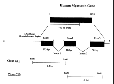
Figure 1 represents a conceptual illustration of the human
myostatin gene. The human myostatin gene contains three exons
and two introns, which encode 1.lkb of myostatin cDNA. When a
human Pi-derived Artificial Chromosome (PAC) library was
screened using a 740 bp probe encoding human myostatin exon 1
and 2, one positive clone with a 120 kb insert was identified.
After digesting the clone with EcoRI, two human genomic
subclones were isolated. One of the subclones, Clone C11,
contained 0.37 kb of exon 1, 1.53 kb of intron 1, and a 3.4 kb
sequence containing the human myostatin promoter region.
Figure 2 represents the nucleic acid sequence of the human
myostatin promoter region. The putative transcription factor
binding regions are identified and underlined.
Figure 3 represents the detection of myostatin mRNA by RT-
PCR of RNA samples from human skeletal muscle, rhabdomyosarcoma
cells, and prostate smooth muscle cells using human myostatin-
specific primers, and for G3PDH using G3PDH-specific primers.
Amplified myostatin gene (1.1 kb) was observed in both human
skeletal muscle cells (lane 1) and rhabdomyosarcoma cells (lane
2), but not in prostate smooth muscle cells (lane 3). Amplified
G3PDH gene (0.9 kb) was observed in skeletal muscle (lane 4),
CA 02375820 2001-12-05
WO 00/77206 PCT/US00/15868
7
rhabdomyosarcoma cells (lane 5), and prostate smooth muscle
cells (lane 6 ) .
Figure 4 represents the luciferase reporter constructs.
The 3.4 kb human myostatin promoter region was cloned into the
XhoI and HindIII sites of a luciferase reporter pGL3-enhancer
vector (Promega, Madison, WI), which contained a luciferase
reporter gene and a SV40 enhancer element for increasing the
transcription level.
Figure 5 represents results from a luciferase assay for the
human myostatin promoter region in human skeletal muscle cells
and rhabdomyosarcoma cells.
DETAILED DESCRIPTION OF THE INVENTION
As noted above, the subject invention relates to the
identification and isolation of a promoter which is involved in
activating or regulating expression of the myostatin gene.
Myostatin negatively regulates the growth and development of
skeletal muscle.
In particular, the subject invention relates to methods
which may be used to identify compounds that inhibit the
myostatin promoter activities. The myostatin promoter region or
nucleic acid sequence which regulates the expression of
myostatin is linked to the luciferase reporter gene. Thus, if
one is able to identify compounds that inhibit the activity of
the myostatin promoter region to prevent expression of
luciferase, one may then prevent the promoter region from
functioning, thereby preventing expression of myostatin.
The identification of compounds which inhibit the myostatin
promoter activity and luciferase production may be carried out
by the use of drug screening assays. Initially, a vector is
CA 02375820 2001-12-05
WO 00/77206 PCT/US00/15868
8
created comprising an isolated DNA sequence encoding the
promoter region of myostatin, which is linked to the luciferase
reporter gene. The vector may be, for example, a plasmid, a
bacteriophage or a cosmid. The vector is then introduced into
host cells under time and conditions suitable for activation of
the myostatin promoter. The host cells may be prokaryotic or
eukaryotic cells. Preferably, eukaryotic cells are utilized,
for example, cell lines with muscle lineage. Examples include
human skeletal muscle cells, human rhabdomyosarcoma cells, and
rat L6 or L8 cells. The host cells are then exposed to the test
composition thought to block activation of the myostatin
promoter and luciferase gene expression. The cells are also
exposed to a substrate for luciferase. One then measures the
quantity of signals or light emitted from the luciferase-
substrate reaction. If the amount of signals produced by the
host cells, exposed to the composition in question, is lower
than that produced by control cells (i.e., cells which have not
been exposed to the composition), then the composition has
inhibited the activity of the myostatin promoter, and will be
useful in inhibiting the expression of the myostatin gene. If
the amount of signals produced by the treated cells is equal to
that produced by the control cells, the composition has not
inhibited the activity of the myostatin promoter and will not
prevent myostatin gene expression.
Once compositions have been identified which inhibit the
activity of the myostatin promoter, such compositions may be
administered to patients having any type of condition involving
muscle wasting, for example, Acquired Immunodeficiency Syndrome
(AIDS), cancer, Multiple Sclerosis and aging. The
pharmaceutical composition may comprise a therapeutically
CA 02375820 2001-12-05
WO 00/77206 PCT/US00/15868
9
effective amount of the inhibitor and an appropriate
physiologically acceptable carrier (e.g., water, buffered water
or saline). The dosage, form (e.g., suspension, tablet,
capsule, etc.), and route of administration of the
pharmaceutical composition (e.g., oral, topical, intravenous,
subcutaneous, etc.) may be readily determined by a medical
practitioner and may depend upon such factors as, for example,
the patient's age, weight, immune status, and overall health.
Additionally, the present invention also encompasses
compositions comprising antibodies derived using purified
myostatin protein or a portion thereof which may be administered
with, for example, an appropriate carrier (e.g., water, buffered
water or saline). Subsequent to administration of the
antibodies, they may bind to expressed myostatin in the body in
order to form a complex, thereby preventing the expressed
myostatin from negatively regulating muscle development. The
antibodies themselves, as well as portions thereof, are also
encompassed within the scope of the present invention, as well
as assays which comprise such antibodies or portions thereof.
It should be noted that the above pharmaceutical
compositions and antibodies may be utilized for veterinary
applications (e.g., for preventing muscle wasting in aging or
diseased animals) or for agricultural applications (e.g., for
increasing the meat production in livestock, since animals
without myostatin exhibit a much larger muscle mass). For
example, the therapeutic composition which inhibits the activity
of the myostatin promoter may be administered to mammals such
as, for example, horses, cows, sheep, goats, cats, dogs and
pigs.
CA 02375820 2009-04-08
The present invention also covers two methods, using
purified myostatin and/or myostatin antibodies, which identify
compositions that inhibit the synthesis and the secretion of the
myostatin protein. In the sandwich method, a mammalian
5 monoclonal and/or polyclonal antibody (e.g., rabbit or mouse)
against the mature form of myostatin is coated on a solid
Tm
surface (e.g., the Immulon-4 plate (Dynatech Laboratories INC.,
Chantilly, VA)). The surface wil:i be blotted by a known blotting
agent, for example, Bovine Serum Albumin (BSA), and washed.
10 Samples (e.g. supernatants from human skeletal muscle cells
treated with or without test agents) or known concentrations of
purified mature form of myostatin are added to the surface
(e.g., plate). After myostatin binds to the antibody or
antibodies, the surface will be washed, and then incubated with
a mammalian monoclonal and/or polyclonal antibody (e.g., goat,
rabbit or mouse) against myostatin. The binding of the second
anti-myostatin antibody will be detected by use of an indicator
reagent which comprises an antibody conjugated with a signal
generating compound, for example, an enzyme. A substrate for
the enzyme is also added if an enzyme is utilized. For example,
horseradish peroxidase (HRP) and its substrate 0-
Phenylenediamine hydrochloride (OPD) may be utilized. In
particular, the enzyme-substrate reaction generates a detectable
signal or change, for example, color, which may be read, for
example, in a Microplate Reader. Examples of signal generating
compounds, other than an enzyme which may be utilized include,
for example, a luminescent compound, a radioactive element, a
visual label and a chemiluminescent compound. Known
concentrations of purified myostatin are used to generate a
standard curve. The concentration of myostatin in the unknown
CA 02375820 2001-12-05
WO 00/77206 PCTIUSOO/15868
11
samples (e.g. supernatants from human skeletal muscle cells
treated with or without test agents) can be determined using the
standard curve. The test agents which decrease the myostatin
concentration in supernatants are potentially useful for
inhibition of myostatin synthesis/secretion.
In the competitive method, a fixed amount of human
myostatin is coated on a solid surface, for example, the
Immulon-4 plate. The plate will be blotted by, for example, BSA
or another known blotting agent, and washed. Samples (e.g.,
supernatants from human skeletal muscle cells treated with or
without test agents) or known concentrations of purified mature
form of myostatin are added to the plate along with a mammalian
monoclonal and/or polyclonal antibody (e.g., goat, rabbit or
mouse) against myostatin. The plate will be washed, and then
incubated with an indicator reagent comprising an antibody
conjugated with a signal generating compound, for example, an
enzyme (or the entities described above). If an enzyme is used,
a substrate for the enzyme is also provided. The enzyme may be,
for example, horseradish peroxidase (HRP) The substrate may
therefore be O-Phenylenediamine hydrochloride (OPD)). Again,
the enzyme-substrate reaction generates a detectable change or
signal, for example, color, which can be read in, for example, a
Microplate Reader. Known concentrations of purified myostatin
may be used to generate a standard curve. The concentration of
myostatin in the unknown samples (e.g. supernatants from human
skeletal muscle cells treated with or without test agents) can
be determined using the standard curve. The test agents which
decrease the myostatin concentration in supernatants are
potentially useful for inhibition of myostatin
synthesis/secretion. Known concentrations of myostatin, or
CA 02375820 2001-12-05
WO 00/77206 PCT/US00/15868
12
myostatin in the sample, compete with myostatin protein coated
on the plate in binding to myostatin antibodies. When more
myostatin is present in the sample, less signal is generated.
If a test agent is able to block myostatin synthesis/secretion,
the amount of myostatin in that particular sample will be less
than in the control, and the signal in that sample will be more
than in the control.
Additionally, the present invention covers an Affinity-
Selection method, using purified myostatin in a filtration
assay, to identify compositions that bind to myostatin to
prevent myostatin from binding to its receptors, thus preventing
myostatin from functioning. Briefly, purified myostatin is mixed
with several test compounds. The mixture is passed through a
filter which only allows certain molecular weight molecules to
pass through. Compositions that bind to myostatin will be
retained by the filter. The unbound compounds are not retained
and can be separated from the bound compositions. The
structures of the compositions which bind to myostatin are
determined, for example, by Mass Spectrometry.
Furthermore, the present invention also encompasses a
receptor binding method using radiolabeled myostatin to bind to
cells or membranes prepared from tissues or cells containing
myostatin receptors. In this manner, one may identify
compositions that block myostatin from binding to its receptors,
thus preventing myostatin from functioning. In particular, the
purified recombinant myostatin protein from bacteria, insect or
mammalian cells is radiolabeled ( [125 I] , [3H] , [14C] , etc. ) . The
radiolabeled myostatin is then incubated with cells or membranes
prepared from tissues or cells which contain myostatin receptors
in the presence or absence of the test composition.
CA 02375820 2001-12-05
WO 00/77206 PCT/US00/15868
13
Radiolabeled cells and membranes are then separated from non-
radiolabeled cells and membranes by separation methods such as,
for example, filtration and centrifugation. The amount of
myostatin binding to cells or membranes is determined by
counting radioactivity. A decrease in radioactivity in the
presence of a test composition indicates that the composition
inhibits myostatin binding, and thus is useful in inhibiting
myostatin function.
The present invention may be illustrated by the use of the
following non-limiting examples:
EXAMPLE I
IDENTIFICATION OF THE NUCLEOTIDE SEQUENCE ENCODING THE HUMAN
MYOSTATIN PROMOTER AND POTENTIAL TRANSCRIPTION FACTORS BINDING
REGIONS
1. Cloning of human myostatin cDNA. Human myostatin cDNA was
amplified from human skeletal muscle 5'-plus cDNA library
(Clontech, Palo Alto, CA) by PCR using specific primers,
5'- ATG CAA AAA CTG CAA CTC TGT GTT T -3' and 5'- TCA TGA
GCA CCC ACA GCG GTC -3'. PCR products were cloned into
eukaryotic TA cloning vector pCR3.1 (Invitrogen, Carlsbad,
CA). Insertion was confirmed by DNA sequencing.
2. Cloning of the human myostatin promoter region. The EcoRI
and HindIII fragment (740 bp) of human myostatin cDNA
(Figure 1), which covers exon 1 and 2 of human myostatin
gene, was sent to GenomeSystems Inc. (St. Louis, MO) as a
probe to screen the human Pl-derived Artificial Chromosome
(PAC) library. One positive clone with 120 kb insert was
identified and confirmed by genomic Southern blot using the
same probe. The 120 kb insert was digested with EcoRI
CA 02375820 2001-12-05
WO 00/77206 PCT/US00/15868
14
restriction enzyme, and subcloned into plasmid pZero
(Invitrogen, Carlbad, CA). Two positive subclones with 5-7
kb insert were identified (Figure 1) . Sequencing results
indicate that clone 10 contains exon 2 and part of intron 1
and 2. Clone 11 (5.3 kb) contains exon 1, part of intron
1, and a 3.4 kb 5' untranslated region - the putative
myostatin promoter region. Program MatInspector V2.2
(Gesellschaft fur Biotechnologische Forschung mbH,
Braunschweig, Germany) was searched to identify potential
transcription factor binding regions (Figure 2).
MatInspector is a software that allows fast scanning of
sequence data for consensus motifs. MatInspector uses the
core similarity, the matrix similarity and the Ci vector
created by MatInd to calculate similarity index. The
potential transcription factor binding sites were selected
when both core similarity and matrix similarity reach 0.95.
The details of the program are illustrated in Quandt et
al., Nucleic Acids Research 23:4878-4884, 1995.
3. Identification of Human skeletal muscle cells or other cell
lines of muscle lineage which express myostatin. Total RNA
from human skeletal muscle cells and rhabdomyosarcoma cells
was isolated using Trizol reagent (Life Technologies,
Gaithersburg, MD) and used as templates for reverse
transcription. The myostatin gene was amplified by PCR
using human myostatin specific primers, 5'- ATG CAA AAA CTG
CAA CTC TGT GTT T -3' and 5'- TCA TGA GCA CCC ACA GCG GTC -
3'. Total RNA from human prostate smooth muscle cells were
also tested. Primers for endogenous G3PDH gene was used to
test the integrity of total RNA. The PCR products were
CA 02375820 2001-12-05
WO 00/77206 PCTIUSOO/15868
analyzed on a 1% agarose gel (Figure 3) The 1.lkb PCR
products, 1.lkb being the correct size for myostatin,
indicated that both human skeletal muscle cells and
rhabdomyosarcoma cells express myostatin.
5
EXAMPLE II
LUCIFERASE ASSAY
10 The luciferase reporter gene assay was designed for
quantitative analysis of mammalian gene expression. The coding
region for firefly (Photinus pyralis) luciferase was linked to
the 3.4 kb 5'untranslated region (5-UTR) of the human myostatin
gene. The construct was transiently transfected into human
15 skeletal muscle cells or human rhabdomyosarcoma cells, and after
48 hours, luciferase activity was measured. To assay for the
luciferase activity, luciferin and Mgz+-ATP were added to
cellular extracts, and the production of light was monitored.
The luciferase activity is increased if the 3.4 kb 5'-UTR region
has promoter activity. Therefore, the luciferase activity may
be used as an indicator (reporter) of the function of the
upstream promoter region.
The 3.4 kb human myostatin 5' untranslated region was
cloned into XhoI and HindIII sites of pGL3-enhancer vector
(Promega, Madison, WI), linked to the luciferase reporter gene
(Figure 4). The construct was transiently transfected into
human skeletal muscle cells or human rhabdomyosarcoma cells
using Superfect reagents (Qiagen, Inc., Santa Clarita, CA).
Another reporter gene such as 9-galactosidase gene or Renilla
luciferase gene was co-transfected as control for transfection
efficiency. Cells were lysed in lysis buffer after 48 hours, and
CA 02375820 2001-12-05
WO 00/77206 PCT/US00/15868
16
cellular extracts were assayed for luciferase and (3-gal activity
using detection kit [e.g. LucLite (Packard, Meriden, CT),
luminescent (3-galactosidase detection Kit II (Clontech, Palo
Alto, CA), or dual-luciferase reporter assay systems (Promega,
Madison, WI)]. The pGL3-enhancer parental vector was used as a
negative control and the pGL3-control vector with SV40 promoter
was used as a positive control for the luciferase activity. The
luciferase activity was counted on a luminescent light detector
such as a MicroBeta counter or a Luminometer (EG & G Life
Sciences-Wallac, Turku, Finland), and the result was normalized
by transfection efficiency. There was about a 5-fold increase
of luciferase activity for the human myostatin promoter
construct in comparison to its parental vector in human skeletal
muscle cells (Figure 5A). In human rhabdomyosarcoma cells, the
increase of luciferase activity was about 7-fold (Figure 53).
As expected, the luciferase activity was not increased in human
prostate smooth muscle cells (Figure 5A), which do not express
myostatin.
In view of the above results, the luciferase reporter
construct containing the 3.4 kb human myostatin promoter region
may be stably or transiently transfected into mammalian cell
lines such as SV-40-transformed human skeletal muscle cells or
rhabdomyosarcoma cells and others for high through-put
screening. Luciferase activity may be used as an indicator for
selecting compounds which inhibit expression of the myostatin
gene.
EXAMPLE III
ENZYME-LINKED IMMUNOSORBENT ASSAY (ELISA) FOR HIGH THROUGHPUT
SCREENING
CA 02375820 2001-12-05
WO 00/77206 PCT/US00/15868
17
Sandwich or competitive ELISA may be used to identify
compounds which inhibit human myostatin protein synthesis and/or
secretion. In the sandwich ELISA, for example, a rabbit or
mouse monoclonal and/or polyclonal antibody against the mature
form of myostatin is coated on the Immulon-4 plate (Dynatech
Laboratories, Inc., Chantilly, VA), and the plate is blotted by
BSA, and washed. Samples (e.g., supernatants from human skeletal
muscle cells treated with or without test agents) or known
concentrations of purified mature form of myostatin are added to
the plate. After myostatin protein binds, the plate is again
washed, and then incubated with a goat or rabbit or mouse
monoclonal and/or polyclonal antibody against myostatin. The
binding of the second anti-myostatin antibody will be detected
by an antibody conjugated with horseradish peroxidase (HRP)
using 0-Phenylenediamine hydrochloride (OPD) as the substrate.
Known concentrations of myostatin are used for generating the
standard curve.
In the competitive ELISA, human myostatin protein is coated
on the Immulon-4 plate, and the plate is blotted by BSA, and
washed. Samples (e.g., supernatants from human skeletal muscle
cells treated with or without test agents) or known
concentrations of purified mature form of myostatin or myostatin
peptides are added to the plate along with a goat or rabbit or
mouse monoclonal and/or polyclonal antibody against myostatin.
The plate is again washed, and then incubated with an antibody
conjugated with horseradish peroxidase (HRP) using 0-
Phenylenediamine hydrochloride (OPD) as the substrate. The
known concentrations of myostatin are used for generating the
standard curve.
Human skeletal muscle cells or other cell lines of muscle
CA 02375820 2001-12-05
WO 00/77206 PCT/US00/15868
18
lineage which synthesize and secrete myostatin may be used to
test compounds thought to inhibit the synthesis or secretion of
myostatin. Cells are incubated with test compounds for a period
of time, e.g., 6-48 hours. The amount of myostatin in the
medium of cells is then determined by ELISA. A decrease in the
amount of myostatin indicates that the test compound is
effective in inhibiting the synthesis and secretion of
myostatin, whereas an increase in the amount of myostatin or
maintenance of the same level of myostatin indicates that the
test compound is not effective in inhibiting the synthesis and
secretion of myostatin.
EXAMPLE IV
PRODUCTION OF RADIOLABELED MYOSTATIN FOR USE IN RECEPTORS
BINDING ASSAY
The purified recombinant myostatin protein from bacteria,
insect or mammalian cells is radiolabeled ([125I] ,[3H] ,[14C] ,
etc.). The radiolabeled myostatin is then incubated with cells
or membranes prepared from tissues or cells which contain
myostatin receptors in the presence or absence of the test
composition. The amount of myostatin binding to membranes is
determined by counting radioactivity. A decrease in
radioactivity in the presence of a test composition indicates
that the composition inhibits myostatin binding, and thus is
useful in inhibiting myostatin function.
EXAMPLE V
PURIFIED MYOSTATIN USED IN AFFINITY SELECTION
CA 02375820 2001-12-05
WO 00/77206 PCT/US00/15868
19
The purified recombinant myostatin protein from bacteria,
insect or mammalian cells is used in binding test compositions.
In particular, test compositions that bind to myostatin are
retained by filter, and the structure is determined by Mass
Spectophotometry. A test composition that binds to myostatin is
useful in inhibiting the binding of myostatin to its receptor,
thus inhibiting myostatin function.
CA 02375820 2002-09-20
19.1
SEQUENCE LISTING
<110> Abbott Laboratories
Wu-Wong, Jinshyun R.
Wang, Jiahong
<120> THE MYOSTATIN GENE PROMOTER AND
INHIBITION OF ACTIVATION THEREOF
<130> 412-109
<140> 2,375,820
<141> 2000-06-09
<150> US 09/329,685
<151> 1999-06-10
<160> 3
<170> FastSEQ for Windows Version 4.0
<210> 1
<211> 3435
<212> DNA
<213> Homo sapiens
<400> 1
ttctctaccc actcacccta atgatgcagt actgtcctgt ctccttggtg ataagaactg 60
ccagaactgg gtctccagca gtcagactac attgaagttt cctatagctg gtgagccctt 120
ctattctggg gcctcaggaa ggttgcaatc actgccactg gagaaggaat aaaacttact 180
taaatttctt cagttcttct tcacccattc aatactgttc tctagagaag ttgattagat 240
atagtcaatt ctccctattc atagtagtta tgttctataa agtcattgcc aacactgagt 300
tagcaaatat tgaactattg ttcccagggg aaaaacaggg ttagttgagc ctctggtcat 360
aacatttaca tcacccaatc aatatataac cttgttttat gtatgttttt gtttacaaat 420
acattattta atatatattg ttcattcatt aacactgaac tcacagccag cagcactata 480
actcaggcct gaatgatgct tatctagcac atgcattttt tcatgagaac ttttttccct 540
taggcatatc acagcctttt aaaattgtgg taaatataca caacatttag catcttaacc 600
atttttaggt gtacagttcg gtggcattaa gcacattcac actgttgtgt aaccatcacc 660
accattcatc ttcagaaatt tttcatcttc ccagactgaa actctgtatc tatcacacag 720
taactacccc tcagtgcctc acccagtccc tggcaaccac catgctactt tccattgcag 780
ctttcctgtg attaggaaca taggaacacc acgcagcacg tcagcactat gcttgggggc 840
catttttaaa aagcaaaatc aataagagga gcaataaaaa aagaagcaca aaatatgtga 900
aaacatggca ctaaatagac gggaaaaagg gaatttgttt atagtatgag agctgaaaca 960
aaaaggcgga accctgcctt gtttgacctc atctgggaac ctgtgcgtca gataggactc 1020
aaattttttg ccgctctgtg aatgccagcg aatgactgaa aaatgctagg aatattgagt 1080
ttcatagata aattttagca agtaggcaaa tttgtgaata cagaaatcat gaataataag 1140
aattgtctgt tagcggaaat gacaaccttt gttcactttt ttccaggtga taagaataaa 1200
CA 02375820 2002-09-20
19.2
gagctggaac atgtcttgta acctggctgc cttagaaaat tttacttgcc tcacaggcct 1260
agaaagtagg ctttgtgact gataattggc agctatgacc tgaagcagtt ctagttcatg 1320
tggagcataa ttttaagcat aatctcaaac ccttctgcat aaaacaaaga gcaagcactc 1380
aaatgccagt tatcaattac ttactatatg acaggtgcca tattcagcaa tttacatgca 1440
ttattaaatt atatcccccc aaaaccctat gaggaagcta aagtttaggg aagttaagta 1500
tctcatccat tatcacatag ttagaagtgg caaagttgag atttgaactc aggtctatct 1560
gactccagag cctgagttct caattcaact gctatacaat tctaagcata ttaaaaaaaa 1620
agtttgactt acttggaact gtatagatgc atgtgttaca atgatcataa catttgaaag 1680
atttacacat tgaaaaatga atttaccaaa caaataaaac cttagaagcc agatctaata 1740
ttgtcccata acaaagagta tctgaaatcc tcagggcatc tggtttgtgt ctggttttcc 1800
ttaatcttta atgatgagca aatccaatgc attatgtaag gccatttttt tctcaagaga 1860
tgtagatacc tcttaagaat ttgatgaaaa tgcattaact tttcaggcta ctgagttgca 1920
ttttagtgca ccgaggcagt aaattagtgt acagtgtgca aaaatggtag tgactttaaa 1980
aataaatatt tgatatgagc cactgtattc tcttggaaaa aaaaagtaat ggactaaatc 2040
tcttaggaat ccttagcttc ccaaaaagga gtaggaaaaa gaaatctcct ttggcctaga 2100
aatatcttct gtttcttgct ggctatgttt gcttagctct ttaatagttc atttgactag 2160
atcttgtggc tcccaaagct aaggttgaac gtttgatccc tacagaggcc acttaaattt 2220
agagaacaaa aagctctatt ctctgctccc agactttacc ccaaatccct gccaggtgtc 2280
tgccctctgg tcaaaatgag aaattggcaa aggggtgcaa acatatcgca gtattgggaa 2340
acaacaaaag gtcacccctt tatcatgatg ctctttctct tttatgtgct cataatattc 2400
tgatataatt tatagagaat agatactgca ctttttactc tctggatatt tactgctgga 2460
aatctgaggc aaactgtaat aatctctgcc atgccagtta taaaattcat tatcttagtc 2520
tatgttcaga gatttttcta ctagctggca ttaccctctt ggtaataaac aatgaaaaac 2580
acatcttctg agttatgtta atctgcatct ttagaatagg aaataatagc actcagtcaa 2640
aagttcagta taattttcat attaataaaa gacatgaaac tatgtaaaaa taattccatg 2700
cacaatatgt tataataaca atgacttcca atatttacta agaatttagt cagaaaacaa 2760
gtttctcaaa ttatagatga aaattctcaa ctagtatcat aatcttaact tttaattcag 2820
gtcttcctaa tttttatttt cctaattact tggcactaaa aataatttaa tacaacaaat 2880
aaaaatattt tctacttcaa atacttgcct aaacaatata aaatcatttt agtttttgag 2940
gaagtaatat ttcatatttt aaatatgtag tataaattaa aattgactta tttaaattac 3000
aataagagtt gtgtgaggat tagtaagatt taagtacagt ttatattatt gccaacatag 3060
acttttgttt ttcaaatgtc acaaatatct tttattattt gtagatttat ttcttttatg 3120
aagtagtcaa atgaatcagc tcacccttga ctgtaacaaa atactgcttg gtgacttggg 3180
acagacaggg ttttaacctc tgacagcgag attcattgtg gagcaagagc caatcataga 3240
tcctgacgac acttgtctca tctaagttgg aatataaaaa gccacttgga atacagtata 3300
aaagattcac tggtgtggca agttgtctct cagactgtac atgcattaaa attttgcttg 3360
gcattactca aaagcaaaag aaaagtaaaa ggaagaaaca agaacaagaa aaaagattat 3420
attgatttta aaatc 3435
<210> 2
<211> 25
<212> DNA
<213> Homo sapiens
<400> 2
atgcaaaaac tgcaactctg tgttt 25
CA 02375820 2002-09-20
19.3
<210> 3
<211> 21
<212> DNA
<213> Homo sapiens
<400> 3
tcatgagcac ccacagcggt c 21