Note: Descriptions are shown in the official language in which they were submitted.
CA 02376019 2001-12-19
WO 01/00096 PCT/US00/15743
1
TIBIAL PLATEAU RESECTION GUIDE
FIELD OF THE INVENTION
The present invention is directed to an apparatus useful as a tibial plateau
resection
guide and methods for its use in arthroplastic surgery of the knee. More
particularly, the
invention relates to an apparatus which utilizes adjustable rods in order to
fix a bone saw
guide to the anterior portion of a patient's proximal tibia.
BACKGROUND OF THE INVENTION
In replacing the knee joint which has been damaged due to disease or trauma,
it is
important that the damaged bone at the proximal end of the tibia be removed by
cutting it at
an appropriate varus/valgus angle and at an appropriate flexion/extension
angle. In this
manner, the bone cut will be in the correct varus/valgus and flexion/extension
alignment,
and the proximal end of the tibia can then receive an implant or prosthesis to
reconstruct a
functioning knee joint. Proper fit and function of the implant will depend on
the accuracy
of the cut.
Many devices for determining the correct angle of the bone cut are known in
the art.
The known devices typically include a cutting block which guides a saw blade
and an
anterior telescoping rod or similar device which extends to a position
adjacent the
approximate center of the anterior face of the patient's ankle or talus to
allow the surgeon
to duplicate the mechanical axis of the tibia as a reference guide for the
proper alignment of
the cutting block with the mechanical axis.
Johnson et al., U.S. Patent No. 5,451,228 (Johnson) discloses a tibial
resector guide
having an angularly adjustable head controlled by a thumb actuated slide
mechanism. The
tibial resector guide disclosed by Johnson includes only one telescoping rod
to reference the
mechanical axis, but no external side rod or similar means to reference the
mid-coronal
plane.
Ferrante et al., U.S. Patent No. 5,342,367 (Ferrante) discloses a tibial
cutting guide
which does not include any means for external referencing, such as extending
rods.
Bowman et al., U.S. Patent No. 4,952,213 (Bowman) discloses using an
intramedullary rod connected to a pivot device carrying the bone saw guide.
There is no
CA 02376019 2001-12-19
WO 01/00096 PCT/US00/15743
2
external referencing rod discussed in Bowman -- rather, the reference used is
the
intramedullary rod inserted deep into the bone canal.
Petersen, U.S. Patent No. 5,342,368 (Petersen '368) discloses a proximal
tibial
resector guide including an intramedullary rod which is attached at its
proximal end to a bar
provided for the cutting saw guide. There is no external referencing rod
disclosed in
Petersen -- rather, the reference used is the intramedullary rod inserted deep
into the bone
canal.
Petersen, U.S. Patent 4,524,766 (Petersen '766) discloses a surgical knee
alignment
system including a tibial resection saw guide which is mounted on one
telescoping external
rod used to reference the mechanical axis. There is no external side rod or
similar means
disclosed to reference the mid-coronal plane.
Petersen, U.S. Patent No. 5,395, 377 (Petersen '377) discloses an
extramedullary
proximal tibial guide that includes a distal end carrying a vertically
adjustable ankle bracket
as well as an ankle pointer, and a proximal end carrying a saw guide. There is
no external
side rod or similar means disclosed to reference the mid-coronal plane.
Wehrli, U.S. Patent No. 4,938.762 (Wehrli) discloses a reference system for
the
implantation of condylar total knee prostheses, including a tibial resection
saw guide. The
Wehrli system utilizes as a main reference point a screw placed in the pelvis,
and includes a
number of screws placed into the tibia. Telescoping rods attached to the
pelvic bone screw
and the tibial bone screw are utilized to position the tibial resection saw
guide.
A drawback of the use of intramedullary rods as references is that the anatomy
of
many patients does not permit an intramedullary rod to be fully inserted.
Also, with both intramedullary and extramedullary tibial resection guides, a
drawback of the use of a single, anterior guide rod is that the surgeon lacks
a side reference
guide to provide a means of reliably and accurately referencing the mid-
coronal plane. The
present invention addresses this need by providing both anterior and side
guide rods to
reference both the mechanical axis and mid-coronal plane.
Additionally, during total knee replacement surgery, the patella is normally
evened
to the lateral side of the knee which may interfere with a side referencing
rod. The present
invention provides a side referencing frame that is attached to the first
distally extending
guide rod assembly anteriorly to the patella, thus allowing lateral placement
without
CA 02376019 2001-12-19
WO 01/00096 PCT/US00/15743
3
interfering with the patella, while at the same time allowing multiple planes
to be
referenced during alignment, such as the mid-coronal plane or longitudinal
axis of the
fibula, while maintaining a constant angle to the plane of resection.
Citation or identification of any reference in Section 2 or any section of
this
application should not be construed as an admission that such reference is
available as prior
art to the present invention. The teachings of these patents are incorporated
by reference
herein.
SUMMARY OF THE INVENTION
The present invention is directed to an apparatus useful as a tibial plateau
resection
guide and methods for its use in arthroplastic surgery of the knee. The
apparatus has an
alignment system that allows for the independent establishment of two separate
geometric
planes to be used for the accurate placement of a cutting guide for use in
removing
damaged bone at the proximal end of the tibia during knee arthroplasty. The
axis formed
by the intersection of these two planes is intended to duplicate the
mechanical axis of the
tibia. An angular relationship between the bone and the cutting block is
established by
fixing a number of adjustable parameters, thereby allowing a surgeon to make a
cut in the
transverse plane of the tibia at specific, preferred varus/valgus and
flexion/extension angles
relative to the duplicated mechanical axis.
The present invention consists of a first and second guide rod assembly with
the
second distally extending guide rod assembly attached to the first distally
extending guide
rod assembly anteriorly to the patella, thus allowing lateral placement
without interfering
with the patella. A side extending rod, used for flexion/extension alignment
of the cutting
instrument, is adjustable in the anterior-posterior direction, thus allowing
multiple planes to
be referenced during alignment, such as the mid-coronal plane or longitudinal
axis of the
fibula while maintaining a constant angle to the plane of resection.
In one embodiment, the apparatus of the present invention comprises an
inverted L-
shaped first distally extending guide rod assembly placed in the sagittal
plane and a
reversed L-shaped second distally extending guide rod assembly placed in the
transverse
plane to which a slidably attached side bar is perpendicularly coupled. The
two frame
assemblies are coupled to one another, preferably removably coupled,
preferably below the
tibial plateau, at a constant angle to one another (e.g., 90 degrees). The
first distally
CA 02376019 2001-12-19
WO 01/00096 PCT/US00/15743
4
extending guide rod assembly, which is placed in the sagittal plane, has a
first end and a
second end, and has a pivot block, a pivot arm, a support arm, a removably
attached
cutting block (cutting saw guide) and a telescoping rod which can be extended
to the center
of the anterior face of the distal end of the tibia, i.e., the center of the
talus or ankle. The
second distally extending guide rod assembly placed in the transverse plane
has a first end
and a second end, and has a side bar, a top bar and a rod which can be
slidably extended
through the side bar to the center of the subject's malleolus.
In use, the two interlocking frame assemblies are assembled together, one
adjacent
the anterior portion of the tibia and one adjacent either the lateral or
medial portion of the
tibia. The frame assemblies may be assembled prior to anchoring the assemblies
or the
second distally extended guide rod assembly may be combined with the first
distally
extending guide rod assembly after the first distally extending guide rod
assembly has been
secured to the tibia. While the surgeon may secure both frame assemblies to
the tibia after
they have already been coupled" it is anticipated that most surgeons will
secure the first
distally extending guide rod assembly first, followed by attaching the second
distally
extending guide rod assembly to the first distally extending guide rod
assembly. Most
surgeons prefer to position the second distally extending guide rod assembly
on the lateral
side, or the outer of the leg, rather than the medial side, or the inner side
of the leg, to
reference the fibular axis. However, the present invention may be adapted for
the second
distally extending guide rod assembly to be used on either the lateral or
medial side of the
tibia.
The first distally extending guide rod assembly may be secured to the tibia in
various manners. In one embodiment, the pivot block may be anchored to the
proximal end
of the tibia by means of a primary pin inserted approximately into the
intramedullary canal
area. In another embodiment, the anchor block may be anchored by tamping
securing pins
on the anchor block into the tibia. Alternatively, a hole may be drilled in
the proximal end
of the tibia in the location of the intramedullary canal. An intramedullary
rod may then be
placed through the pivot block into the intramedullary canal to anchor the
guide assembly.
In yet another embodiment, the first distally extending guide rod may be
anchored through
pins inserted through the cutting block.
CA 02376019 2001-12-19
WO 01/00096 PCT/L1S00/15743
Where the first distally extending guide rod assembly is anchored above the
tibial
plateau, the frontal telescoping rod is then extended downward and rotated
using a rod
hinge in the required direction until the end of the rod is placed directly
over the end of the
tibia. The rod hinge can then be maintained in a fixed position by turning a
thumb screw.
5 The pivot block is fixed in place by means of a secondary pin which anchors
it to the tibial
plateau and prevents the apparatus from rotating. A cutting block is removably
attached,
either at a fixed or at an adjustable angle, to a proximal portion of the
first distally
extending guide rod assembly. Adjustments to the angle of each frame assembly
and to the
height of the cutting block can be made and locked into place.
The two frame assemblies have a fixed angle between them. In the preferred
embodiment, the fixed angle between the frame assemblies is 90 degrees,
thereby providing
direct and reliable references to the mid-coronal plane and to the mechanical
axis. This
facilitates the identification of the tibial mechanical axis. However, the
angle may vary
according to the preference of the user.
The present invention also provides for a method of using the tibial resection
guide
in knee arthroplasty. The method includes attaching a first frame assembly to
the tibia and
coupling a second frame assembly to the first assembly which interlocks the
assemblies,
preferably at angle of 90 degrees. A frontal telescoping rod which extends
downwards
from the first distally extending guide rod assembly is placed adjacent to the
approximate
center of the distal end of the tibia or talus. A slidably attached rod which
extends
downwards from the second distally extending guide rod assembly is placed in
the
approximate center of the malleolus. In order to set the extending rod in the
center of the
malleolus, the surgeon may have to slide the extending rod along the top bar
of the second
distally extending guide rod assembly. The stylus is then used to measure the
resection
guide height.
BRIEF DESCRIPTION OF THE DRAWINGS
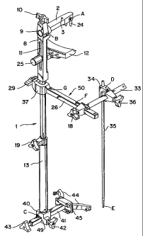
FIG. 1 is a perspective view of one type of Extra Medullary Tibial Resection
Guide;
FIG. 2 is a perspective view of another type of Extra Medullary Tibial
Resection
Guide;
CA 02376019 2001-12-19
WO 01/00096 PCT/US00/15743
6
FIG. 3 is an anterior view of a tibia and fibula showing the positional
relation of the
mechanical axis with respect to the resection plane and to the first distally
extending guide
rod assembly;
FIG. 4 is a side view of a tibia and fibula showing the positional relation of
the mid
coronal plane with respect to the resection plane and to the reference rod of
the second
distally extending guide rod assembly;
FIG. 5 is a perspective view of another type of Extra Medullary Tibial
Resection
Guide; and
FIG. 6 is a perspective view of one type of Intra Medullary Tibial Resection
Guide.
DETAILED DESCRIPTION OF THE INVENTION
The present invention is directed to a tibial alignment system that allows the
independent establishment of two separate geometric planes to be used for
angular reference
in the cutting of the tibial plateau during partial or total knee
arthroplasty. The reference
planes intersect one another at a fixed relative angle. The axis formed by the
intersection
of these two planes is intended to duplicate the mechanical axis of the tibia,
which
represents an imaginary line connecting the approximate center of the proximal
and distal
ends of the bone. A predetermined angular relationship between the
reconstructed
mechanical axis (see FIG. 3) and the resection plane (see FIG. 3), defined by
the surface of
the cutting block, determines the specific varus/valgus angle of the bone cut.
Additionally,
a predetermined angular relationship between the mid-coronal plane (see FIG.
4) and the
resection plane (see FIG. 4) determines the specific flexion/extension angle
of the bone cut.
By determining these angles the surgeon can resect the tibia optimally to
allow for the most
precise fit of a knee prosthesis, maximizing the performance, comfort and wear
of the
prosthesis.
Figure 1 shows a preferred embodiment of extramedullary tibial resection
guide. In
this embodiment, preaffixed securing pins (24) anchor the first distally
extending guide rod
assembly (1) to the top of the tibia. The first distally extending guide rod
assembly (1) is
composed of four main elements: anchor block (2), support arm (8), cutting
block (12),
and frontal telescoping rod (13). In this embodiment the securing pins (24)
are affixed to
the anchor block (2). The anchor block (2) is attached to the support arm (8).
After the
CA 02376019 2001-12-19
WO 01/00096 PCT/US00/15743
7
surgeon has positioned the cutting block (12) in close proximity to the front
of the tibia and
in the desired orientation, a secondary anchor pin (not shown) is placed in a
secondary hole
or aperture (3) to secure the entire first distally extending guide rod
assembly (1) in place.
The sagittal reference plane where the first distally extending guide rod
assembly (1) lies is
defined by points A, B, and C. Point A lies in the approximate center of the
tibial plateau,
point B is the intersection of the support arm (8) and anchor block (2), and
point C lies at
the distal tip of the frontal telescoping rod (13), which is placed at the
approximate center
of the anterior face of the distal end of the tibia or ankle.
The anchor block (2) fits into the aperture (9) on the support arm (8) and
locked in
place with a thumbscrew (10). The support arm (8) has an elongated aperture
(11) in the
approximate center of the support arm (8). A screw (25) fits through the
elongated
aperture ( 11 ) and allows for the tightening of the cutting block ( 12)
against the support arm
(8). This allows the cutting block (12) to be stabilized at an optimal
position along the tibia
based on where the surgeon determines the cut should be made. A stylus (not
shown) is
used to set the depth of the cut at the level desired by the surgeon.
The support arm (8) is attached at its distal end to a frontal telescoping rod
(13)
which has an adjustable length. The frontal telescoping rod (13) can be fixed
at a position
to adjust the length of the first distally extending guide rod assembly (1),
as determined by
the surgeon, by tightening a thumbscrew (19). The distal end of the
telescoping rod (13)
may be secured to the ankle through ankle clamp (40) which is affixed to the
distal end of
the.telescoping rod (13). The ankle clamp (40) is adjustable to contact the
ankle by sliding
the ankle clamp rod (41) through an aperture (49) and can be fixed in place by
tightening a
thumbscrew (42). The ankle clamp attachments (44) can be horizontally moved on
the end
(45) of ankle clamp rod (41) to best adjust to the ankle and can be fixed in
placed with a
thumbscrew (43). The second distally extending guide rod assembly (50) is
removably
attached to the first distally extending guide rod assembly (1) and is secured
thereon by an
attachment lever or a thumbscrew (29).
The second distally extending guide rod assembly (50) is made up of several
parts,
including: side bar (26); connection (37); top bar (33); rod anchoring block
(34); and an
extending rod (35). The second distally extending guide rod assembly (50) has
a side bar
(26) with a first end and second end.
CA 02376019 2001-12-19
WO 01/00096 PCT/US00/15743
8
The side bar (26) is connected to the first distally extending guide rod
assembly (1)
at its first end through a connection (37) that may be a c-clamp type
connection such as that
shown. Alternatively, side bar (26) may have a dovetail connection (such as in
FIG. 2
(217)) to engage grooves (see FIG. 2 (238)) of the support arm (8) below the
cutting block
(12). With this variation, the anterior half and posterior half of the grooved
members (see
FIG. 2, grooved members (227) and (228)) are connected to a thumbscrew or
lever arm
such that turning the thumbscrew or activating the lever arm causes the
anterior half and
posterior half to close on the dovetail connection of the side bar, gripping
it in place.
At its second end, the side bar (26) is connected to top bar (33), which
extends
perpendicularly in the transverse plane from the side bar (26), the transverse
plane being
defined by points D, F, and G. The top bar (33) has a first end and a second
end. The
first end of the top bar (33) is connected to the side bar (26). The top bar
(33) may slide
back and forth on the side bar (26) in the coronal plane to adjust the
distance of the top bar
(33) from the leg of the patient. The coronal reference plane being defined by
points A, D,
and E. Point A lies on the approximate center of the tibial plateau at the
very top of the
tibia. Point D lies at the top of a rod anchoring block (34). Point E lies at
the distal end of
an extending rod (35), at the approximate center of the malleolus.
The top bar (33) may be secured at a position on the side bar (26) by
tightening a
thumbscrew (18) or a similar functioning lever arm. Proximate the second end
of the top
bar (33) a rod anchoring block (34) is connected thereto. The rod anchoring
block (34)
may slide on the top bar (33) to adjust its position thereon.
The proximal end of an extending rod (35) which extends downwardly
perpendicular to the top bar (33) is slidably coupled to the proximal end of
the rod
anchoring block (34). In order to facilitate the placement of the distal end
of the extending
rod (35) adjacent the malleolus, the surgeon may slide the top bar (33) along
the side bar
(26) and/or slide the rod anchoring block (34) along the top bar (33) which
merely varies
the position of the distal end of the extending rod (35) and does not affect
any other
parameter. When the distal end of the extending rod (35) is brought in close
proximity to
the appropriate center of the malleolus on the lateral face of the distal end
of the tibia,
thumbscrews (18) and (36) may be tightened to secure the position of the
extending rod
(35).
CA 02376019 2001-12-19
WO 01/00096 PCT/US00/15743
9
Figure 2 shows an alternative preferred embodiment of an extramedullary tibial
resection guide. In this embodiment, a primary anchor pin (224) anchors the
assembly to
the approximate center of the top of the tibia. The first distally extending
guide rod
assembly (201) is composed of five main elements: pivot block (202), pivot arm
(204),
support arm (208), cutting block (212), and frontal telescoping rod (213).
In this embodiment the primary anchor pin (224) runs through the pivot block
(202)
at a hole or aperture (205). The pivot block (202) itself is pivotally
attached to the
posterior end of the pivot arm (204) near a hinge point (206), which allows
rotation of the
pivot arm about the axis in the direction of the sagittal plane. Additionally,
the pivot block
(202) and attached pivot arm (204) can swivel about hole (205) after the
insertion of the
primary anchor pin (224). After the surgeon has positioned the cutting block
(212) in close
proximity to the front of the tibia, a secondary anchor pin (not shown) is
placed in
secondary hole or aperture (203) to secure the entire first distally extending
guide rod
assembly (201) in place. At the anterior end of the pivot arm (204), a
thumbscrew or lever
(207) can be turned to tighten the grip of the pivot arm (204) against the
pivot block (202).
The pivot arm (204) extends through the aperture (209) of the support arm
(208)
and may slide back and forth on the pivot arm (204), and is tightened against
the pivot arm
by a thumbscrew (210) or similarly functioning lever located adjacent the
aperture (209).
The support arm (208) has an elongated aperture (211) in the approximate
center of the
support arm (208). A screw (225) fits through the elongated aperture (211) and
allows for
the tightening of the cutting block (212) against the support arm (208). This
allows the
cutting block (212) to be stabilized at an optimal position along the tibia
based on where the
surgeon determines the cut should be made. A stylus (not shown) is used to set
the depth
of the cut at the level desired by the surgeon.
The support arm (208) is attached at its distal end to a frontal telescoping
rod (213)
which can adjust the length of the first distally extending guide rod assembly
(201).
Alternatively, the frontal telescoping rod (213) can be fixed at a position
determined by the
surgeon by tightening a thumbscrew (see FIG. 1 thumbscrew (19)). Optionally,
the distal
end of the first distally extending guide rod assembly (201) may be secured to
the ankle
through an ankle clamp assembly such as that shown in FIG. 1 where an ankle
clamp (40)
would be affixed to the distal end of the first distally extending guide rod
assembly (201).
CA 02376019 2001-12-19
WO 01/00096 PCT/US00/15743
The second distally extending guide rod assembly (250) is made up of several
parts,
including: side bar (226); connection (237); top bar (233); rod anchoring
block (234); and
an extending rod (235). The second distally extending guide rod assembly (250)
has a side
bar (226) with a first end and second end.
5 The side bar (226) is connected to the first distally extending guide rod
assembly
(201) at its first end through a connection (237) that may be a dovetail
connection (217) to
engage grooves (238) of the support arm (208) below the cutting block (212).
With this
type of connection, the anterior half (227) and posterior half (228) of the
grooved members
are connected to a thumbscrew or lever arm (229) such that turning the
thumbscrew or
10 activating the lever arm (229) causes the anterior half (227) and posterior
half (228) to close
on the dovetail connection (217) of the side bar (226), gripping it in place.
Alternatively,
the side bar (226) may be connected to distally extending guide rod assembly
(201) through
a c-clamp connection as described above in FIG. 1 and as displayed in FIG. 5.
At its second end, the side bar (226) is connected to top bar (233), which
extends
perpendicularly in the transverse plane from the side bar (226), the
transverse plane being
defined by points D, F, and G. The top bar (233) has a first end and a second
end. The
first end of the top bar (233) is connected to the side bar (226). The top bar
(233) may
slide back and forth on the side bar (226) in the coronal plane to adjust the
distance of the
top bar (233) from the leg of the patient. The coronal reference plane as
being defined by
points A, D, and E.
The top bar (233) may be secured at a position on the side bar (226) by
tightening a
thumbscrew (218) or a similar functioning lever arm. Proximate the second end
of the top
bar (233) a rod anchoring block (234) is connected thereto. The rod anchoring
block (234)
may slide on the top bar (233) to adjust its position thereon.
The proximal end of an extending rod (235) which extends downwardly
perpendicular to the top bar (233) is slidably coupled to the proximal end of
the rod
anchoring block (234). In order to facilitate the placement of the distal end
of the
extending rod (235) adjacent the malleolus, the surgeon may slide the top bar
(233) along
the side bar (226) and/or slide the rod anchoring block (234) along the top
bar (233) which
merely varies the position of the distal end of the extending rod (235) and
does not affect
any other parameter. When the distal end of the extending rod (235) is brought
in close
CA 02376019 2001-12-19
WO 01/00096 PCT/US00/15743
11
proximity to the approximate center of the malleolus on the medial face of the
distal end of
the tibia, thumbscrews (218) and (236) may be tightened to secure the position
of the
extending rod (235).
Figure 5 shows another alternative embodiment of an extramedullary tibial
resection
guide. In this embodiment, the tibial resection guide is not anchored above
the tibia.
Instead, the first distally extending guide rod assembly (501) is anchored by
pins inserted
through the apertures (512a) in the cutting block (512) into the tibia. The
first distally
extending guide rod assembly (501) is comprised of three main elements:
support arm
(508), cutting block (512), and frontal telescoping rod (513).
In this embodiment, instead of a pivot block, a guide bar (502) is used to
approximate the position of the intramedullary canal. The guide bar (502) has
an elongated
aperture (505) with which the position of the guide bar (502) may be adjusted
to align the
end of the guide bar (502) with approximately the intramedullary canal. When
the guide
bar (502) is in position, the guide bar (502) may be secured in place using a
securing screw
(510) which secures the guide bar (502) to the proximal end of the support arm
(508). At
the proximal end of the support arm is an adjustment screw (511) which is on a
threaded
rod (515) on the support arm (508). The cutting block (512) has several
apertures (512a)
extending through the cutting block (512) for inserting pins to secure the
first distally
extending guide rod assembly (501) in a position along the tibia based on
where the surgeon
determines the cut should be made. A stylus (502) is used to set the depth of
the cut at the
level desired by the surgeon.
The support rod (508) is attached at its distal end to a frontal telescoping
rod (513)
which can be used to adjust the length of the first distally extending guide
rod assembly
(501). The frontal telescoping rod (513) can be fixed at a length determined
by the surgeon
by tightening a thumbscrew (519). Optionally, the distal end of the
telescoping rod (513)
may be secured to the ankle through an ankle clamp assembly such that an ankle
clamp
(540) is affixed to the distal end of the telescoping rod (513). The ankle
clamp (540) is
adjustable to contact the ankle by sliding the ankle clamp rod (541) through
an aperture
(549) and can be fixed in placed by tightening a thumbscrew (542). The ankle
clamp
attachments (544) can be horizontally moved on the end (545) of ankle clamp
rod (541) to
best adjust to the ankle and can be fixed in place with a thumbscrew (543).
CA 02376019 2001-12-19
WO 01/00096 PCT/US00/15743
12
The second distally extending guide rod assembly (550) is made up of several
parts,
including: side bar (526); connection (537); top bar (533); rod anchoring
block (534); and
an extending rod (535). The second distally extending guide rod assembly (550)
has a side
bar (526) with a first end and second end.
The side bar (526) is connected to the first distally extending guide rod
assembly
(501) at its first end through a connection (537) that may be a c-clamp
connection such as
that described in the FIG. 1 embodiment or a dovetail connection such as that
described in
the FIG. 2 embodiment.
At its second end, the side bar (526) is connected to top bar (533), which
extends
perpendicularly in the transverse plane from the side bar (526), the
transverse plane being
defined by points D, F, and G. The top bar (533) has a first end and a second
end. The
first end of the top bar (533) is connected to the side bar (526). The top bar
(533) may
slide back and forth on the side bar (526) in the coronal plane to adjust the
distance of the
top bar (533) from the leg of the patient. The coronal reference plane being
defined by
points A, located on the approximate center of the tibial plateau, D, and E.
The top bar (533) may be secured at a position on the side bar (526) by
tightening a
thumbscrew (518) or a similar functioning lever arm. Proximate the second end
of the top
bar (533) a rod anchoring block (534) is connected thereto. The rod anchoring
block (534)
may slide on the top bar (533) to adjust its position thereon.
The proximal end of an extending rod (535) which extends downwardly
perpendicular to the top bar (533) is slidably coupled to the proximal end of
the rod
anchoring block (534). In order to facilitate the placement of the distal end
of the
extending rod (535) adjacent the malleolus, the surgeon may slide the top bar
(533) along
the side bar (526) and/or slide the rod anchoring block (534) along the top
bar (533) which
merely varies the position of the distal end of the extending rod (535) and
does not affect
any other parameter. When the distal end of the extending rod (535) is brought
in close
proximity to the approximate center of the malleolus on the lateral face of
the distal end of
the tibia, thumbscrews (518) and (536) may be tightened to secure the position
of the
extending rod (535).
Figure 6 shows a preferred embodiment of an intramedullary tibial resection
guide.
In this embodiment, an intramedullary rod (624) anchors the assembly to the
approximate
CA 02376019 2001-12-19
WO 01/00096 PCTNS00/15743
13
center of the top of the tibia. The first distally extending guide rod
assembly (601) is
composed of five main elements pivot block (612), pivot arm (604), support arm
(608),
cutting block (612), and frontal telescoping rod (not shown) such as that in
FIG. 2.
In this embodiment the intramedullary rod (624) runs through the pivot block
(602)
at a hole or aperture (605). The pivot block (602) itself is pivotally
attached to the
posterior end of the pivot arm (604) near a hinge point (606), which allows
rotation of the
pivot arm about the axis in the direction of the sagittal plane. Optionally,
the
intramedullary rod (624) and the aperture (605) may be sized and dimensioned
so that the
pivot block (602) and attached pivot arm (604) can swivel about hole (605)
after the
insertion of the intramedullary rod (624). After the surgeon has positioned
the cutting
block (612) in close proximity to the front of the tibia, a secondary anchor
pin (not shown)
is placed in secondary hole or aperture (603) to secure the entire first
distally extending
guide rod assembly (601) in place. At the anterior end of the pivot arm (604),
a
thumbscrew or lever (607) can be turned to tighten the grip of the pivot arm
(604) against
the pivot block (602).
The pivot arm (604) itself fits through an aperture (609) near the proximal
end of
the support arm (608). The pivot arm (604) extends through aperture (609) of
the support
arm (608) and may slide back and forth on the pivot arm (604), is tightened
against the
pivot arm by a thumbscrew (610) or similarly functioning lever located
adjacent the
aperture (609). The support arm (608) has an elongated aperture (611) in the
approximate
center of the support arm (608). A screw (625) fits through the elongated
aperture (611)
and allows for the tightening of the cutting block (612) against the support
arm (608). This
allows the cutting block (612) to be stabilized at an optimal position along
the tibia based on
where the surgeon determines the cut should be made. A stylus (not shown) is
used to set
the depth of the cut at the level desired by the surgeon.
The support arm (608) may be attached at its distal end to a frontal
telescoping rod
(not shown) such as that in FIG. 2 which can adjust the length of the first
distally extending
guide rod assembly (601).
The second distally extending guide rod assembly (650) is made up of several
parts,
including: side bar (626); connection (637); top bar (633); rod anchoring
block (634); and
CA 02376019 2001-12-19
WO 01/00096 PCT/US00/15743
14
an extending rod (635). The second distally extending guide rod assembly (650)
has a side
bar (626) with a first end and second end.
The side bar (626) is connected to the first distally extending guide rod
assembly
(601) at its first end through a connection (637) that may be a c-clamp type
connection such
as that shown. Alternatively, side bar (626) may have a dovetail connection
(such as in
FIG. 2 (217)) to engage grooves (see FIG. 2 (238)) of the support arm (608)
below the
cutting block (612). With this variation, the anterior half and posterior half
of the grooved
members (see FIG. 2, grooved members (227) and (228)) are connected to a
thumbscrew or
lever arm such that turning the thumbscrew or activating the lever arm causes
the anterior
half and posterior half to close on the dovetail connection of the side bar,
gripping it in
place.
At its second end, the side bar (626) is connected to the top bar (633), which
extends perpendicularly in the transverse plane from the side bar (626), the
transverse plane
being defined by points D, F, and G. The top bar (622) has a first end and a
second end.
The first end of the top bar (633) is connected to the side bar (626). The top
bar (633) is
connected to the side bar (626). The top bar (633) may slide back and forth on
the side bar
(626) in the coronal plane to adjust the distance of the top bar (633) from
the leg of the
patient. The coronal reference plane being defined by points A, D, and E.
Point A lies on
the approximate center of the tibial plateau at the top of the tibia. Point D
lies at the top of
a rod anchoring block (634). Point E lies at the distal end of an extending
rod (635), at the
approximate center of the malleolus.
The top bar (633) may be secured at a position on the side bar (626) by
tightening a
thumbscrew (618) or a similar functioning lever arm. Proximate the second end
of the top
bar (633) a rod anchoring block (634) is connected thereto. The rod anchoring
block (634)
may slide on the top bar (633) to adjust its position thereon.
The proximal end of an extending rod (635) which extends downwardly
perpendicular to the top bar (633) is slidably coupled to the proximal end of
the rod
anchoring block (634). In order to facilitate the placement of the distal end
of the
extending rod (635) adjacent the malleolus, the surgeon may slide the top bar
(633) along
the side bar (626) and/or slide the rod anchoring block (634) along the top
bar (633) which
merely varies the position of the distal end of the extending rod (635) and
does not affect
CA 02376019 2001-12-19
WO 01/00096 PCT/US00/15743
any other parameter. When the distal end of the extending rod (635) is brought
in close
proximity to the approximate center of the malleolus on the lateral face of
the distal end of
the tibia, thumbscrews (618) and (636) may be tightened to secure the position
of the
extending rod (635).
5 The first distally extending guide rod assembly is positioned against the
tibia in the
sagittal plane After tamping of the securing pins of FIG. 1 into the top of
the tibia,
placement of the primary anchor pin of FIG. 2 through the pivot block aperture
into the
intramedullary canal, or placement of the intramedullary rod through the pivot
block
aperture into the intramedullary canal, the first distally extending guide rod
assembly is
10 positioned in close proximity to the anterior face of the tibia with the
cutting block touching
the patient's leg. Optimal placement of the second distally extending guide
rod assembly in
the sagittal plane can vary due to surgeon preference. The lateral side of the
knee is
preferred in order to reference the fibular axis. In the embodiments of FIG. 6
and FIG. 2,
once the pivot block is positioned, the secondary anchoring pin is placed into
the bone to
15 stabilize and prevent any rotation of the pivot block. In one aspect of the
present invention,
the support arm is attached to the pivot arm prior to placement of the pivot
block into the
bone. In another aspect of the invention, the support arm is attached to the
pivot arm after
placement of the pivot block into the bone.
After the pivot block is secured by an anchoring pin inserted through aperture
in
pivot block and the first distally extending guide rod assembly is attached,
the frontal
telescoping rod is extended downward and the end of the rod is placed directly
over the
talus. The angle formed between the plane defined by points A, B, C and the
cutting block
is preferably constant. The cutting block is removably attached to the support
arm; hence,
rotation of one causes equal rotation of the other.
Additionally, due to the fixed angular relationship between the extending rod
and
the cutting block, varying the flexion/extension angle of the cutting block
also varies the
flexion/extension angle of the side extending rod by the same amount. Thus, in
order to
locate the distal end of the side rod in close proximity to the malleolus, the
surgeon may
vary the flexion/extension angle of the cutting block.
After all alignments are made, the surgeon may wish to reassess one or more of
the
set angles and/or reference points and/or anchoring pin locations, according
to personal
CA 02376019 2004-09-15
WO 01/00096 PCT/US00/15'143
16 w
preference and/or patient anatomy. Then, a plurality of stabilizing pins (not
shown) are
inserted into the anterior portion of the tibia through selected stabilizing
pin apertures in the
cutting block. The cutting block is then detached from the first distally
extending guide rod '
assembly and both frame assemblies are removed, leaving behind only the
cutting block. A
proper tibial plateau resection plane for a saw blade to follow is thereby
referenced by the
top of the cutting block.
In another embodiment of the invention, the second distally extending guide
rod
assembly is configured and dimensioned so as to be attachable to an existing
tibias resection
guide assembly. The existing tibial resection guide may be any device
attachable to the
tibia which extends in the sagittal plane and contains a cutting block
disposed generally
perpendicular to the guide, such as those disclosed in U.S. Patent No.
5,451,228 or
4,524,766, described above.
The invention described and claimed herein is not to be limited in scope by
the
specific embodiments herein disclosed since these embodiments are intended as
illustrations
of several aspects of the invention. Any equivalent embodiments are intended
to be within
the scope of this invention. Indeed, various modifications of the invention in
addition to
those shown and described herein will become apparent to those skilled in the
art from 'the
foregoing description. Such modifications are also intended to fall within the
scope of 'the
appended claims.