Note: Descriptions are shown in the official language in which they were submitted.
CA 02376173 2001-11-30
01805 (PC"TlJPOI/02018)
Specification
METHOD AND APPARATUS FOR DIAGNOSING
THE PRESENCE OR ABSENCB OF MASTITIS BY USING
VISUAL LIGHT RAYS AND/OR NEAR INFRARED RAYS
Technical Field to Which the Liven ion pertains
The present invention relates to a method and an apparatus for
diagnosing the mastitis based on visual light and/or near infrared spectra
from urine, raw milk or mammary gland of cows.
Prior art technigue
The number of somatic cells in raw milk is an important factor
for the mastitis diagnosis. Heretofore, a direct microscopy method, a
CMT modified method, and a fluorometry have been used for measuring
the number of the somatic cells.
At present, a fluorometrical type somatic cell counter
(Fossomatic) is used to measure the number of the somatic cells in the
raw milk. This apparatus can calculate and display the number of the
somatic cells per 1 rnl through mixing a buffer solution and a dying
liquid (ethidium bromide solution) to the raw milk, fluorescently staining
cell nuclei of the somatic cells, scatteredly applying the resulting
mixture to a peripheral portion of a disc continuously rotated with use of
a microsyringe, and automatically measuring the number of the somatic
cells with the fluorescent microscope.
In Japan, it is prescribed that if the number of the somatic cells
is 300,000 or more per 1 ml in the measurement of the raw milk with the
fluorometrical type somatic cell counter (Fossomatic), the cow is judged
to suffer the mastitis, and prohibited from being milked.
Problems to be~olvod lZ,y th~~v~~ion
However, the conventional mastitis diagnosis method based on
the measurement of the somatic cells with the fluorometrical type
somatic cell counter has various problems to be solved, in that (1 ) the
raw milk taken needs to be subjected to preliminary treatment with the
addition of chemicals such as the buffer solution and the dying liquid;
(2) the raw milk sample cannot be measured in a non-destructive manner
-1-
CA 02376173 2001-11-30
01805 (PCT/JPOI/02018)
(3) the raw material is likely to be influenced with another substance; (4)
the prices of the chemicals are high, which is disadvantageous from the
standpoint of the cost performance; and (5) skillful technical [method is
required to handle the apparatus and the sample.
It is an object of the present invention to provide a measuring
method and apparatus and a judgment method therefore, which perform
the diagnosis of the mastitis at a high precision in a short time through
the optical measurement of the visual light and/or near infrared spectra
from urine, raw milk or a mammary gland of a cow.
Mleasures to solye the problems
The present invention relates to the method for diagnosing
mastitis of cows, comprising the steps of irradiating visual light rays
and/or near infrared rays in a wavelength range of 400 to 2500 nm into
urine, raw milk or a mammary gland of a cow, detecting an intensity of
transmitted light rays, reflected light rays or transmitted and reflected
light rays from said urine; raw milk or mammary gland, effecting
multivariate regression analysis, and diagnosing the presence of the
mastitis of the cow. With respect to the visual light rays and/or the near
infrared rays to be used for tha detection, those in a wavelength judged
effective for the diagnosis of the mastitis are selected.
According to the present invention, the absorbance, which
varies depending upon the number of the somatic cells is urine, raw milk
or mammary gland, can be determined by detecting the intensity of the
transmitted light rays, reflected light rays or transmitted and reflected
light rays from the urine, raw milk or mammary gland of the cow. Thus,
the mastitis of the cow can be diagnosed by performing the multivariate
regression analysis, and diagnosing the presence of the mastitis of the
cow. Therefore, it is no need to effect the conventionally troublesome
pretreatments, to use expensive chemicals, etc. and to skillfully handling
samples. The intensity of the light rays, etc, reflected from the
mammary gland means the intensity of the reflected light rays, etc, from
the tissues of the living body including mammal cells. It is considered
that mammary gland cells (including the raw milk) and the living tissues
-2-
CA 02376173 2001-11-30
. ' 01805 (PCf/JP01/02018)
are milky and cuvette, respectively, in the mammary gland. As to the
measurement of the transmitted light rays, for example, the incident light
rays are applied to a one right side of the mammary gland through an
optical fiber, and the transmitted light rays (on a side of the detector) are
measured through another optical fiber applied to an another left side of
the mammary gland, while the optical fiber is applied to a left side.
The light rays in the near infrared range pass an even thick path in
mammary gland depending upon the wavelength range.
The wavelength of the visual light rays and the near infrared
rays to be used for the diagnosis of the mastitis in the present invention
ranges from 400nm to 2500 nm. if the visual light rays and the infrared
rays in a range of 400nm to 1100 nm are used, a silicon light detector is
used. If the near infrared rays in the wavelength range of 700nm to
2500 nm are used, a light detector of such as PbSe, InGaAs or GaAs is
used.
Since the visual light rays and the near infrared ray in the
range of 400 nm to 700 nm have noises it is preferable to use the near
infrared rays in the range of 700 to 2500 nm among the above-mentioned
wavelength range of the visual light rays and the near infrared rays.
Further, since the raw milk contains various ingredients such as water,
proteins, fat, carbohydrates, minerals, ete. and light rays are absorbed
principally by water as the main ingredient at various wavelength regions,
it may be feared that such will interrupt the measurement of the near
infrared spectra. However, the water-related influence is smaller in the
wavelength region of 700 to 2500 nm as compared with those in the other
wavelength region. In the wavelength region of 1100 to 2500 nm,
changes in the absorbance of the somatic cells in the urine, raw milk or
mammary gland appear as first harmonic tone or combination tone of
molecular vibrations. Therefore, the measurement is preferably made
with the near infrared rays in the wavelength range of 1100 nm to
2500 nm, which enables measurement of the somatic cells in the urine,
raw milk or mammary gland in a short time.
Moreover, since the intensity of the light absorption in the
-3-
CA 02376173 2001-11-30
01805 (J'CT/J1?01/02018)
urinc> raw milk or mammary gland is relatively small in the wavelength
region of the infrared rays, the thickness of the sample can be ensured at
a few or several mm in the measurement of the transmitted light rays or
the transmitted and reflected light rays. Therefore, it is easy to handle
and set a sample container.
The mastitis of the cows can be readily diagnosed at high
precision through the optical measurement of the urine, raw milk or
mammary gland and the data processing based on the multivariate
regression. The method for the diagnosis of the cow mastitis, which
uses the optical measurement values for the urine, raw milk or mammary
gland and data processing based on the multivariate regression analysis,
will be explained later.
The present invention is characterized in that the incident light
rays, . transmitted light rays, reflected light rays or transmitted and
reflected light rays from the urine, raw milk or mammary gland in the
optical measurement is scanned over the wavelengths by using a
spectroscope, and the multivariate regression analysis is applied to the
spectroscopic spectra obtained.
According to the present invention, since substantially con-
tinuous spectroscopy spectra having a high wavelength resolution can be
obtained through scanning over the wavelengths with use of the
spectroscope, such a large amount of data as required for the data
analysis can be obtained. For example, if the scanning is effected in the
wavelength region of 1100 to 2500 nm at a wavelength resolution of
2 nm, ?O1 data points can be taken in per one scanning, resulting in
enhanced precision of the data analysis.
The present invention also relates to the apparatus for
diagnosing mastitis of cows, comprising: (1) a near infrared ray
generator for generating visual light rays and/or near infrared rays in a
wavelength range of 400nm to 2500 nm; (2) an optical system far
introducing the visual light rays and/or near infrared rays into urine, raw
milk or a mammary gland of a cow; (3) a detector for detecting an
intensity of transmitted light rays, reflected light rays or transmitted and
-4-
CA 02376173 2001-11-30
01805 (1'CT/1P01/02018)
reflected light rays from said urine, raw milk or mammary gland; (4) and
a data processor for receiving signals from said detector, and effecting
multivariate regression analysis to diagnose the presence of the mastitis
in cow.
The mastitis diagnosis apparatus according to the present
invention preferably further comprises an optical fiber for leading visual
light rays and/or near infrared rays from said urine, raw milk or a
mammary gland of the cow to the light detector, so that the intensity of
transmitted light rays, reflected light rays or transmitted and reflected
light rays from said urine, raw milk or mammary gland is detected with
said detector through the optical fiber.
The utilization of the optical fiber can provide a portable,
compact mastitis diagnosis apparatus.
The cow mastitis-diagnosing apparatus preferably further
comprises a feeder for introducing said raw milk into a raw milk sample
container via an in-line or at line.
The provision of the feeder for introducing said raw milk into
the sample container via an in-line or at line enables the continuous
measurement of the visual light rays and/or near infrared rays with the
lapse of time.
The cow mastitis-diagnosing apparatus further comprises a
sample container for holding the raw milk, and a temperature controller
for stabilizing the milk inside the sample container to a given temper-
ature.
Stabilization of the temperature of the raw milk in the sample
container can prevent variations in absorbance of the raw milk due to
temperature, which can enhance the precision in the diagnosis of the
mastitis. When the mammary gland is measured to diagnose the
mastitis, the mammary gland is held with a milking machine, and the tem-
perature is controlled in the same way as mentioned above, if necessary.
Brief Description of the Drawings
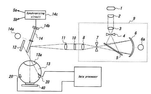
Fig. 1 is a construction view of a raw milk spectra-measuring
apparatus as one embodiment of the present invention.
-5-
CA 02376173 2001-11-30
01805 (PGT/1P01/02018)
Fig. 2 is a block diagram constituting an example of an electric
construction of the raw milk spectra-measuring apparatus.
Fig. 3 is a sectional view of a sample holder 40.
Fig. 4 is a graph showing an example of near infrared ray
spectra of a number of raw milk samples in a wavelength range of
400 nm to 1100 nm.
Fig. 5 is a graph showing an example of near infrared ray
spectra of a number of raw milk samples in a wavelength range of
1100 nm to 2500 nm.
Fig. 6 is a figure for illustrating a procedure for performing a
PLS method as one form of multivariate regression analyses according to
the present invention.
Fig. 7 gives graphs showing the number of PLS factors used in
respective calculations and analysis errors.
Fig. 8 is a graph showing the correlation between the number
of somatic cells presumed by the PLS method and the actual number of
the somatic cells when raw milk samples from cows were used.
Fig. 9 is a graph showing the correlation between the number
of somatic cells presumed by the PLS method and the actual number of
the somatic cells when urine samples from cows were used.
Embodiments of working the resent ~;~vention
The mastitis-diagnosing apparatus according to the present
invention will be explained with reference to Fig. 1.
Fig. 1 is a construction view of a raw milk spectra-measuring
apparatus as one embodiment of the present invention. As viewed in
the light-progressing direction, this apparatus comprises a light source
for generating measuring light rays, a lens 2 for making light rays from
the light source 1 in parallel to one another, a spectroscope 9 for taking
out desired light rays through separating the light rays from the light
source 1, a filter 10 for cutting off a high light portion of the light rays
emitted from the spectroscope, a lens 11 for collecting the separated
light rays, a reflection mirror 12 for reflecting the light rays from the
lens 11, a light chopper 14 interposed between the lens 11 and the
-6-
CA 02376173 2001-11-30
ol8os ~PCruPOlro2o18~
reflection mirror 12, an integrating sphere 132 formed of a light-
diffusing material, a sample holder 40 for holding a sample, etc.
The light source 1 is constituted by a tungsten halogen lamp or
the like, which generates a wide wavelength range of the light rays
including near infrared rays. The spectroscope 9 comprises a lens 3 for
collecting the incident light rays, a slit 4 for regulating the size and the
amount of the light ray flux, a reflection mirror 5 for reflecting the light
rays having passed the slit 4, a diffraction grating 6 having a curved
surface, a motor 6a for controlling the diffraction angle of the diffraction
grating 6, a slit 7 for passing only a desired light portion among the light
rays diffracted at the diffraction grating 6, a light-emitting lens 8 for
emitting the diffracted light rays in parallel to one another, etc. Only
the light rays with the desired wavelength can be selectively taken out by
the angular controlling with the motor 6a.
The light chopper 14 is designed in the form of a rotary disc in
which light-reflecting sections and light-permeating sections are
alternatively arranged, and the light rays coming from the lens 11 is
periodically reflected or passed by rotating the optical chopper 14
through driving a motor 14a. A chopper sensor 14b detects the rotary
phase of the light chopper 14, and a synchronizing circuit 14c outputs
synchronizing signals Sa and Sb indicative of the reflected and passing
states for the light rays from the lens 11, respectively, based on the
signals from the chopper sensor 14b.
The integrating sphere 13 comprises an incident light window
13a opened upwardly, a light-emitting window 13b opened downwardly,
and plural light detectors 20 for converting amounts of received light
rays to electric signals. The integrating sphere 13 functions to diffuse
the light rays entering the sphere to reduce measurement errors.
The detector 20 is constituted by PbS or the like, which has a sensitivity
in the near infrared ray region. The sample holder 40 is arranged near
the light-emitting window 13b.
If the light rays, which are separated by the spectroscope 9, are
reflected with the light chopper 14, the light rays come into the sample
CA 02376173 2001-11-30
. ~ 01805 (PCT1JP(11/02018)
holder 40 as it is through the integrating sphere 13 via the incident light
window 13a. As a result, return light rays diffuse in the integrating
sphere 13, so that a part of the light rays are received with the light
detectors 20. On the other hand, if the light rays separated with the
spectroscope 9 passes the light chopper 14, the light rays are reflected
with the reflection mirror 12, so that the light rays enter obliquely into the
integrating sphere 13 via the incident light window 13a. Consequently,
the light rays are diffused without roaching the sample, and a part of that
light rays are received by the light detector 20. The above operation of
the chopper takes out signals influenced with the sample and those not
influenced with the sample.
Fig. 2 is an example of a block diagram showing an electric
construction of the data processor of the raw milk spectra-measuring
apparatus. Detection signals from light detectors 20 are amplified with
an amplifier 21, and inputted to a sample holding circuit 22 for sampling
with synchronizing signals Sa and a sample holding circuit 23 for
sampling with synchronizing signals Sb. The sample holding circuit 22
holds a signal voltage only during a sampling time period when the light
rays enter the sample from the spectroscope 9, whereas the sample
holding circuit 23 holds the electcic signals only during the sampling
time period when the light rays do not enter the sample from the
spectroscope 9. Then, autgut signals from the samgle holding circuits
22 and 23 arc logarithmically converted with logarithmically converting
circuits 24, 25, respectively, which are subjected to subtraction between
them in a subtraction circuit 26. Disturbance components can be
removed through detection in synchronization with the light chopper 14.
Output signals from the subtraction circuit are quantized with
an AD (analogueldigital) converter 27, which is led into a personal
computer (PC) 30 in which various programs are installed to effect data
processing according to the multivariate regression method. To the PC
30 are connected a keyboard 28 for inputting data, a display 29 for
displaying the data, etc.
Fig. 3 is a sectional view showing the construction of a sample
_g_
CA 02376173 2001-11-30
01805 (PCT/JP01/02018)
holder 40. This sample holder 40 fits to the configuration of the light-
emitting window 13b of the integrating sphere 13. The sample holder,
which is made of a heat-conductive material such as aluminum,
comprises a sample container 41 for holding a liquid sample SP such as a
raw milk, a transparent cover glass plate 42 for covering an opening of
the sample container 41, a Peltier element for heating or cooling the
sample container 41, a temperature sensor 45 for controlling the temper-
ature of the sample container 41, a temperature-controlling circuit 44 for
stabilizing the temperature of the sample SP by driving the Peltier
element based on temperature signals from the temperature sensor 45, etc..
When the light rays reflected from the light chopper 14 enter
the sample SP via the cover glass plate 42, they return into the integrat-
ing sphere 13 again after being attenuated and scattered depending upon
the absorption spectra of the sample SP. Consequently, a part of the
returned light rays are received by the light detector 210 where they are
converted to electric signals.
Since the absorbance of the raw milk is sensitive to changes in
temperature and less influence of fat in the raw milk has to be achieved,
significance of the measurement may be lost if the measurement
environmental temperature changes every measurement. Thus, according
to this embodiment, the temperature of the sample SP is stabilized by the
temperature feed-back system constituted by the temperature sensor 45,
the temperature-controlling circuit 44 and the Peltier element 43, thereby
enhancing the measuring precision.
Fig. 4 is a graph showing an example of near infrared spectra
of the raw milk wherein an ordinate gives absorbances represented by
figures obtained by logarithmically converting reciprocals of light
reflectances, and an abscissa denotes wavelengths (nm). A curve
corresponds to an absorption spectra obtained by scanning over a
wavelength of 400 nm to 1100 nm with use of the spectroscope 9 in
Fig. 1. In Fig. 4, results obtained by measuring plural raw milk samples
are displayed in an overlapped state. Fig. 5 is also a graph showing an
example of near infrared spectra of the raw milk wherein an ordinate
-9-
CA 02376173 2001-11-30
01805 (PCT/JPO1 /02018)
gives absorbances represented by figures obtained by logarithmically
converting reciprocals of light reflectances, and an abscissa denotes
wavelengths (nm). A curve corresponds to an absorption spectra
obtained by scanning over a wavelength of 1100 nm to 2500 nm with use
of the spectroscopy 9. In Fig. 5, results obtained by measuring plural
raw milk samples are displayed, while overlapped.
All the curves are attributable to absorption spectra of water,
and large peaks particularly near 1400 nm to 1500nm and near 1850 nm
to 2050 nm are attributable to molecular vibrations of water.
The above explanation is made on the transmission and
reflection type construction where the light rays to be measured pass the
sample SP, reflected at the inner surface of the sample container 41 and
pass the sample SP again. In addition, measurement may be also made
by a transmission type where the sample container 41 is made of a
transparent material, and a transmitted lights having passed the sample
SP are detected or a reflection type where ..the light rays reflected from
the surface of the sample SP are measured.
The above explanation is made on the construction example
where the spectroscope 9 is arranged between the light source 1 and the
sample SP and the light rays to enter the sample SP are separated.
In addition, a construction example may be used, where the spectroscope
9 is arranged between the sample SP and the light detector 20, and the
transmitted light rays from the sample SP or the transmitted and reflected
light rays are split.
Next, the multivariate regression analysis will be explained.
According to the multivariate regression method, relationship
between a certain quantitative property of samples (corresponding to
concentration or number of somatic cells in this application, for example)
and one or more explanatory variants is established. In this application,
the explanatory variable means a near infrared spectrum measured.
However, the near infrared spectrum is actually constituted by not a
single absorbance but absorbances at so many wavelengths. Therefore,
the invention method in which the quantitative property (number of the
-10-
CA 02376173 2001-11-30
01805 (PCT/JPOI/02018)
somatic cells in this application) of the samples known from utterly
different measurements are presumed by using near infrared spectra as
multivariate data is a so-called multivariate regression analysis. In this
application, the multivariate regression analysis is used to find out the
relationship to the number of the somatic cells as the property to be
presumed, based on the near infrared spectra as the multivariate data.
In the following, a PLS method (Partial Least Squares
Regression) will be explained as one of the multivariate regression
analyses (References: Tetsuro Aijima, "Chemometrics" published by
Maruzen in 1991, and H. Martens & T. Naes, "Multivariate Calibration"
published by John Willy, New York in 1991). As in Fig. 4, if scanning
is effected at a resolution of 2 nm over a wavelength range of 700 nm to
1100 nm, 201 data are obtained per one scanaing.
As a pretreatment for the spectra, a Kubelka-Munk conversion
in which an apparent reflection index is converted to a rstio between an
absorptivity K(~.)and a scattering index S(~.), a smoothing treatment such
as moving average, a one- or two-dimensional differential treatment, a
base line correction treatment or the tike is effected, if necessary.
Fig. 6 is an explanatory figure for illustrating a procedure for
implementing the PLS method according to the present invention.
An absorbance at one wavelength point is one-dimensional data
constituted by plural components (somatic cells, proteins, lipids, ete. in
the raw milk here or constituting components of the mammary gland),
and ono spectrum includes multi-dimensional data at M wavelength
points in one scanning over the wavelengths. Therefore, one spectra
data constitutes 1-dimensional data x M orthogonal coordinate spaces in
the multivariate analysis of the somatic cells.
Next, variations in spectra data X and those in the number of
somatic cells are represented by factor scores tl, t2, t3, ---, ta, ---, th
mutually orthogonal to one another with use of a orthogonal type PLS
method. Fig. 6 shows a procedure for forming factor parameters of an
a-th factor. E is a spectra residual matrix (matrix of number of wave-
length data paints x sampled number of samples), t a factor score, w a
-11-
CA 02376173 2001-11-30
01805 (PCT/1P01/02018)
loading weight, and p and q X- and Y-loadings, respectively.
A spectra residual matrix Ea-1 for an a-,1 factor is represented
as a sum of a product between a factor score to and a loading weight wa
of the a-th factor, a product between a factor score to and a X-loadiag,
and the spectra residual matrix Ea of the a-th factor. An error matrix
fa-1 of the somatic cells for the (a-1)th factor is represented by a sum of
a product between the factor score to and the Y-loading qa and an error
matrix fa of the number of the somatic cells of the a-th factor.
Therefore, as an analysis procedure, the factor score ta, the
loading weight wa, the X-loading pa and the Y-loading qa for the (a-1)th
factor are calculated based on the spectra residue matrix Ea-1 and the
somatic cell number error matrix fa-1 for the (a-1)th factor, and then the
spectra residue matrix Ea and the somatic cell number error matrix fa are
calculated.
More concretely, a first residual matrix EO = X - Xm (Suffix m
means "average value") of the spectra data and a first error matrix f0 = Y
- Ym of the number of the somatic cells are calculated, and parameters tl,
wl, p1 and ql of the first factor are calculated. Then, a spectra residual
matrix E1 and a somatic cell number error matrix f1 are newly formed by
removing an influence of the first factor. Next, parameters t2, w2, p2
and q2 of the second factor are calculated by using the spectra residual
matrix E1 and the somatic cell number error matrix fl. Then, a spectra
residual matrix E2 and a somatic cell number error matrix f2 are newly
formed by removing an influence of the second factor. Next,
parameters t3, w3, p3 and q3 of the thiid factor are calculated by using
the spectra residual matrix E2 and the somatic cell number error matrix
f2. Then, a spectra residual matrix E3 and a somatic cell number error
matrix f3 are newly formed by removing an influence of the third factor.
Subsequently, calculations of parameters for each factor and formations
of each of residual matrixes and error matrixes are repeated in the same
manner consecutively until no effective factors are extracted.
Fig. 7 gives graphs showing the number of PLS factors and
analysis errors, which graphs are related to the number of the somatic
-12-
CA 02376173 2001-11-30
01805 (PCT/JP01 /02018)
cells. The ordinate gives the Log number of the somatic cells.
Fig. 7(a) gives data (SEC: Standard Error of Calibration) obtained by
measuring the number of the somatic cells when a sample having a
known number of the somatic cells, and Fig. 7 gives data (SEP: Standard
Error of Prediction) obtained by measuring the number of the somatic
cells when a sample having an unknown number of the somatic cells was
used.
From these graphs, it is seen that when the PLS factor is 5,
that is, when error calculations are repeated until the 5th factor, both of
the SEC value and the SEP value converge in an error range of about
0.25.
When the PLS factors up to the 5th one in Fig. 7 are used, a
model which enables the number of the somatic cells to be calculated
inversely from the near infrared spectra can be constructed. Therefore,
a presumed number of somatic cells of a sample can be calculated by
applying a near infrared spectrum related to the unknown number of the
somatic cells to this model.
Fig. 8 is a graph showing the correlation between a presumed
number of somatic cells according to the PLS method and the actual
number of the somatic cells. The abscissa gives the actual number of
the somatic cells, and the ordinate the presumed number of the somatic
cells. From this figure, it is seen that there is a strong correlation in
that figures between both cases are distributed in an almost linear
fashion. This shows that the multivariate analysis according to the PLS
method is extremely effective.
Next, a MLR method and a PCR method will be described.
According to the MLR method, a linear regression formula is constructed
with respect to the number of somatic cells obtained in the measurement
for a known sample by using only absorbances at plural specific
wavelengths among new infrared spectra consisting of absorbances as
multi-variations. An unknown number of somatic cells can be predicted
by applying a near infrared spectrum for the unknown number of the
somatic cells to be predicted to the thus constructed linear regression
-13-
CA 02376173 2001-11-30
01805 (PCT/IP01 /02018)
formula. In the PCR method, substantially the same calculation is
effected as in the PLS method (Reference: Yoshikatu Miyashita 8c Sin-
ichi Sasaki, "Kemometria - Recognition and Multivariate analysis of
chemical patterns" published by Kyoritsu Publisher in 1995).
Fig. 9 is a graph showing a correlation between a presumed
number of somatic cells according to the PLS method and the actual one
when urines of cows were used. Urine spectra were determined by the
same measuring apparatus and the same measuring method as in the
above raw milk spectra. From Fig. 9, it is sccn that there is a strong
correlation in that the actual numbers of the somatic cells in the abscissa
and the presumed numbers of the somatic cells in the ordinate are
distributed in an almost linear fashion. This shows that the multivariate
analysis according to the PLS method is extremely effective.
Effects of the Invention
As mentioned above in detail, according to the present
invention, the number of the somatic cells can be measured by detecting
the intensity of transmitted light rays, reflected light rays or transmitted
and reflected light rays from urine, raw milk or mammary gland, and
effecting the multivariate regression analysis of the obtained absorbance.
The number of the somatic cells caa be readily measured at a
high precision by the optical measurement of urine and data processing
of the raw milk or the mammary gland.
- 14-