Note: Descriptions are shown in the official language in which they were submitted.
CA 02376312 2001-12-10
WO 00/74607 PCT/US00/15654
KEYED INTERVERTEBRAL DOWEL
BACKGROUND OF THE INVENTION
1. Technical Field
The present disclosure relates to an intervertebral implant for spinal
fusion and more particularly, to an intervertebral dowel having at least two
radially
extending tabs for securing the dowel within a receiving bed formed in the
intervertebral space.
2. Background of Related Art
The spine is a flexible column formed of a series of bone called
vertebrae. The vertebrae are hollow and piled one upon the other, forming a
strong
hollow column for support of the cranium and trunk. The hollow core of the
spine
houses and protects the nerves of the spinal cord. The different vertebrae are
connected together by means of articular processes and intervertebral, fibro-
cartilages.
In general, a vertebral body is made of a cortical shell enclosing a
cancellous (spongy)
bone core. The portion of the cortical bone shell facing the surface of the
disk is the
endplate.
The intervertebral fibro-cartilages are also known as intervertebral disks
and are made of a fibrous ring filled with pulpy material. The disks function
as spinal
shock absorbers and also cooperate with synovial joints to facilitate movement
and
maintain flexibility of the spine. When one or more disks degenerate through
trauma,
-1-
WO 00/74607 CA 02376312 2001-12-10
PCT/US00/15654
spondylolisthesis or other pathologies, nerves passing near the affected area
may be
compressed and are consequently irritated. The result may be chronic and/or
debilitating back pain. Various methods and apparatus, both surgical and non-
surgical,
have been designed to relieve such back pain.
One method designed to relieve such back pain is interbody spinal
fusion. Typically, interbody spinal fusion involves distracting adjoining
vertebrae of
the spine so that the nerve root canal sizes are increased and nerve
irritation is
eliminated or reduced. In order to maintain the adjoining vertebrae in a
distracted
state, at least one intervertebral implant is inserted into a receiving bed
formed between
the vertebrae. The implant is positioned to engage the adjoining vertebrae to
maintain
the vertebrae at a fixed degree of distraction.
Preferably, the implant should stabilize the intervertebral space and
become fused to adjacent vertebrae in order to prevent the implant and
adjacent
vertebrae from moving. The implant must also provide spinal load support
between the
vertebrae. Further, during the time it takes for fusion, i.e. biological
fixation of the
vertebrae, to be completed, the implant should have enough structural
integrity to
maintain the space without substantial degradation or deformation of the
implant. The
implant should also have sufficient stability to remain in place prior to
actual
completion of bone ingrowth fusion. The implant should include structure which
maintains the implant in position between the vertebrae while bone ingrowth is
occurring. To facilitate rapid bone growth, and thus quick fusion, the implant
may
-2-
WO 00/74607 CA 02376312 2001-12-10 pCT/US00/15654
include or be provided with a bone growth supporting material. Obviously, the
material from which the implant is constructed should be a biocompatible
material and,
preferably, interact biologically with the body's own naturally occurring
tissues.
A variety of different types of intervertebral implants have been
developed to perform this function including spinal fusion cages, threaded
bone dowels
and stepped bone dowels. An exemplary implant is disclosed in U.S. Patent
Application filed on even date herewith, under Certificate of Express Mail
Label No.
EL260888076US, and entitled "Ramp-Shaped Intervertebral Implant" , the entire
disclosure of which is incorporated by reference herein.
Common deficiencies in some of the prior art implants may include
expulsion of the implant from between adjacent vertebrae, difficulty in
inserting the
implant into position, and/or lack of ability to allow incorporation of
implant into the
body. Also, in some prior art spinal fusion methods utilizing implants, the
vertebrae
may need to be distracted to a large extent in order to position the implant
between the
vertebrae.
Accordingly, a need exists for an improved intervertebral implant which
is configured to prevent the likelihood of expulsion or retropulsion during
normal
patient activity, provide ease of insertion and include structure to
facilitate
incorporation of the implant into the body. Furthermore, need exists for an
improved
intervertebral implant which can be inserted between vertebrae without
excessive
distraction of the vertebrae and a method of installing such an implant.
-3-
WO 00/74607 CA 02376312 2001-12-10
PCT/US00/15654
SUMMARY
In accordance with the present disclosure, an intervertebral implant
having tabbed securing structure is provided. The intervertebral implant
includes a
substantially cylindrical body portion and at least one pair of radially
extending tabs
that are configured to engage vertebral bodies.
By engaging the vertebrae, the tabs reduce the likelihood that expulsion
or retropulsion might occur. This is particularly significant in that where an
implant is
pushed out of place, damage to vital structures including neural (the spinal
cord and
existing nerve roots) and vascular (the aorta and inferior vena cava) can
occur resulting
in possible injury or death. Additionally, the tabs assist in preventing
migration of the
implant due to rotation of the adjacent vertebrae.
The tabs may take the form of various shape and constructions, such as,
for example, smooth rounded, wedge shaped, cam shaped, toothed, or threaded,
etc.
In alternate embodiments, two diametrically opposed pairs of tabs are provided
on the
cylindrical body portion. In various embodiments, a throughbore or a plurality
of
throughbores extend from a top surface of the implant to the bottom surface of
the
implant providing a space for boney bridging to occur between the vertebrae
which are
intended to be fused. The throughbore(s) is dimensioned to receive growth
factors or
other grafting materials to stimulate bone healing. The pairs of tabs may be
provided
adjacent the opening of the throughbore or may be offset 90° from the
openings of the
throughbore. In one embodiment of an intervertebral implant, the cylindrical
body
portion is tapered.
-4-
CA 02376312 2001-12-10
WO 00/74607 PCT/US00/15654
In an alternate embodiment, the implant has an abbreviated body portion
and does not include a throughbore.
In another embodiment, the tabs are formed by inserting a cortical plug
through the throughbore. Preferably, the cylindrical body portion includes a
slot
formed in one end thereof for receipt of an insertion tool and a bore
extending between
the slot and into the throughbore for facilitating insertion and facilitating
injection into
the throughbore of any desirable material, such as, for example, bone growth
stimulants, autograft, allograft, demineralized bone matrix, or other bone
grafting
materials.
Further, alternate embodiments may include body portions having shapes
other than cylindrical, such as, those having rectangular, oval, mufti-sided,
etc., cross-
sections.
In a preferred embodiment, the implant is formed from a cortical ring
allograft cut from the diaphysis of a long bone. By utilizing bone or bone-
derived
materials as the implant material, the implant has the added advantage of
facilitating
incorporation of the implant into the body. The implant can be formed by
milling the
top and bottom surfaces of a cortical ring to form the substantially
cylindrical body
portion and a pair of radially extending wings. The implant is further milled
such that
the radially extending wings are formed into tabs each of which is spaced a
predetermined distance from the end of the cylindrical body portion.
Additionally,
each tab may be milled so as to form the desired camming, wedge, threaded,
etc.
-5-
CA 02376312 2001-12-10
WO 00/74607 PCT/US00/15654
shape. The implant is milled such that the intramedullary canal of the
cortical ring
defines a throughbore in the cylindrical body portion of the implant.
Alternatively, the
implant may be formed of any biocompatible material such as titanium and
titanium
alloys, stainless steel, carbon fiber, ceramics, etc. having the requisite
strength
requirements via any known process, i.e., molding, machining, etc. Further, it
is
preferable that the implants be surface demineralized prior to use by exposing
them to
acid or other demineralizing solutions.
Preferably, the bone should be surface demineralized prior to use.
Where partially or surface demineralized bone is utilized, such bone can be
obtained
employing known demineralization techniques, e.g., those employing strong
acids such
as hydrochloric acid as described in Reddi et al., Proc. Nat. Acad. Sci. 69,
pp. 1601-
1605 (1972), the entire disclosure of which is incorporated herein by
reference. The
extent of demineralization is a function of the strength of the acid solution,
the shape of
the bone and the duration of the demineralization treatment as disclosed in
Lewandrowski et al., J. Biomed. Materials Res., 31, pp. 365-372 (1996) the
disclosure
of which is incorporated by reference herein. The use of partially or surface
demineralized bone is beneficial since such substances exhibit greater initial
osteogenic
and/or osteoinductive activity than fully mineralized bone.
There is also disclosed a method of inserting the tabbed implant between
adjacent vertebrae. The method involves forming a stepped bore between
adjacent
vertebrae, providing an intervertebral implant having a cylindrical body
portion and at
-6-
CA 02376312 2001-12-10
WO 00/74607 PCT/US00/15654
least one pair of diametrically opposite radially extending tabs extending
from the
cylindrical body portion and inserting the implant between adjacent vertebrae
such that
the tabs are in alignment with the space defined between adjacent vertebrae.
The
method further includes positioning the implant such that the tabs are within
the
enlarged areas of the stepped bore and rotating the implant such that the tabs
enter the
enlarged or stepped area of the bore. This provides a greater ease of
insertion over
other styles of implants, such as, for example, threaded implants.
BRIEF DESCRIPTION OF THE DRAWINGS
Various preferred embodiments are described herein with reference to
the drawings wherein:
FIG. 1 is a perspective view of one embodiment of the presently
disclosed intervertebral implant;
FIG. 2 is a side view of the intervertebral implant shown in FIG. 1;
FIG. 3 is a top view of the intervertebral implant shown in FIG. l;
FIG. 4 is a front view of the intervertebral implant shown in FIG. 1;
FIG. 5 is a side view of a long bone;
FIG. 6 is a perspective view of a ring cut from the long bone shown in
FIG. 5;
FIG. 7 is a side view of the ring shown in FIG. 6;
FIG. 8 is a perspective view of the ring after the top surface has been
milled;
CA 02376312 2001-12-10
WO 00/74607 PCT/US00/15654
FIG. 9 is a perspective view of the ring after the bottom surface has
been milled;
FIG. 10 is a perspective view of the ring after the side walls have been
machined;
FIG. 11 is a perspective view of the ring after the radially extending
wings have been machined to form tabs;
FIG. 12 is a an end view of the vertebral space with a stepped hole
drilled therein;
FIG. 13 is a side view of the vertebral space shown in FIG. 12;
FIG. 14 is an end view of the vertebral space of FIG. 12 with one
embodiment of the presently disclosed intervertebral implant inserted therein;
FIG. 15 is a perspective view similar to FIG. 14 with the intervertebral
implant rotated 90°;
FIG. 16 is a side view of the intervertebral space similar to FIG. 13 with
the intervertebral implant inserted and rotated 90 ° ;
FIG. 17 is a perspective view of another embodiment of the presently
disclosed intervertebral implant;
FIG. 18 is a side view of the intervertebral implant shown in FIG. 17;
FIG. 19 is a top view of the intervertebral implant shown in FIG. 17;
FIG. 20 is a front view of the intervertebral implant shown in FIG. 17;
_g_
CA 02376312 2001-12-10
WO 00/74607 PCT/US00/15654
FIG. 21 is a perspective view of another embodiment of the presently
disclosed intervertebral implant;
FIG. 22 is a side view of the intervertebral implant shown in FIG. 21;
FIG. 23 is a top view of the intervertebral implant shown in FIG. 21;
FIG. 24 is a front view of the intervertebral implant shown in FIG. 21;
FIG. 25 is a perspective view of another embodiment of the presently
disclosed intervertebral implant;
FIG. 26 is a side view of the intervertebral implant shown in FIG. 25;
FIG. 27 is a top view of the intervertebral implant shown in FIG. 25;
FIG. 28 is a front view of the intervertebral implant shown in FIG. 25;
FIG. 29 is a perspective view of another embodiment of the presently
disclosed intervertebral implant;
FIG. 30 is a side view of intervertebral implant shown in FIG. 29;
FIG. 31 is a top view of the intervertebral implant shown in FIG. 29;
FIG. 32 is a front view of the intervertebral implant shown in FIG. 29;
FIG. 33 is a perspective view of another embodiment of the presently
disclosed intervertebral implant;
FIG. 34 is a side view of the intervertebral implant shown in FIG. 33;
FIG. 35 is a top view of the intervertebral implant shown in FIG. 33;
FIG. 36 is a front view of the intervertebral implant shown in FIG. 33;
-9-
CA 02376312 2001-12-10
WO 00/74607 PCT/US00/15654
FIG. 37 is a perspective view of another embodiment of the presently
disclosed intervertebral implant;
FIG. 38 is a side view of the intervertebral implant shown in FIG. 37;
FIG. 39 is top view of the intervertebral implant shown in FIG. 37;
FIG. 40 is a front view of the intervertebral implant shown in FIG. 37;
FIG. 41 is a perspective view of another embodiment of the presently
disclosed intervertebral implant;
FIG. 42 is a side view of the intervertebral implant shown in FIG. 41;
FIG. 43 is a top view of the intervertebral implant shown in FIG. 41;
FIG. 44 is a front view of the intervertebral implant shown in FIG. 41;
FIG. 45 is a perspective view of another embodiment of the presently
disclosed intervertebral implant;
FIG. 46 is a side view of the intervertebral implant shown in FIG. 45;
FIG. 47 is a top view of the intervertebral implant shown in FIG. 45;
FIG. 48 is a front view of the intervertebral implant shown in FIG. 45;
FIG. 49 is a perspective view of another embodiment of the presently
disclosed intervertebral implant;
FIG. 50 is a side view of the intervertebral implant shown in FIG. 49;
FIG. 51 is a top view of the intervertebral implant shown in FIG. 49;
-10-
WO 00/74607 CA 02376312 2001-12-10
PCT/LTS00/15654
FIG. 52 is a front view of the intervertebral implant shown in FIG. 49;
FIG. 53 is a perspective view of another embodiment of the presently
disclosed intervertebral implant;
FIG. 54 is a side view of the intervertebral implant shown in FIG. 53;
FIG. 55 is a top view of the intervertebral implant shown in FIG. 53;
FIG. 56 is a front view of the intervertebral implant shown in FIG. 53;
FIG. 57 is a perspective view of another embodiment of the presently
disclosed intervertebral implant;
FIG. 58 is a side view of the intervertebral implant shown in FIG. 57;
FIG. 59 is a top view of the intervertebral implant shown in FIG. 57;
FIG. 60 is a front view of the intervertebral implant shown in FIG. 57;
FIG. 61 is a perspective view of another embodiment of the presently
disclosed intervertebral implant body portion with a rectangular cross-
section;
FIG. 62 is a perspective view of another embodiment of the presently
disclosed intervertebral implant body portion with an oval cross-section; and
FIG. 63 is a perspective view of another embodiment of the presently
disclosed intervertebral implant body portion with a multi-sided cross-
section.
DETAILED DESCRIPTION OF PREFERRED EMBODIMENTS
Preferred embodiments of the presently disclosed intervertebral implant
will now be described in detail with reference to the drawings, in which like
reference
numerals designate identical or corresponding elements in each of the several
views.
-11-
CA 02376312 2001-12-10
WO 00/74607 PCT/US00/15654
The spinal interbody fusion devices or intervertebral implants according
to the present disclosure are intended to be placed between adjacent vertebrae
in an
attempt to correct a debilitating degeneration of the spinal structure. In
humans, the
device may be used predominantly in the lumbar region of the spine, but is
adjustable
for use in the thoracic and cervical regions as well. When in place, the
device supports
and maintains an appropriate distance between vertebrae and causes bone tissue
to form
and become integral with the device. Consequently, the intervertebral space
becomes
filled with autologous bone tissue and forms an integral rigid bone
construction
between adjacent vertebrae. While the disclosed implants and methods are
discussed in
terms of humans, it is contemplated that the disclosed implants and methods
may fmd
beneficial use in veterinary applications.
The disclosed intervertebral implants are formed with a tabbed
configuration which allows the implants to be inserted between the vertebrae
and
twisted or rotated to secure the implant in position between the vertebrae.
This has the
resultant benefits of reduced likelihood of expulsion. Furthermore, the
implants
disclosed herein also allow insertion of the implant between the vertebral
space without
excessive distraction between the vertebrae.
Referring now to FIGS. 1-4, there is illustrated one embodiment of the
presently disclosed intervertebral implant shown generally as 10. Briefly,
intervertebral implant 10 includes a substantially cylindrical body portion 12
having a
pair of diametrically opposed and radially extending tabs 14 and 16.
Cylindrical body
-12-
CA 02376312 2001-12-10
WO 00/74607 PCT/US00/15654
portion 12 has a first end 18 and a second end 20. Tab 14 has first and second
engaging or retaining surfaces 22a and 22b which are stepped or longitudinally
spaced
a predetermined distance from first end 18 and second end 20, respectively.
Similarly,
tab 16 has a pair of retaining surfaces 24a and 24b which are similarly
stepped or
longitudinally spaced from a first end 18 and second end 20 respectively.
Retaining
surfaces 22a, 22b and 24a, 24b are configured to engage a portion of adjacent
vertebrae
when installed therebetween.
As shown, tabs 14 and 16 extend only along a limited extent of the
circumference of a cylindrical body portion 12. Preferably, tabs 14 and 16 are
radially
spaced 180° apart. Tab 14 includes a rounded side surface 26 and tab 16
includes a
rounded side surface 28.
As shown, implant 10 includes a throughbore 30 which has a
longitudinal axis substantially perpendicular to the longitudinal axis of
implant 10.
Further, implant 10 may be provided with perforations instead of, or in
addition to,
throughbore 30. Where implant 10 is formed of bone, the perforations assist in
facilitating biological attachment and eventual incorporation of the implant
into adjacent
vertebrae.
Implant 10 further includes an installation slot 32 machined or milled in
first end 18. A second bore 34 extends between slot 32 and throughbore 30.
Second
bore 34 is provided for mating of the implant with an insertion tool.
Throughbore 30
is dimensioned to receive bone particles and/or biocompatible osteoinductive
or
-13-
CA 02376312 2001-12-10
WO 00/74607 PCT/US00/15654
osteoconductive material. These materials may include cancellous bone,
cancellous
bone particles, ceramics, polymers, composites, BMP, etc.
Intervertebral implant 10 can be constructed from a broad range of
biocompatible materials such as, for example, surgical stainless steel,
titanium,
ceramic, hydroxyapatite, polymer, carbon fiber, tantalum, etc. Preferably,
implant 10
is constructed from a human and/or animal cadaver bone. Intervertebral implant
10,
appropriately sized, can be used in cervical, thoracic and lumbar spinal
fusion
procedures. For cervical spinal fusion procedures, in which implants are
typically
between 8 to 15 mm in length and 10 to 14 mm in diameter, bone is preferably
obtained from the fibula, radius, ulna or humerus bones. For thoracic and
lumbar
spinal fusion procedures in which implants are typically 10 to 30 mm in length
and/or
diameter and about 10 to 14 mm in height, bone is preferably obtained from the
humerus, femur or tibia. The sources of cortical bone for the bone-derived
implant are
preferably allogenic but also include xenogenic sources such as bovine and
porcine
bone.
Additionally, the bone may be subjected to penetration with osteogenic
or demineralization agents during manufacture of the implant.
Alternatively, as discussed above, intervertebral implant 10 can be
molded or machined from other biocompatible materials including composites
made of
bone as discussed in U.S. Patent No. 5,899,939 to Boyce et al., the entire
disclosure of
which is incorporated by reference herein.
-14-
CA 02376312 2001-12-10
WO 00/74607 PCT/US00/15654
Referring now to Figs. 5-11, in one preferred embodiment,
intervertebral implant 10 is manufactured in accordance with the procedure
disclosed in
U.S. Patent Application filed on even date herewith under Certificate of
Express Mail
Label No. EL260888080US and entitled, "Intervertebral Implant", the entire
disclosure
S of which is incorporated by reference herein. In general, implant 10 is
manufactured
from a ring C formed by making transverse cuts through a long bone D along
lines A
and B as illustrated in FIG. 5. Next, the top 36 of ring C is machined using a
milling
device (not shown) having a dome or crown configuration to shape one side of
ring C
to have a semi-cylindrical portion 38 with two radially extending flats 40
(Fig. 8).
Ring C is flipped over and the same milling procedure is formed on a bottom 42
of
ring C as shown in Fig. 9. Next, the front and side surfaces are machined to
flatten the
side surface to reconfigure femoral ring C to have a generally rectangular
configuration
(Fig. 10). Finally, tabs 14 and 16 are formed by machine flats 40 so as to
provide
stepped surfaces from first and second ends 18 and 20 (Fig. 11). Additionally,
further
milling may be performed to provide rounded side surfaces 26 and 28 on tabs 14
and
16 respectively. It should be noted that throughbore 30 may be formed from a
medullary canal through the long bone and further milled to provide a uniform
throughbore 30 through ring C. While not shown, first end 18 may be further
milled
and/or drilled to provide installation slot 32 and bore 34 extending between
installation
slot 32 and an interior of throughbore 30. As discussed above, intervertebral
implant
10 need not be formed from cadaveric bone but rather may be formed from any
-15-
CA 02376312 2001-12-10
WO 00/74607 PCT/US00/15654
biocompatible material. As such, other known processes, such as molding
techniques
may be used to manufacture the implant.
Installation of implant 10 between a pair of adjacent vertebrae will now
be described. Referring to Figs. 12-16 and initially to Figs. 12-13, there is
illustrated
a pair of adjacent vertebrae X and Y defining intervertebral space Z
therebetween. The
endplate is stronger bone than is the cancellous core. Thus, cuts in the
vertebral bodies
permit the tabs of the implant to extend past the endplate and into the softer
bone
beneath. A caroming approach for some of the following disclosed embodiments
of the
implant tabs allows the cancellous bone to be compressed against the implant
thereby
providing additional frictional resistance against implant movement. A drill
or other
known devices and methods are utilized to form a stepped hole or bore E
between the
adjacent vertebrae preferably by milling or machining. Examples of such
devices and
procedures are disclosed in U.S. Patent No. 5,445,639, the entire disclosure
of which
is incorporated by reference herein. Stepped hole E preferably has narrow
diameter
portion F adjacent the outer surface of the vertebrae and enlarged portion G
interior to
the vertebrae. In preparation for use, intervertebral implant 10 may be
demineralized
as discussed hereinabove and mounted on suitable installation devices.
Referring now to Fig. 14, once installed on an insertion device,
intervertebral implant 10 is inserted between vertebrae X and Y such that tabs
14 and
16 are aligned with the intervertebral space Z. Intervertebral implant 10 is
inserted
into the drilled hole a sufficient distance such that tabs 14 and 16 align
with the
-16-
CA 02376312 2001-12-10
WO 00/74607 PCT/US00/15654
enlarged portion G of bore E. Implant 10 is subsequently rotated approximately
90°
such that tabs 14 and 16 rotate into enlarged portion G. As noted above,
retaining
surfaces 22a and 22b on tab 14 and retaining surfaces 24a and 24b on tab 16
engage
edges of enlarged portion G of bore E and prevent expulsion of the implant
from
between the adjacent vertebrae A and B. It should be noted that the entire
procedure
may be accomplished without any substantial or excessive distraction between
adjacent
vertebrae. While the present disclosure provides installation slot 37 and bore
32 for
receipt of an installation device, it is within the contemplated scope of the
present
disclosure to provide implant 10 with other structure to allow insertion and
rotation of
the implant by various insertion tools.
Referring now to FIGS. 17-19, there is disclosed an alternative
embodiment of an intervertebral implant. Intervertebral implant 50 is similar
to
implant 10 described above and generally includes cylindrical body portion 52
having a
throughbore 54 formed therein. An installation slot 56 is provided in a first
end 58 and
a bore 60 extends from slot 56 to the interior of throughbore 54 similar to
that
described above with respect to implant 10.
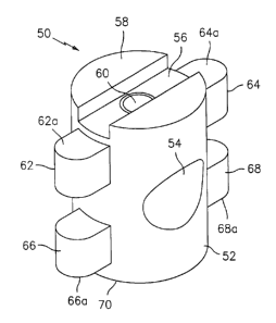
Implant 50 includes a pair of radially extending first tabs 62 and 64
adjacent to, and longitudinally displaced from, first end 58 and a pair of
second tabs 66
and 68 adjacent to, and longitudinally spaced from, a second end 70 of
cylindrical body
portion 52. Thus, first tabs 62 and 64 as well as second tabs 66 and 68 are
stepped
from first and second ends 58 and 70 respectively. First tabs 62 and 64
include
-17-
WO 00/74607 CA 02376312 2001-12-10
PCT/US00/15654
engaging surfaces 62a and 64a for engaging an edge of stepped bore in a
drilled
vertebrae. Similarly, second tabs 66 and 68 also include engaging surfaces 66a
and
68a for engaging an interior of a bore drilled in bone or vertebrae. Similar
to that
disclosed with regard to implant 10, first tabs 62 and 64 as well as second
tabs 66 and
68 may have a generally rounded profile.
Intervertebral implant 50 is formed in the manner disclosed above with
respect to implant 10 and is similarly installed in a stepped bore drilled in
adjacent
vertebrae. The stepped bore may have only a single enlarged area or may
include two
separate enlarged areas to accommodate the first and second tabs as the
intervertebral
implant is rotated into place.
Referring now to FIGS. 21-24, there is disclosed another alternate
embodiment of an intervertebral implant similar to that of implant 50.
Intervertebral
implant 80 includes a generally cylindrical body portion 82 having a
throughbore 84
formed therethrough. An installation slot 86 is provided along with a bore 88
extending between installation slot 86 and an interior of throughbore 84.
Implant 80
includes a pair of radially extending first tabs 90 and 92 as well as a pair
of radially
extending second tabs 94, 96. In contrast to implant 50, first tabs 90, 92 and
second
tabs 94, 96 are formed on cylindrical body portion such that they are
generally
perpendicular to slot 86 and are adjacent to throughbore 84:
In the presently disclosed embodiments where the tabs are adjacent to
the throughbore, a different method of forming the implant from bone is
necessary.
-18-
WO 00/74607 CA 02376312 2001-12-10
PCT/US00/15654
The bone will initially be cut parallel to the long axis of the long bone to
permit the
tabs to extend in a plane that transects the medullary canal. Subsequently,
the presently
disclosed methods of milling or machining the bone are performed to form the
body
portion and tabs. An installation shaft and bore between the installation slot
and
throughbore may be formed.
Referring now to FIGS. 25-28, there is disclosed another embodiment of
an intervertebral implant which includes specific wedging structure to prevent
the
implant from moving longitudinally within a bore. Implant 100 generally
includes a
cylindrical body portion 102 having a throughbore 104 formed therein. Similar
to
previous embodiments, implant 100 is provided with an installation slot 106
and a bore
108 extending between installation slot 106 and throughbore 104. Implant 10
also
includes a pair of radially extending first anterior tabs 110, 112 and a pair
of radially
extending second tabs 114, 116. As shown, first tabs 110 and 112 have curved
wedge
surfaces 118, 120. Similarly, second tabs 114 and 116 also include curved
wedge
surfaces 122 and 124. Wedge surfaces 118 and 120 of first tabs 110 and 112
curve
away from a first end 126 of implant 10 and wedge surfaces 122, 124 of second
tabs
114 and 116 curve away from a second end 128 of implant 100. The provision of
wedge surfaces on the tabs provides a range of ramming contact with the
interior of a
stepped bore drilled in adjacent vertebrae to thereby prevent expulsion of the
implant.
Referring now to FIGS. 29-32, there is disclosed a further alternate
embodiment of an intervertebral implant which includes progressive, radial
ramming
-19-
CA 02376312 2001-12-10
WO 00/74607 PCT/US00/15654
structure which, upon rotation of the implant, cams the implant into position
within a
stepped bore. Specifically, intervertebral implant 130 includes a cylindrical
body
portion 132 having a throughbore 34 formed therethrough. An installation slot
136
may be provided along with a bore 138 extending between installation slot 136
and
throughbore 134. Implant 130 additionally includes first tabs 140 and 142
formed
adjacent first end 144 and second tabs 146 and 148 formed adjacent a second
end 150.
As illustrated, first tabs 140 and 142 as well as second tabs 146 and 148 have
a
generally, progressively curved shape such as a spline shape or one defined by
a
polynomial-defined curve. Thus, first tabs 140, 142 include progressive
caroming
surfaces 152, 154. Second tabs 146 and 148 include progressive caroming
surfaces 156
and 158. Implant 130 may be formed in a manner similarly described above with
respect to implant 10.
Upon installation of implant 130, between adjacent vertebrae, implant
130 is rotated and progressive caroming surfaces 152, 154 and 156, 158 engage
walls
of the stepped bore in progressive fashion to firmly wedge implant 130 within
the
stepped bore and prevent any loosening or further rotation or reverse rotation
of
implant 130 within the stepped bore. The provision of progressive caroming
surfaces
allows for the use of implant 130 in bores which may not have been drilled
precisely or
to a constant/consistent diameter. Further, as noted above, caroming structure
on the
disclosed implants allows the tabs to compress the spongy bone to gain
additional
frictional force to secure the implant between the vertebrae.
-20-
CA 02376312 2001-12-10
WO 00/74607 PCT/US00/15654
Referring now to FIGS. 33-36, there is disclosed another alternate
embodiment of an intervertebral implant including caroming surfaces provided
on tabs
so as to allow the implant to be caromed within a stepped bore formed in
adjacent
vertebrae upon rotation of the implant. Specifically, implant 160 includes a
cylindrical
body portion having a throughbore 164 and installation slot 166 and a bore 168
extending between installation slot 166 and throughbore 164. A pair of
radially
extending first tabs 170, 172 and a pair of radially extending second tabs
174, 176 are
formed on cylindrical body portion 162. First tabs 170 and 172 have relatively
flat
caroming surfaces 178 and 180, respectively, formed thereon, while second tabs
174,
176 also include relatively flat caroming surfaces 182, 184, respectively,
formed
thereon. As with implant 130, rotation of implant 160 within a stepped bore
causes the
caroming surfaces 178, 180 and 182, 184 to engage sidewalls of the stepped
bore and
cam the implant therein to prevent further rotation. As with all prior
embodiments,
first tabs 170 and 172 also include caroming surfaces 170a, 172a and second
tabs 174,
176 include caroming engaging surfaces 174a, 176a to engage edges of stepped
bore
and prevent expulsion of the implant after it has been rotated into position
within the
stepped bore.
Referring now to FIGS. 37-40, there is disclosed a further alternate
embodiment of an intervertebral implant. Intervertebral implant 190 generally
includes
a cylindrical body portion 192 having a throughbore 194. Implant 190 includes
first
tabs 196 and 198 spaced a predetermined distance from first end 200 of
cylindrical
-21-
CA 02376312 2001-12-10
WO 00/74607 PCT/US00/15654
body portion 192. Implant 190 additionally includes second tabs 202 and 204
positioned adjacent and spaced a distance from second end 206 of cylindrical
body
portion 192. Implant 190 includes caroming structure formed on the first and
second
tabs which permits rotation of the implant in either direction upon
installation.
S Specifically, first tabs 196 includes opposed inclined caroming surfaces
208a and 208b
and first tab 198 also includes opposed inclined caroming 210a and 210b.
Similarly,
second tab 202 includes opposed inclined caroming surfaces 212a and 212b and
second
tab 204 includes opposed inclined caroming surfaces 214a and 214b. The opposed
inclined caroming surfaces allow the implant to be rotated in either direction
and still
achieve a caroming function within a stepped bore. As with prior embodiments,
first
tabs 196 and 198 include bore engaging surfaces 196a and 198a respectively.
Similarly, second tabs 202 and 204 include bore engaging surfaces 202a and
204a
respectively. Implant 190 may preferably be provided with an installation slot
216 and
a bore 218 extending between slot 216 and throughbore 194.
Referring now to FIGS. 41-44, there is disclosed a further alternate
embodiment of an intervertebral implant. Implant 220 generally includes
cylindrical
body portion 222 having a throughbore tube 224 defined therein. First tabs 226
and
228 and second tabs 230 and 232 extend radially from cylindrical body portion
222.
The first and second tabs of implant 220 include threaded structure which
allows the
implant to engage precut threads in a stepped bore formed between adjacent
vertebrae
or to act as teeth to cut into bone and thereby secure implant 220 within a
stepped bore
-22-
CA 02376312 2001-12-10
WO 00/74607 PCT/US00/15654
between adjacent vertebrae. Alternatively, the tabs may be grooved but not
necessarily
threaded. Specifically, first tab 226 includes a threaded surface 234 and
first tab 228
includes a threaded surface 236. Similarly, second tab 230 includes a threaded
surface
238 and second tab 232 includes a threaded surface 240. It should be noted
that the
number of threads on any individual tab may differ from the number on an
adjacent or
diametrically opposed tab. Preferably, an installation slot 242 is provided
having a
bore 244 extending between slot 242 and into throughbore 224.
Referring now to Figs. 45-48, there is disclosed an asymmetrical
embodiment of an intervertebral implant. Implant 250 generally includes a
cylindrical
body portion 252 having a first end 254 and a second end 256. A throughbore
258
extends through implant. A first tab 260 is provided a predetermined spaced
distance
from first end 254 and a second tab 262 is provided a predetermined spaced
distance
from second end 256. As shown, first and second tabs 260, 262 are radially
spaced
approximately 180°. First and second tabs 260, 262 may be of any of the
previously
described shapes in the prior embodiments and include respective camming
and/or
abutment bone engaging surfaces. Additionally, implant 250 may be provided
with an
installation slot 269 and a bore 266 and be formed in accordance with the
previously
described methods and of same or similar materials.
Referring now to Figs. 49-52, there is disclosed an intervertebral
implant 270 designed to utilize a plug, which may be formed from cortical
bone, to
form the tabs. Implant 270 generally includes a cylindrical body portion 272
formed in
-23-
CA 02376312 2001-12-10
WO 00/74607 PCT/US00/15654
accordance with the above described method such that the medullary canal
provides a
throughbore 274 in implant 270. A cortical plug 276 formed by turning on a
lathe,
milling, or other appropriate machining process. Plug 276 is positioned within
throughbore 274 which may be suitably drilled or otherwise prepared to receive
plug
276 such that first and second ends 278, 280 of cortical plug 276 extend
radially
outward from body portion 272. First and second ends 278, 280 thus form tabs
which,
when installed by the above described method, engage edges of a stepped bore
formed
in adjacent vertebrae. An installation slot 282 may be formed in an end 284 of
body
portion and a bore 286 extends between slot 282 and throughbore 274.
Referring now to Figs. 53-56, there is disclosed an alternate embodiment
of an intervertebral implant with a substantially shortened body portion.
Implant 290 is
designed to be provided in various diameters such that two or more implants
290 of
differing diameters may be used together to introduce the appropriate lordosis
into the
spine. Implant 290 generally is similar to the above described implants except
that the
length of a cylindrical body portion 292 is substantially abbreviated or
shortened.
Implant 290 may include any of the previously described versions of tabs and
preferably first and second tabs 294, 296. Implant 290 may also include an
installation
slot 298 and bore 300 extending between slot 298 and end face 302 of body
portion
292. However, it is not contemplated that implant 290 have a throughbore and
thus
implant 290 may be formed from bone extending up to, but not including, the
-24-
CA 02376312 2001-12-10
WO 00/74607 PCT/US00/15654
medullary canal of a long bone. Further, various body portion configurations,
such as,
for example, tapered, semi-conical, etc. are also envisioned.
Referring now to Figs. 57-60, there is disclosed another embodiment of
an intervertebral implant. Implant 310 generally includes a tapered
cylindrical body
portion 312 having a first end 314 and a second end 316. The diameter of first
end
314 is smaller than the diameter of second end 316. Implant 310 may be formed
by the
disclosed method and include a throughbore 318, an installation slot 320 and a
bore
322 extending from slot 320 to throughbore 318. Additionally, implant includes
first
tabs 324, 326 and second tabs 328, 330.
As best shown in FIGS. 61-63, various body portions other than
cylindrical are within the contemplated scope of the present disclosure. These
body
portions may include a body portion 340, having a rectangular cross-section
(FIG. 61),
a body portion 350 having an oval cross-section (FIG. 62), a body portion 360
having a
multi-sided cross-section (FIG. 63), etc. The embodiments disclosed in FIGS.
61-63
may obviously include structure similar or identical to that provided in
previously
described embodiments such as, for example, throughbores, installation slots,
bore and
all the various configurations and orientations of tabs.
It will be understood that various modifications may be made to the
embodiments disclosed herein. For example, differing or alternate tab
constructions
may be provided on a single implant. Additionally, the various configurations
may be
combined on individual tabs. Therefore, the above description should not be
construed
-25-
CA 02376312 2001-12-10
WO 00/74607 PCT/US00/15654
as limiting, but merely as exemplifications of preferred embodiments. Those
skilled in
the art will envision other modifications within the scope and spirit of the
claims
appended hereto.
-26-