Note: Descriptions are shown in the official language in which they were submitted.
CA 02376480 2001-12-12
WO 00/77491 PCT/CA00/00690
FIBER OPTIC MULTITASKING PROBE
FIELD OF THE INVENTION
The present invention relates to optical fiber probes for sensing
parameters simultaneously at different positions along the probe to provide a
spatial profile of the parameters. More particularly, the invention relates to
optical fiber probes for measuring multiple dosimetric parameters
simultaneously
at different positions along the probe in diagnostic and/or therapeutic
applications related to photodynamic therapy.
to
BACKGROUND OF THE INVENTION
Photodynamic therapy (PDT) dosimetry is currently based on two
principal approaches, termed explicit and implicit dosimetry as discussed in
for
example Wilson BC, Patterson MS, Lilge L. (1997), Extrinsic and intrinsic
Dosimetry For Photodynamic Therapy; Lasers in Medical Science 12: 182-
199. For explicit dosimetry the three parameters governing PDT efficacy
(fluence-rate, photosensitiser concentration, and molecular oxygen
concentration) need to be monitored throughout the treatment volume. However,
the current available fiber optic based dosimeters enable detection of
2 o parameters only at a single location, requiring several detectors, see for
example Lilge L., Molpus K., Hasan T. and Wilson B.C., (1998) Intraperitoneal
Photodynamic Therapy In A Murine Xenograft Model Of Human Epithelial
Ovarian Carcinoma: Light Dosimetry And Biological Response, Photochem.
Phtobiol. 83: 281-288, resulting in clinically unacceptable invasive
procedures.
As described in previous work, Lilge L., Haw T., Willson B.C. (1993)
Miniature Isotropic Optical Fibre Probes For Quantitative Light Dosimetry
In Tissue, Phys. Med. Biol. 38: 215-230, fluence-rate detectors need to
provide
good sensitivity and isotropy of response in order to quantify the light
intensity,
called fluence-rate, in a turbid media such as tissue. Using fluorescent dyes
with
1
CA 02376480 2001-12-12
WO 00/77491 PCT/CA00/00690
a fiber optic provides good response with isotropy provided by the inherently
isotropic fluorescence emission by molecules. It was shown that the
fluorescence intensity transmitted via the optical fiber to an opto-electronic
detector is correlated to the fluence-rate at the position of the fluorophores
s inside the tissue. However, individual calibration of the optical fiber
detector
probe response is required prior to use. The efficiency and efficacy of such
procedures would be enhanced by being able to monitor the radial dependence
of fluence rate, photosensitizer fluorescence and molecular oxygen during the
procedure.
Other use of lasers for therapeutic applications is well known, see for
example Wilson BC. Wyman Dr, Malon ED, Tracy R, Farrell T (1991) Energy
Delivery And Control For Interstitial Laser Hyperthemia And Laser
Photocoagulation of Solid Tumors In Vivo; Proc. Soc. Photo-opt. Instr. Eng.
1599: 333-342. Here, the invention provides advantages through measuring the
15 fluence rate profile, or for example, measuring the distribution of
exogenous
fluorophores.
United States Patent No. 5,082,630 discloses a fiber probe for immuno-
testing that uses fluorophore tags bound to an intermediate molecule which in
turn is bound to a protein coating on a fiber core. A light beam from the
fiber
2 o excites fluorescence in the fluorophores and when biomolecules being
detected
displace the fluorophores the fluorescent intensity decreases thereby
indicating
the presence of the biomolecules.
United States Patent No. 5,275,160 shows a fiber probe with a single dye
contained in a modified tip for radiance dosimetry. United States Patent No.
25 5,483,958 shows a fiber optic probe using a solid state fluorescent probe
joined
to the end of the optical fiber.
United States Patent No. 5,173,432 discloses a fiber optic sensor for the
detection of p02 using a luminescent dye encapsulated in a polymer matrix
attached to the end of the optical fiber using an 02 permeable membrane.
2
_ CA 02376480 2001-12-12
26-09-2001
i~:~44 HI_~_ a ~wHLIMR'HcR, = cP0 CAOOOOt~9C
wa____ __
United States Patent No. 3,~141,53p discloses a photocharnotherapy
dosimeter having a chemical cell at the~end of the optical fiber and United
States
Patent No. 5,851,225 discloses providing a las~r probe for PDT applications
having madifled surface configurations foncreating tight emission at different
wave lengths.
United States Patent No. 5,837,196 discloses using an array of different
binsensors comprised ~of biological binding partners to detect two or more
different species of biological partners. This system reties upon a fiber
bundle to
convey the information to the detector from the distal ends of the fibers.
1 o There is therefore a need for a single optical fiber probe capable of
pertanning several independent tasks simultaneously.
SUAIIMARY OF THE INVENTION
It is an object of the present invention to provide a multitasking optical
1s fiber probe, capable, of measuring the fluence-rate, photosensitizer
fluorescence . .
and pox at several different positions along the probe and to provide
increased
information relevant to PDT dvsim~try. ~ . . . . _ _ . , . .
it is also an object of the present invention to provide an optical fiber
probe that can be employed in other laser based therapeutic applications such
2 o as Barren's esophagus and irltsrstitial laser hyperthermia.
An advantage of the mukitasking probe of the present invention is that it
can be used to provide the parameters required for PDT fvr the entire tissue
volume in question. The probes of the present invention provide for
measurement of a single parameter at multiple locations along the axis of a
25 single optical fiber as wall as providing for measurement of multiple
parametet5
at multiple locations along the axis of the fiber.
in one aspect of the invention there is provided a multitasking optical fiber
probe for measuring dosimetric parameters, comprising;
an optical fiber including at least two sensor zones spaced along a length
3 0 , of said optical fiber, each sensor zone including an effective
photoac4ve
3
EmPf.ceit:~6/09/tA,MENDED SHEET
1-m~f nr 17~ D fll7~
CA 02376480 2001-12-12
26-0~-200 i ~0~ i'-_'_':44 ~. HILL ~ S~HUMRCh-i'er ~ cF0 _ ~I G/~oooOt'9y
urn r
constituent having an emission spectrum distinguishable in time domain or
spectral domain from emission spectra of photoacfive const~uants in all other
sensor zones, each photaactive constituent emitting light responsive to
excitation
by light incident on each sensor zone on the exterior of said fiber with some
of
the emitted light being coupled into said optical fiber; and
detection means optically coupled to said optical fiber, said detection
means being connected to processing means for deeonvoluting emission
spectrum from the photoactive constituent in each of said at least two sensor
zones to measure said dosimetric parameters along the length of the fiber.
1o In this aspect of the invention the sensor zones may be defined by spaced
circumferential slots formed in an outer cladding of said optical fiber, the
phvtosctive constituent being retain~ad in said slots.
Alternatively, the sensor zones may be formed by diffusing a photoactive
constituent into the fiber core at spaced locations along the length of the
optics(
fiber.
In another aspect of the invantlon than: is provided a method of
nioasuring dvsimetric parameters in tissue, comprising; _ . _ _ . ., _ _
providing an optics! fiber having at least two sensor zones along a length
of the optical fiber, each sensor zone including an effsctiye photoactive
2o constituent having an emission spectrum distinguishable in time domain yr
spe~ral domain from emission spectra of photoactive constituents in a!1 other
sensor zones, each photoactive constituent emitting light responsive to
exc'tta'~on
by light incident on each sensor zone on the exterior of said fiber with some
of
the emitted light being coupled into said optical fiber; and
detecting atui deconvoluting emission spectrum from the photoactive
constituent in each of said at least two sensor zones to measure said
dosimetric
parameters along the length of the fiber.
The dosimetric parameters include fiuence rates calculated from emission
spectra from the sensor zones in which the photoactive constituent is a
3o fluorophore, p02 calculated from emission spectra from the sensor zones in
4
AMENDED SHEET
EmPf .zei t:~6/031~~", ",..,~ I-rtlpt _nr ~ ~ i~ v nm
CA 02376480 2001-12-12
26-OS-200 ~ ~~'_ ~:='~ r~I~_' ~; S:=N:IMa:=Hcrr: ~ ~P~7 CAOOOOu9l
.. NO . _ _ _ _ ._..."
which the photoactive constituent is a photolumineacent phosphor, and
photosens'itizer concentration calculated from fluorescence radiation incident
on
said distal end face emitted by photosensitizers located in said tissue.
BRIEF DESCRIPTION GF THE DRAWINGS
. The invention will now be described, by way of non-limiting examples only,
refetence being had to the accompanying drawings, in which:
Figure 1 is a cross sectional view of a fiber optic probe constructed in
1 o accordance with the present invention;
Figure 2 is a cross sectional uiew of an al~mati're embodiment of a fiber
optic probe;
2D
34
5
AMENDED SHEET
Empf .zei t:26/t~9/~~..1 ,~,.~,,,, rmpt _nr _' 1 ih D MR
CA 02376480 2001-12-12
WO 00/77491 PCT/CA00/00690
Figure 3 is cross sectional view of another alternative embodiment of a
fiber optic probe;
Figure 4 is a plot of fluorescence emission intensity versus wavelength for
seven exemplary fluorophores used in the multitasking probes;
Figure 5a is a plot of fluorescence intensity verses wavelength giving the
average normalized fluorescence emission spectra of a sensor illuminated from
different azimuth angles;
Figure 5b is a plot of fluorescence emission intensity versus wavelength
giving the normalized fluorescence emission spectra of 10 individually
to manufactured probes, showing independence of manufacturing, hence,
responsivity calibration for each sensor is possible;
Figure 6 shows a polar diagram showing the isotropy of detector
response;
Figure 7 shows the emission spectra of a fiber optical detector probe
15 comprised of 2 sensors for individually exposed sensors and combined
exposure
of both sensors;
Figure 7a shows the emission spectra of a fiber optical detector probe
comprised of 2 sensors for individually exposed sensors and combined exposure
of both sensors;
2o Figure 8 is a perspective view of another embodiment of the fiber optic
probe constructed in accordance with the present invention specifically
adapted
to use in photodynamic therapy in Barrett ~ s esophagus; and
Figure 9 is cross sectional view of another alternative embodiment of a an
optical fiber probe in accordance with the present invention.
DETAILED DESCRIPTION OF THE INVENTION
Fiber Optic Probes For Fluence-Rate Dosimetry And Quantification Of p02
Fluence-rate is a physical quantity describing the power density [W/cm2j
of the PDT treatment light. Due to multiple scattering of light by biological
tissue,
6
CA 02376480 2001-12-12
WO 00/77491 PCT/CA00/00690
the radiation is traveling in all directions. The latter is also called
isotropic
radiation. To accurately determine the power density at a position P, inside a
light scattering medium such as tissue, the unit area is defined by the
surface of
a sphere with radius of 0.28 cm at position P. The signal measured is
s fluorescence intensity which is a function of the fluorescence quantum yield
of
the fluorophore used in the optical fiber and its concentration as well as the
fluence-rate of the exciting radiation. The first two parameters are fixed for
each
detector probe, hence the fluorescence intensity is a direct function of the
fluence-rate.
1o The partial pressure of oxygen inside the tissue being monitored and/or
treated is referred to as p02. This is different from the oxygen saturation in
the
arterial blood as measured by pulse oxymeters. The oxygen pressure is
determined by the supply via the vasculature (or in case of the skin by
diffusion
from the outside of the skin) and the loss due to metabolic activity of the
cells.
15 For PDT molecular oxygen is required. However, PDT results in additional
consumption of oxygen and possibly in reduction of supply due to vaso-
constriction or obstruction due to hemorrhaging. The molecular oxygen will
interact with the phosphor on the optical fiber and allow, next to the
phosphorescence decay of the excited triplet state, another non-radiative
2o deactivation mechanism. This interaction will reduce the probability of a
phosphorescence event and hence the phosphorescence lifetime. Thus, the
concentration of the available oxygen is directly correlated with this non-
radiative decay of the triplet state and hence directly correlated with the
phosphorescence lifetime.
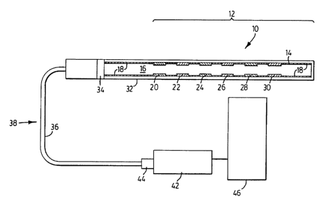
25 Referring to Figure 1, a fiber optic multitasking probe 10 is shown for
fluence-rate dosimetry in turbid media. The probe 10 includes a probe head
portion 12 comprising an optical fiber 14 having a core 16 encased by the
cladding 18 and several fluorphore filled etch zones 20, 22, 24, 26, 28 and
30.
The different fluorophores each preferably have different wavelength
7
CA 02376480 2001-12-12
WO 00/77491 PCT/CA00/00690
sensitivities. A removable translucent cover 32 which slides over the optical
fiber
14 prevents leakage of fluorophores from the probe into the tissue while
allowing
exposure of the fluorophores by the PDT treatment light. Probe 10 also
includes
an x-ray opaque marker 34 for providing placement verification by x-ray
diagnostic tools and a protective cover 36 (for example TEFLON or other
optically transparent and physiologically acceptable material) along the
length of
the fiber portion 38 adjoining the probe head portion 12 to a spectrometer 42.
The fiber portion 38 is connected to the spectrometer 42 using an industry
standard terminating end 44. The spectrophotometer 42 allows wavelength
to selection of the emitted light transported through the optical fiber 14 and
is
connected to a computer 46 for controlling the output of the PDT light source
and handling the optical data from the different fluorophore zones.
The PDT treatment light, the intensity of which is the parameter to be
measured, is acting as the excitation source for the fluorophore filled etch
zones
(20, 22, 24, 26, 28 and 30) and originates from a PDT light source spaced away
from probe 10. A portion of the fluorescence produced in the fluorophores in
each zone is guided via the optical fiber out of the tissue and delivered to
the
spectrophotometer. To be able to separate the contributions from the various
etch zones, fluorophores with sufficiently different emission spectra
(intensity
2o and/or spectral shape which convey the optical information) are used. As
described above the signal measured (fluorescence intensity) is a function of
the
fluorescence quantum yield of the fluorophore used in the optical fiber and
its
concentration (both fixed in each probe) as well as the fluence-rate of the
exciting radiation e.g. the PDT treatment light. Hence the fluorescence
intensity
is a direct function of the fluence-rate.
Figure 2 shows a probe 60 similar to probe 10 to provide multitasking for
fluence-rate dosimetry in turbid media but modified to provide simultaneously
both fluence-rate and photosensitizer quantification. Photosensitizers are
exogenous dyes which are administered to a patient for the purpose of treating
8
CA 02376480 2001-12-12
WO 00/77491 PCT/CA00/00690
with photodynamic therapy. Next to generating cytotoxic products upon
illumination, the photosensitizer also emits fluorescent light. The probe 60
is
designed so that probe head portion 58 includes a fluorophore containing zone
62 placed at the distal end 64 of the probe. The exposed distal end 64 of the
fiber 14 enables coupling of the photosensitiser fluorescence into the fiber
optical probe 60. The spectra of the emitted fluorescence is significantly
different
from that of the fluorophores embedded into the fiber itself.
As described above, the fluorophore containing zone 62 is quantifying the
fluence-rate at this position. All photosensitizers fluoresce, after being
excited by
to the PDT-treatment light fluence-rate. The photosensitizer fluorescence
intensity
is given by the excitation fluence rate (measured through fluorophore at
position
62 as described above) its fluorescence quantum yield, which is fixed, and the
photosenstizer concentration, e.g. the quantity of interest. Hence, the
fluorescence intensity is directly correlated with the photosensitizer
concentration. Based on the known fluence rate at this position, the known
fluorescent quantum yield and the measured fluorescence intensity, the
photosensitizer concentration is calculated.
Probe 10 shown in Figure 1 measures only the fluence-rate in the tissue,
while probe 60 shown in Figure 2 in addition also measures the photosensitizer
2o fluorescence.
For quantification of p02, the phosphorescence lifetime may be employed
(see for example Lo LW, Koch C.J, Wilson DF ( 1996) Calibration of Oxygen-
Dependent Quenching Of The Phosphorescence Of Pd-mesotetra (4-
Cabocyphynyl) Porphine: A Phosphor With General Application Of Measuring
2s Oxygen and Vanderkooi et al. J. of Biol. Chem. Vol 262 5476-82 1987).
Referring now to Figure 3, there is shown a fiber optic multitasking probe 70
for
simultaneous fluence-rate and p02 dosimetry in turbid media. The probe 70
comprised of the optical core is similar to probes 10 and 60 but is modified
so
the optical fiber core 16 includes a sensor zone 72 comprising a
9
CA 02376480 2001-12-12
WO 00/77491 PCT/CA00/00690
photoluminescent phosphor between the distal end portion 64 containing
fluorophore zone 62 and fluorophore zone 28, see the blowup in Figure 3. A
control unit 76 comprises a light source 78 for phosphor excitation (for
example
any source emitting in the blue that can be activated very rapidly, such as an
LED), a dichroic beam splitter 80 to separate selection detector 82 and fast
opto-
electronic detector 84 for lifetime quantification. In the embodiment in which
the
probe 70 operates as a dual fluence-rate and p02 detector, the control unit 76
will also house a spectrophotometer 42. Other embodiments of the probe may
include several different phosphor containing sensor zones along the fiber 14
to each with its own spectral signature or alternatively probes may be
produced in
which all the zones contain different phosphors if the application of the
probe is
only for measuring p02 whether or not photosensitizer concentrations are being
measured.
When photoluminescent phosphors are used the optical information is
encoded in the spectral shape and lifetime. Table 1 provides an exemplary, non-
limiting list of phosphors that may be used with the fiber probes. The
phosphors
are embedded in a membrane which is brought into contact with the core of the
optical fiber and exposed to the tissue. Some of the light emitted by the
phosphors is transmitted down the fiber 14 to be interrogated at the
2 o spectrometer 42 and computer 46. The fluorescence lifetime of triplet
oxygen
measurement is made using a short pulse excitation (-100 nsec) followed by
nanosecond time resolution of the decay. The excitation wavelength is provided
by the spectrophotometer 42 in control unit 76 and is absorbed by all employed
phosphors. The phosphors used are selected on the basis that they exhibit
sufficiently unique emission spectra to provide spatial resolution and
quantification of lifetimes. Cross talk between the fluorescence and
phosphorescence measurements is avoided because the phosphors do not
absorb at the wavelengths corresponding to the PDT treatment wavelength.
Therefore the light used in the PDT does not induce any phosphorescence.
CA 02376480 2001-12-12
WO 00/77491 PCT/CA00/00690
Also, the fluorescence decay being several orders of magnitude faster than the
phosphorescence, does not affect the phosphorescence lifetime after a few
nsec. However, this approach requires that illumination with the PDT treatment
light needs to be suspended during phosphorescence measurements.
A common feature of the different embodiments of the multitasking probe
disclosed herein are the different spaced sensor zones along the length of the
optical fiber filled with a photoactive constituent. Methods of generating the
sensor zones include, but are not limited to; etching of the original material
either mechanically, chemically or optically, and applying a photoactive
1o constituent doped material (polymethylmethacrylate (PMMA), cyanoacrylate
and
others); diffusion of the photoactive constituent, such as fluorophores, into
the
core material, for example by heating and chemically softening the raw
material.
The depth of the etched zone ranges from 0.20 micrometer to full core
diameter.
For example, a probe can be produced by etching one or more zones in a fiber
1s by laser induced plasma or mechanical abrasion using miniature tools and
filling
of the etched zones) by applying a fluorophore doped polymer mix (the latter
may be comprised of either the optical fiber monomer or a cyanoacrylate
adhesive) and polishing the fiber upon completion of the polymerization
process.
The probes preferably use plastic optical fibers for ease of manufacturing and
2o safety of handling outside and inside the patient.
The basic principle of the present invention involves using one or more
different sensor zones along the length of the fiber each with a different
photoactive constituent having a sufficiently unique emission spectra
(spectral or
temporal) to enable deconvolution of the emission spectra by the computer and
25 therefore correlation of the detected parameter with the position of the
sensor
zone along the length of the optical fiber. In the broadest form of the
invention
the probe is embodied by only one sensor zone located at some point along the
length of the fiber spaced away from the end face of the fiber. When two or
more
sensor zones are used one of the sensor zones may be located on the planar
11
CA 02376480 2001-12-12
WO 00/77491 PCT/CA00/00690
end face at the end of the fiber.
The important feature that enables operation of the present probe as a
sensor for detecting factors at any given position along the length of the
fiber is
to incorporate the photoactive constituent into the sensor zone in the bulk of
the
fiber or on the fiber surface located at the given position along the fiber.
Whether on the surface of the fiber or incorporated in the bulk of the fiber
the
key is the photoactive constituent is incorporated in such a way so that a
portion
of the light emitted by the photoactive constituent responsive to interaction
of the
latter with the factor of interest is guided via the optical fiber out of the
tissue and
to delivered to the spectrophotometer. In the preferred embodiments in which
multiple sensor zones are disposed along the length of the fiber, photoactive
constituents with sufficiently unique emission spectra (intensity and/or
spectral
shape which convey the optical information) are used in the different sensor
zones so that the different spectra can be deconvoluted so that the
contributions
from the various etch zones can be distinguished. More than one different
photoactive constituent could be incorporated into a single sensor zone for
measuring several factors in the vicinity of the sensor zone.
Examples of seven different fluorophores and their spectral properties
that may be used in the present invention for PDT applications are shown in
2 o Table 2. All fluorophores absorb the PDT treatment wavelength, here 630 to
690
nm, but show sufficiently different emission spectra so the detected spectra
can
be numerically deconvoluted to extract the intensity of each fluorophore. Thus
the fluence-rate at all points doped with fluorophore can be extracted, one of
which will be close to the distal end (see Figure 2). The fluorescence
emission of
the seven example fluorophores used in the multitasking probes are shown in
Figure 4. All spectra were measured in an optical fiber containing only one
active sensor. It will be understood that the list of candidate fluorophores
in
Table 2 is not exhaustive and other fluorophores being currently developed may
be employed in the present fluence rate fiber optical probes. For use in other
12
CA 02376480 2001-12-12
WO 00/77491 PCT/CA00/00690
light (300-900nm) based therapeutics, the fluorophore absorption needs to
match the treatment wavelength. For example, in the case of interstitial laser
hypothermia (ILH) discussed hereinafter, a fluorophore is required that is
able to
absorb between 800 to 900 nm.
Figure 5a shows the average normalized fluorescence emission spectra
of a sensor illuminated from different azimuth angles, showing that the
emission
spectra shape is independent of azimuth angle of illumination. Figure 5b shows
the normalized fluorescence emission spectra of ten individually manufactured
multitasking probes, showing independence of manufacturing, and hence,
to responsivity calibration for each sensor is possible.
Figure 6 shows a typical polar diagram showing the isotropy of detector
response. The reduction of responsivity in the for,nrard and backward
direction
of the fiber is due to shielding of the sensor by the fiber itself. This
shielding
encompasses only 6% of the total surface volume.
Figures 7 and 7a show the emission spectra of an optical fiber detector
probe comprised of 2 sensors for individually exposed sensors and combined
exposure of both sensors. The data shown in Figures 5 to 7a illustrates the
efficacy of the multitasking probes disclosed herein.
While the optical fiber probes have been described using fluorophores
2 o and photoluminescent phosphors as the photoactive constituents in the
sensor
zones, it will be understood that other materials could be used depending on
the
factors) being detected. For example, sensor zone containing chemiluminescent
compounds as the photoactive constituent may be used for the detection of
adenosine triphosphate (ATP) and for the detection of hydrogen peroxide. In
either case the chemical to be detected supplies the energy required for
photon
emission. Further examples for the use of chemiluminescence are in the
detection of choline and phosphaolipase D activity, phosphate ions and
immunoenzymes given by Ruach P, Ferri En Girotti S, Rauchova H, Carrea G,
Bovara R, Fini F Analyytical Biochem. 245 133-40 1997, Nakamura H, Ikebukuro
13
CA 02376480 2001-12-12
WO 00/77491 PCT/CA00/00690
K, NcNiven S, Karube I, Yamamoto H, Hayashi K, Suzuki M Kubo M, in
Biosensors And Bioelectronics 12 956-66, 1997; and Ospipov A, Aeitseva NV,
Egorov EM, Biosensors And Bioelectronics 11 881-7, 1996, respectively. The
chemiluminescent compound may be provided by using choline oxidase (Ch0)
and horseradish peroxidase (HRP) immobilized on Eupergit C (polymer beads of
methacrylamide, -methylene-bis-methacrylamide, and allyl-glycidyl-ether) or be
catalyzed by arthromyces ramosus peroxidase. In embodiments employing
chemiluminescent compounds the unique emission spectrum and intensity
characteristic of each chemiluminescent reaction encodes the spatial location
to and concentration information.
Additionally, for applications of the present invention involving
measurement of radioactivity, the photoactive constituents in some or all of
the
sensor zones would be scintillator compounds in which the unique intensity of
each scintillator compound encodes the information. Organic scintillators may
be
used with compatible optical fibers in producing the probes.
The optical fiber probes of the present invention may be used in a variety
of applications in addition to PDT in mammals. For example, they may be used
as environmental sensors for use in normally unaccessible areas. The length of
the fibers may be as long as required for use in, for example, bodies of water
as
2o environmental sensors where pH is being measured as function of water
depth.
The fibers constructed for use as radiation detectors may be wrapped around
pipes and the like and radioactive leaks may be detected since the optical
controller deconvolutes the optical data to correlate the emission spectra
with
the spatial location of the photoactive constituent along the fiber. The
length of
the photoactive constituent along the fiber will be given by the required
spatial
resolution of the application.
Photodynamic Therapy in Barrett's Esophagus Or Solid Tumors
The present invention provides two specific fiber optic probe systems for
the treatment of Barrett's esophagus and interstitial treatment of tumors.
14
CA 02376480 2001-12-12
WO 00/77491 PCT/CA00/00690
Referring to Figure 8, an applicator 100 is shown for the treatment of
Barrett's
esophagus. Probe 100 comprises a cage 102 made of superelastic wires 104
with multitasking optical probes 106 mounted on the cage wires. The applicator
100 optionally may include a bulbous end portion 108 that enables anchoring of
s the device at the gastro-esphageal-junction. The probes 106 are guided
through
the gastroscope 112 and are connected to the spectroscope 42 and data is
processed in the CPU 46. The wires 104 are made out of superelastic metals,
such as nitenol wire. The cage applicator 100 is introduced via the working
channel 110 of a standard gastroscope 112 and once in the lumen of the
1o esophagus the cage expands the esophagus to enable homogenous illumination
of the esophagus, see Overholt B.F., Panjehpour M. (1996), Photodynamic
Therapy In Barrett's Esophoagus; J. Clin. Laser Med. Surg. 14: 245-249. In
one embodiment eight 0.5 mm diameter wires comprise the cage 102. The total
diameter of the cage and the probes is about 2.8 mm.
15 An applicator for interstitial tumor treatment is shown at 200 in Figure 9.
Applicator 200 comprises a cylindrical isotropic light emitter 202 and two
multitasking probes 204 and 206. Light emitter 202 is preferably an optical
fiber
diffuser which may emit light having a uniform intensity distribution along
the
length of the diffuser. The two probes 204 and 206 measure the fluence-rate
2 o gradient at 2 different positions in the glow field of the isotropic
emitter probe
202. These three optical fibers are combined and introduced via a single
hypodermic needle 210. The probes are connected to the spectrograph 42 and
the data evaluated via CPU 46. The size between the individual detector
elements is selected to be comparable to the mean free path of the photons to
25 be measured in the tissue. Hence, the detector is not integrating too much
over
the gradient of the fluence-rate.
In one embodiment a multitasking probe 204 is located at the proximal
end portion of the applicator 200 and a second probe 206 is located at the
distal
end portion of the applicator with both probes parallel to the axis of the
CA 02376480 2001-12-12
WO 00/77491 PCT/CA00/00690
cylindrical source 202. The radial dependence of the three parameters, fluence-
rate, photosensitizer concentration and molecular oxygen are those measured
during the procedure. In this arrangement the ovid shaped fluence rate
profiles
generated by the cylindrical source in the target tissue are measure along the
long axis of the distribution. The total outer diameter of the assembly is
preferably less than 1 mm, where the source has an outer diameter of less than
0.5 mm and the two detectors are 0.25 mm in diameter.
The multitasking probes of the present invention are very advantageous
over previous probes because they can be used to provide the parameters
to required for PDT for the entire tissue volume in question. The multitasking
probes disclosed herein can provide for measurement of a single parameter at
multiple locations along the axis of a single optical fiber as well as
providing for
measurement of multiple parameters at multiple locations along the axis of the
fiber.
The foregoing description of the preferred embodiments of the invention
has been presented to illustrate the principles of the invention and not to
limit
the invention to the particular embodiment illustrated. It is intended that
the
scope of the invention be defined by all of the embodiments encompassed
within the following claims and their equivalents.
25
16
CA 02376480 2001-12-12
WO 00/77491 PCT/CA00/00690
Table 1
Phosphore Excitation maximum Solvent
Pd Coproporphyrin I 394 nm DMSO
to
Pd Coproporphyrin III 398 nm DMSO
Pd TPPS4 408 nm Acetone
Pd tetra(N-methyl-4- 425 nm DMSO
pyridyl)porphine
Table 2
fluorophore used solvent absorption peak fluorescence
[nm] peak [nm]
DTTC DMSO 772 802
LD 700 DMSO 598 / 648 697
LD 800 DMSO 626 / 686 700
LDS 750 MeOH 580 696
LDS 821 DMSO 601 754
Oxazine 720 DMSO 636 694
3 5 Oxazine 750 DMSO 668 694 / 735
17