Note: Descriptions are shown in the official language in which they were submitted.
CA 02377418 2002-03-19
DEVICE AND METHOD FOR REGISTERING
A POSITION SENSOR IN AN ANATOMICAL BODY
FIELD OF THE INVENTION
This invention relates to devices and methods to insert
position sensors into an anatomical body. More particularly,
the present invention relates to a device and method for
registering a position sensor inserted in an anatomical body.
BACKGROUND OF THE INVENTION
Position sensors have been increasing in accuracy and
decreasing in size. This has made position sensors for use in
tracking portions of an anatomical body during surgical
procedures more feasible.
However, in order to accurately track areas of interest
in an anatomical body, it is necessary to rigidly fix the
position sensor near or at a location of interest in the
anatomical body. It is also necessary to then register the
position sensor with the anatomical body. A position sensor is
registered to an anatomical body by correlating the position
of the position sensor in the anatomical body to the
determined position of the position sensor in the frame of
reference. At that time, the location of interest in the
anatomical body can be tracked in a fixed frame of reference,
such as the operating room frame of reference, by determining
the position of the position sensor.
A number of position sensors have been used in the past.
CA 02377418 2002-03-19
- 2 -
Recently, magnetic sensor coils or fibre optic sensors that
are reasonably small, and therefore can be substantially
unobtrusively inserted into an anatomical body, have been
successfully used.
However, the prior art suffers from the disadvantage that
it is difficult to register the position sensors to the
anatomical body. Methods for registering the position sensor
in the anatomical body have included obtaining an image of the
anatomical body after insertion of the position sensor and
attempting to register the position sensor to the anatomical
body from the acquired image. However, this suffers from the
disadvantage that the position sensor is not always easily
identifiable in the acquired image. Furthermore, while it may
be possible to determine the position of the position sensor
in the anatomical body, sufficient information may not be
available from the image to determine and register both the
position as well as orientation of the position sensor.
Because of this, it may not be possible to determine all of
the degrees of freedom, such as movement along the x, y, z
axes, as well as three orientation coordinates, namely pitch,
yaw and roll.
In some embodiments, it may be desirable that the
position sensor be permitted to move relative to the
anatomical body. However, in most cases, it is preferred that
there be no relative movement between the position sensor and
the location of interest in the anatomical body. Most prior
art devices and methods for registering the position sensor in
an anatomical body suffer from the disadvantage that there may
be relative movement between the position sensor and the
CA 02377418 2002-03-19
- 3 -
anatomical body. Accordingly, in addition to a method and
device for accurately registering the position of the position
sensor in an anatomical body, there is a need in the art for a
device and method to reliably fix the position sensor to the
anatomical body, thereby avoiding relative movement during the
procedure. There is also a need in the art for reliable
devices and methods to insert the position sensor into the
anatomical body.
SUMMARY OF THE INVENTION
Accordingly, it is an object of this invention to at
least partially overcome the disadvantages of the prior art.
Also, it is an object of this invention to provide an improved
type of device and method that facilitates simple and accurate
registration of a position sensor within an anatomical body.
It is also an object of this invention to provide an improved
type of device and method to minimize relative movement
between the position sensor and the anatomical body.
Accordingly, in one of its aspects, this invention
resides in an apparatus insertable into an anatomical body,
said apparatus comprising an insertable portion for holding a
position sensor that can transmit a signal indicative of its
position in a frame of reference; fiducial markings on the
insertable portion, said fiducial markings being detectable by
an imaging modality when the insertable portion is inserted in
the anatomical body; wherein, after insertion in the
anatomical body, the fiducial markings can be detected by the
imaging modality to facilitate registration of the position
sensor held in the insertable portion to the anatomical body.
CA 02377418 2002-03-19
- 4 -
In a further aspect, the present invention resides in a
method of registering a position sensor to an anatomical body,
said method comprising the steps of: fixing a position sensor
to an insertable portion of an apparatus, said insertable
portion having fiducial markings thereon; inserting the
insertable portion of the apparatus to a location of interest
in the anatomical body; and detecting the fiducial markings on
the insertable portion of the apparatus to facilitate
registration of the position sensor in the insertable portion
to the anatomical body.
In a further aspect, the present invention resides in a
device for facilitating tracking of an apparatus in an
anatomical body, said device comprising an insertable portion
for holding a position sensor that can transmit a position
signal indicative of its position in a frame of reference;
wherein the apparatus can insert the insertable portion into
the anatomical body, and, the position signal transmitted from
the position sensor indicates the position of the apparatus
near the position sensor.
In one aspect, the apparatus comprises a fixing mechanism
for releasably fixing the insertable portion of the apparatus
to the anatomical body at a location of interest. The fixing
mechanism can be any type of mechanism that can releasably and
rigidly fix the insertable portion to the anatomical body,
such that the insertable portion will not become easily
displaced during the procedure. In one embodiment, the fixing
mechanism comprises a screw that screws into a part of the
anatomical body, or, at least one barb that can become fixed
-
CA 02377418 2008-07-28
- 5 -
to the anatomical body near the location of interest.
The fixing mechanism may also comprise an inflatable
member that inflates when the insertable portion is at the
location of interest. More preferably, the inflatable member
has lobes that can inflate to fix the catheter, such as in a
passageway near the location of interest, while still
permitting fluid flow around the apparatus, such as a
catheter, and through the passageway.
In a further preferred embodiment, the fixing mechanism
comprises a plurality of movable fingers having gripping
elements. The movable fingers have a collapsed configuration
where the gripping elements are near the apparatus, such as a
catheter or a needle, and permit insertion of the catheter
into the anatomical body and near the location of interest.
The movable fingers also have a deployed configuration where
the gripping elements engage a surface of a passageway in the
anatomical body to fix the insertable portion to the
anatomical body near the location of interest, but permit
fluid flow around the apparatus and through the passageway.
In a further aspect, the present invention provides an
apparatus insertable into an anatomical body, said apparatus
comprising: an insertable portion for holding a position
sensor that can transmit a signal indicative of its position
in a frame of reference; fiducial markings on the insertable
portion, said fiducial markings being detectable by an imaging
modality when the insertable portion is inserted in the
anatomical body and permitting the position and orientation of
the insertable portion to be determined, said insertable
CA 02377418 2008-07-28
- 5a -
portion substantially rigidly holding the position sensor at a
known spatial position with respect to the fiducial markings;
wherein, after insertion in the anatomical body, the fiducial
markings can be detected by the imaging modality to facilitate
registration to the anatomical body of the position sensor
rigidly held in the insertable portion by correlating the
fiducial markings detected by the imaging modality to the
determined position of the position sensor in the frame of
reference.
In a still further aspect, the present invention provides
a device for facilitating tracking of an apparatus in an
anatomical body, said device comprising: an insertable portion
for holding a position sensor that can transmit a position
signal indicative of its position in a frame of reference;
fiducial markings on the insertable portion, said fiducial
markings being detectable by an imaging modality to facilitate
registration of the position sensor held in the insertable
portion to the anatomical body; wherein the apparatus can
insert the insertable portion into the anatomical body, and,
the position signal transmitted from the position sensor
indicates the position of the apparatus near the position
sensor; and wherein the insertable portion substantially
rigidly holds the sensor at a known spatial position with
respect to the fiducial markings; and wherein, after insertion
of the insertable portion in the anatomical body, an image of
the insertable portion can be obtained by the imaging
modality, and, the position sensor can be registered to the
anatomical body by correlating the fiducial markings detected
by the imaging modality to the determined position of
the position sensor in the frame of reference.
CA 02377418 2008-07-28
- 5b -
One advantage of the present invention is that the
fiducial markings on the insertable portion permit the
position, and preferably orientation, of the insertable
portion to be accurately determined with respect to the
anatomical body. As the position of the position sensor with
respect to the insertable portion, and therefore the fiducial
markings, is known, the position sensor can then be registered
to the anatomical body by correlating the position of the
fiducial markings in the images to the determined position of
CA 02377418 2002-03-19
- 6 -
the position sensor in the fixed frame of reference.
A further advantage of the present invention is that the
fixing mechanism preferably substantially rigidly fixes the
insertable portion of the apparatus to the anatomical body.
In this way, once the position sensor has been registered,
relative movement between the anatomical body and the position
sensor is decreased.
Further aspects of the invention will become apparent
upon reading the following detailed description and drawings
that illustrate the invention and preferred embodiments of the
invention.
BRIEF DESCRIPTION OF THE DRAWINGS
In the drawings, which illustrate embodiments of the
invention:
Figure 1A is a symbolic representation of a position
sensor system as known in the art.
Figure 1B is an illustration of a position sensor system
as known in the art.
Figure 2A illustrates an apparatus comprising a catheter
having an inflatable member according to one embodiment of the
present invention.
Figure 2B is an enlarged detailed drawing of the
guidewire and catheter shown in Figure 2A.
CA 02377418 2002-03-19
- 7 -
Figure 2C is a detailed drawing of a catheter having an
inflatable member according to a further embodiment of the
present invention.
Figure 2D is a drawing of the catheter shown in Figure 2C
with the inflatable member inflated.
Figure 3A is a front section view of a catheter having an
inflatable member according to a further embodiment of the
present invention.
Figure 3B is a side section view of the catheter shown in
Figure 3A.
Figure 3C is a side view of an alternate embodiment of
the inflatable member shown in Figure 3B.
Figure 4A is a side view of a catheter in a deployed
configuration according to a further embodiment of the present
invention.
Figure 4B is a side view of the catheter shown in Figure
4A in a collapsed configuration.
Figure 5 shows a side view of the rigid portion of the
catheter according to a further embodiment of the present
invention.
Figure 6A shows a detailed view of the catheter according
to a further embodiment of the present invention.
CA 02377418 2002-03-19
- 8 -
Figure 6B shows a side view of the catheter shown in
Figure EA attached to a cannulated screwdriver.
Figure 7 illustrates an insertable portion in the form of
a stylette insertable in a needle, according to a further
embodiment of the present invention.
Figures 8a and 8b illustrates an apparatus comprising a
needle and an insertable portion in the form of a stylette
releasably fixed into the needle according to a further
embodiment of the present invention.
Figure 8c illustrates the needle shown in Figures 8a and
8b, but with the insertable portion removed.
Figure 9 illustrates an apparatus according to one
embodiment of the invention having a needle, a stylette and
the insertable portion in the form of a secondary stylette.
Figure 10 illustrates a needle with therapy or sensor
zones according to a further embodiment of the present
invention.
Figure 11 illustrates an embodiment of the present
invention having two position sensors longitudinally displaced
according to a further embodiment of the present invention.
Figure 12 illustrates an insertable portion for insertion
in a needle with the insertable portion having a fixing
mechanism.
CA 02377418 2002-03-19
- 9 -
Figures 13a, 13b and 13c illustrate the deployment of the
fixing mechanisms shown in Figure 12.
DETAILED DESCRIPTION OF THE PREFERRED EMBODIMENTS
Figures lA and IB show a conventional position sensor
system, shown generally by reference numeral 10. The system
comprises a control unit 12 that is connected to a field
generator 14 and a host computer 16. The host computer 16 can
be a user supplied work station. The field generator 14
generates a complex electromagnetic field within a frame of
reference. A position sensor 20 within the frame of reference
can sense the complex electromagnetic field. The system 10
also comprises a sensor interface unit 18 that interfaces the
control unit 12 to the position sensor 20. It is understood
that these components may be integrated together. For
example, the sensor interface unit 18 may be combined with the
control unit 12.
The position sensor 20 generally comprises a sensor
element, such as a magnetic sensor coil 21, which reacts to,
or senses, the complex electromagnetic field generated by the
field generator 14. As the position sensor 20 moves in the
electromagnetic field generated by the field generator 14, the
sensor coil 21 generates a position signal Sp that is
indicative of the position of the position sensor 20.
Generally, the sensor coil 21 will react to changes to both
the position and orientation of the position sensor 20 in the
frame of reference. In this way, the position signals Sp
generated by the sensor coil 21 are indicative of both
CA 02377418 2002-03-19
- 10 -
position and orientation of the position sensor 20. The
position signals Sp are received by the sensor interface unit
18 and converted to a form which can be understood by the host
computer 16.
Thus, the position and orientation of the position sensor
20 can be determined in the frame of reference of the field
generator 14. The frame of reference of the field generator 14
is generally a fixed frame of reference, such as the frame of
reference of the operating room.
In order for the position sensor 20 to be of assistance
in tracking or determining the position and orientation of a
location of interest in an anatomical body, it is necessary
that the position sensor 20 be registered with respect to the
location of interest in the anatomical body. In addition, it
is often desirable that the position sensor 20 is fixed in
some way to the anatomical body so that, once the position
sensor 20 is registered to the anatomical body, the position
sensor 20 will not move with respect to the anatomical body.
Figures 2A, 2B, 2C and 2D show an apparatus, shown
generally in Figure 2A by reference numeral 100, to facilitate
inserting, fixing and registering a position sensor 20 into
the anatomical body according to one embodiment of the present
invention. The apparatus 100 in this embodiment comprises the
sensor coil 21 held within an insertable portion 90. In this
embodiment, the apparatus 100 comprises a catheter 110 for
inserting the insertable portion 90 into the anatomical body.
The catheter 110 moves along a guidewire 60, as is known in
the art. The catheter 110 will move into a passageway of the
CA 02377418 2002-03-19
- 11 -
anatomical body until the position sensor 20 is near a
location of interest in the anatomical body. The location of
interest is the part of the anatomical body whose position is
desired to be determined and tracked during a procedure.
In this embodiment, the apparatus 100 also comprises
electrical leads 26 which extend from the position sensor 20,
and in particular, the sensor coil 21, outwards through a plug
connector 24, shown in Figure 2A. The electrical leads 26
transmit the position signals Sp from the sensor coil 21 of
the position sensor 20 to the sensor interface unit 18, the
control unit 12, and to the host computer 16. The plug
connector 24 connects to the sensor interface unit 18 of the
system 10.
The apparatus 100 also comprises a fixing mechanism,
shown generally by reference numeral 28. In the embodiment
shown in Figure 2B, the fixing mechanism 28 comprises an
inflatable member 30, such as a balloon, which expands
radially outwards from the longitudinal axis of the insertable
portion 90 when inflated. The catheter 110 also comprises an
inflatable opening 44 that is used in this embodiment for
providing air, or other fluid, to the inflatable member 30. It
is understood that if a different type of fixing mechanism 28,
other than an inflatable member 30, is used, an inflatable
opening 44 may not be required or may be substituted with an
alternate activation pathway or method. The catheter 110 also
comprises a guide path opening 40, as is known in the art.
In operation, the catheter 110 is inserted into the
anatomical body by placing the catheter 110 over the guidewire
CA 02377418 2002-03-19
- 12 -
60 at the distal end, as is known in the art. The insertable
portion 90 of the catheter 110 will then be lead to a
passageway near the location of interest in the anatomical
body. In the preferred embodiment, once the insertable
portion 90 is near the location of interest, the inflatable
member 30 will be inflated by passing air through the
inflatable opening 44. The inflatable member 30 will then
expand radially to the sides of the passageway, thereby
rigidly fixing the insertable portion 90 to the passageway
near the location of interest.
Once the insertable portion 90 is fixed to a part of the
anatomical body near the location of interest, an image of the
location of interest can be made using a known imaging
modality. It is noted that the insertable portion 90 has
fiducial markings 70 which are detectable by the imaging
modality after insertion of the insertable portion 90 into the
anatomical body. Detection of the fiducial markings 70 by the
imaging modality facilitates registration of the position
sensor 20 to the anatomical body.
Preferably, the position sensor 20 is held in the
insertable portion 90 so that the distance and orientation of
the position sensor 20 with respect to the fiducial markings
70 will remain constant. To accomplish this, it is generally
preferred that the insertable portion 90 is rigid or
substantially rigid so that the distance and orientation of
the position sensor 20 with respect to the fiducial markings
70 will remain substantially constant. In general, the
insertable portion 90 should be as rigid as required by the
particular position sensor 20 being used. For instance, if
CA 02377418 2002-03-19
- 13 -
the position sensor 20 can detect movement, such as bending,
then the insertable portion 90 can be less rigid, provided the
position sensor 20 could compensate for this lack of rigidity
by sensing this bending movement. In a preferred embodiment,
the insertable portion 90 is substantially rigid so as to
maintain the position and orientation of the fiducial markings
70 with respect to the insertable portion 90 substantially
constant.
In a preferred embodiment, the imaging modality can
comprise CT scans, fluoroscopic images and ultrasound images.
When the imaging modality comprises CT scans and fluoroscopic
images, the fiducial markings 70 preferably comprise a radio-
opaque substance. The fiducial markings 70, as well as the
insertable portion 90 and the catheter 110, should also be
made from a substance that does not react with anatomical
bodies. Preferably, when the imaging modality comprises CT
scans and fluoroscopy, it is preferred that the fiducial
markings 70 comprise tantalum.
During the procedure, the fiducial markings 70 can
perform a dual purpose. Firstly, the fiducial markings assist
in guiding the catheter 110 to an anatomical body near the
area of interest when viewed with a fluoroscope, for example.
Secondly, the fiducial markings 70 facilitate registration by
quantitatively co-locating the position sensor 20 in the image
with the detected position of the position sensor 20 in the
frame of reference. In other words, by determining the
location of the fiducial markings 70 in the image, and knowing
the distance and orientation of the fiducial markings 70 with
respect to the position sensor 20 which is rigidly held in the
____________________________ - __________________________________
CA 02377418 2002-03-19
- 14 -
insertable portion 90 of the catheter 110, an automatic
registration can be made between the determined position of
the position sensor 20 using the position sensor system 10 and
the determined position of the position sensor 20 using the
fiducial markings 70 in the image. The fiducial markings 70
detected by the imaging modality can be used to determine the
position and orientation of the position sensor 20 in the
image, and therefore the anatomical body. As stated above,
the insertable portion 90 is preferably rigid or semi-rigid,
as required to determine the position and orientation of the
position sensor 20 from the fiducial markings 70.
In order to facilitate registration, it is preferred that
the fiducial markings 70 comprise at least two markings 71, 72
which permit the position and orientation of the insertable
portion 90 to be determined in the image. As shown in Figure
2B, the at least two markings 71, 72 are bands around the
insertable portion 90. Furthermore, in order to increase the
accuracy, it is preferred that the at least two markings 71,
72 be located at opposed ends of the insertable portion 90,
and preferably as far apart as possible, while remaining on
the insertable portion 90. To assist in registering the
position sensor 20, the shadow of the position sensor 20, such
as the sensor coil 21 in this embodiment, can be used as a
further marker in the image obtained by the imaging modality.
As shown in Figure 2B, the at least two markings 71, 72
are bands which extend around the circumference of the
insertable portion 90. In addition, the fiducial markings 70
may comprise a bead 73 as shown in Figure 2C. A band 74 would
preferably still be used in association with the bead 73 as
the band 74 can be used to more easily identify the
CA 02377418 2002-03-19
- 15 -
orientation of the insertable portion 90. In addition, the
fiducial markings 70 may comprise a series (not shown) of
beads 73, which can also be used to identify the orientation.
Also, a series of bands 74 may be used to increase accuracy.
The fiducial markings 70 should have any position,
orientation or shape that permits determination of the
position and orientation of the position sensor 20 held by the
insertable portion 90 by the imaging modality. While a number
of fiducial markings 70 may be used in the insertable portion
90, at least two markings 71, 72 will generally be required to
determine both the position and orientation of the insertable
portion 90. If, in a particular situation, only the position
and not the orientation need be determined, one fiducial
marking 70 may be used. Furthermore, in some cases, the
sensor element in the position sensor 20 may not be able to
determine six degrees of movement. For instance, when the
sensor element is a magnetic sensor element 21, as shown in
Figures 2A, 2B, 2C and 2D, it may be difficult to determine
roll using the system 10. In this case, if the position
sensor 20 cannot determine roll, it is not necessary to use
the fiducial markings 70 which can identify the roll
orientation of the position sensor 20. Accordingly, the
fiducial markings 70 may only be required to identify some of
the six degrees of freedom, and may not be required for all
three positions or all three orientations, depending on the
particular situation. Furthermore, the fiducial markings 70
need not be two separate markings, but rather may be a single
complex marking, a series of interconnected markings, or a
single marking, provided some positional and/or orientational
information can be obtained.
If the system 10 cannot determine all of the degrees of
CA 02377418 2002-03-19
- 16 -
freedom of the position sensor 20, it may be necessary to
place more than one position sensor 20 near the location of
interest in order to track the motion of the location of
interest in all of the degrees of freedom. It is also
understood that in some situations, it will not be necessary
to track all of the possible positions and orientations of the
location of interest. In these situations, the fact that a
particular sensor element (not shown) of the position sensor
20 cannot track one degree of freedom, such as the roll, may
not adversely affect use of the position sensor 20 and may not
require additional position sensors 20 to be used.
Figures 2C and 2D also illustrate use of the inflatable
member 30. In particular, Figure 20 shows the inflatable
member 30 in the deflated position, and Figure 2D shows the
inflatable member 30 in the inflated position. It is
understood that inflating the inflatable member 30 will fix
the insertable portion 90 to a passageway or other part of the
anatomical body.
In the preferred embodiment illustrated in Figures 20 and
2D, the apparatus 100 comprises an electromagnetic shield 45.
The electromagnetic shield 45 may be a braided mesh, as is
known in the art, to shield the electrical leads 26 from
spurious electromagnetic signals. It is understood that the
electromagnetic shield 45 is particularly preferable in cases
where the power level of the position signal Sp is not great,
and therefore even minor spurious electromagnetic fields could
adversely affect the position signal Sp.
While the inflatable member 30 can be placed in manmade
CA 02377418 2002-03-19
- 17 -
passageways, such as drilled holes into bones, the inflatable
member 30 is also well suited for natural occurring
passageways in the body, such as arteries, veins and airways.
In smaller veins, particularly where the blood flow is not to
a crucial part of the body, or alternate blood paths are
available, the inflatable member 30 may encompass the entire
circumference of the insertable portion 90 as shown in Figures
2A, 2B, 20 and 2D. In this case, inflating the inflatable
member 30 may occlude the passageway preventing fluid flow
through the passageway.
In some cases, however, it is not desirable to completely
occlude a passageway. This would be the case, for example, in
an airway or main artery. In these cases, it is preferable
that the inflatable member 30 comprises at least one lobe
portion 3. In the preferred embodiment illustrated in Figure
3A, the inflatable member 30 comprises three lobe portions 3a,
3b, 3c and 3d, located at 90 degrees with respect to each
other and around the insertable portion 90. Figure 3A
illustrates the inflatable member 30 in the inflated
configuration, and thereby fixing the insertable portion 90 to
a part of the anatomical body, in this case the passageway
300. It is understood that in the deflated configuration, the
lobes 3a, 3b, 3c and 3d of the inflatable member 30 would be
near the rigid portion 90 in order to permit insertion of the
insertable portion 90 into the anatomical body.
It is apparent from Figure 3A that by utilizing the lobe
portions 3a, 3b, 3c, 3d, fluid can still flow around the
insertable portion 90 of the apparatus 100, which in this
embodiment comprises the catheter 110, and therefore through
CA 02377418 2002-03-19
- 18 -
the passageway 300. In this way, use of lobe portions 3 to
fix the insertable portion 90 will not greatly adversely
affect the fluid flow in a location of interest. This may be
particularly important depending on the pathology of the
location of interest and the nature of the passageway 300.
Figure 3A also shows in cross-section the sensor coil 21
located within the insertable portion 90. Figure 3A also
shows the guidewire path 40 through which the guidewire 60
passes and the inflatable opening 44 through which fluid can
pass in order to inflate the inflatable member 30, which in
this case comprises the three lobes 3a, 3b, 3c, 3d, attached
to a catheter 110 in this embodiment. It is understood that
an apparatus 100 other than a catheter 110 could also be used
with the inflatable member 30, such as an endoscope (not
shown).
Figure 3B shows a side view of the embodiment illustrated
in Figure 3A. As Figure 3B is a side view, only lobes 3c and
3a are illustrated. It is understood that lobes 3d and 3b
would be entering out of and into the page, respectively.
Figure 3B also shows the fiducial markings 70 at opposed ends
of the insertable portion 90.
Figure 3C shows a further embodiment of the present
invention where the lobes, in this embodiment identified by
reference numerals 3x and 3y, are barbed shaped. The barb
shaped lobes 3x and 3y more rigidly secure the insertable
portion 90 to the passageway 300. It is understood that while
two of the lobe portions 3 are barb shaped lobes 3x, 3y, the
insertable portion 90 may also have other lobes 3 which may
_ ______________________________________________
CA 02377418 2002-03-19
- 19 -
not be barb shaped, but rather may be shaped similar to lobes
3d and 3b.
In a further preferred embodiment, the barbed shaped
lobes 3x, 3y may also have a barbed end 33 for engaging the
surface 301 of the passageway 300. The barbed ends 33 may be
made of more rigid material in order to permit engaging of the
barb shaped lobes 3x, 3y to the surface 301 of the passageway
300. The barbed ends 33 would move towards the insertable
portion 90 when the barb shaped lobes 3x, 3y are deflated.
Figures 4A and 413 illustrate a further embodiment of the
present invention. As shown in Figure 4A, the catheter 110
comprises a fixing mechanism 28 having a plurality 400 of
longitudinally extending fingers 410. The plurality 400 of
longitudinally extending fingers 410 is fixed to the rigid
portion 90 of the catheter 110 at point 430. The
longitudinally extending fingers 410 also comprise a pivoting
connection 440, such as a hinge, permitting the longitudinally
extending fingers 410 to pivot about point 430.
Each longitudinally extending finger 410 has a gripping
element 420 for gripping a surface 301 of the passageway 300
when the insertable portion 90 is near the location of
interest. As also illustrated in Figures 4A and 4B, the
insertable portion 90 has a bead 73 and band 74 as the
fiducial markings 70. While the gripping element 420 is shown
in Figures 4A and 413 as being at the end of the longitudinally
extending fingers 410, it is understood that the gripping
element 420 could be at any location along the longitudinally
extending fingers 410, such as at a bend (not shown) in a mid-
_
ak 02377418 2002-03-19
- 20 -
portion.
Figure 4B shows the longitudinally extending fingers 410
in the collapsed configuration where the gripping elements 420
are near the catheter 110. In the collapsed configuration,
the insertable portion 90 of the catheter 110 can be more
easily inserted into the anatomical body. Once the insertable
portion 90 is near the location of interest, the
longitudinally extending fingers 410 can be moved to the
deployed configuration, shown in Figure 4A, where the gripping
elements 420 engage the surface 301 of the passageway 300.
The longitudinally extending fingers 410 may be moved from the
collapsed configuration to the deployed configuration in any
manner. For instance, the movement may be performed by use of
magnetic field acting on the finger 410. In a preferred
embodiment, as illustrated in Figures 4A and 4B, the fingers
451 are moved from the collapsed configuration to the deployed
configuration by mechanically moving the supports 450 for each
of the longitudinally extending fingers 410. The supports 450
can be extended by signals received in any manner, including
mechanically, electrically, pneumatically, hydraulically or
through the use of light signals transmitted through fibre
optics.
It is apparent from Figure 4A that when the
longitudinally extending fingers 410 are in the deployed
configuration, fluid can still flow around the catheter 110,
and therefore through the passageway 300. In other words,
deployment of the longitudinally extending fingers 410 does
not occlude the passageway 300. Once the procedure is
completed, the fingers 410 can be moved from the deployed
CA 02377418 2002-03-19
- 21 -
configuration to the collapsed configuration in the same
manner in which they were deployed.
Figure 5 shows an alternate embodiment where the fixing
mechanism 28 comprises barbs 200. The embodiment illustrated
in Figure 2E has three barbs 201, 202, 203, but it is
understood that even one barb 201 may be sufficient. The
barbs 200 form part of the insertable portion 90 that holds
the position sensor 20. In this embodiment, the barbs 200
form an integral part of the insertable portion 90. Fiducial
markings 70 are shown on barbs 201, 203, which are near
opposed ends of the insertable portion 90.
Fixing mechanisms 28, such as the barbs 200, could be
used in association with different parts of the anatomical
body, and in particular bone. For example, a manmade
passageway, such as a drilled hole, could be made in a bone,
such as a spine segment, the skull or the femur. The
insertable portion 90 could then be inserted into the drilled
hole such that at least one of the barbs 200 fixes the
insertable portion 90 to the bone. Depending on whether the
procedure would need to be repeated in the near future, and
the size of the insertable portion 90, it may be preferable to
maintain the insertable portion 90 within the bone for an
extended period of time.
In a further embodiment, illustrated in Figures 6A and
6B, the insertable portion 90 comprises a screw structure 610.
The screw structure 610 is designed to screw the insertable
portion 90 into a passageway within the anatomical body.
Preferably, such a passageway would be made within bone so
CA 02377418 2002-03-19
- 22 -
that the screw structure 610 can grip into the edges of the
passageway and draw the insertable portion 90 within the
passageway to rigidly fix it therein. As shown in Figure 6A,
the position sensor 20 is contained within the insertable
portion 90. Figure 6B illustrates a cannulated screwdriver
620 which can be used to rotate the insertable portion 90 so
that the screw structure 610 can engage the edges of the
passageway to rigidly fix the insertable portion 90 therein.
In this embodiment, it is particularly preferable to have an
insertable portion 90 to withstand the torque forces
encountered when the cannulated screwdriver 620 rotates the
screw structure 610.
It is understood that the present invention also
contemplates a method for registering a position sensor 20 to
an anatomical body as described above. In particular, the
present invention relates to a method of fixing a position
sensor 20 to an insertable portion 90 of a catheter 110, the
insertable portion 90 having fiducial markings 70 as described
above. The method also comprises inserting the insertable
portion 90 of the catheter 110 to the location of interest in
the anatomical body. The fiducial markings 70 are then
detected on the insertable portion 90 of the catheter 110 to
facilitate registration of the position sensor 20 in the
insertable portion 90 to the anatomical body.
As described above, it is understood that detecting the
registration step includes detecting the fiducial markings 70
on the insertable portion 90. In the case where an imaging
modality is used, the fiducial markings 70 would be detected
on the insertable portion 90 by viewing the image obtained
. . .
CA 02377418 2002-03-19
- 23 -
through the image modality. It is understood that other types
of methods and means could be used to detect the fiducial
markings 70 on the insertable portion 90 once the insertable
portion 90 has been inserted to the location of interest.
The position of the position sensor 20 in the frame of
reference can then be determined using the system 110. Having
obtained a determined position of the position sensor 20 in
the frame of reference using the system 10, the position
sensor 20 can then be registered to the anatomical body by
correlating the detected fiducial markings 70 to the
determined position of the position sensor 20. In this way,
the position sensor 20 can be registered to the anatomical
body and movement of the position sensor 20 in the frame of
reference can then be used to track movement of the location
of interest in the anatomical body.
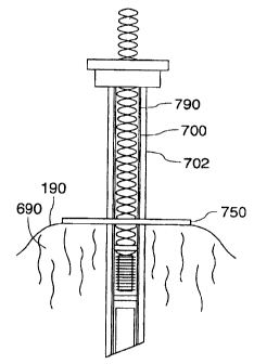
Figure 7 shows a further embodiment of the present
invention where the insertable portion 90 comprises a stylette
790 which can be inserted into a needle 702, as shown in
Figures 8a and 8b. The stylette 790 comprises a hollow
stylette 700 as is known in the art. The hollow stylette 700
holds a position sensor 20 comprising a sensor coil 21, as
discussed above. The sensor coil 21 is connected to sensor
leads 26 for transmitting the position signal Sp from the
sensor coil 21 of the position sensor 20 to the sensor
interface unit 18 the control unit 12 and the host computer
16, similar to the manner described above. The position
signals Sp generated by the sensor coil 21 is indicative of
both the position and orientation of the position sensor 20
held within the hollow stylette 700.
ak 02377418 2002-03-19
- 24 -
The stylette 790 also comprises fiducial markings 70. The
fiducial markings 70 are detectable by an imaging modality
when the stylette 790 is inserted into the anatomical body.
The fiducial markings 70 can be any markings that permit the
position and orientation of the stylette 790 to be determined
from images obtained by imaging modality, as discussed above.
In one preferred embodiment, as shown in Figure 7, the
fiducial markings comprise two bands 710, 712.
The stylette 790 also comprises a stylette hub 770. The
stylette hub 770 on the hollow stylette 700 mates with the
corresponding needle hub 760 on the needle 702. In this way
the hollow stylette 700 can be releasably fixed to the needle
702. This also ensures that the orientations of the hollow
stylette 700 and the needle 702 are consistent.
This is the case in part, because the tip 701 of the
stylette 790 is cut in a manor consistent with a tip 703 of
the needle 702 and therefore they are preferably oriented
together.
Generally, a stylette is made of a wire (not shown) that
simply blocks the central cavity of needle 702. But because
this stylette 790 comprises a hollow stylette 700, a stylette
plug 708 is preferably used at the tip 703 of the needle 702
to prevent entry of tissue 690 into the hollow stylette 700.
Figure 8a shows the stylette 790 releasably fixed within
the needle 702. As shown in Figure 8a, a sliding washer 750
is present on the needle 702, for contacting the tissue 690 of
CA 02377418 2002-03-19
- 25 -
the anatomical body 190, as shown in Figure 8b. Accordingly,
as shown in Figures 8a and 8b, the stylette hub 770 is
initially mated with the needle hub 760 to releasably fix the
needle 702 to the hollow stylette 700 and to ensure that their
orientations are consistent. The needle 702 can then be used
to insert the stylette 790 into the anatomical body 190. At
this time, the sliding washer 750 may be slid down the needle
702 to contact the tissue 690 of the anatomical body 190 as
shown in Figure 8b.
At this time, an imaging modality may be used to detect
the fiducial markings 70 on the stylette 790. In this way,
the position sensor 20 can be registered to the anatomical
body 190 by correlating the fiducial markings 70 detected by
the image modality to the determined position of the position
sensor 20 in the frame of reference, as discussed above.
Accordingly, this is an additional embodiment of the
present invention where the apparatus 100 comprises a stylette
790 which can be inserted into the anatomical body 190 using
the needle 702. In this embodiment, the position sensor 20 can
be used to ensure that the needle 702 has been inserted into
the location of interest in the anatomical body 190. At that
time, the stylette 790 may be removed as shown in Figure Sc
and the needle 702 used to deliver a drug, therapeutic agent,
or monitor activity at the location of interest. A biopsy
needle (not shown) could also be inserted through the needle
702 shown in Figure 8c so that the biopsy needle protrudes
from the tip of the needle 702 to take a biopsy from the
tissue 690, such as parts of organs, tumours or body fluids.
CA 02377418 2002-03-19
- 26 -
It is understood that once the needle 702 is in the
location of interest the stylette 790 is no longer required
for guidance and may be removed so that the needle 702 can be
used in a procedure. However, if after the procedure the
needle 702 is required to be moved again, or the movement of
the anatomical body 190 around the needle 702 is to be
tracked, the stylette 790 may be reinserted into the needle
702 for a dynamic or partial dynamic reference of the needle
702 in the anatomical body 190. It is understood that the
stylette 790 may need to be reregistered if it has been
reinserted into the needle 702.
In a further embodiment, as shown in Figure 10, the
needle 702 may contain on a surface 830 having therapy or
sensor zones 820. The therapy or sensor zones 820 can
likewise be inserted to a location of interest in the
anatomical body 190 using a position sensor 20 in the stylette
790. In Figure 10, a stylette 790 has been shown removed from
the needle 702, although this need not necessarily be done.
Rather, the stylette 790 has been shown removed in Figure 10
for ease of illustration.
Once the needle 702 is in the location of interest or
near the location of interest, the therapy or sensor zones 820
can be activated. In other words, the therapy or sensor zones
820 can either provide a therapy to the location of interest,
or, sense activities in the location of interest. For
instance, the therapy or sensor zones 820 located on the
surface 830 may be capable of measuring a second unrelated
parameter such as electrical activity, pressure, temperature,
radiation or any other type of parameter including content or
CA 02377418 2002-03-19
- 27 -
concentration of other types of substances. The therapy or
sensor zones 820 could also be used to deliver a therapeutic
substance. Such therapeutic substances can include energy in
the form of heat, electrical signals, radio frequency (RF)
energy, high frequency sound, ultrasound, microwave, x-rays,
particle beams laser energy through a fibre optic cable, or
other types of radiation. The therapy or sensor zones 820
could also be used to scan the inside of blood vessels or
other anatomical features, such by using magnetic resonance.
In a further embodiment, the therapy or sensor zones 820 may
facilitate delivering a therapy such as a drug or radioactive
seed that can be inserted into the needle 702 if the stylette
790 has been removed.
As illustrated in Figure 10, the stylette 790 comprises
therapy/sensor leads 826 for transmitting the therapeutic
signals ST from the therapy or sensor zones 820 indicative of
the sensed second parameter as discussed above. The
therapy/sensor leads 826 may also transmit control signals Se
to control the therapy/sensor zones 820.
It is understood that the stylette 790 preferably holds
the position sensor 20 within the needle 702 such that the
position and orientation of the fiducial markings 70 with
respect to the insertable portion 90 remains substantially
constant. It is understood that because the needle 702 is
generally more rigid than the hollow stylette 700, the
rigidity of the needle 702 will assist in maintaining the
position and orientation of the fiducial markings 70 with
respect to stylette 790 substantially constant.
CA 02377418 2002-03-19
- 28 -
Figure 9 shows a further embodiment of the present
invention comprising a secondary stylette 704 for holding
secondary sensor 705. In this embodiment, the secondary
stylette 704 contains a secondary sensor 705 and the secondary
stylette leads 729 transmit the sensor signals Ss generated by
the secondary sensor 705. Accordingly, in this embodiment,
the apparatus 100 comprises a needle 702 and a hollow stylette
700 releasably fixed within the needle 702, as well as the
secondary stylette 704. The stylette 790 is initially
releasably fixed within the needle 702. Once the needle 702
has been inserted to or near a location of interest, the
stylette 790 can be removed and the secondary stylette 704
inserted into the needle 702. The secondary sensor 705 of the
secondary stylette 704 can then sense a secondary parameter,
likely other than position, and transmit sensory signals Ss
through the secondary leads 729 to the sensor interface unit
18 or another interface unit (not shown).
Figure 11 illustrates an embodiment where the position
sensor 20 of the stylette 790 comprises a first position
sensor 720 and a second position sensor 722. The first
position sensor 720 has a first sensor coil 721 for generating
a position signal S1 indicative of the position of the first
position sensor 720 in the fixed frame of reference. Likewise,
the second position sensor 722 has a second sensor coil 723
for generating a second position signal Sp2indicative of the
position of the second position sensor 722 in the frame of
reference. Preferably, the first position sensor 720 is
longitudinally displaced from the second position sensor 722.
This is the case, in part, so that the first position sensor
720 can be displaced from the second position sensor 722.
CA 02377418 2002-03-19
- 29 -
Because the stylette 790 is designed to fit within the needle
702, the position sensors 720, 722 can best be displaced by
displacing them longitudinally axis of the stylette 790.
Also, in a preferred embodiment, a secondary sensor or therapy
device 820 is longitudinally located between the first
position sensor 720 and the second position sensor 722. This
permits the position of the secondary sensor or therapy device
820 to be more clearly determined, as it is located between
the two position sensors 720, 722.
Because the position sensors 720, 722 are displaced to
such an extent, it is generally preferred that each of the
position sensors 720, 722 have their own fiducial markings 70a
and 70b. The fiducial markings 70a, 70b operate in a manner
as described above, to facilitate registration of the position
sensors 720, 722 in the anatomical body 190 by correlating the
fiducial markings 70a, 70b detected by the imaging modality to
the determined position of the position sensors 720, 722
irrespectively in the frame of reference. As discussed above,
the stylette 790 may bend when the needle 702 has been
removed. Because of this, it is generally necessary to have
fiducial markings 70a, 70b associated with each position
sensor 720, 722, to accommodate for any bending by the
stylette 790.
The position signals S1, Sp from the first and second
position sensors, 720, 722 travel along the sensor leads 826
to the sensor interface unit 18, as discussed above. The
stylette 790 illustrated in Figure 11 also comprises leads
826. The leads 826 travel from the secondary sensor or
therapy device 820 to an interface unit, which may also be the
,
CA 02377418 2002-03-19
- 30 -
sensor interface unit 18, for analysing the signals ST from
the secondary sensor or therapy device 820. Alternatively,
the therapy signals ST may enter a separate interface
device (not shown). In addition, control signals Sc may be
sent to the secondary sensor or therapy device 820 to control
its operation.
It may be desired to have the stylette 790 fixed to the
anatomical body 190 at or near a location of interest. This
could be the case, for example, if the position sensor 20 is
to dynamically reference the tissue 690. In a preferred
embodiment, the stylette 790 comprises a fixing mechanism 28.
The fixing mechanism 28 can comprise a simple mechanical
element such as barbs 728 illustrated in Figure 7. The
barbs
728 can be used, for example, to fix the stylette 790 in the
tissue 690 such as the liver or muscle.
In a further preferred embodiment, the fixing mechanism
28 comprises deployable stabilization members 828 as
illustrated in Figure 12. The deployable stabilization
members 828 are preferably located near the position sensor
coil 21 to fix the position sensor 20 to the anatomical body
190 at or near a location of interest.
Figures 13a, 13b and 13c illustrate operation of the
deployable stabilization members 828. As illustrated in
Figure 13a, when the stylette 790 is releasably fixed within
the needle 702, the deployable stabilization members 828 are
in a collapsed configuration. In Figure 13b, the needle 702
is near or at the location of interest and the -stylette hub
770 can be removed thereby releasing the needle 702 from the
CA 02377418 2002-03-19
- 31 -
stylette 790 and permitting the needle 702 to slide out.
Removing the needle 702 causes the deployable stabilization
members 828 to move to a deployed configuration, as shown in
Figure 13c. In the deployed configuration, the stylette 790
is releasably fixed to the anatomical body 190 near or at the
location of interest. The position sensor 20 can now sense
the position of the location of interest. Once the procedure
is completed, the stylette 790 can be removed by pulling the
stylette 790 from the anatomical body 190.
It is understood that the present device can be used in
any anatomical body. For instance, the device 100 can be used
on a living human body, as well as a cadaver, such as during
an autopsy. Furthermore, the device 100 may be used in non-
human anatomical bodies, such as in veterinary use on animals.
It is also understood that the position sensor 20 may be
inserted for any reason. For example, the position sensor 20
may be inserted to assist in treatment, diagnosis or
monitoring. This is illustrated, for example, at least in
Figure 11 discussed above.
While the present invention has been described in terms
of a position sensor 20 comprising a particular type of sensor
element, namely the magnetic sensor coil 21, it is understood
that the present invention is not limited to this type of
sensor. Rather, any type of position sensor, which can sense
movement in at least some degrees of freedom, can be used. In
particular, fibre optic position sensors, which sense changes
in light, could also be used. Furthermore, while the present
invention has been described in terms of a magnetic sensor
CA 02377418 2002-03-19
- 32 -
coil 21 connected to electrical leads 26, it is understood
that the electrical leads 26 may not be required. For
instance, if a fibre optic position sensor 20 is utilized,
electrical leads 26 may be replaced by fibre optic cables (not
shown). In this case, the electromagnetic shielding 45 may
not be needed. It is further understood that the present
invention is not limited to position sensors 20 which require
an electrical lead 26 or fibre optic (not shown) to transmit
the position signals Sp indicative of the movement of the
position sensor 20. Rather, the position signals Sp may be
transmitted wirelessly directly from the position sensor 20 to
a position sensor receiver (not shown) in the frame of
reference.
It is also understood that reference has been made to
placing the insertable portion 90 near the location of
interest and at the location of interest. It is understood
that, in this context, near the location of interest also
includes at the location of interest, and, how near the
position sensor 20 can be placed to the location of interest
would change with each situation and depend on the pathology
and part of the anatomical body 190 which is in the location
of interest. For instance, if a location of interest
comprising an organ such as the kidney, position sensor 20 may
easily be placed at the location of interest by being placed
within passageways 300, such as veins, within the kidney.
Conversely, if the location of interest comprises the spinal
cord, the spinal cord may be tracked by placing position
sensors in spine segments near the spinal cord. Accordingly,
it is understood that both "near the location of interest" and
"at the location of interest" refer to placing the insertable
CA 02377418 2002-03-19
- 33 -
portion 90 in a location which can best track the location of
interest in the anatomical body 190 for the procedure being
performed.
It is understood that the insertable portion 90 is rigid
to the extent required to keep the position sensor 20 in a
known position and orientation with respect to the fiducial
markings 70 during the registration procedure. In other
words, if the position sensor 20 can be flexible, so that it
can be bent and still operate, then the insertable portion 90
can be more flexible. Accordingly, the insertable portion 90
is as rigid as necessary for the position sensor 20 to
operate. Furthermore, it is contemplated that the insertable
portion 90 could be rigid for a predetermined period of time,
such as during registration, and could be more flexible at
other times, such as by removing a removable rigid member (not
shown) temporarily forming parts of the insertable portion 90.
It is also understood that while the invention has
disclosed a number of different fixing mechanisms 28 for
fixing the insertable portion 90 containing a position sensor
20, the fixing mechanisms 28 are not limited to this
embodiment. Rather, the fixing mechanisms 28 could be used
whether or not the catheter 110 contains a position sensor 20,
and regardless of the use of the catheter 110.
It will be understood that, although various features of
the invention have been described with respect to one or
another of the embodiments of the invention, the various
features and embodiments of the invention may be combined or
used in conjunction with other features and embodiments of the
CA 02377418 2002-03-19
- 34 -
invention as described and illustrated herein.
Although this disclosure has described and illustrated
certain preferred embodiments of the invention, it is to be
understood that the invention is not restricted to these
particular embodiments. Rather, the invention includes all
embodiments that are functional, electrical or mechanical
equivalents of the specific embodiments and features that have
been described and illustrated herein.
-