Note: Descriptions are shown in the official language in which they were submitted.
CA 02377974 2001-12-21
WO 01/80791 PCT/IBO1/00839
LASER ABLATION ZONE RESTRICTION SYSTEM AND METHOD
BACKGROUND OF THE INVENTION
Cross-Reference to Related Application
This application claims priority from commonly owned provisional application
Serial No. 60/199,641, filed April 25, 2000, "Use of Graphical User Interface
for
Protection of LASIK Flap and Hinge during LASIK Surgery."
Field of the Invention
The present invention relates to systems and methods for correction visual
aberrations, and, more particularly, to such systems and methods for defining
a
region to be restricted from ablation.
Description of Related Art
Laser-in-situ-keratomileusis (LASIK) is a common type of laser vision
correction method. It has proven to be an extremely effective outpatient
procedure
for a wide range of vision correction prescriptions. The use of an excimer
laser
allows for a high degree of precision and predictability in shaping the cornea
of the
eye. Prior to the LASIK procedure, measurements of the eye are made to
determine
the amount of corneal material to be removed from various locations on the
corneal
surface so that the excimer laser can be calibrated and guided for providing
the
corrective procedure previously determined by the measurements. Prior to the
procedure, a microkeratome is typically used to make a thin, shallow incision
in the
cornea from the side, top, or bottom to create a hinged flap. During surgery
the
hinged flap is opened, the flap is positioned on or outside the hinge, and the
excimer
laser is then used to ablate corneal tissue commensurate with the
predetermined
corrective procedure.
Ablation is typically carried out discretely at each (x,y) coordinate along
the
cornea by a laser beam delivery and eye tracking system such as described in
U.S.
Patent Nos. 5,980,513; 5,849,006; and 5,632,742, and application Serial No.
09/566,668, all of which are commonly owned with the present invention, and
the
disclosures of which are herein incorporated by reference.
Preferably the size of the flap is sufficient for performing the ablation
substantially without risk of ablating the hinge or flap. In prior art methods
the
CA 02377974 2001-12-21
WO 01/80791 PCT/IBO1/00839
2
surgeon could elect to allow the flap/hinge to be ablated, which is not to be
desired,
or to attempt to cover the flap/hinge, such as with a surgical instrument or
partial
contact lens, which may result in unwanted ablated material being deposited
info the
corneal bed.
In addition, a surgeon may desire to protect a region of the cornea from
ablation for other reasons, which will be detailed in the following. Further,
it would
be desirable to protect other areas of the eye from ablation exposure.
SUMMARY OF THE INVENTION
It is therefore an object of the present invention to provide a system and
method for protecting a selected region of an eye from exposure to an ablating
laser.
It is a further object to provide such a system and method for protecting a
hinge and flap created by a microkeratome.
It is another object to provide such a system and method that are software-
driven.
It is an additional object to provide such a system and method that function
through a graphical user interface.
These and other objects are achieved by the present invention, one
embodiment of which comprises a system for protecting a sector of tissue from
exposure to surgically directed radiation. The system comprises a processor
and
input means, a camera, and an output screen in electronic communication with
the
processor.
A software package resident on the processor has means for receiving
camera data containing an image of a region of tissue. The tissue region
includes
at least a portion of a predetermined area desired to receive therapeutic
radiation.
The software package also has means for routing the image for display on the
screen
and means for superimposing on the displayed image first indicia indicative of
the
predetermined area. Means are also included for receiving via the input means
data
on a location of a sector of the tissue desired to be protected from the
radiation and
for superimposing on the displayed image second indicia indicative of the
sector.
In a specific embodiment of the system the tissue comprises an eye and the
predetermined area comprises at least a portion of the cornea. This is not
intended
CA 02377974 2001-12-21
WO 01/80791 PCT/IBO1/00839
3
as a limitation, however, and alternate tissue sites, such as internal organs,
skin
could also be irradiated using the graphical user interface of the system.
The method of the present invention, which is for protecting a sector of
tissue
from exposure to surgically directed radiation, comprises the step of
receiving an
image of a region of tissue and displayingthe image. First indicia are
superimposed
on the displayed image, the first indicia indicative of the predetermined
area. Data
are received on a location of a sector of the tissue desired to be protected
from the
radiation, and second indicia are superimposed on the displayed image, the
second
indicia indicative of the sector.
In a preferred embodiment the method is employed on an eye, as for the
system discussed above.
The features that characterize the invention, both as to organization and
method of operation, together with further objects and advantages thereof,
will be
better understood from the following description used in conjunction with the
accompanying drawing. It is to be expressly understood that the drawing is for
the
purpose of illustration and description and is not intended as a definition of
the limits
of the invention. These and other objects attained, and advantages offered, by
the
present invention will become more fully apparent as the description that now
follows
is read in conjunction with the accompanying drawing.
BRIEF DESCRIPTION OF THE DRAWINGS
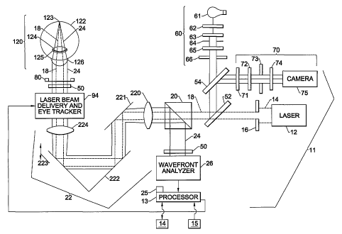
FIG. 1 is a schematic diagram of the system of the present invention.
FIG. 2 is a logic flow diagram for the data flow.
FIG. 3 illustrates an exemplary graphical user interface of the present
invention.
DETAILED DESCRIPTION OF THE PREFERRED EMBODIMENTS
A description of the preferred embodiments of the present invention will now
be presented with reference to FIGS. 1-3.
An exemplary layout of the system 90 for performing optical measurements
and a corrective procedure is illustrated in FIG. 1. This layout is not
intended as a
CA 02377974 2001-12-21
WO 01/80791 PCT/IBO1/00839
4
limitation, and alternate systems adapted to achieve laser ablation may also
be
envisioned by one of skill in the art. The eye tracker and laser beam delivery
unit 94
comprises a processor 13 in electronic communication with a graphical display
14
having means for receiving inpufi from an operator, such as by means of an
input
device such as a mouse 15 in electronic communication with the processor 13.
Under various conditions, which will described in the following, a portion of
the
eye may be desired to be "screened" from the ablation beam. In the example of
a
graphical user intertace 50 illustrated in FIG. 3, an ablation zone 20 is
indicated by
a cross-hatched area. In a preferred embodiment, the graphical user interface
50
shown would be in color to provide improved contrast against the eye, and the
ablation zone 20 would comprise a color that would stand out against the eye,
such
as yellow.
The flap and hinge in this illustration will have been made along the lower
left-
hand quadrant of the eye 22, a portion 23 of which resides within the ablation
zone
20. In order to protect this portion 23, a protected sector 24 is defined
using the
mouse 15 and input into a software package 25 resident on the processor 13,
which
creates the protected sector 24. The protected sector 24 on a color screen 14
would
also be a contrasting color, such as blue. The software package 25 then
prevents
the protected sector 24 from the impinging ablation beam by inhibiting the
excimer
laser from firing in this sector 24. If the flap is sufficiently large that
the flap and hinge
do not intersect the ablation zone 20, this definition of a protected sector
24 would
not be required.
In a preferred embodiment of the invention, configuration parameters allowfor
an additional sector adjacent the protected sector 24 to be protected also in
order to
compensate for the finite extent of the laser pulses. Without this additional
sector,
a laser shot fired just outside the protected sector 24 may still ablate, to
some
degree, an area just within the protected sector 24. Similarly, an
extracorneal region,
such as a conjunctiva or sclera, can be protected from unwanted exposure by,
for
example, a large, decentered treatment at least a portion of the shots of
which could
otherwise have extended beyond the cornea.
CA 02377974 2001-12-21
WO 01/80791 PCT/IBO1/00839
The graphical user interface 50 of FIG. 3 also has a reticle 26 including a
generally circular portion 27 overlying the edge of the cornea 28 and a cross-
hair 29,
the intersection point 30 positioned generally at a central point of the
cornea 28.
Data 31 are also provided giving dimensional and positional information.
In addition to protecting the flap and hinge region of the eye, it may also be
contemplated to use the present invention in the following:
~ Avoidance of reexposure of a pre-existing treated zone of tissue, such
as might occur during a retreatment for decentration
~ Avoidance of reexposure of tissue during a reoperation to complete an
aborted treatment
~ Avoidance of laser exposure of scarred tissue or thin areas where such
an exposure would lead to undesired clinical consequences
~ Definition and creation of multiple zones of treatment, such as may be
desired to create a multifocal cornea for amelioration of presbyopia
~ Definition and creation of a multifocal cornea by ablation of nearsighted
treatment in the midperiphery and a central zone of distance correction
~ Definition and creation of a multifocal cornea by ablation of alternating
annuli of distance and near corrections, starting with distance correction in
the
center of the cornea
~ Definition and creation of a multifocal cornea by ablation of a central
distance zone, a midperipheral zone of middle-distance correction (typically
3 ft), and an outer zone of near correction
In addition, one of skill in the art will recognize that the systems and
methods
of the present invention are amenable for use with other radiative treatments,
such
as localized irradiation of a tumor, lithotrypsy, removal of a skin
disfigurement, or
cauterization.
In the foregoing description, certain terms have been used for brevity,
clarity,
and understanding, but no unnecessary limitations are to be implied therefrom
beyond the requirements of the prior art, because such words are used for
description purposes herein and are intended to be broadly construed.
Moreover, the
embodiments of the apparatus illustrated and described herein are by way of
CA 02377974 2001-12-21
WO 01/80791 PCT/IBO1/00839
6
example, and the scope of the invention is not limited to the exact details of
construction.
Having now described the invention, the construction, the operation and use
of preferred embodiment thereof, and the advantageous new and useful results
obtained thereby, the new and useful constructions, and reasonable mechanical
equivalents thereof obvious to those skilled in the art, are set forth in the
appended
claims.