Note: Descriptions are shown in the official language in which they were submitted.
11.5ET.2001 1?:Z9 . SIB MILRNO +39-0280633200 NR.380 P.3i3
11-09-2001 CA 02378064 2002-O1-17 IT0000301
,'
WO OL853I7 PC?/IT00100301
. ~_. _ I .
~~CTROSURGI~ r.fi'~OR.TIJMOR'f~~~,_T _BY
.
~Q~,7 AEOU~ZtCY"
the present invention relates to as electrosurgical probe far tlae ~
tceby sadiofrequency energy, and pa~al~dy to a probe coatevia~ag a
of aeedk-shaped elect~c~odes whose tips can be expanded at tha tmmr
to be so as to affect a volume of catuoer tissue which is ss Iarge cg
possc'Erlc.
The tumor treatment by hypetthermia which is induced by radiofrequency
energy or other enetBy farms is already known in medieiae. Electsosur$ical
14 probes provided with needle-sloped electrodes which, by pinto the
cancer tissue, cause its neeros'ss, have already beau developed. WO 96129146
describes electrosurgical probes comprising a muttiplicixy of independent
needle
shaped electrodes which are pushed inside the tisane to be arexal by making
them
come out from the-point of a metal cannula inseated into the patient's body.
'Ibis
is obtained by usi~~g electrodes formed of thin metal wires having as arched
end
~ P .a« , ~~ ore, pP"~
eiaetroprob~'~which ~aLso formed of aaaula o mul~plicriy
of f~lifo~r electrodes liaviug arched tips with elastic memory which ate
compressed inside the caanula in a stretehcd po 'satiov and which expand
themselves whey they are pushed oat of the esnnula iase~d ias3de the tisstu w
be treated. Zhe fili~m electrodes are arranged iaaide the caaaula ammzd a
camtal
nucleon so that, when their tips are pushed out from the point of the caaaula,
the
cancer tissue is ai~Cted ui a regular volume wliicli is as aseimilable as
possible to ° ~ : :: : , _ _ . - -
a sphere. ' - .° :' y~.
H~owevez, none of the presently lmomi elecxro~cai probes is capa'fle of
creating sa electrical field having a sea! spherical shape, by them it is at
the most
posssble to obtain as elorrzical field having an ellipsoidal shape since, as
it is
pravyded with el8stieal memory which is kept in a snbet~ially str~c~hed
condition inside said eannula sad is released, .expaa.diung ~itsel~ when.. ~
eleet~odes are pushed out of the caanula is order to penetrate into the tissue
to be
treated. Object of said expavsioa is that awvr ~ 4 csr ' to be tmated
which is as lar as oasible is a$eoted. O desonb et
AMENDED SHEET
CAADCAAI/.C'7CTT 11 ('CD 1~.'1A ~mnnnnnvn-rrtr .. urn .~ n.
CA 02378064 2002-O1-17
WO 01/05317 PCT/IT00/00301
-2-
apparent from figures 4 and 5 of W098/52480, only a small portion of the
distal
end of the metal caunula, which participates to the formation of the
electrical
field, is surrounded by the arched ends of the filiform electrodes.
Anotlier drawback of the known electrosurgical probes is that tlieir use
involves the risk that the arched ends of the electrodes, by penetrating into
the
cancer tissue under the impulse of the suitable control, may go beyond the
appointed target and penetrate also in vital structures, for instance a blood
vessel,
adjacent to the portion to be treated.
Tlierefore, object of the present invention is providing an electrosurgical
probe of the type with multiple needles liaving an arched point witli elastic
memory which is free from the above mentioned drawbacks. Said object is
achieved by the electrosurgical probe liaving the features specified in claim
1.
Further features are specified in the following claims.
The electrosurgical probe according to the present invention eliminates the
first of the above mentioned drawbacks of the probes according to the prior
art,
because it has the important feature that the electrodes, when tliey are
outside the
cannula, are arranged like the meridians of an ideal sphere wliose diameter is
formed of a long segment of the cannula distal portion, which is not coated by
the
insulating material. Accordingly, also the cannula in its distal portion
participates
in creating the active field of the radiofrequency. As a matter of facts, when
the
needle-sliaped electrodes protrude fi-om the cannula, the arched portion of
each
electrode forms au arc of 180° wliose two ends are located near to the
two ends of
the uncoated distal portion of the caunula, tliat is, near to the two poles of
the ideal
sphere of wliicli tliat portion of the cannula forms the diameter.
Tlie second of the above mentioned drawbacks of the known electrosurgical
probes is eliminated by means of the probe according to the present invention
because it lias the feature that the expansion of the arched tips of the
electrodes is
controlled by traction of the electrodes and not by thrust like in the known
probes.
In otlier words, whereas in the known probes the expansion of the filiform
electrodes is controlled by a movement in the same direction of penetration of
the
electrodes into the patient's tissues, in the probe according to the present
invention
CA 02378064 2002-O1-17
WO 01/05317 PCT/IT00/00301
-3-
the expansion is controlled by traction, that is by a movement in the opposite
direction with respect to that of penetration of the electrodes into the
patient's
tissues. Resultingly, the flee end of each filiform electrode will have the
tendency
to converge, after the expansion movement, towards the distal end of the metal
cannula, thus avoiding the risk that it may diverge towards vital structures
and
perforate them. Tliis is due to the fact that in the probe according to the
present
invention, in the rest position thereof, both the filiform electrodes and the
rod
wliicli controls tliem liave the same direction but they are turned in
different
directions after the expansion.
Besides eliminating the above mentioned drawbacks of the probes according
to the prior art, the electrosurgical probe according to the present invention
offers
anotlier important advantage, tliat is avoiding, during said operation, that
the
electrodes come out accidentally from the probe point during the positioning
operation of the probe itself into the tissue. This is due to the fact that
the
operation for causing the electrodes expansion takes place by retraction with
a
movement in the opposite sense witli respect to the positioning movement, and
not in the same sense like in the prior ait.
A fizrtlier advantage of the electrosurgical probe according to the present
invention with respect to the prior art is that no fi~ee space between the
distal end
of the cannula and the content thereof is provided. As a matter of fact, said
fi~ee
space, which is present in the known probes, may cause undesired phenomena of
core boring of healthy tissue of the patient during the probe positioning
operation.
The structure of the probe according to the present invention allows also to
close
the distal end of the cannula and to confer it a pointed tip.
These and other advantages of the electrosurgical probe according to the
present invention will be evident to those skilled in the art fiom the
following
detailed description of one embodiment thereof with reference to the
accompanying drawings, wherein:
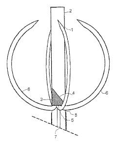
- figure 1 shows an enlarged and partially sectioned view in side elevation
of the canuula of the probe according to the present invention;
- figure 2 shows a similar view of the same cannula of figure l, but with
CA 02378064 2002-O1-17
WO 01/05317 PCT/IT00/00301
-4-
the arclied tips of the electrodes taken out from the cannula and in the
working position; and
- figure 3 sliows a view in scale of a complete electrosurgical probe with
the electrodes in the working position.
Witli reference to figure 1 there is shown that the electrosurgical probe
according to the present invention comprises a metal cannula 1, of a known
kind,
inside which a liead 3 is placed, having the upper end 4 preferably pointed
like a
flute moutlipiece. Also the point of cannula 1 can be pointed like a flute
moutlipiece like the upper end 4 of head 3. In this way, the point of head 3
can
coincide in the rest position witli the point of cannula 1 without any free
space
between the two points. This structural measure avoids the undesirable
phenomena of core boring of healthy parencliymatous tissue whicli occur by
using
the probes according to the prior art wherein a free space between the piston
head
wliich pushes the electrodes and the point of the cannula whicli contains
tliem
necessarily exists. It is obvious tliat the point of liead 3 in the rest
position can
also protrude from the end 2 of the cannula, because tliis is open. However,
constructive variants are possible, with a closed and sliaip point of cannula
2, or
with an open canuula 2 and a pointed head 3 like in figure 2. Otlier
embodiments
are obviously possible in order to fix the base of the electrodes to the
distal end of
tlieir control rod so that in the stretched position they are side by side
with said
axis.
From base 5 of liead 3 a multiplicity of filiform electrodes 6, as well as
control rod 7 of head 3, branch off downwards. This is the most innovative and
advantageous feature of the electrosurgical probe according to the present
invention, witli respect to tliat of the prior art wherein the electrodes in
the
stretched position form the prolongation of the electrode control rod and are
not at
the side tliereof. The filiform electrodes liaving elastic memory of shape are
already known in the art and therefore they do not need a detailed
description.
The filiform electrodes 6 are arranged on the side and parallel to rod 7 in a
stretched position with tlieii~ downward turned tips whicli are near or
sliglitly
above a multiplicity of lioles 8 arranged along the circumference of cannula
1.
CA 02378064 2002-O1-17
WO 01/05317 PCT/IT00/00301
-5-
The number of holes 8 corresponds to the number of electrodes 6 so tliat each
electrode 6 has a relevant hole 8 for coming out of canuula 1 when rod 7 of
head 3
is pulled downwards. Holes 8 are arranged in such a way that each of the
electrode points can pass through the relevant hole 8 in order to get out of
cannula
1 under the thrust of liead 3 wlien this is pulled downwards. As soon as rod 7
is
pulled downwards, electrodes 6 come out of holes 8 of canuula 1, gradually
regaining their naturally arched shape by virtue of their elastic memory, thus
assuming in the end the configuration shown in figure 2.
Witli reference to figure 2, there is shown that each electrode 6, after
passing through the relevant hole 8, under the tlwust of liead 3 wliich is
pulled
downwards by means of control rod 7, has passed nearly completely through the
relevant holes 8, thus penetrating in the cancer tissue to be treated or
surrounding
it. In the course of said penetration, by viirtue of the elastic memory of
which
filiform electrodes 6 are provided, tliese expand tliemselves wliile bending
until
they assume the position shown in the figure. As it can be seen, eacli
electrode 6
has assumed a position which greatly resembles that of a meridian of an ideal
sphere. Said shape is much more regular tlian those which can be obtained by
the
electrosurgical probes according to the prior au. Said regularity depends
substantially on the fact that filiform electrodes 6 are not only pushed
inside the
cancerous tissue, like in the known electrosurgical probes, but they are also
guided from the lower ends of the relevant holes 8 suitably provided along the
circumference of cannula 1.
Holes 8 can have any suitable shape for favoring the coming out of the
needle-shaped electrodes and for guiding them upwards as soon as they come out
from cannula 1. The prefeiTed sliape for holes 8 is the one slightly
lengthened in
the direction of the length of the needle-shaped electrode so as to favor the
coming out thereof from cannula 1. The lower rim of the hole is provided with
a
profiled upward-turned cross-section wliich forms an upward-directed guide
plane
for the needle-sliaped electrode which helps it in its expansion until it
reaches the
position shown in the figure.
Obviously, the number of the holes depends on the number of the filiform
CA 02378064 2002-O1-17
WO 01/05317 PCT/IT00/00301
-6-
electrodes. Their number varies according to the needs and can vary between
two
and twenty. In the embodiment shown in the figures 1-3 they are four. When the
number of the filiform electrodes is very higli, it is preferable that holes 8
are not
circumferentially aligned on the surface of cannula l, but arranged according
to an
elicoidal line or on more parallel circumferences.
Cannula 1 is provided with an insulating coating 9 of a plastic material
whose upper edge is positioned slightly under the last of holes 8. In tliis
way, the
exposed portion of metal cannula 1 during working forms the diameter of the
ideal
sphere created by the envelope of the needle-shaped electrodes.
With reference to figure 3 there is sliown au electrosurgical probe according
to the present invention in the working position. In said position, the
filiform
electrodes 6 are already expanded and their points liave reached a position
very
close to the distal end 2 of cannula 1. The coming out of electrodes 6 from
the
inside of cannula 2 has been caused by moving away head 3 fi~om distal end 2
of
the cannula. Said movement away has been achieved by pulling control rod 7 by
means of knob 9 which is thus progressively removed fi~om handle 10 which is
internally provided with a room suitable for housing stem 11 of knob 9.
In figure 3 there is also shown the independent lateral needle 14 which
works as a support for one or more thermistors of the teletliermometnc system
applicable to the probe according to the present invention. Needle 14 comes
out of
cannula 1 tlirough a suitable hole 15 made on the cannula and is controlled by
cursor 12 wliicli is partially housed inside knob 10. Further to lateral
needle 14,
the probe according to the present invention can be provided with one or more
similar needles. Each one of needles 14 carrying thernustors can be rendered
radiofrequency active at discretion, thus enlarging the extent of the
thermolesion.
Obviously, the probe according to the present invention can be completed
with the necessary connections to the radiofrequency generator and with all
the
other attachments necessary for its working, maintenance and use, as well as
for
the telethermometric check of the thermal lesion during the treatment.
In a preferred embodiment of the present invention, a thermistor has been
applied on each end of the non insulated portion of cannula 1. A third
thermistor
CA 02378064 2002-O1-17
WO 01/05317 PCT/IT00/00301
_'7_
lias been advantageously applied also on the insulated portion of canuula l,
immediately under holes 8.
Rigid cannula 1 can be replaced by a flexible tube in a portion comprised
between the line of the holes 8 and handle 10. Said embodiment allows the
probe
to be used as a catheter.
