Note: Descriptions are shown in the official language in which they were submitted.
CA 02379670 2002-O1-17
WO 01/12255 PCT/US00/22213
-1-
DILATION BALLOON HAVING MULTIPLE DIAMETERS
Description
Technical Field
This invention relates generally to surgical devices, and more particularly
to catheters, dilators and other devices for establishing, restoring or
enlarging lumens
in the body, especially in the intestines and esophagus.
Backctround of the Invention
A variety of body lumens are subject to undesired strictures or narrow
regions. For example, blood vessels can be blocked or narrowed by
atherosclerosis,
while esophageal strictures can arise from individual anatomical differences,
or from
diseases such as connective tissue disorder. Procedures for dilating or
enlarging
such strictures or narrowed regions often entail the use of a balloon dilation
catheter.
Such catheters include a deflated balloon which can be positioned across a
particular
stricture or narrowed region, and which is then inflated with a fluid in order
to widen
the lumen without trauma to the wall of the lumen.
A variety of balloon catheters and dilators are known which include a
balloon attached to the distal end or' a catheter tube or shaft, and which
also include
a stainless steel or nitinol wire stiffener extending through the catheter
shaft and
balloon. Balloons for dilating esophageal, pyloric, or colonic strictures can
be made
of a semi- or non-compliant material that permits sufficient expansife force
to dilate
the stricture. Non-compliant materials, such as polyethylene terephthalate
(PET), are
preferred over semi-compliant or compliant materials because they are much
less
prone to "dog-boning", a situation in which the resistance of the stricture
forces the
fluid in the balloon to either side, therefore providing comparatively less
radial or
expansile force than would a standard non-compliant balloon.
While dilation of stenoses in blood vessels is usually performed as a one
step procedure, there is often a clinical advantage in being able to dilate
esophageal
WO 01/12255 CA 02379670 2002-O1-17 PCT/US00/22213
-2-
and other gastrointestinal strictures using a series of progressively larger
balloons so
as to avoid tearing or perforation of the luminal wall. The disadvantage of
sequentially introducing larger balloons is that multiple introductions
increase risk to
the patient and prolongs the procedure. One factor determining the length of
the
procedure is the difficulty in being able to precisely position and reposition
the
balloon at the stricture. Additionally, patient discomfort is naturally a
concern when
multiple catheter introductions are required. What is needed is a dilation
balloon that
can efficiently and effectively perform staged dilation of a stricture while
minimizing
risk and discomfort to the patient.
_Summary of the Invention
The foregoing problems are solved and a technical advance is achieved in
an illustrative dilation balloon catheter comprising a single non-compliant
balloon,
made of polyethylene terephthalate (PET) or another suitable material, that is
formed,
such as over a mold, to include a plurality of longitudinal sections, each
having a
different diameter at the center of the section. The balloon can be attached
to the
distal end of a catheter made of a polymer, such as polyurethane, using a
bonding
means such as a UV adhesive. In one embodiment used in conjunction with an
endoscope to dilate esophageal, colonic, and pyloric strictures, the dilation
balloon
comprises three sections with the distal section having the smallest diameter.
A
wire guide, e.g., of a nitinol (NiTi) alloy, can extend through the lumen of
the
catheter, the balloon, and extend distally, encased in a protective polymer
jacket,
to aid in cannulation of the stricture.
To cannulate a stricture of a body lumen such as the esophagus, the
balloon portion is advanced from the endoscope and the stricture is dilated
using the
> distal (smallest) section. The balloon is usually deflated, then the second,
intermediate section, which is about 2 mm larger that the first, is advanced
over the
stricture and inflated. Finally, the proximal section, which is yet another 2
mm
larger, can be used to make a third dilation of the stricture, if desired,
before the
balloon catheter is removed from the patient. This staged series of inflation
helps
avoid tearing or perforating of the particular body lumen being dilated, while
the
WO 01/12255 CA 02379670 2002-O1-17 pCT~S00/22213
-3-
single balloon allows a single introduction into the patient for the
procedure, rather
than requiring three separate introductions of different-sized balloons. In
addition,
the single balloon can be attached to a smaller diameter catheter, since it is
does not
have to be multi-lumen, an important advantage when being used in endoscopy.
In one aspect of the invention, the central portion of each balloon section
is depressed to form a waist that helps the balloon to center itself over the
stricture.
This waist, normally 2-6 mm narrower than the adjacent portions of the
section, can
be configured to include an abrupt change in diameter, creating somewhat of a
dumbbell-shaped balloon section, or it may be more gradual in transition. In
an
illustrative embodiment of a three section balloon, the adjacent portions of
the
intermediate section are basically shared with the distal adjacent portion of
proximal
section and the proximal adjacent portion of the distal section, respectively.
The
number of sections is determined by the number of different central portions
or
waists of the balloon, rather than the number of adjacent portions, which are
often
going to be one greater in number than the central portions.
In another aspect of the invention, the longitudinal positions of the
different balloon sections can be marked with indicia that can be observed
under
fluoroscopic imaging and/or via the endoscope. The indicia can be imprinted
on, or
incorporated into the wire guide that extends through the balloon, using ink,
bands,
or other means. Additionally, the indicia can be directly printed on, or
applied to the
balloon surface (e.g., using thin radiopaque foil). The indicia, which
preferably marks
the center of the balloon section, can be different for each balloon section,
or it can
be the same.
Brief Description of the Drawing
Embodiments of the present invention will now be described by way
of example with reference to the accompanying drawings, in which:
FIG. 1 depicts a pictorial view of the illustrative embodiment of the
present invention;
FIG. 2-3 depict side views of an embodiment of the present invention
being deployed from an endoscope;
WO 01/12255 CA 02379670 2002-O1-17 PCT/US00/22213
-4-
FIG. 4 depicts a side view of an alternative embodiment of the present
invention;
FIG. 5-6 depict side views of alternative embodiments of the present
invention having indicia to facilitate positioning of the device; and
FIG. 7 depicts a side view of an embodiment of the present invention
showing alternatively shaped balloon sections.
Detailed Description
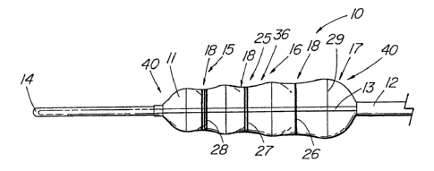
FIG. 1 depicts a pictorial view of the illustrative embodiment of the
present invention of a dilation balloon catheter 10 comprising a single non-
compliant
balloon 11 mounted distally to a catheter 12 with a lumen 31 extending
therethrough that also contains a wire guide 13 which extends the length of
the
balloon 1 1. The lumen 31 of the catheter 12 serves as the inflation lumen for
the
balloon 1 1, which is normally filled with saline or water to a pressure of 40-
100 psi
about 275-690 kPa), typically about 60 psi (413 kPa). The wire guide 13
extends
i beyond the balloon 1 1 to form a distal portion 14 that facilitates
cannulation of a
stricture for placement of the balloon. The balloon 1 1 is longitudinally
divided into
three sections 15,16,17, each having a different maximum outer diameter when
inflated and each in communication with one another. Naturally, it is within
the
scope of this invention to have any number of different diameter sections from
two
up to as many as is practical for a given procedure. It is also within the
scope of the
invention to have one or more additional balloons, separate from the multiple-
section
balloon, that can be separately inflated via a different lumen, and possibly
be of a
different shape, diameter, or material than that of the primary balloon of the
present
invention. In the illustrative embodiment, the first diameter section 15,
comprising
approximately the distal one-third of the balloon, has the smallest diameter.
The
second diameter section 16 and third diameter section 17 comprising the middle
and
proximal portions, respectively, of balloon 1 1 , are progressively larger in
diameter.
In the illustrative balloon 10, which is used for dilating esophageal,
pyloric, or
intestinal strictures, the respective sections 15,16,17 of balloon 1 1 have
diameters
0 of 18, 16, and 14 mm as measured from the midpoint or central portion 36 of
each
CA 02379670 2002-O1-17
WO 01/12255 PCT/US00/22213
-5-
section, with the range for balloon sections appropriate for particular these
anatomical sites being generally within the 4 to 25 mm range. The typical
balloon
length for esophageal use would be about 8 cm, while a 5.5 cm length would be
appropriate for colonic and pyloric dilation.
In the illustrative embodiment of FIG. 1 , the balloon is made of non-
compliant material such as polyethylene terephthalate (PET), irradiated
polyethylene,
or nylon. For the use described, the thickness of the material should ideally
fall
within the range of .005 to .02" (.13 to .5 mm) to provide a dilation balloon
that will
exert sufficient force against the luminal wall without causing rupture, yet
still fit
within the channel of an endoscope. The balloon of the illustrative embodiment
can
be formed using well-known techniques. One method includes heating a tube of
PET, then stretching and inflating the material within a mold to create the
desired
final shape. For example, a tube having an O.D. of .150" (3.8 mm) and a wall
thickness of .008" to.015" f.2 to .38 mm) can be used to produce a 14-16-18
mrn
diameter balloon. After convection heating of a central portion of the tube
for about
15-45 seconds, the heat source is retracted and a mold of the fully distended
shape
of the balloon is placed over the tube. The tube is stretched along its
longitudinal
axis to create a thin-walled portion corresponding to the final length of the
balloon.
At that point, pressurized gas is introduced through one end of the tube, the
tube
being sealed at the other end, thereby expanding the heated tube to conform
with
the inner surface of the mold. After a brief interval, the gas is partially
released to
a point above 1 atmosphere such that when the mold is retracted, the balloon
remains inflated in its generally distended shape. After a brief cooling
period the
balloon is ready to be removed and bonded to the catheter. In the illustrative
embodiment, the PET balloon is bonded to a catheter made of polyurethane
(PELLETHANE°, Dow Corning Co.) using a UV adhesive. Other appropriate
medical
device adhesives can be used as well.
While the balloon of FIG. 1 includes three main section 15,16,17, each
having a different nominal diameter, the sections themselves are not of a
uniform
diameter. To assist the balloon in centering over a stricture and maintaining
its
WO 01/12255 CA 02379670 2002-O1-17 PCT/US00/22213
-6-
position during inflation, a depression or waist 18 is formed at the central
portion 36
of each section 15,16,17. As used in the specification and claims herein, the
measurements of the diameter of the balloon sections 40 as defined, are taken
about
the waist 18 of the central portion 36 at each section's midpoint. This value
represents the nominal diameter of the balloon section with the widest point
of the
adjacent portions 32,33 of the section 15 being 2-6 mm greater in diameter.
That
difference between the two points 36 and 32 or 33, is 2 mm in the illustrative
embodiment. It is the number of different central portions 36 that determine
the
number of sections in the balloon, not the number of adjacent portion 32,33,
which
in the embodiment of FIG. 1 appear a four distinct enlarged sections
surrounding the
three waists 18 of the respective central portions.
FIG. 4 depicts an alternative embodiment of the balloon in which the
sections 15,16,17 of the balloon are dumbbell shaped. In this embodiment the
waist
18 is more abruptly defined relative to the adjacent sections 32,33, which are
more
spherical in shape than in the embodiment of FIG. 1. An embodiment without a
well-
defined waist 18 at the central portion 36 is depicted in FIG. 7. In the this
embodiment, each of the sections 40 are of substantially of the same diameter,
or
are only slightly concave at the center portion 36.
To provide the balloon catheter sufficient rigidity, a wire guide 13 is
included within the lumen of the catheter 12 and the balloon 1 1. The wire
guide
does not completely fill the lumen such that fluid can adequately traverse the
lumen
to inflate the balloon. Alternatively, a multiple lumen catheter can be used
with the
wire guide being situated within a lumen that is separate from the inflation
lumen.
The preferred wire guide material is a superelastic alloy such as nitinol
(NiTi alloy),
i although a standard stainless steel wire guide can be used. A .023" (.58 mm)
diameter nitinol wire guide offers good rigidity for introduction into the
gastrointestinal system and to cannulate strictures, while remaining highly
flexible
within a tortuous navigational path. The distal portion 14 includes the distal
portion
of the wire guide 13 that is coated or encased in a polymer such as
polyurethane.
WO 01/12255 CA 02379670 2002-O1-17 pCT~S00/22213
_7_
To make the distal portion 14 less traumatic to tissue, the nitinol wire guide
13 is
ground to a gradual taper over the distal 5 cm of the device.
FIG. 2 depicts deployment of the balloon catheter 10 from the accessory
channel of a standard endoscope 19. The endoscope 19 serves as an outer
constraining device for introducing the balloon catheter 10 to the target
site. The
uninflated balloon 1 1 is shown partially advanced from the end of the
endoscope 19.
When used with an endoscope, the balloon 11 is normally deployed completely
before it is inflated, primarily to avoid damaging the accessory channel of
the scope.
Clinical use of the dilation balloon catheter is depicted in FIG. 3.
Initially,
the stricture is examined and sized using the endoscope. In the illustrative
example,
a dilation balloon catheter 10 is selected having three sections wherein the
distal
section 15 (the smallest section) is sized approximately 2 mm larger than the
stricture opening 34. It is clinically important when dilating many types of
strictures
that dilation be conducted in stages, rather attempt dilating the stricture in
a single
step, such as how an angioplasty procedure is performed. Gradual dilation will
prevent the esophagus, colon, or other body lumen from tearing or perforating
from
the expansile force of the balloon. Following inspection of the stricture, the
dilation
balloon catheter 10 is advanced through the scope to the site of the stricture
34 and
is cannulated by the distal portion 14 of the balloon catheter 10. As shown in
the
figure, the distal section 15 is positioned with the waist 18 over the
stricture 34 and
then inflated. The waist naturally centers over the narrowest part of the
stricture
34 and keeps the balloon section 15 from slipping to one side or another.
After
dilation, the balloon 1 1 is then partially deflated. The middle section 16,
which is
2 mm larger that the distal section, is advanced over the stricture 18 and the
balloon
reinflated. If desired, the proximal section 17 is used as a third dilation of
the
stricture. For gastrointestinal strictures, many physicians consider 6 mm (or
three
inflations) to be the maximum amount of dilation that can be safely performed
without risking damage to the body lumen being treated. Alternatively, it
would also
be possible to locate the largest end of the balloon at the distal end,
advance the
distal 15 and/or middle 16 sections beyond the stricture 18, and initially
dilate using
05-11-2001 5 pCT CA 02379670 2002-O1-17 REPLACEMENT pA US002221
..~..~i
_ _ ~'~I7J~lPfi
s ~tl pn CO
py
a middle 16 or proximal 17 sections, then moving proximally. The disadvantage
of
this is that you will possibly advance much of the balloon catheter past the
stricture
without the benefit of having been able to inspect this distal this distal
region
endoscopically, thereby adding potential risk to the procedure.
FIGs. 5-6 depict alternative embodiments of the present invention in which
a system of indicia 21,25 are placed on the balloon catheter 10 to enable the
clinician to orient a given section of the balloon to the stricture or other
desired site,
either under fluoroscopy or via direct visualization (e.g., through an
endoscope): In
the embodiment of FIG. 5, the series of indicia 21 are placed on the wire
guide 13
for orienting the balloon 11 to the desired location. The marks are placed at
each
section 15,16,17 of the balloon 11 and can each be identical vary in shape,
color,
or number as in the illustrative example. As depicted, the first wire guide
indicium
22 comprises a single band that corresponds to the center portion 36 or waist
18 of
the proximal balloon section 17, while the second wire guide indicium 23
comprises
a double band identifying the intermediate 16 section. The third wire guide
indicium
24 comprises a triple band that corresponds to the center portion 36 or waist
18 of
the distal section 15. As used herein, "indicium" is defined can include a
single
identifier, such as a band, dot, number, color, etc., or combination of
markings (i.e.,
double bands, dots, etc.) that is used to designate the location of a single
balloon
section. These markings or indicia 21 can be made radiopaque to assist the
physician in positioning the balloon under a fluoroscope. Bands or other
indicia made
of a material such as gold, platinum, or tantalum can be applied to the outer
surface
of the wire guide or metal. Other types of radiopaque materials can also be
applied
to or deposited on the surface of the wire, such as an ink, paint, or polymer
containing barium or tantalum, etc. As an alternative to providing varying
numbers
of bands, dots, etc, to mark the different balloon sections, numbering or
lettering can
be used, especially if the purpose of the indicia is to be viewable by the
endoscope.
FIG. 6 depicts an alternative embodiment having system of indicia for
positioning of the balloon, comprising markings 25 that are imprinted on the
balloon
1 1 material. Much like the embodiment of FIG. 5, a first balloon indicium 26,
. 05-11-2001 CA 02379670 2002-O1-17 US0022213
. . . __ . 5 PCT REPLACEMENT PA__
- 9 - Co~firmati
ors CopY
comprising a single stripe, encircles the balloon 11 at the waist 19 of the
proximal
balloon section 17, while the second and third balloon indicia 27,28,
comprising
double and triple stripes, identify the middle and distal balloon sections
15,16,
respectively. The stripes can comprise metal particles that are deposited on
the
outer surface using well-known techniques. Typically, a .002" (.05 mmf thick
deposit with provide sufficient radiopacity. Alternatively, thin strips of a
radiopaque
material, such as a microthin metal foil, can be applied to the balloon, or a
radiopaque
material can be imprinted directly on the balloon material.
Except where the teachings differ, one can look to U.S. Patent No.
7 0 5,681,344 to Kelly for additional details of the construction and use of
an
esophageal dilation balloon made of PET and having a nitinol wire guide. The
balloon
of the '344 patent is similar to the present invention, with the primary
difference
being that the Kelly balloon is of a single diameter. Any other undisclosed or
incidental details of the construction or composition of the various elements
of the
disclosed embodiment of the present invention are not believed to be critical
to the
achievement of the advantages of the present invention, so long as the
elements
possess the strength or flexibility needed for them to perform as disclosed.
The
selection of these and other details of construction are believed to be well
within the
ability of one of even rudimentary skills in this area, in view of the present
disclosure.
25