Note: Descriptions are shown in the official language in which they were submitted.
CA 02380111 2002-02-O1
WO 01/08717 PCT/US00/21288
CONTROLLED RELEASE IMPLANTABLE DEVICES
Field of the Invention
The invention relates to implantable devices for delivering drugs to a
desired location within a body.
Background of the Invention
Drugs can be delivered systemically, e.g., by oral ingestion, or can be
delivered locally directly to a site of disease. Some drugs are most effective
if
delivered repeatedly, over a period of time, or delivered steadily, e.g.,
using an
implantable device.
Summary of the Invention
The invention relates to new implantable devices specially designed to
deliver drugs to desired locations adjacent to unique target sites in bone,
cartilage,
ligaments, muscle, and other internal body tissues and structures, and to
provide a
controlled release of a wide variety of drugs. In some embodiments, the
devices also
perform a mechanical function, e.g., attaching tissue to a support structure,
such as
bone.
In general, in one aspect, the invention features an implantable device for
attaching tissue to a support structure inside a body and for delivering a
drug to a
target location near the support structure. The device includes a first
portion that
engages the tissue, and a second portion that engages the support structure. A
section
of the device defines an internal cavity that has a size and shape for
containing a
controlled release agent that includes the drug. Alternatively, instead of
defining an
internal cavity, at least a portion of the section can be formed from a
material that
comprises the controlled release agent that includes the drug. The section can
be part
of the first portion, part of the second portion, or a separate section
connected to either
the first portion, the second portion, or both.
Embodiments of this aspect of the invention may include one or more of
the following features. The device can include the controlled release agent.
The
controlled release agent can be a mixture of a polymer and the drug, e.g.,
microspheres of the polymer containing the drug. The controlled release agent
can be
configured to release the drug for a period greater than, e.g., two days or
five weeks.
CA 02380111 2002-02-O1
WO 01/08717 PCT/US00/21288
The agent can also be configured to release the drug intermittently over a
period of
time.
The drug can be, e.g., a down-regulatory cytokine, such as interleukin-10,
a pain killer, such as lidocaine, platelet derived growth factor, an
antibiotic, a
hormone, a prostaglandin, a protein, a peptide sequence, or a nucleic acid.
The
polymer can be, e.g., a polyanhydride, a polylactide, a polyglycolide, a
polylactic
acid, a polyglycolic acid, a polyorthoester, a polyorthocarbonate, a
polyacetal, a
polymer derived from alpha hydroxycarboxylic acids and lactones, a polymer
derived
from condensation of divinyl ethers and polyols, an e-caprolactone polymer,
ethylene
vinyl acetate copolymer, and other co-polymers of the above listed polymers,
such as
50:50 poly(DL-lactide-co-glycolide).
The second portion of the device can be configured to penetrate the tissue,
e.g., with a pointed end. The section can be degradable by bodily fluids. In
addition,
the section can have an aperture that exposes the interior cavity to bodily
fluids when
the device is implanted in the body. A membrane permeable to bodily fluids and
to
the drug only when the drug is dissolved or suspended in bodily fluids can
cover the
aperture. The section can also include the first and/or the second portion.
The tissue can be soft tissue or bony tissue, and the support structure can
include bone.
In another aspect, the invention features an implantable device for
delivering a drug to a desired location inside a body. The device includes a
rigid
exterior that has a tapered end for penetrating tissue within the body, and a
projection
for engaging tissue within the body. The device also includes an internal
cavity in
fluid communication with the rigid exterior. The cavity has a size and shape
for
containing a controlled release agent that includes the drug.
Embodiments of this aspect of the invention may include one or more of
the following features. The rigid exterior has a pointed, arrow-shaped head
that
includes both the tapered end and the projection. The arrow-shaped head can
have a
shaft and two projections, each projection having a first pointed end and a
second end
connected to the shaft. The first ends are movable between a first position
flush with
the shaft, and a second position displaced away from the shaft.
In another aspect, the invention features an implantable staple for
delivering a drug to a desired location within a body. The staple includes at
least two
prongs that penetrate and engage tissue, and a shaft connecting the two
prongs. The
-2-
CA 02380111 2002-02-O1
WO 01/08717 PCT/US00/21288
shaft has an internal cavity that has a size and shape for containing a
controlled
release agent that includes the drug. The shaft can include a material
degradable by
bodily fluids.
The invention also features an implantable device for delivering a drug to a
target location. The device includes an elongated rod curved in a generally
helical
shape. The helical shape tapers to a point that in use penetrates soft tissue,
and the
helical shape forms a conical interior space configured to contain a solid
controlled
release agent that includes the drug.
In another aspect, the invention features an implantable device for
delivering drug to a target location. The device has a body that includes a
controlled
release agent that includes the drug, and has a through-hole for passage of a
guide
wire therethrough.
Embodiments of this aspect of the invention may include one or more of
the following features. The body includes a shell that surrounds the
controlled release
agent. The shell has a head and a shaft, and defines a bore. The bore contains
a
medicament core that includes the controlled release agent, and defines the
through
hole. The device can further include a tissue engaging projection connected to
the
shaft.
Furthermore, the invention includes an implantable suture anchor for
delivering a drug to a desired location in a body. The suture anchor includes
an
exterior shell that defines a hole for passage of a suture therethrough, and
an internal
cavity within the shell in fluid communication with the hole. The cavity has a
size
and shape for containing a controlled release agent that includes the drug.
The anchor
also includes a membrane covering the hole to retain the agent within the
cavity. The
membrane is permeable to bodily fluids and to the drug when the drug is
dissolved or
suspended in bodily fluids. The exterior shell of the anchor can include a
material
that is degradable by bodily fluids.
In another aspect, the invention features an implantable bone screw that
has a rigid, threaded shaft for penetrating bone, and an internal cavity
within the shaft.
The internal cavity has a size and shape for containing a controlled release
agent that
includes a drug.
Embodiments of this aspect of the invention may include one or more of
the following features. The bone screw can include the controlled release
agent, and
the controlled release agent can be a mixture of the drug and a polymer, the
mixture
-3-
CA 02380111 2002-02-O1
WO 01/08717 PCT/US00/21288
formulated to provide controlled release of the drug. The shaft of the bone
screw can
define an aperture that opens into the cavity. The aperture can be covered by
a
membrane that is permeable to bodily fluids and the drug only when the drug is
dissolved or suspended in bodily fluids. The aperture can be located on a
cylindrical
threaded wall of the shaft. In addition, the shaft can define a plurality of
apertures
that open into the cavity.
The invention also features an implantable anchor for delivering a drug to
a desired location in a body. The anchor includes a laterally expandable
shaft, a
plurality of prongs connected to a distal end of the shaft. The prongs are
movable
between a contracted position and an expanded position, and form an interior
hollow
space configured to contain a controlled release agent that includes the drug.
In another aspect, the invention features an implantable suture anchor for
delivering a drug to a desired location within a body. The suture anchor
includes a
pellet formed from a mixture of the drug and a polymer, where the mixture is
formulated for controlled release of the drug, and a suture passing through
the pellet
for implanting the pellet within the body.
Further, the invention includes a splaying implantable device for
delivering a drug to a desired location within a body. The device includes a
pellet that
comprises a controlled release agent which includes the drug, and a splaying
anchor
connected to the pellet. The anchor has at least two prongs that in use
penetrate soft
tissue. A distance separating the two prongs increases when the prongs are
inserted
into the tissue.
In another aspect, the invention features an implantable staple for
delivering a drug to a desired location within a body. The staple is formed
from a
material comprising a mixture of the drug and a polymer, where the mixture is
formulated for controlled release of the drug. The staple includes at least
two prongs
for penetrating soft tissue and a shaft connecting the two prongs.
The invention also features an implantable device for delivering a drug to a
desired location inside a body formed from, e.g., woven or braided threads.
The
device includes a section formed from a sheet of one or more polymer threads
molded
to form the section. The section of the device defines an internal cavity that
has a size
and shape for containing a controlled release agent that includes the drug.
Embodiments of this aspect of the invention may include one or more of
the following features. The one or more threads can be woven to form the
sheet, or
-4-
CA 02380111 2002-02-O1
WO 01/08717 PCT/US00/21288
compressed to form a mesh sheet. The device can include the controlled release
agent, and the controlled release agent can be a cylindrical pellet that
includes the
drug.
In another embodiment, the invention includes a method of attaching
tissue to a support structure and delivering a drug to a target location
inside a body.
The method includes: (a) obtaining one of the implantable devices described
above;
and (b) implanting the device within the body by engaging the second portion
with the
support structure, and the first portion with soft tissue, such that the agent
releases the
drug to the desired location over time. In this method, the device can be made
from a
material degradable by bodily fluids.
In another aspect, the invention features a method of treating inflammatory
disease. The method includes obtaining an implantable device that in use
contains a
down-regulatory cytokine, e.g., interleukin-10, and implanting the device in
proximity
to a site of inflammation in the body. The implantable device then releases
the down-
regulatory cytokine to the site of inflammation.
This aspect of the invention may include one or more of the following
features. The implantable device can contain a sustained release formulation
that
includes the down-regulatory cytokine, such that the device releases the down
regulatory cytokine steadily over a period of time greater than, e.g., two
days, greater
than five days, or greater than five weeks. The sustained release formulation
can be a
mixture of the drug and a polymer, e.g., microspheres that include the drug
and the
polymer.
Embodiments of the invention may include one or more of the following
advantages. By engaging an internal body structure in proximity to a target
area, the
implantable devices focus delivery of the drug to a target area. The devices
are
specially designed to remain engaged with internal body structures near the
target site,
allowing controlled, e.g., continuous, sustained or intermittent, release of a
drug to a
target site.
The rigid exteriors of certain embodiments of the invention protect the
controlled-release agent, avoiding rupture of the agent and promoting
controlled
release of the drug. The devices formed entirely from a drug-polymer mixture
have
the advantage of being formed from a single, unitary piece.
The devices allow controlled, e.g., sustained, release of a drug to a target
site over periods of, e.g., several hours, one or more days, several weeks,
months, or
-5-
CA 02380111 2002-02-O1
WO 01/08717 PCT/US00/21288
longer. Other devices control the release of a drug to provide one or more
doses per
day for several days to weeks or months.
Many of the devices perform a second function in addition to sustained
release of a drug. For example, the tissue staples and T-fixes described below
can be
used for wound closure, and the bone screws and soft tissue tacks can be used,
e.g., in
ligament replacement surgeries.
The microsphere conglomerates of certain embodiments are relatively
simple to manufacture and promote steady release of specific amounts of a drug
when
exposed to bodily fluids.
The devices obviate the need for systemic delivery of drugs, or repeated
injections with needles to a target area. For the embodiments relating to
delivery of
down-regulatory cytokines such as IL-10, targeting therapy to a site of
inflammation
is particularly desirable, since IL-10 has a short lifespan, and since
systemic delivery
of IL-10 could potentially interfere with proper functioning of the immune
system.
As used herein, a "body" is a human or animal body, unless specifically
described as one or the other.
"Bodily fluids" are liquids within a body which may or may not include
cells. For example, blood, digestive fluids, lymphatic fluids, plasma, and
waste fluids
are all "bodily fluids."
"Soft tissue" is any tissue found in a body that is less rigid than bone. For
example, muscle, tendons and ligaments, and organs are all made from "soft
tissue."
A "support structure" is a structure within the body that has sufficient
structural integrity to support an attached implantable device. Bone is an
example of
a support structure. Rigd artificial structures implanted in the body, such as
plastic or
metal plates or screws, can also serve as support structures.
Unless otherwise defined, all technical and scientific terms used herein
have the same meaning as commonly understood by one of ordinary skill in the
art to
which this invention belongs. Although methods and materials similar or
equivalent
to those described herein can be used in the practice or testing of the
present
invention, suitable methods and materials are described below. All
publications,
patent applications, patents, and other references mentioned herein are
incorporated
by reference in their entirety. In case of conflict, the present
specification, including
definitions, will control. In addition, the materials, methods, and examples
are
illustrative only and are not intended to be limiting.
-6-
CA 02380111 2002-02-O1
WO 01/08717 PCT/US00/21288
Other features and advantages of the invention will be apparent from the
following detailed description, and from the claims.
Brief Description of the Drawings
Fig. 1A is a perspective view of a drug-polymer T-fix with a splaying
anchor.
Fig. 1B illustrates the T-fix of Fig. 1A implanted into tissue.
Fig. 2 is a perspective view of an alternative drug-polymer T-fix having a
suture passed therethrough rather than a splaying anchor.
Fig. 3A is a perspective view of an implantable drug-polymer plug.
Fig 3B is a cross-sectional view of the plug of Fig. 3A.
Fig. 4 is a perspective view of an implantable drug-polymer staple.
Fig. 5A is a perspective, diagrammatic view of a drug delivery T-fix
having a rigid exterior.
Fig. 5B is a schematic illustrating implantation of the T-fix of Fig. 5A into
a knee.
Fig. 6A is a perspective view of a drug delivery bone screw.
Fig. 6B is a schematic illustrating implantation of the bone screw of Fig.
6A into a knee.
Fig. 7A is a perspective view of an apertured drug delivery bone screw and
a drug-polymer pellet for insertion into the bone screw.
Fig. 7B is a sectional view of the bone screw and pellet of Fig. 7A.
Fig. 8A is a perspective view of a drug delivery plug and delivery probe,
shown separated.
Fig. 8B is a perspective view of the plug and probe of Fig. 8A, shown
attached to each other.
Figs. 8C-8F illustrate implantation of the plug of Fig. 8A using the probe
of Fig. 8A.
Fig. 9A is a perspective view of a drug delivery soft tissue tacker and a
drug-polymer pellet.
Fig. 9B is a perspective view of a drug delivery soft tissue tacker made
from a woven polymer fabric, and the drug-polymer pellet of Fig. 9A.
Fig. 10 is a perspective view of a drug delivery soft tissue staple and a
drug-polymer pellet.
_7_
CA 02380111 2002-02-O1
WO 01/08717 PCT/US00/21288
Fig. 11 is a perspective view of a drug delivery helical anchor and a drug-
polymer pellet.
Fig. 12A is an exploded view of a drug delivery implantable disk.
Fig. 12B is a perspective view of an apparatus for implanting the disk of
Fig.l2A.
Fig. 13A is a perspective view of a drug delivery soft tissue tack with a
drug-polymer medicament core.
Fig. 13B is a sectional view of the tack of Fig. 13A.
Fig. 14A is a perspective view of a expandable drug delivery anchor with a
plug partially inserted therein.
Fig. 14B is a perspective view of the anchor of Fig. 14A with the plug
fully inserted.
Fig. 14C is a sectional view of the anchor and plug of Fig. 14A.
Fig. 1 S is a partially perspective, partially sectional view of a
microsphere.
Fig. 16A is a sectional view of a sectored drug-polymer pellet configured
for intermittent release of the drug.
Fig. 16B is a sectional, end view of a layered drug-polymer pellet
configured for intermittent release of the drug.
Fig. 17 is a diagrammatic, sectional view of a mold for compressing a
drug-polymer powder into a pellet.
Detailed Description
Embodiments of the invention relate to a family of implantable devices for
delivering a drug to a target site. Each device includes a drug-polymer
mixture
formulated for controlled release of the drug, and a portion constructed to
engage or
affix to one or more specific internal body structures, such as soft tissue or
bone. As
described below, the devices have a variety of shapes and sizes.
The devices can be used to treat a variety of localized conditions. For
example, as described in the Examples provided below, inflammatory disease can
be
treated directly at a site of inflammation by implanting a device containing a
mixture
of a polymer and interleukin-10 (IL-10).
_g_
CA 02380111 2002-02-O1
WO 01/08717 PCT/US00/21288
Implantable Devices
The implantable devices described herein include a mixture of a drug and a
biodegradable polymer, and a portion for engaging or affixing the device to
internal
body tissue, such as muscle tissue, or a support structure, such as a bone,
for an
extended period of time without significant shifting or drifting from the
target site.
As described below, the drug-polymer mixture is formulated to release the drug
in a
controlled fashion, e.g., steadily or in specified pulses, over an extended
period of
time.
The devices can generally be divided into two groups: those having at
least a portion constructed from the drug-polymer mixture, and those which
include
an exterior and a cavity for containing the drug-polymer mixture. The
structure and
operation of representative shaped implantable devices, the structure and
operation of
representative cavity containing, or "hollow" implantable devices, and
suitable
materials and methods of manufacture for both groups of devices are described
below.
Shaped ImQlantable Devices
The shaped implantable devices are constructed from a drug-polymer
mixture molded into a desired shape, or include at least a portion made of
such drug-
polymer mixtures.
Fig. 1A illustrates a T-fix 110. T-fix 110 has a pellet 112 formed from a
drug-polymer mixture, and a splaying anchor 114 formed from a flexible,
absorbable
polymer, such as polyglycolic acid or polylactic glycolic acid. Anchor 114 has
two
flexible prongs, 116a, 116b, for penetrating soft tissue near a target site.
Each prong
116a, 116b forms an angle a with a longitudinal axis A of T-fix 110. When T-
fix 110
is at rest, outside of tissue, angle a is, e.g., about 10 °. Each prong
116a, 116b also
includes a pointed barb 117a, 117b.
Referring to Fig. 1B, T-fix 110 is affixed to soft tissue 118 by inserting
prongs 116a, 116b. Soft tissue 118 can be, e.g., a muscle, or an internal
organ such as
an intestinal wall. As they are inserted, prongs 116a, 116b splay, increasing
angle a
to, e.g., about 30°. Barbs 117a, 117b hold T-fix 110 in place within
tissue 118.
Alternatively, a T-fix can be attached to a desired target site using a
suture,
rather than a splaying anchor. Referring to Fig. 2, a suture T-fix 130
includes a pellet
132 and a suture 134. T-fix 130 can be attached to a target site by wrapping
suture
-9-
CA 02380111 2002-02-O1
WO 01/08717 PCT/US00/21288
134 around an internal structure, such as bone 136, or by passing suture 134
through
tissue 118.
Figs. 3A and 3B illustrate a plug-shaped implantable device 150. Plug 150
is formed from a drug-polymer mixture, and has a generally conical shape. The
plug
includes longitudinal through-hole 152 sized and shaped for passage of a guide
wire
therethrough.
In operation, a guide wire or guide pin is passed into tissue 118 and into
contact with, e.g., a bone. The tip of the guide wire makes a small cavity in
the bone,
and remains pressed against the bone. A drill or other tool is then passed
over the
guide wire, and used to widen the cavity, such that a dimension of the bone
cavity is
wide enough to receive, e.g., a portion of distal end 154 of plug 150, or the
entire plug
150. After the drill widens the bone cavity, plug 150 is passed over the guide
wire
and into the cavity. Other known techniques of using guide wires for
positioning can
also be used.
Guide wires used with plug 150 are generally less than 0.1 inches in
diameter, e.g., about 0.031 inches to 0.094 inches, but most frequently about
0.031 to
0.062 inches. Hole 152, therefore, generally has a diameter less than 0.2
inches, e.g.,
about 0.035 to 0.1 inches.
Rather than drilling a cavity in bone, a surgeon can press plug 150 directly
into soft tissue, or can wedge the plug into a gap between internal body
structures,
e.g., between muscle and bone, or between bones in a knee or wrist. Plug 150
can
also include a bioabsorbable plastic shell surrounding the drug-polymer
mixture to
add stability to the plug. Referring to Fig. 4, a staple 170 formed of a drug-
polymer
mixture has two prongs 172a, 172b. Prongs 172a, 172b have arrow-shaped heads
174a, 174b for engaging soft tissue. As with the T-fix 110 shown in Fig. 1,
staple 170
can be attached to various types of internal soft tissue 118, including
muscle, and
organ walls. Staple 170 can be affixed to soft tissue 118 using a staple gun
(not
shown) loaded with multiple staples 170.
Staple 170 can be used, e.g., for wound closure after a surgical procedure.
The drug included in the drug-polymer mixture forming the staple can be a pain
killer,
such as lidocaine, an antibacterial agent to prevent infection, or an agent
that
promotes healing of the wound.
-10-
CA 02380111 2002-02-O1
WO 01/08717 PCT/US00/21288
Hollow Implantable Devices
The hollow implantable devices generally include a rigid exterior designed
to penetrate an internal body structure, such as a bone, muscle, or soft
tissue, and a
hollow portion or cavity for containing a drug-polymer mixture.
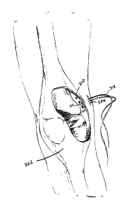
Referring to Fig. 5A, a rigid T-fix 310 includes a cylindrical shell 312
defining a hollow interior 314. Shell 312 also defines two holes 316a, 316b
for
passage of a suture 318 therethrough. A drug-polymer mixture (not shown),
either in
powder form or in the form of one or more solid or semi-solid pellets, is
loaded into
interior 314. A membrane 320 retains the drug-polymer mixture within interior
314
prior to implantation. Membrane 320, however, is permeable to bodily fluids
and to
the drug, when the drug is dissolved or suspended in bodily fluids.
As shown in Fig. 5B, rigid T-fix 310 is implanted within a location in the
body, e.g., a knee 322, by creating a hole 324 in skin and muscle and passing
T-fix
310 through hole 324, with the aid of suture 318. Rigid T-fix 310 can then be
affixed
to soft tissue or tied to a bone, as described above with reference to Fig. 2.
Once in
place, bodily fluids enter interior 314 through membrane 320, and dissolve the
drug-
polymer mixture. The drug is then carried out of T-fix 310 by the bodily
fluids, and
delivered to the nearby target site. Like staple 170, supra, T-fix 310 can be
used for
wound closure.
Referring to Fig. 6A, a bone screw 340 includes a threaded shaft 342, a
pointed tip 344, and an open end 346. Shaft 342 defines a hollow interior (not
shown).
As with rigid T-fix 310, a drug-polymer powder or pellet is loaded into the
hollow
interior, and a membrane 348 covers open end 346 and retains the drug-polymer
mixture within the interior. Membrane 348, like membrane 320 is permeable to
bodily fluids and to dissolved drug, but not to solids.
Referring to Fig. 6B, bone screw 340 can be drilled into bone, e.g., a knee
bone 350, using various drilling tools known in the art. To facilitate
implanting bone
screw 340 into bone, the opening at end 346 and membrane 348 can be moved to a
point along a side 351 of shaft 342. In this arrangement, the hollow interior
could be
a transverse cavity rather than a longitudinal bore. End 346 could then be
solid, and
could include a section configured to receive a drilling tool.
Bone screw 340 can also be drilled or manually twisted into soft tissue,
such as muscle.
-11-
CA 02380111 2002-02-O1
WO 01/08717 PCT/US00/21288
Referring to Figs. 7A and 7B, a bone screw can also have apertures to
release the drug. Bone screw 370 includes threads 372, open end 374, hollow
interior
376, and cross holes 378. A pellet 380 made from a drug-polymer mixture is
loaded
into hollow interior 376 through open end 374. Pellet 380 can be held in place
within
hollow interior 376 by a membrane, or by sealing open end 374. As shown in
Fig.
7B, cross holes 378 expose pellet 380 to the exterior, allowing bodily fluids
to reach
and dissolve pellet 380.
Bone screws 340 and 370 can be used, e.g., in ligament replacement
surgeries, or other surgical procedures that commonly employ bone screws. The
drug
in the drug-polymer pellets can be an agent that promotes healing, or promotes
adhesion of the ligament replacement to bone.
Referring to Figs. 8A-8F, an implantable plug 410 includes a hollow core
412, a pointed end 414, and retractable engagement wings 416a, 416b. A pellet
418
made from a drug-polymer mixture is loaded into hollow core 412. Pellet 418
has a
length L, less than the length Lz of hollow core 412, such that pellet 418
does not
entirely fill core 412. Plug 410 includes openings 419a, 419b under wings
416a, 416b
which expose pellet 418 to the exterior.
Plug 410 is implanted into soft tissue using a delivery probe 420. Probe
420 has an external shell 422 and hollow interior tube 424. Shell 422 and tube
424
can be made from any rigid material, such as a metal or hard plastic. Interior
tube 424
has an external diameter approximately equal to the internal diameter of
hollow core
412, such that tube 424 can be snugly fit within core 412. Interior tube 224
is slidable
within shell 422 in the direction of arrows A and B. Shell 422 has an open end
423.
In operation, plug 410, with pellet 418 pre-loaded in core 412, is loaded
into probe 420 by retracting wings 416a, 416b and inserting core 412 into tube
424.
Alternatively, plug 410 can be pre-loaded into probe 420 during manufacture.
Tube
424 is then slid in the direction of arrow A to retract plug 410, until plug
410 is fully
within shell 422, as shown in Fig. 8C. Next, probe 420 is inserted into soft
tissue 118,
as shown in Fig. 8D. Tube 424 is then pushed in the direction of arrow B such
that
plug 410 is pushed out of shell 422, as shown in Fig. 8E. Once plug 410 leaves
shell
422, wings 416a, 416b partially expand into tissue 18. Shell 422 and tube 424
are
then extracted from tissue 18 by pulling shell 422 and tube 424 in the
direction of
arrow A, as shown in Fig. 8F. When tube 424 is pulled in the direction of
arrow A,
wings 416a, 416b engage tissue 18 and prevent plug 410 from moving in the
direction
-12-
CA 02380111 2002-02-O1
WO 01/08717 PCT/US00/21288
of arrow A. Consequently, tube 424 slides out of hollow core 412, leaving plug
410
implanted within tissue 18. Bodily fluids then reach pellet 418 through
openings
419a, 419b and slowly dissolve pellet 418, delivering the drug to the nearby
target
site.
Fig. 9A illustrates a hollowed soft tissue tacker 440. Tacker 440 includes
a generally cylindrical body 442, an arrow-shaped head 444, engagement
projections
446a, 446b, and an open back end 448. Body 442 defines a hollow, cylindrical
cavity
450 communicating with opening 451 of open back end 448. Body 442 also defines
four holes, two of which, 452a, 452b, are shown in Fig. 10. The holes allow
cavity
450 to communicate with the exterior.
In operation, a pellet 454 made from a drug-polymer mixture is inserted
into cavity 450 through back end 448. Pellet 454 can be retained in cavity 450
by
covering opening 451 with a permeable membrane (not shown). Tacker 440 is then
inserted into soft tissue near a target site, arrow-shaped head 444 first.
Head 444 and
projections engage the soft tissue, holding tacker 440 in place.
When tacker 440 is inserted into soft tissue, back end 448 remains above
the tissue, exposing opening 451 to bodily fluids in a body cavity adjacent to
the
tissue. Alternatively, tacker 440 can be fully inserted into the tissue.
Bodily fluids
then enter cavity 450 through opening 451 and through the four holes,
dissolving
pellet 454 and delivering the drug to the target site.
The soft tissue tacker can also be made from a woven fabric, rather than
from an apertured solid shell. Referring to Fig. 9B, a tacker 460 is made from
a
woven fabric 462, where the threads that form fabric 462 are made from a
biodegradable polymer. In tacker 460, bodily fluids enter an internal cavity
464
through gaps 466 in fabric 462, rather than through holes in a solid shell.
The
tightness of the weave of fabric 462 controls the size of gaps 466 and,
therefore, the
speed at which the drugs reach the target site. Woven fabrics such as fabric
462 can
be used in embodiments other than soft tissue tacker 460 to house drug-polymer
pellets.
Referring to Fig. 10, a soft tissue staple 470 includes two penetration arms
472a, 472b, and a connecting arm 474 attaching arm 472a to arm 472b.
Penetration
arms 472a, 472b include arrow-shaped heads 476a, 476b, and connecting arm 474
defines a cavity 478 and an opening 480. In operation, a pellet 482 made from
a drug-
polymer mixture is inserted into cavity 478 through opening 480. Pellet 482
can be
-13-
CA 02380111 2002-02-O1
WO 01/08717 PCT/US00/21288
retained within cavity 478 by covering opening 480 with a permeable membrane
(not
shown). Once pellet 482 has been inserted, staple 470 is inserted into soft
tissue near
a target site, arrow-shaped heads 476a, 476b first. When inserted, connecting
arm
474 rests against soft tissue, but does not penetrate the tissue.
Alternatively, staple
470 can be fully inserted into the tissue. Bodily fluids then enter opening
480 and
dissolve pellet 482, delivering the drug to the target site.
Figure 11 illustrates a helical soft tissue anchor 510. Helical anchor 510 is
made from a strip 512 of material, e.g., a polymer, such as polyglycolic acid
or
polylactic glycolic acid, or a metal, such as stainless steel or titanium,
twisted into a
helical shape. Helical anchor 510 tapers to a pointed end 514 for penetrating
soft
tissue. Helical anchor 510 defines an open back 516 and a conical-shaped
interior
518 for receiving a tapered pellet 520. In operation, pellet 520 is inserted
into interior
518 through open back 516, and helical anchor 510 is then inserted into soft
tissue,
pointed end 514 first. Helical anchor 510 can be either pushed or twisted into
the soft
tissue. Bodily fluids then reach pellet 520 through slits 522 and open back
516,
dissolving pellet 520 and delivering the drug to a nearby target site.
Alternatively, a helical anchor, e.g., a metal helical anchor, can be
machined, and then the drug-polymer mixture can be molded around the helix. In
addition, the helical anchor can be manufactured entirely from a drug-polymer
mixture that slowly degrades or dissolves to release the drug into bodily
fluids over
time.
Referring to Fig. 12A, an implantable disk 540 includes a crown-shaped
base 542, a wafer 544 made from a drug-polymer mixture, and a permeable
membrane cover 546. Cover 546 has a diameter D~ approximately equal to a
diameter DB of base 542. Base 542 includes four arrow-shaped projections 548a,
548b, 548c, 548d for engaging soft tissue.
In operation, wafer 544 is placed inside rim 550 of base 542. Base 542
can have a shelf (not shown) for receiving wafer 544, or wafer 544 can be
attached to
the interior 552 of rim 550. Membrane cover 546 is then placed over wafer 544,
holding wafer 544 within base 542. A second cover (not shown) can also be
placed
over wafer 544 on the opposite side of wafer 544. Alternatively, disk 540 can
be pre-
assembled during manufacture.
- 14-
CA 02380111 2002-02-O1
WO 01/08717 PCT/US00/21288
After assembly, disk 540 is placed against internal soft tissue by inserting
projections 548a, 548b, 548c, 548d into the tissue. Bodily fluids reach wafer
544
through cover 546, dissolve wafer 544, and deliver the drug to a nearby target
site.
Fig. 12B illustrates an apparatus 560 for affixing disk 540 to tissue.
Apparatus 560 includes an interior cylindrical block 562 slidable within an
exterior
tube 564. Block 562 and tube 564 can be made from any rigid material, such as
a
metal or hard plastic. Exterior tube 564 has an inside diameter DE
approximately
equal to diameter D8 of base 542, such that base 542 fits snugly within
exterior tube
546. Interior block 562 has a diameter D~ less than diameter DB. In operation,
disk
540, fully assembled, is loaded into second tube 564. In Fig. 12B, disk 540 is
shown
in dashed lines inside apparatus 560. Apparatus 560 is then inserted into the
body,
e.g., through an orifice or a surgically created opening, and pressed against
internal
soft tissue near a target site. Interior block 562 is then slid in the
direction of arrow A,
forcing disk 540 out of exterior tube 564 and into the tissue. Apparatus 560
is then
withdrawn from the body, leaving disk 540 attached to internal soft tissue.
Referring to Figs. 13A and 13B, a drug delivery tack 610 includes a shell
611 that forms a shaft 612 and a head 614. Shaft 612 includes exterior ribs
616a,
616b, 616c and a tapered end 617. Head 614 includes a jagged edge 624 for
engaging
soft tissue or bone.
The shell 611 defines a hollow interior bore 618 that extends
longitudinally throughout the shaft and the head. A medicament core 620 made
from
a drug-polymer mixture fills bore 618. A narrow hole 622 is drilled through
medicament core 620 for insertion of a guide pin therethrough. Hole 622 has a
diameter of, e.g., less than 0.1 inches, and most commonly between about 0.03
and
0.08 inches.
Tack 610 is used to affix soft tissue to a support structure. For example,
tack 610 can be used to tension and attach a tendon to muscle, or a ligament
to bone.
To use tack 610 to attach a ligament to bone, a guide pin (not shown) is
inserted
through hole 622 until the pin pierces the ligament. The pin is then moved
transversely toward the bone, and inserted into a pre-drilled hole in the
bone. Tack
610 is then slid over the pin and forced into the hole in the bone, tapered
end 617 first,
until jagged edge 624 engages the bone (or nearby soft tissue). The guide pin
is then
removed, leaving the tack in place, and the ligament secured to the bone. A
similar
-15-
CA 02380111 2002-02-O1
WO 01/08717 PCT/US00/21288
procedure is used to attach a tendon to muscle, or other soft tissue to a
support
structure.
After tack 610 has been inserted and the guide wire has been withdrawn,
bodily fluids enter hole 622 through opening 626 and dissolve medicament core
620,
delivering the drug to the nearby target site.
Figs. 14A-14C illustrate an expansion anchor 640 for delivering a drug.
Expansion anchor 640 includes a shaft 642 defining an internal bore 644. Shaft
642
has an end 646 that includes four serrated prongs 648a, 648b, 648c, 648d.
Shaft 642
is made from a flexible, bioabsorbable polymer, such as polyglycolic acid or
polylactic glycolic acid, allowing radial expansion of bore 644 by, e.g.,
flexing prongs
648a, 648b, 648c, 648d. Anchor 640 also has a head 650 attached to shaft 642.
An
interior side 652 of head 650 has a retention ring 654.
A plug 656 holding a drug-polymer pellet 658 is configured to be
insertable within bore 644. Plug 656 has a groove 660 sized and shaped to
receive
retention ring 654.
In operation, plug 656 is first partially inserted into bore 644, until an end
662 of pellet 658 reaches ridges 664 within bore 644. Fig. 14A shows plug 656
partially inserted. Next, anchor 640 is inserted into soft tissue near a
target site, until
shaft 642 is fully within the tissue. Once anchor 640 has been inserted, plug
656 is
pushed further into bore 644, until groove 660 catches ring 654. Pushing plug
656
further into bore 644 causes prongs 648a, 648b, 648c, 648d to flex, radially
expanding
a portion of bore 644 and exposing pellet 658, as shown in Fig. 14B. Bodily
fluids
then dissolve pellet 658 and deliver the drug to the nearby target site.
Materials and Manufacture
The drug-polymer mixture in each of the above implantable devices can
be, e.g., a conglomerate of drug-polymer microspheres, a sponge-like polymer
matrix
in which molecules of drug are embedded, or a solidified drug-polymer mixture,
e.g.,
an emulsion or dispersion.
Referring to Fig. 15, in the microsphere conglomerate embodiment, each
microsphere 710 includes small amounts of a drug 712 suspended within a
polymer
substrate 714. The individual microspheres form a "powder" that can be
compressed
to form the shapes of the shaped implantable devices of Figs. 1-4, or to form
a pellet
which can be inserted into the hollow portions of the hollow implantable
devices of
-16-
CA 02380111 2002-02-O1
WO 01/08717 PCT/US00/21288
Figs. 5-14. Such a conglomerate of drug-polymer microspheres will biodegrade
slowly, from the exterior inward, and will therefore steadily release small
amounts of
the drug over an extended period of time.
The pellet can also be configured to release doses of the drug
intermittently. For example, referring to Fig. 15A, in devices where the
pellet is only
exposed to bodily fluids at one end (e.g., bone screw 340 of Fig. 6), a pellet
850 can
be constructed from alternating sectors of drug-polymer mixture 852 and
placebo 854.
Bodily fluids would dissolve drug-polymer sectors 852 and placebo sectors 854
in
succession, causing intermittent release of the drug. Referring to Fig. 15B,
pellet 870
is constructed from layers of drug-polymer mixture 872 and placebo 874. Pellet
870
would allow intermittent release of the drug in devices such as T-fix 110 of
Fig. 1A,
and helical anchor 510 of Fig. 11. In addition, varying layers can be used to
release
different drugs or different dosages of the same drug.
Alternatively, the microspheres can be left in powder form and loaded into
the hollow implantable devices.
A powder including drug-polymer microspheres can be manufactured
using known techniques. For example, as described in detail in the Examples
below,
a drug is dissolved in a polymer-methylene chloride mixture (or a polymer
ethyl
acetate mixture) to form an inner emulsion. The inner emulsion is then poured
into
and mixed with an aqueous polyvinyl alcohol solution to form a second
emulsion.
The resulting double emulsion is then mixed with polyvinyl alcohol and placed
on a
magnetic stirrer for two-three hours until the methylene chloride evaporates,
leaving
microspheres. The resulting microspheres are then washed repeatedly using a
centrifuge, frozen with liquid nitrogen, and placed in a lyophilizer to form a
powder
composed of microspheres.
Other known methods of encapsulating drugs within microspheres can also
be used. See, ~, Cohen et al., "Controlled Delivery Systems for Proteins Based
on
Poly(Lactic/Glycolic Acid) Microspheres," Pharm. Research. 8:713-20 (1991)
(similar to method described above, except that the inner emulsion is poured
into and
mixed with a polyvinyl alcohol-methylene chloride solution rather than simply
a
polyvinyl alcohol solution); DeLuca et al., U.S. Patent Nos. 5,160,745 and
4,741,872
(a vinyl derivative of a polymer, a water soluble monovinyl monomer, and a
drug
macromolecule are emulsified in water, and the polymer and monomer are co-
polymerized such that the macromolecule is entrapped therein); Mathiowitz et
al.,
-17-
CA 02380111 2002-02-O1
WO 01/08717 PCT/US00/21288
U.S. Patent No. 5,718,921 (polymer dissolved in a volatile organic solvent,
drug
dispersed in the solution, mixture suspended in an organic oil, and the
organic solvent
extracted, creating microspheres); and Kent et al., U.S. Patent No. 4,675,189
(polymer
water-in-oil solution phase separated by addition of silicone oil, causing
polymer to
deposit as droplets onto surface of water-polypeptide microdroplets,
encapsulating the
polypeptide).
In making the drug-polymer microspheres, buffers, such as sucrose and
cyclodextrin, can be added. The buffers serve several purposes. First, they
act as a
cushion for the IL-10 when the microspheres are compressed into pellets,
reducing
denaturing of the IL-10. Second, the buffers dissolve more quickly than the
polymer,
creating tunnels in the microspheres to facilitate escape (release) of the IL-
10.
Inclusion of buffers, therefore, can lead to an initial "burst" of IL-10
release during,
e.g., the first 24 hours after implantation, followed by sustained release of
a smaller
amount of IL-10 over days, weeks, or longer.
Various polymers can be used for encapsulating drugs in microspheres.
Preferably, the polymers are biocompatible and degradable when placed within
human tissue. Such polymers include, e.g., polyanhydrides, polylactides,
polyglycolides, polylactic acid, polyglycolic acid, polyorthoesters,
polyorthocarbonates, polyacetals, polymers derived from alpha
hydroxycarboxylic
acids and lactones, polymers derived from condensation of divinyl ethers and
polyols,
e-caprolactone polymers, and various other polymers described in the above
incorporated references. In addition, co-polymers of some of the above
polymers,
such as poly(DL-lactide-co-glycolide) can be used to encapsulate certain
drugs.
Various drugs and combinations of drugs can be encapsulated by polymers
for delivery using the claimed devices. For example, anti-inflammatory agents,
such
as down-regulatory cytokines, can be used to treat inflammatory disease, as
described
in the Examples below. Pain medications, such as lidocaine, can be used to
treat
localized pain. Other possible drugs include platelet derived growth factor,
antibiotics, hormones, prostaglandins, insulin, adrenalin, xylocaine,
morphine,
corticoid compounds, atropine, cytostatic compounds, estrogen, androgen,
interleukins, digitoxin, biotin, testosterone, heparin, cyclosporin,
penicillin, vitamins,
anti-platelet activating agents, somatostatin, SOMATRIPTANT"', triptorelin,
diazepam, other protein based drugs, peptide sequences (which are generally
more
-18-
CA 02380111 2002-02-O1
WO 01/08717 PCT/US00/21288
heat resistant and last longer than full proteins), nucleic acid based drugs
and
therapies, and other drugs described in the incorporated references.
Instead of encapsulating the drug within polymeric microspheres, the
polymer and drug can simply be mixed together in powdered form, and then
compressed into pellets. Non-microsphere pellets would also release small
amounts of
the drug steadily, over an extended period of time, as the polymer in the
pellet
biodegrades. Alternatively, liquid or semi-solid drugs and polymers can be
mixed and
then extruded into rods that can be cut into short pellets.
To create a non-microsphere drug-polymer mixture, an emulsion including
a drug and a polymer can be frozen with liquid nitrogen and then placed in a
lyophilizes. This process is similar to the microsphere formation process
described in
detail in the Examples below, except that the drug polymer emulsion is not
stirred
with a magnetic stirrer.
Alternatively, a drug can be dissolved in a mixture of methylene chloride
and ethylene vinyl acetate copolymer. A small amount of the resulting solution
is then
placed in a mold that has been frozen with liquid nitrogen. The frozen mold is
then
placed in a vacuum chamber to dissolve the solvent, leaving only a film of the
ethylene vinyl acetate and the drug. The film, which is typically rubbery and
somewhat adhesive, can be rolled tightly into a pellet for insertion into an
implantable
device.
Other techniques for mixing drugs and polymers into sustained release
formulations can also be used. See, e.~, Cohen et al., "Sintering Techniques
for the
Preparation of Polymer Matrices for the Controlled Release of Macromolecules,"
J.
Pharm. Sciences, 73:1034-37 (1984) Briefly, drug and polymer powders are mixed
at
a temperature below the glass transition point of the polymer. The resulting
mixture
is then compressed at a temperature above the glass transition point, forming
the
matrix.
In the non-microsphere embodiment, many of the polymers mentioned
above can be used, in addition to other polymers, such as ethylene-vinyl
acetate
copolymer and some non-biodegradable polymers.
The microspheres or the non-microsphere drug-polymer mixture are
compressed into shapes or pellets using simple molds and a press, e.g., a
Carver press.
The amount of pressure required to shape a powder into an implantable device
having
-19-
CA 02380111 2002-02-O1
WO 01/08717 PCT/US00/21288
a desired shape will depend on the size of the device and the particular drug-
polymer
mixture.
The rigid exteriors of the devices illustrated in Figs. 5-14 can be made
from a variety of materials, depending on the nature of the implantable
device. The
rigid exteriors of the bone screws of Figs. 6 and 7, for example, are
typically made
from a biocompatible metal, such as titanium, cobalt, chromium, stainless
steel, or
other alloys. The rigid exteriors of the devices of Figs. 5 and 8-14, however,
can be
manufactured from a rigid, biodegradable polymer, such as polyglycolic acid or
polylactic glycolic acid, a hard, non-binding surgical grade plastic, such as
DELRINT"", or a non-biodegradable polymer, ceramic, or metal.
The shaped implantable devices of Figs. 1-4 can be formed by
compressing drug-polymer powders into the desired shape, as described below
with
reference to Fig. 16. The hollowed implantable devices of Figs. 5-14 can be
formed
using techniques known in the art, including deposition of a molten polymer
into a
mold, or extrusion. The devices can also be formed from several separate
pieces
melded together using heat.
The permeable membranes of the embodiments of, e.g., Figs. 5, 6, and 12
can made from, e.g., any membrane material known in the art. The size and
density
of the pores in the membranes can be varied, depending on the drug and the
desired
drug delivery rate. In general, the membranes will have micron ratings of
greater than
0.5 (for filtering suspended solids, but not dissolved large molecules). Other
micron
sizes are possible, depending on the application. Membranes can be purchased
from,
e.g., RGF ENVIRONMENTAL, West Palm Beach, Florida.
The sizes of the devices of Figs. 1-14 can vary. Generally, the longest
dimension of each device will range from about 1.5 mm to I cm or larger, e.g.,
2 mm,
5 mm, I cm, 2 cm, or 5 cm.
The invention is further described in the following Examples, which do not
limit the scope of the invention described in the claims.
-20-
CA 02380111 2002-02-O1
WO 01/08717 PCT/US00/21288
Examples
In the following Examples, interleukin-10 ("IL-10") was encapsulated in
microspheres of 50:50 poly(DL-lactide-co-glycolide). The resulting microsphere
powder was compressed into pellets, and also tested for biological activity.
The
results of these Examples establish that IL-10 can be incorporated into
implantable
devices such as those described above for localized, controlled release of IL-
10
directly to a site of inflammation.
Example l: Encapsulation of IL-10 within Polymer Microspheres
In three separate experiments, IL-10 was encapsulated within 50:50
poly(DL-lactide-co-glycolide) microspheres.
In each experiment, the materials and equipment were as described in
Table 1.
Table 1
MATERIAL/DEVICE DESCRIPTION
Polymer powder (50:50 poly(DL-lactide-BOEHRINGER INGELHEIM (Henley
co-glycolide)) Chemicals), Cat. No. RG503.
Polyvinyl alcohol ALDRICH CHEMICAL CO., Cat.
No.
18,953-7; 96% hydrolized
Ethyl Acetate ALDRICH CHEMICAL CO., Cat.
No.
27,098-9; 99.8% anhydrous
Recombinant Human IL-10 ENDOGEN, INC., Cat. No. R-IL10-25
Methylene Chloride (Dichloromethane)ALDRICH, Cat. No. 27,099-7;
99.8%
anhydrous
Human Serum Albumin CALBIOCHEM, Cat. No. 12666;
Type:
Fraction V
Hydroxypropyl Beta CycloDextrinAM. MAIZE-PRODUCTS CO.,
Hammond, Indiana
Sonicator VibraCellT"~ Sonicator
Homogenizer Silverson L4R Homogenizer
Centrifuge IEC model Centra GP8
Lyophilizing chamber I Labconco
-21 -
CA 02380111 2002-02-O1
WO 01/08717 PCT/US00/21288
IL-10 (recombinant human interleukinENDOGEN, Cat. No. R-IL10-25
10)
Methylene Chloride (Dichloromethane)ALDRICH, Cat. No. 27, 099-7;
99.8%
anhydrous
HSA (human serum albumin) CALBIOCHEM, Cat. No. 12666;
Type:
Fraction V
The remaining chemicals and equipment are standard laboratory supplies
available
from numerous sources.
First Experiment
First, two separate sets of IL-10 solution, polymer powder solution, and
homogenized polyvinyl alcohol solution were prepared as follows.
Ten micrograms of IL-10 previously bulked with bovine serum albumin
("BSA"), and 25 micrograms of pure IL-10 were placed in separate vials. Next,
1 ml
of chilled phosphate buffered solution, pH 7.4 ("PBS"), was added to the 25
microgram vial, and 400 ~1 of PBS was added to the 10 microgram vial. The
solutions were mixed and then chilled.
Two 50 mg of samples of 50:50 poly(DL-lactide-co-glycolide) ("polymer
powder") were placed in two separate test tubes. One ml of methylene chloride
was
added to each tube, and the resulting polymer solutions were chilled.
Two separate beakers, one labeled "w/ BSA" and one labeled "w/o BSA"
were prepared. To each beaker, 100 ml of 1% polyvinyl alcohol was added and
placed in a homogenizer at 5800 rpm for several minutes.
Next, 100 ,u1 of the 10 microgram IL-10 solution was added to the first
polymer solution test tube. The test tube was sonic pulsed in the sonicator
for about 5
pulses (40% duty cycle), and the resulting emulsion was added to the beaker
labeled
"w/ BSA" while still homogenizing at 5800 rpm. Homogenization was continued
for
an additional 1 minute, and the beaker was then moved to a magnetic stirrer
set at a
speed of about 4.5. Similarly, 100 microlitres of the 25 microgram IL-10
solution
was added to the second polymer solution test tube, pulsed, and added to the
beaker
labeled "w/o BSA." This beaker was also homogenized for an additional 1
minute,
and then moved to the magnetic stirrer. At this point, microspheres could
already be
observed through a microscope.
-22-
CA 02380111 2002-02-O1
WO 01/08717 PCT/(JS00/21288
After two hours in the stirrer, the two beakers were removed from the
magnetic stirrer. The resulting solutions were then poured into four 50 ml
centrifuge
vials: two labeled "w/ BSA" and two labeled "w/o BSA." The vials were
centrifuged
at 1500 rpm (program 6). After centrifuging for 5 minutes, the vials were
removed,
the liquid was poured off the top, and distilled water was added to return the
total
volume in each vial to about 30 ml. The vials were then centrifuged for an
additional
five minutes. Once again, the vials were removed, the liquid was poured from
the
top, and distilled water was added to return the volume to 20 ml. The vials
were
centrifuged again for 5 minutes, and distilled water was added to bring the
total
volume to 5-10 ml per vial.
The vials were then dipped into a bucket of liquid nitrogen until frozen,
covered with KIMWIPES and a rubber band, and placed in a lyophilizing chamber.
The chamber was attached to the lyophilizer, and the vents to vacuum were
opened
until the reading reached 100 microns Hg.
The result was 70 mg of fine, white powder composed of microspheres of
50:50 poly(DL-lactide-co-glycolide) entrapping IL-10 (40 mg of microspheres
with
BSA, 30 mg of microspheres without BSA).
Second Experiment
In this experiment, sucrose and CycloDextrin buffers were added to
polymer/IL-10 microsphere mixtures. The sugar buffers serve two purposes.
First,
they act as a cushion during pressing of IL-10 powder into pellets, thereby
protecting
the IL-10 from being denatured by the pressure. Second, the sugar buffers,
which are
larger than IL-10 molecules, form "tunnels" in the microsphere pellets after
the
powder is compressed, facilitating release of the IL-10 after implantation.
The experiment was performed as follows. First, 100 ml of 1 % polyvinyl
alcohol was poured into six beakers and chilled using an ice bath. The beakers
were
labeled "MeCI/std," "MeCI/su," "MeCI/CD," "EtAc/std," "EtAc/su," and
"EtAc/CD."
Ten grams of powdered human serum albumin ("HSA") was combined
with distilled water to make a stock HSA solution having a concentration of
lOmg/lml. (The HSA helps protect the IL-10 from becoming denatured.) One
hundred u1 of the stock HSA was combined with 400 u1 of distilled water and
added
to the vial containing the 25 ug of IL-10. The mixture was mixed gently using
a
VORTEX GENIE, and then chilled.
- 23 -
CA 02380111 2002-02-O1
WO 01/08717 PCT/US00/21288
Six 50 mg samples of polymer powder were placed in six test tubes
labeled "MeCI/std", "MeCI/su", "MeCI/CD", "EtAc/std", "EtAc/su", and
"EtAc/CD."
One ml of methylene chloride was then added to each of the three "MeCI" test
tubes,
and 1 ml of ethyl acetate was added to each of the three "EtAc" test tubes.
The tubes
were then chilled. The methylene chloride easily dissolved the polymer, but
the ethyl
acetate did not. The VORTEX GENIE was used to help dissolve the polymer in
each
test tube.
Next, 100 u1 of distilled water was poured into three vials labeled "std,"
"su," and "CD." Ten mg of sucrose was added to the "su" vial and 10 mg of
CycloDextrin was added to the "CD" vial. The contents of the vials were then
mixed.
Approximately 125 u1 of the IL-10 solution were added into each of the 3
vials. The
contents were mixed gently using the Vortex Genie only on low settings.
The six beakers (each containing 100 ml PVA) were placed in the
homogenizer at 4600-4700 rpm for several minutes.
Approximately half of the "std" IL-10 solution, about 112 u1, was added to
the "MeCI/std" polymer solution test tube, and the other half was added to the
"EtAc/std" polymer solution test tube. Similarly, the "su" and "CD" IL-10
solutions
were divided into the corresponding polymer solutions: "MeCI/su" and
"EtAc/su,"
and "MeCI/CD" and "EtAc/CD." The test tubes were then sonic pulsed in the
sonicator (20% duty cycle) for 4 pulses while keeping them on ice.
Each emulsion was added to the correspondingly labeled PVA-filled
beaker while still homogenizing at 4600-4700 rpm. Homogenization was continued
for an additional 1 minute. The beakers were then moved to a magnetic stirrer,
set at
a speed of 6. At this point microspheres could already be observed through a
microscope.
After 2-3 hours, the beakers were removed from the magnetic stirrer.
Twelve 50 ml centrifuge vials were labeled as follows: "MeCI/std 1," "MeCI/std
2,"
"MeCI/su 1," "MeCI/su 2," "MeCI/CD 1," "MeCI/CD 2," "EtAc/std 1," "EtAc/std
2,"
"EtAc/su 1," "EtAc/su 2," "EtAc/CD 1," and "EtAc/CD 2." The solutions from the
beakers were poured into the corresponding 12 vials. The vials were then
centrifuged
for 5 minutes at 1500 rpm (program 6).
Next, the vials were removed from the centrifuge and liquid was carefully
poured off the top. The solid (microspheres) residue of the two vials marked
"MeCI/std 1" and "MeCI/std 2" were combined by adding some distilled water to
the
-24-
CA 02380111 2002-02-O1
WO 01/08717 PCT/OS00/21288
vials to re-suspend the microspheres, and then pouring one vial into the
other. In the
consolidated vial, more distilled water was added to reach a total volume of
about 30
ml. This "washing" process was repeated for the other 10 vials. After all were
combined, only 6 vials remained. The vials were then centrifuged as before.
The
microspheres were then washed again (liquid poured off the top and spheres re-
suspended with distilled water), and additional distilled water was added to
reach a
total volume of about 25 ml in each of the six vials. The vials were then
centrifuged
and washed once more, and distilled water was added to reach a total volume of
about
5 ml in each of the 6 vials.
The vials were then dipped into a bucket of liquid nitrogen until frozen,
covered with KIMWIPES and a rubber band, and placed in a lyophilizing chamber.
The chamber was attached to the lyophilizer, and the vents to vacuum were
opened
until the reading reached 100 microns Hg.
The remaining IL-10 solution was saved for the biological activity tests
described below.
Third Experiment
In this experiment, two different variations of polymer/IL-10 microsphere
mixtures were created.
First, 100 ml of 1 % polyvinyl alcohol was poured into four beakers and
chilled using an ice bath. The beakers were labeled "MeCI/su A," "MeCI/su B,"
"MeCI/CD A," "MeCI/CD B."
Next, as in the second experiment, I O grams of powdered HSA was
combined with distilled water to make a stock HSA solution having a
concentration of
I Omg/l ml. One hundred ~l of the stock HS A was combined with 400 u1 of
distilled
water and added to the vial containing the 25 ug of IL-10. The mixture was
mixed
gently using a Vortex Genie, and then chilled.
Four 100 mg samples of polymer powder were placed in four test tubes
labeled "MeCI/su A," "MeCI/su B," "MeCI/CD A," "MeCI/CD B." Two ml of
methylene chloride were then added to each of the two MeCI test tubes. The
Vortex
Genie was used to help dissolve the polymer.
Next, 20 mg of sucrose was added to the "su" vial, 20 mg of CycloDextrin
was added to the "CD" vial, and about 250 u1 of the IL-10 solution was added
to each
- 25
CA 02380111 2002-02-O1
WO 01/08717 PCT/US00/21288
of the two vials. The contents were then mixed gently using the Vortex Genie
on low
settings only. About 250 u1 of distilled water was added to each vial.
The four beakers (each containing 100 ml PVA) were placed in
homogenizer at 4600-4700 rpm, for several minutes.
Approximately half of the "su" IL-10 solution, about 250 u1, was added to
the "MeCI/su A" polymer solution test tube, and the other half was added to
the
"MeCI/su B" polymer solution test tube. Similarly, the "CD" IL-10 solution was
divided into the corresponding polymer solutions: "MeCI/CD A" and "MeCI/CD B."
The test tubes were then sonic pulsed in the sonicator (20% duty cycle) for S-
6 pulses
while keeping them on ice.
Each emulsion was added to the correspondingly labeled PVA-filled
beaker while still homogenizing at 4600-4700 rpm. Homogenization was continued
for an additional I minute. The beakers were then moved to a magnetic stirrer,
set at
a speed of 6. At this point microspheres could already be observed through a
microscope.
After 2-3 hours, the beakers were removed from the magnetic stirrer.
Eight SO ml centrifuge vials were labeled as follows: "MeCI/su AI," "MeCI/su
A2,"
"MeCI/su B1," "MeCI/su B2," "MeCI/CD A1," "MeCI/CD A2," "MeCI/CD B1," and
"MeCI/CD B2." The solutions from the beakers were poured into the
corresponding 8
vials. The vials were then centrifuged for 5 minutes at 1500 rpm (program 6).
Next, the vials were removed from the centrifuge and liquid was carefully
poured off the top. The solid residue (microspheres) of the two vials marked
"MeCI/su A1" and "MeCI/su A2" were combined by adding some distilled water to
the vials to re-suspend the microspheres, and then pouring one vial into the
other. In
the consolidated vial, more distilled water was added to reach a total volume
of about
ml. This "washing" process was repeated for the other 6 vials. After all were
combined, only 4 vials remained. The vials were then centrifuged as before.
The
microspheres were then washed again (liquid poured off the top and spheres re-
suspended with distilled water), and additional distilled water was added to
reach a
30 total volume of about 25 ml in each of the four vials. The vials were then
centrifuged
and washed once more, and distilled water was added to reach a total volume of
about
5 ml in each of the 4 vials.
The vials were then dipped into a bucket of liquid nitrogen until frozen,
covered with KIMWIPES and a rubber band, and placed in a lyophilizing chamber.
-26-
CA 02380111 2002-02-O1
WO 01/08717 PCT/US00/21288
The chamber was attached to the lyophilizer, and the vents to vacuum were
opened
until the reading reached 100 microns Hg.
Example 2: Compression of IL-10/Polymer Powder into Pellets
Microsphere powder obtained from the first experiment of Example 1 was
compressed into four disk-shaped pellets as follows. Referring to Fig. 16, a
disk-
shaped mold 910 includes a removable top 912, a removable bottom 914, and a
body
916 defining a bore 918. To load the mold, top 912 was removed from bore 918
in
the direction of arrow A, and 10 mg of microsphere powder containing BSA was
loaded into bore 918. Top 912 was reinserted into bore 918 over the powder,
and
twisted to compress the powder. Mold 910 was then subjected to 1500 pounds of
force from a Carver Press (not shown) for seven minutes, creating a 5 mm
diameter
flat disk pellet.
Five additional pellets were prepared in the same manner: one more using
microsphere powder with BSA, two using microsphere powder without BSA, and two
using pure polymer powder. Mold 910 was cleaned between each use with
methylene
chloride.
To test the structural integrity of the pellets, sutures were successfully
passed through each of the four different microsphere pellets.
Example 3: Testin, o~ f Polymer/IL-10 Microspheres for Biol~,ical Activity
A series of tests were performed to verify that microspheres formed by the
second and third experiments of Example 1 released encapsulated IL-10 when
placed
in a biological environment, and that the released IL-10 will inhibit
production of
TNF-a.
To test release of IL-10 in a biological medium, IL-10 microspheres were
first incubated with Dulbecco's Modified Eagle Medium (DMEM) at 37°C.
The
medium and microspheres were kept on a rocker to prevent the microspheres from
settling. After a predetermined amount of time (e.g., 3 hours) the medium and
microspheres were removed and centrifuged. The supernatant was collected, and
an
enzyme-linked immunosorbent assay (ELISA) was used to measure the amount of IL-
10 in the supernatant. The amount of IL-10 found in the supernatant was
recorded as
IL-10 released during the "0-3 hrs" interval. The microspheres were then
returned to
the medium and incubated further. At another predetermined time (e.g., after
21
-27-
CA 02380111 2002-02-O1
WO 01/08717 PCT/US00/21288
additional hours), the medium and microspheres were removed and centrifuged
again,
and the amount of IL-10 found in the supernatant was recorded as IL-10
released
during the "3-24 hrs" interval. The process was repeated to measure IL-10
released
during subsequent intervals. The data tables for each release experiment,
therefore,
show how much IL-10 was released by each type of microsphere, and when the IL-
10
was released.
ELISAs were also used to measure degradation of IL-10 in the DMEM
over the various time intervals. For the degradation experiments, loose IL-10
(not
encapsulated in microspheres or mixed with polymer) was placed in the DMEM at
an
initial concentration of, e.g., 200 ng/ml. At the end of each time interval,
the
concentration of IL-10 remaining was measured by removing and testing a small
sample of the medium.
To test biological activity of the released IL-10, IL-10 was taken from the
supernatants used to perform the ELISA tests, and added to monocytes to
achieve a
final concentration of 1 ng/ml. Some IL-10 bound to the IL-10 receptors of the
monocytes and became incorporated into the cells. The final cell concentration
was 1
x 106 cells/ml of DMEM.
After 1.5-2 hours, the monocytes were first stimulated with a concentration
of 100 Units/ml of interferon gamma (IFN-y) and then with a concentration of
20
ug/ml of muramyl dipeptide (MDP). The IFN-y increases MDP receptor expression
so that the MDP can bind readily with the cells. Once the MDP binds to the
cell and
is incorporated, it attempts to turn on the TNF-a gene. However, if active IL-
10 is
already in the cell, it will block the TNF-a gene from turning on and
producing TNF-
a.
After 16 hours, the cells were harvested and the culture supernatant was
collected. The TNF-a levels of the cells and supernatant were then tested by a
cloned
mouse fibrosarcoma cell line (LM) bioassay, to determine the extent to which
TNF-a
production was inhibited.
- 28 -
CA 02380111 2002-02-O1
WO 01/08717 PCT/US00/21288
Results for Testing of Micros~heres Formed in Second
Experiment of Example 1
The following ELISA data (Table 2) were obtained for microspheres
formed in the second powder formation experiment of Example 1. The beginning
concentration of IL-10 (in the DMEM) was about 200 ng/ml, for both the
microsphere
encapsulated IL-10 and the loose IL-10.
Table 2
ELISA Results IL-10 released
(ng/ml) per
amount of
time in culture
medium (DMEM)
at
37C
Sample # Name 0-24 hrs. 24-48 hrs.
1 MeCI/std 35 0.024
2 MeCI/su 113 0.8
3 MeCI/CD 101 0.4
4 EtAc/std 15 0.3
5 EtAc/su 61 1.3
6 EtAc/CD 65 1.1
7 Loose IL-10 290 87
These results show that the MeCI/su and MeCI/CD microspheres release
more IL-10 than the other samples during the first 24 hours, but the EtAc/su
and
EtAc/CD microspheres release the most IL-10 over a two day period. T'he
concentration of the loose IL-10 actually increased during the first 24 hours
due to
evaporation of DMEM. The concentration dropped during the second 24 hours,
however, due to degradation of IL-10.
-29-
CA 02380111 2002-02-O1
WO 01/08717 PCT/US00/21288
The LM bioassay results for the IL-10 released during the 0-24 hrs interval
are shown in Table 3 below.
Table 3
Sample # Name TNF-a TNF-a TNF-a Total
Supernate Membrane (pg/ml)
(pg/ml) Bound (pg/ml)
1 Control 47,199 906 48,1 OS
2 MeCI/std 29,207 576 29,783
3 MeCI/su 31,439 0 31,439
4 MeCI/CD 19,999 0 19,999
EtAc/std 29,776 0 29,776
6 EtAc/su 32,736 616 33,352
7 EtAc/CD 21,786 0 21,786
8 IL-10 Control22,951 0 22,951
5
Sample 1, the control, shows that 48,105 pg/ml of TNF-a are produced by
monocytes stimulated with MDP and IFN-Y. To set a benchmark for the
effectiveness of IL-10 on reducing TNF-a production, pure IL-10 was added to
Sample 8, the IL-10 Control. This benchmark shows that the IL-10 reduces the
TNF-
a level from 48,1 OS to 22,951 pg/ml. Samples 2-7 represent the various
microspheres. All lowered the TNF-a levels. The best results were from the
MeCI/CD and EtAc/CD microspheres which actually lowered the TNF-a levels below
the benchmark.
The amount of IL-10 collected from each sample after 48 hours was not
enough to run a bioassay. The second experiment of Example 1, therefore, was
repeated. In this second run, IL-10 release data was gathered for the 0-3
hours
interval, the 3-24 hours interval, the 1-5 days interval, and the 5-12 days
interval. The
ELISA results for this second run are shown in Table 4 below:
-30-
CA 02380111 2002-02-O1
WO 01/08717 PCT/US00/21288
Table 4
ELISA IL-10
Results released
(ng/ml)
per amount
of time
in culture
medium
(DMEM)
at 37~C
Sample Name 0-3 hrs. 3-24 1-5 days 5-12 days
# hrs.
1 MeCI/std 30 0.65 0.076 0.009
2 MeCI/su 82 13 1.26 0.178
3 MeCI/CD 90 5.6 0.39 0.066
4 EtAc/std 18 1.4 0.084 0.023
EtAc/su 74 5.5 0.466 0.095
6 EtAc/CD 77 8 1.03 0.135
These results show sustained release of the IL-10 even though a large
5 portion of the IL-10 is released in the first three hours. Most likely, the
initial burst of
IL-10 release is caused by the inclusion of HSA and the CD and SU sugar
buffers.
The buffers are fairly large molecules, and they tend to dissolve faster than
the
polymer. As the buffers dissolve, they create tunnels in the microspheres,
causing an
initial burst of IL-10 release from the IL-10 mixed with the buffers. Once the
buffers
have dissolved, the IL-10 mixed with the polymer escapes at a steady rate.
The LM bioassay results for the IL-10 released during the 0-3 hours and
the 3-24 hours intervals of the second run are shown in Table 5 below.
Table 5
Sample # Name % Inhibition % Inhibition
0-3 hrs. 3-24 hrs.
1 MeCI/std 6
2 MeCI/su 40 19
3 MeCI/CD 10 17
4 EtAc/std 0
5 EtAc/su 20 14
6 EtAc/CD 28 19
7 IL-10 Control82 60
-31 -
CA 02380111 2002-02-O1
WO 01/08717 PCT/US00/21288
In this test, the MeCI/su and EtAc/CD microspheres were most effective,
especially during the first 3 hours.
In this second testing run, bioactivity was much lower than in the first run,
so another testing experiment was performed. This third run began with an IL-
10
concentration of 500 ng/ml. The ELISA results are shown below in Table 6:
Table 6
ELISA Results IL-10 released (ng/ml) per
amount of
time in culture medium (DMEM)
at
37C
Sample # Name 0-24 hrs.
1 MeCI/su 340
2 MeCI/CD 132
3 EtAc/su 103
4 EtAc/CD 102
5 Loose IL-10 905
These results again show that the MeCI microspheres release more IL-10
during the first 24 hours than the EtAc microspheres. As before, the
concentration of
the loose IL-10 increased due to evaporation of DMEM.
In this third run, appropriate amounts of IL-10 were taken from the same
supernatant used to perform the ELISAs and added to monocytes to achieve a
final
concentration of 10 ng/ml, rather than 1 ng/ml. The LM bioassay results are
shown
below in Table 7.
Tahle 7
Sample # Name % Inhibition
0-24 hrs.
I MeCI/su 23
2 MeCI/CD 8
3 EtAc/su 7
4 EtAc/CD
5 IL-10 Control 85
-32-
CA 02380111 2002-02-O1
WO 01/08717 PCT/US00/21288
In this run, the MeCI/su microspheres were again the most effective during
the first 24 hours.
Results for Testing of Microspheres Formed in Third
Experiment of Example 1
The microspheres formed in the third formation experiment were similarly
tested for biological activity. These tests began with an IL-10 concentration
of 500
ng/ml. The ELISA data obtained are shown below in Table 8.
Table 8
ELISA Results IL-10 released (ng/ml) per
amount of
time in culture medium (DMEM)
at
37C
Sample # Name 0-24 hrs.
1 MeCI/su A 104
2 MeCI/su B 115
3 MeCI/CD A 280
4 MeCI/CD B 240
5 Loose IL-10 779
These results show that the MeCI/CD microspheres release more IL-10
than the MeCI/su microspheres during the first 24 hours.
In this test, as in the third run above, appropriate amounts of IL-10 were
taken from the supernatant of the ELISA releases and added to monocytes to
achieve
a final concentration of 10 ng/ml. The LM bioassay results are shown below in
Table
9.
- 33 -
CA 02380111 2002-02-O1
WO 01/08717 PCT/US00/21288
Table 9
Sample # Name % Inhibition
0-24 hrs.
1 MeCI/su A 56
2 MeCI/su B 43
3 MeCI/CD A 77
4 MeCI/CD B 64
IL-10 Control 96
These results show that the MeCI/CD microspheres were more active than
the MeCI/su microspheres.
Example 4: Testing-of Pellets Formed in Example 2 for Biological Activity
The weights of the four pellets formed in Example 2 were as shown below
in Table 10.
Table 10
Name Powder Weight (g) Pellet Weight (g)
MeCI/su A 0.0105 0.0093
MeCI/su B 0.0101 0.0098
MeCI/CD A 0.0098 0.0085
MeCI/CD B 0.0103 0.0091
The pellets were then subjected to the same ELISA and LM bioassay experiments
described above in Example 3. The ELISA data were as shown below in Table 11.
-34-
CA 02380111 2002-02-O1
WO 01/08717 PCT/US00/21288
Table 11
ELISA Results IL-10 released (ng/ml) per
amount of
time in culture medium (DMEM)
at
37C
Sample # Name 0-48 hrs.
1 MeCI/su A 313
2 MeCI/su B 483
3 MeCI/CD A 708
4 MeCI/CD B 629
IL-10 Control 938
These results show that the CD microspheres released more IL-10 than the
5 su microspheres.
Here, appropriate amounts of IL-10 were taken from the supernatant of the
ELISA releases and added to monocytes to achieve a final concentration of 10
ng/ml.
The LM bioassay results were as shown below in Table 12.
Table 12
Sample # Name % Inhibition
0-48 hrs.
1 MeCI/su A 42
2 MeCI/su B 43
3 MeCI/CD A 30
4 MeCI/CD B 28
5 IL-10 Control 89
In this particular experiment, the MeCI/su microsphere pellets inhibited
TNF-a more effectively than the MeCI/CD pellets.
Both experiment 3 of Example 1 and Example 2 were repeated to create
four additional pellets. The weights of the four new pellets are shown below
in table
13.
-35-
CA 02380111 2002-02-O1
WO 01/08717 PCT/1JS00/21288
Table 13
Name Powder Weight (g) Pellet Weight (g)
MeCI/CD A-2 0.0100 0.0098
MeCI/CD B-2 0.0100 0.0095
MeCI/su A-2 0.0080 0.0073
MeCI/su B-2 0.0105 0.0103
To test the new pellets for biological activity, each was placed in 0.5 ml of
10%
IMDM (Iscove's Modified Dulbecco's Medium) in a 48 well plate at 37°C.
After 24
hours, the plate was centrifuged and 50 ,u1 of the supernatant was collected
and frozen
in two aliquots. The remaining pellet was washed once with phosphate buffer
solution, placed in a fresh 0.5 ml of IMDM, and exposed to ultraviolet light
for 30
minutes for sterilization. The process was repeated every 24 hours for 13
days, and
then every week thereafter for an additional five weeks (until the pellets
were no
longer visible). After all the samples were collected, they were thawed and
tested for
biological activity in the manner described above.
The ELISA results for the new pellets were as shown below in table 14.
Table 14
ELISA IL-10
Results released
(ng/ml)
per
amount
of
time
in
culture
medium
(IMDM)
at
37C
Sample Name Day Day Day Day Day Day
# 1 2 3 4 5 6
1 MeCI/CD 532 22 1 0.23 0.12 0.48
A-2
2 MeCI/CD 353 12 2 0.44 0.24 0.20
B-2
3 MeCI/su 202 5 0.8 0.20 0.13 0.10
A-2
4 MeCI/su 302 13 1.7 0.38 0.15 0.15
B-2
-36-
CA 02380111 2002-02-O1
WO 01/08717 PCT/US00/21288
Table 14 Continued
ELISA IL-10
Results released
(ng/ml)
per
amount
of
time
in
culture
medium
(IMDM)
at
37C
Sample Name Day Days Day 10 Day 11 Day 12
# 7 8-9
1 MeCI/CD 0.11 0.13 0.09 0.05 0.03
A-2
2 MeCI/CD 0.14 0.25 0.14 0.08 0.04
B-2
3 MeCI/su 0.06 0.10 0.07 0.04 0.03
A-2
4 MeCI/su 0.09 0.13 0.11 0.05 0.03
B-2
Table 14 Continued
ELISA IL-10
Results released
(ng/ml)
per
amount
of time
in culture
medium
(IMDM)
at 37C
Sample Name Days Days Days Days Days 42-
# 13-
19 20-26 27-33 34-41 47
1 MeCI/CD 0.175 0.183 0.126 0.056 0.037
A-2
2 MeCI/CD 0.244 0.116 0.176 0.04 0.029
B-2
3 MeCI/su 0.127 0.260 0.146 0.11 0.023
A-2
4 MeCI/su 0.190 0.178 0.148 0.07 0.04
B-2
These results show an initial burst of IL-10 release (probably due to the
presence of the sucrose and cyclodextrin), followed by sustained release over
a period
of seven weeks. The CD pellets released more IL-10 during the first 10 days,
but
during the final 37 days, the CD and su microspheres released at an
approximately
equal rate.
-37-
CA 02380111 2002-02-O1
wo ovos~m rcT~soom2ss
Appropriate amounts of IL-10 were taken from the day 1 ELISA releases
and added to monocytes to achieve a final concentration of 10 ng/ml. The LM
bioassay results were as shown below in table 15.
Table 15
Sample # Name % Inhibition
0-24 hrs.
1 MeCI/CD A-2 55
2 MeCI/CD B-2 44
3 MeCI/su A-2 35
4 MeCI/su B-2 50
5 IL-10 Control 85
These results show the CD and su pellets exhibiting roughly equal
inhibitory activity.
To determine the effect of the HSA and sucrose and cyclodextrin buffers,
another experiment was performed. In this experiment, microspheres were
prepared
in the manner described in Example l, first experiment (no HSA, no
cyclodextrin or
sucrose buffers), and were pressed into pellets in the manner described in
Example 2.
The pellets were then suspended in 0.5 ml of IMDM in a 48 well plate and kept
on a
rocker at 37°C. Supernatants were collected daily for the first 4 days,
and then once
after 8 days. After collecting the supernatants, the remaining pellet was
washed once
with phosphate buffer solution (0.5 ml), and the washing was collected. The
ELISA
results for the supernatants and the washings were as shown below in Table 16.
Table 16
ELISA IL-10
Results released
(ng/ml)
per
amount
of time
in culture
medium
(IMDM)
at 37C
Sample Name Day 1 Day 2 Day Day 4 Days
# 3 5-8
1 Supernate 1.125 0.0724 0.015 0.012 0.013
2 Washing 0 0 0 0 0
-38-
CA 02380111 2002-02-O1
WO 01/08717 PCT/US00/21288
These results show that without the addition of buffers, the initial burst of
IL-10 release is considerably smaller. The ELISA results for the washings
demonstrate that no IL-10 was lost in the washing process.
Example 5: Testine of Pellets for Biological Activity in Rats
In this experiment, the ability of IL-10 microspheres to reduce
inflammation in rats is tested. First, inflammation is induced in rats using
the method
described in Tate et al., "Suppression of Acute and Chronic Inflammation by
Dietary
Gamma Linolenic Acid," J. Rheumatolo~y, 16:729-33 (1989). Briefly, 20 ml of
sterile air is injected subcutaneously into rats to create a subcutaneous air
pouch. Six
days later, monosodium urate crystals are injected into the air pouches to
induce
chronic inflammation. Approximately 10 mg of crystals diluted in 5 ml sterile
saline
is injected into each air pouch.
Next, the rats are treated by implanting IL-10 microsphere pellets in the
rats near the inflammation cite. The therapeutic effect of the pellets is
determined by
monitoring the level of swelling after 12 hours, 24 hours, and then daily. The
level of
swelling is measured by characterizing the level of inflammation on a 0-4
scale, as
described in Tate et al. The rats show steady reduction of swelling as the
pellets
steadily release IL-10 over a period of at least several days.
As controls, some rats are implanted with pellets that do not contain IL-10,
and some are injected with IL-10 microspheres not compressed into pellets. In
addition, some rats in which inflammation is not induced are implanted with IL-
10
microsphere pellets. The rats that do not receive IL-10 treatment show no
significant
reduction in swelling. The rats receiving microspheres not compressed into
pellets
show some initial reduction, but not the steady, sustained reduction
experienced by
the rats receiving IL-10 microsphere pellets.
Other Embodiments
The implantable devices need not employ a drug-polymer mixture to
accomplish controlled release of the drug. For example, the drug could be
loaded into
a device which changes shape when implanted into the body in proximity to a
target
site, e.g., by osmotic absorption of fluid, causing release of the drug to the
target site.
In addition, for the devices having a rigid exterior shell defining a hollow
interior, a
drug might be loaded directly into the hollow portion, without mixing the drug
with a
-39-
CA 02380111 2002-02-O1
WO 01/08717 PCT/US00/21288
polymer. Such an embodiment might be employed, e.g., for short term release of
a
drug rather than long-term sustained release.
For certain types of drugs, a shaped device similar to those described
above can be constructed entirely from the drug. For example, drugs such as an
anti-
adhesion medication might be shaped directly into an implantable device.
It is to be understood that while the invention has been described in
conjunction with the detailed description thereof, the foregoing description
is intended
to illustrate and not limit the scope of the invention, which is defined by
the scope of
the appended claims. Other aspects, advantages, and modifications are within
the
scope of the following claims.
What is claimed is:
-40-