Note: Descriptions are shown in the official language in which they were submitted.
CA 02380243 2002-O1-23
WO 01/09589 PCT/US00/20730
OPTICAL SENSOR HAVING A SELECTABLE SAMPLING DISTANCE FOR
DETERMINATION OF ANALYTES
BACKGROUND OF THE INVENTION
This application is a continuation-in-part of U. S. Serial No. 09/198,049,
filed
to November 23, 1998.
1. Field of the Invention
This invention relates to devices and methods for the determination of the
~s concentration of an analyte in a human tissue. More specifically, this
invention
relates to devices and methods for the non-invasive determination of the
concentration of one or more analytes in vivo in a human tissue, wherein an
optical
property at a given depth in the tissue is significantly affected by a given
analyte.
20 2. ' Discussion of the Art
Non-invasive monitoring of analytes in the human body by optical devices and
methods is an important tool for clinical diagnosis. "Non-invasive"
(alternatively
referred to herein as "N1") monitoring techniques measure in vivo
concentrations of
2s analytes in the blood without taking out a blood sample from the human
body. As
defined herein, a "non-invasive" technique is one that can be used without
removing
a sample from, or without inserting any instrumentation into, the human body.
The
ability to determine an analyte, or a disease state, in a human subject
without
performing an invasive procedure, such as removing a sample of blood or a
biopsy
3o specimen, has several advantages. These advantages include ease in
performing
the test, reduced pain and discomfort to the patient, and decreased exposure
to
potential biohazards. These advantages will promote increased frequency of
testing,
accurate monitoring and control of a disease condition, and improved patient
care.
CA 02380243 2002-O1-23
WO 01/09589 PCT/US00/20730
2
Representative examples of non-invasive monitoring techniques include pulse
oximetry for oxygen saturation (U. S. Patent Nos. 3,638,640; 4,223,680;
5,007,423;
5,277,181; 5,297,548). Another example is the use of laser Doppler flowmetry
for
diagnosis of circulation disorders (Tooke et al, "Skin microvascular blood
flow control
s in long duration diabetics with and without complication", Diabetes
Research, Vol. 5,
1987, pages 189-192). Other examples of NI techniques include determination of
tissue oxygenation (WO 92/20273), determination of hemoglobin (U. S. Patent
No.
5,720,284), and hematocrit (U. S. Patent Nos. 5,553,615; 5,372,136; 5,499,627;
WO
93/13706). Determination of bilirubin was also described in the art (R. E.
Schumacher, "Noninvasive measurement of bilirubin in the newborn", Clinics in
Perinatology, Volume 17, 1990, pages 417-435, and U. S. Patent No. 5,353,790).
Measurements in the near-infrared region of the electromagnetic spectrum
have been proposed, or used, in the prior art. The 600 nm to 1300 nm region of
the
electromagnetic spectrum represents a window between the visible hemoglobin
and
Is melanin absorption bands and the strong infrared water absorption bands.
Light
having a wavelength of 600 nm to 1300 nm can penetrate sufficiently deep into
the
skin to allow use thereof in a spectral measurement or a therapeutic
procedure.
Oximetry measurement is very important for critical patient care, especially
after the use of anesthesia. Oxygenation measurements of tissue are also
important
2o diagnostic tools for measuring oxygen content of the brain of the newborn
during and
after delivery, for monitoring tissue healing, and in sports medicine.
Non-invasive determination of hemoglobin and hematocrit values in blood
would offer a simple, non-biohazardous, painless procedure for use in blood
donation centers. Such techniques could increase the number of donations by
2s offering an alternative to an invasive procedure, which is inaccurate and
may
possibly lead to the rejection of a number of qualified donors. Non-invasive
determination of hemoglobin and hematocrit values would be useful for the
diagnosis
of anemia in infants and mothers, without the pain associated with blood
sampling.
Non-invasive determination of hemoglobin has been considered as a method for
30 localizing tumors and diagnosis of hematoma and internal bleeding (S.
Gopinath, et
al., "Near-infrared spectroscopic localization of intracamerial hematomas", J.
Neurosurgery, Vol. 79, 1993, pages 43-47). Non-invasive determination of
hematocrit values can yield important diagnostic information on patients with
kidney
CA 02380243 2002-O1-23
WO 01/09589 PCT/US00/20730
3
failure before and during dialysis (R. R. Steuer, et al., "A new optical
technique for
monitoring hematocrit and circulating blood volume; Its application in renal
dialysis",
Dialysis and Transplantation, Volume 22, 1993, pages 260-265). There are more
than 50 million dialysis procedures performed in the United States and close
to 80
s million dialysis procedures performed world-wide annually.
Non-invasive diagnosis and monitoring of diabetes may be the most important
potential advantage for non-invasive diagnostics. Diabetes mellitus is a
chronic
disorder of carbohydrate, fat, and protein metabolism characterized by an
absolute
or relative insulin deficiency, hyperglycemia, and glycosuria. At least two
major
io variants of the disease have been identified. "Type I" accounts for about
10% of
diabetics and is characterized by a severe insulin deficiency resulting from a
loss of
insulin-secreting beta cells in the pancreas. The remainder of diabetic
patients suffer
from "Type II", which is characterized by an impaired insulin response in the
peripheral tissues (Robbins, S. L. et al., Pathologic Basis of Disease, 3rd
Edition, W.
is B. Saunders Company, Philadelphia, 1984, p. 972). If uncontrolled, diabetes
can
result in a variety of adverse clinical manifestations, including retinopathy,
atherosclerosis, microangiopathy, nephropathy, and neuropathy. In its advanced
stages, diabetes can cause blindness, coma, and ultimately death.
The concept upon which most NI detection procedures are based involves
2o irradiating a tissue or a vascular region of the body with electromagnetic
radiation
and measuring the spectral information that results from at least one of three
primary
processes: absorption, scattering, and emission. The extent to which each of
these
processes occurs is dependent upon a variety of factors, including the
wavelength of
the incident radiation and the concentration of analytes in the body part.
Signals are
2s measured as a change in reflectance or transmittance of the body part.
Concentration of an analyte, e. g., glucose, hemoglobin or bilirubin is
determined
from the spectral information by comparing the measured spectra to a
calibration
data set. Alternatively the concentration of an analyte is determined by
comparing
the magnitude of the change in signal to the results of calculations based on
a
3o physical model describing the optical properties of the tissue under
examination.
Various categories of non-invasive measurement techniques will now be
described.
N1 techniques that utilize the interaction of a sample with infrared radiation
can be categorized according to three distinct wavelength regions of the
CA 02380243 2002-O1-23
WO 01/09589 PCT/US00/20730
4
electromagnetic spectrum: near-infrared (NIR), mid-infrared (MIR) and far-
infrared
(FIR). As defined herein, NIR involves the wavelength range from about 600 nm
to
about 1300 nm, MIR involves the wavelength range from about 1300 nm to about
3000 nm, and FIR involves the wavelength range from about 3000 nm to about
s 25000 nm. As defined herein, "infrared" (or IR) is taken to mean a range of
wavelengths from about 600 nm to about 25000 nm.
Due to the highly scattering and absorption nature of the human skin and
tissue, light in the 600 nm to 1300 nm spectral range penetrates the skin and
underlying tissues to different depths. The tissue depth at which most of the
io reflectance signal is generated (sampling depth) depends on the wavelength
of light
and positioning of the source and detector. Analyzing the reflected or
transmitted
signal without accounting for the effect of different layers of skin can lead
to
erroneous estimates of the optical properties of the tissue and hence, the
concentration of metabolites determined from these measured properties. The
is stratum corneum, epidermis, dermis, adipose tissue, and muscle layers can
interact
with light differently and contribute separately to the measured signals.
Controlling
the sampling depth of the light and understanding the effect of the different
layers of
the skin on the generated signal are important for the accurate non-invasive
determination of metabolites in tissues. The NIR spectral region has been used
for
2o determination of blood oxygen saturation, bilirubin, hemoglobin,
hematocrit, and
tissue fat content. It is also used for exciting and detecting therapeutic
agents in
photodynamic therapy. At longer wavelengths in MIR region, water absorption
bands are dominant in tissue spectra. There are some narrower spectral windows
in
the 1500 nm to 1900 nm range and the 2100 nm to 2500 nm range, where both in
2s vitro and in vivo tissue measurements have been performed.
Light striking a tissue will undergo absorption and scattering. Most of the
scattered photons are elastically scattered, i. e., they keep the same
frequency as
the incident radiation (e.g., Rayleigh scattering). A small fraction of the
scattered
light (less than one in a thousand incident photons) is inelastically
scattered (Raman
3o scattering). Unless otherwise indicated herein, "scattering" refers to
elastic
scattering.
Because of the multiple scattering effect of tissue, optical measurements of
either transmission or reflectance will contain tissue scattering information,
as well as
CA 02380243 2002-O1-23
WO 01/09589 PCT/US00/20730
absorption information. Tissue scattering information includes cell size and
cell
shape, depth of the tissue layer in which scattering occurs, and refractive
index of
intracellular fluids and extracellular fluid (interstitial fluid). Absorption
information
includes absorption by tissue components, such as hemoglobin, melanin, and
s bilirubin, and the overtone absorption of water, glucose, lipids, and other
metabolites.
One method for measuring elastic light scattering of tissues and turbid media
is spatially resolved diffuse reflectance (SRDR), where detection fibers are
placed at
multiple distances from a light entry point. Reflectance values at different
distances
from the illumination point are used to calculate the absorption and
scattering
to coefficients of the tissue based on photon diffusion theory models or
numerical
calculations such as Monte Carlo simulations. The values of the absorption and
scattering coefficients are then used to correlate with the concentration of
an analyte.
As shown in FIG. 1, light is introduced into the surface of a tissue sample,
such as a body part, at an introduction site. The diffusely reflected light is
measured
is at two or more detection sites located on the surface of the sample (e.g.,
the skin) at
different distances, r, from the introduction site. The dependence of the
intensity of
the diffusely reflected light, i. e., reflectance R, as a function of the
distance between
the detector and the light source in touch with the sample (r) is used to
derive
scattering and absorption coefficients of the tissue sample. These
coefficients, in
2o turn, are correlated with the concentration of analyte(s) (see, for
example, U. S.
Patent No. 5,492,118).
European Patent No. 0843986A2 describes a reflectance spectrophotometer
for blood glucose measurement from human skin. The spectrophotometer intends
to
minimize the influence of undesirable spectral information from the epidermis
by
2s separating the light introduction site and the light detection site. This
undesirable
spectral information is in the form of diffuse surface reflectance that
depends on the
condition of the surface of the skin. In the arrangement disclosed therein,
however,
light penetrates through the epidermis twice - once at the light introduction
site and
once at the light detection site, and its properties will be affected by the
optical
3o properties of the epidermis. The method of European Patent No. 0843986A2 is
based on the erroneous assumption that light penetrating to a lower layer of
the skin
will not be affected by the optical properties of the upper layers. The method
does
not account for both of the scattering and absorption properties of different
skin
CA 02380243 2002-O1-23
WO 01/09589 PCT/US00/20730
6
layers being affected by different tissue analytes and relies mainly on
absorption of
glucose in the 1300-2500 spectral range, which is dominated mainly by water
absorption.
The above prior art methods do not address the effect of skin layers on
signal,
distribution of analytes in these layers, and the effect of each analyte on
the optical
properties of each layer.
The use of absorption and scattering coefficients derived from mathematical
models that assume homogeneous non-layered structures can lead to inaccurate
io determination of analytes in tissue. Further, use of measurement methods
that
average out over several layers and multiple compartments of the skin or other
samples can also lead to complicated and misrepresenting data.
An important variable in an in vivo measurement is the fluctuation of blood
volume at the measurement site. Fluctuation in blood volume at the measurement
is site could result from such factors as lack of anatomical homogeneity,
blood vessel
dilation or constriction due to hormonal control, or change in ambient
temperature. A
change in the volume fraction of the blood can lead to erroneous measurement
if the
concentration of a non-absorbing analyte is calculated from scattering data as
suggested by U. S. Patent No. 5,551,422 and U. S. Patent No. 5,492,118.
2o Scattering of red blood cells and the effect of blood volume on fluid
contents of tissue
affect the values of the scattering coefficients and hence the calculated
concentration of analytes such as glucose determined in the near-IR (600-nm
to1300
nm). In the same manner, changes in scattering values of tissue affect the
calculated values of the absorption coefficient and can affect the calculated
2s concentrations of absorbing analytes, such as hemoglobin, bilirubin, and
colored
therapeutic agents.
Although a variety of techniques have been disclosed in the art, there is
still
no commercially available device that provides non-invasive glucose
measurements
with an accuracy that is comparable to the established invasive methods.
Devices
so for non-invasive measurement of bilirubin and hematocrit have been
commercialized. However, signals obtained by prior art methods operate on the
assumption that the tissue comprises a single uniform layer. As the change in
optical signal due to a weakly absorbing analyte such as glucose is expected
to be
CA 02380243 2002-O1-23
WO 01/09589 PCT/LTS00/20730
7
small, any approximation in the over-simplified skin model or in the
calculation of the
scattering and absorption coefficients will lead to erroneous results. The
signals, for
example, are vulnerable to the effects of top layers of the skin, which are
significantly
different from the deeper layers of the skin in terms of textures, colors, and
other
s properties.
Thus, there is a continuing need for improved NI instruments and methods
that are unaffected by variations in skin structures and layers or account for
the
effect of skin layers. There is also a need for instruments with simple
calibration
schemes that can be set in the factory and periodically checked for accuracy
in the
field.
Co-pending U. S. Application Serial No. 09/198,049, filed November 23, 1998
("Non-invasive sensor capable of determining optical parameters in a sample
having
multiple layers"), assigned to the assignee of this application, describes
methods for
determining optical properties of tissue with multiple layers. The methods
involve the
is use of multiple groups of closely spaced optical fibers that are located at
spatially
resolved measurement sites. Each group yields information on a specific layer
in the
sample that is determined by the distance between the light illumination site
and the
residing site of the group. The layers described in the co-pending application
are
within the depth of 3 mm for human tissue samples. In body parts with a thin
skin
2o such as the forearm or the abdomen, this depth encompasses the stratum
corneum,
the epidermis and the dermis layers.
Skin components affect its optical properties in different ways depending if
they are strongly absorbing, such as hemoglobin, bilirubin and melanin, or
strongly
scattering such as cells and muscle fibers. The color of the human skin is
affected
2s mostly by the contents of hemoglobin, melanin and bilirubin. Densities,
sizes and
shapes of cells and the refractive indexes of intercellular fluids
(interstitial fluid) and
intracellular fluid will affect skin scattering, especially in the relatively
uniform
epidermis and upper dermis. Analytes that may cause changes in the cell sizes
and
shapes and the refractive indexes of fluids can be tracked by measuring the
3o scattering coefficient of these layers. Compounds that may have significant
effect on
these changes in the interstitial fluid are glucose, salts, proteins, fatty
acids, and
water. However, as light gets deeper into the dermis it starts to probe
capillary beds
CA 02380243 2002-O1-23
WO 01/09589 PCT/US00/20730
8
and upper and lower plexus. Further deeper in the subcutaneous tissues, light
interacts with capillaries, veins, various corpuscles, adipose tissues, etc.
SUMMARY OF THE INVENTION
s
We have discovered that the measurement of trans-cutaneous diffuse
reflectance at a single sampling distance can achieve good correlation with
the
concentration of an analyte in a biological sample, such as, for example,
human
tissue. Such correlation has been found to depend on the sampling distance and
io reaches an optimal result at a defined sampling distance for a given
analyte and a
given biological sample.
This invention provides a method for determining the concentration of an
analyte in a biological sample, typically one having a plurality of layers,
is e. g., a sample of human tissue. The method comprises the steps of:
(a) introducing a beam of light into the biological sample at a light
introduction site on a surface of the biological sample;
(b) collecting the light re-emitted from the biological sample at a fight
2o collection site on the surface of the biological sample, the light
collection site located
at a distance from the light introduction site, the distance of the light
collection site
from the light introduction site corresponding to a sampling depth in the
biological
sample, at which sampling depth an optical property of the biological sample
is
significantly affected by the analyte;
2s (c) determining the intensity of the collected light; and
(d) determining the concentration of the analyte from the intensity of the
collected light.
The method involves measuring the light re-emitted at a distance from the
30 light introduction site and correlating the intensity of the re-emitted
light to the
concentration of an analyte. For a given biological sample, the distance
between the
light collection site and a light introduction site (i. e., sampling distance)
corresponds
CA 02380243 2002-O1-23
WO 01/09589 PCT/US00/20730
9
to the depth from the surface into the biological sample at which scattering
and
absorption events significantly affect the intensity of re-emitted light (i.
e., sampling
depth). Prior knowledge about the biological sample determines the optimal
sampling depth for performing a measurement for a specific analyte and the
s corresponding sampling distance needed to reach that optimal sampling depth.
Optimization of the sampling distance, as well as the correlation
relationship, can be
established in a calibration procedure described herein.
In a preferred embodiment of this invention, a method for determining the
concentrations of a plurality of analytes in a biological sample, typically
one having a
to plurality of layers, e. g., a sample of human tissue, comprises the steps
of:
(a) introducing a beam of light into the biological sample at a light
introduction site on a surface of the biological sample;
(b) collecting the light re-emitted from the biological sample at a light
is collection site on the surface of the biological sample, the light
collection site located
at a distance from the light introduction site, the distance of the light
collection site
from the light introduction site corresponding to a sampling depth in the
biological
sample, at which depth an optical property of the biological sample is
significantly
affected by one analyte of the plurality of analytes;
20 (c) determining the intensity of the collected light;
(d) determining the concentration of the one analyte of the plurality of
analytes from the intensity of the collected light; and
(e) repeating steps (a), (b), (c), and (d) for at least another analyte of the
plurality of analytes.
The method of this invention is applicable for an arrangement wherein a
single light introduction site and one or more light collection sites are
employed. The
method of this invention is also applicable for an arrangement wherein a
single light
collection site and one or more light introduction sites are employed. In
either
3o variation, the method is capable of determining the concentration of at
least one
component of a sample of human tissue having a plurality of layers, wherein
each of
these layers has different properties that are affected differently by the
concentration
of analytes in the tissue.
CA 02380243 2002-O1-23
WO 01/09589 PCT/US00/20730
Another aspect of this invention involves a method whereby the selection of
the sampling distance at which each analyte is determined is accomplished
automatically by means of a programmable device. At the time of measurement,
the
sampling distance and the wavelengths) of the incident light are selected by a
computer, based on an input that includes the specific analyte to be
determined and
the prior knowledge about the sample.
In another aspect, this invention provides an apparatus for determining the
concentration of at least one analyte in a biological sample, typically one
having a
plurality of layers, e. g., a sample of human tissue. The apparatus comprises:
io
(a) a means for introducing a beam of light into the biological sample at a
light introduction site on a surface of the biological sample;
(b) a means for collecting light re-emitted from the biological sample at at
least one light collection site on the surface, the at least one light
collection site
is located at a predetermined sampling distance from the light introduction
site, the
predetermined sampling distance corresponding to a sampling depth, at which
sampling depth an optical property of the biological sample is significantly
affected
by the analyte;
(c) a means for determining the intensity of the light collected at each light
2o collection site; and
(d) a means for determining the concentration of the at least one analyte
from the intensity of the light collected at one of the light collection
sites.
In an alternative of this apparatus, the apparatus comprises:
(a) a means for introducing a beam of light into the biological sample at at
least one light introduction site on a surface of the biological sample;
(b) a means for collecting the light re-emitted from the biological sample at
a light collection site on the surface, the at least one light introduction
site being
located at a predetermined distance, as measured on the surface, from the
light
collection site, each predetermined distance corresponding to a predetermined
sampling depth in the biological sample;
CA 02380243 2002-O1-23
WO 01/09589 PCT/US00/20730
11
(c) a means for determining the intensity of the light collected at the light
collection site; and
(d) a means for determining the concentration of at least one analyte from
the intensity of the light collected at the light collection site.
In another aspect, a non-stationary illumination and detection system can be
used and the sampling distance can be selected by moving a single illuminating
element on the skin surface via a mechanism similar to a compact disk (CD)
player
read head. With a single light collecting element fixed at a given light
collection site,
Io the illuminating element can be moved to a predetermined position and
thereby
illuminate a site on the skin surface that is at a desired distance from the
light
collection site. Mechanisms for directing a light beam to predetermined
sampling
distances include beam steering devices such as moving mirrors or prisms.
Alternatively, a system can comprise a stationary illuminating element and a
~s movable light collection element.
This invention provides the following advantages over techniques that use a
spatially resolved diffuse reflectance measurement (U. S. Patent Nos.
5,075,695;
5,492,118; and 5,551,422):
(1 ) This invention accounts for the effect of the layers of tissue samples on
2o the measurement.
(2) Selection of sampling distance, and , hence sampling depth, allows
collection of optimal analyte signal relative to interfering signal for each
analyte and
each individual.
(3) This invention incorporates both absorption and scattering information
2s and allocates appropriate balance between both types of information to
maximize the
effectiveness of analyte determination.
(4) In the normal mode of operation of this invention, signal detection relies
on measurement at only one sampling distance, thereby simplifying the
instrumentation.
30 (5) The method of this invention directly correlates the intensity of light
collected to the concentration of an analyte and consequently eliminates the
need for
an algorithm for handling results based on assumptions such as the diffusion
theory
approximation or the complex Monte Carlo modeling computation. This invention
CA 02380243 2002-O1-23
WO 01/09589 PCT/US00/20730
12
also eliminates the errors associated with the conversion of reflectance
values to
scattering and absorption coefficients through empirical or semi-empirical
algorithms.
BRIEF DESCRIPTION OF THE DRAWINGS
s
FIG. 1 is a schematic diagram illustrating (1) an arrangement of light
collection
sites with respect to the light introduction site and (2) the sampling depth,
d, for a
given sampling distance, r.
FIG. 2 is a diagram illustrating the layers of tissues in the skin.
to FIG. 3 is a block diagram illustrating a device of this invention.
FIG. 4A is a diagram illustrating a bifurcated optical fiber bundle.
FIG. 4B is a series of diagrams showing portions of the bifurcated optical
fiber
bundle of FIG. 4A.
FIG. 5 is a diagram illustrating the nominal separation distances, r, between
is light collection sites and the light introduction site.
FIG. 6 is an illustration of the correlation coefficient and standard error of
calibration for the non-invasive determination of hematocrit as a function of
sampling
distance.
FIG. 7 is a calibration diagram for hematocrit measurement. The sampling
2o distance was 1.84 mm.
FIG. 8 is a calibration diagram for glucose measurement in a meal tolerance
test. The sampling distance was 0.92 mm.
FIG. 9 is a Clark error grid presentation of calibration results in glucose
measurement. The sampling distance was 0.92 mm.
2s
DETAILED DESCRIPTION
As used herein, "biological sample" includes, but is not limited to, a sample
of
3o intact or excised human tissue, such as, for example, a sample of intact or
excised
human skin, a human body part. Due to biological activities, the
concentrations of
components of a given biological sample may change over time. Repeated in vivo
measurements of the biological sample may be required to monitor such changes.
CA 02380243 2002-O1-23
WO 01/09589 PCT/US00/20730
13
The expression "tissue optics" refers to the study of light propagation in
biological
tissues. The expression "optical properties" refers to the absorption,
scattering,
emission, and depolarization properties of the tissues. The expression
"optical
parameter" refers to a parameter that describes and defines an optical
property of a
s medium and its components. Examples of optical parameters include absorption
coefficients, scattering coefficients, anisotropy factors, transport optical
mean free
path, extinction coefficients of analytes. The expression "scattering media"
refers to
media that both scatter light and absorb light. The expression "absorption
coefficient
" (i.e., Via) refers to the probability of light absorption per unit path
length. The
io expression "scattering coefficient " (i.e., ~S) refers to the probability
of light scattering
per unit path length. The expression "anisotropy factor" (i.e., g) refers to
the average
cosine of the scattering angle for a multiply scattered photon. The expression
"reduced scattering coefficient " (i.e., ~S') refers to the probability of
equivalently
isotropic (uniform in all directions) scattering per unit path length. The
reduced
is scattering coefficient is related to the scattering coefficient ps and the
anisotropy
factor g by the relationship ~S' _ (1-g) ~5. The expression "penetration
depth" (i.e., b)
refers to the rate of decay of light intensity in scattering media with
respect to the
path traveled by the light in the same direction as the incident light.
Penetration
depth 8 is the reciprocal of the effective attenuation coefficient Jeff, i.e.,
8 = 1/~eff.
2o The expression "Monte Carlo simulation" refers to a numerical method that
can be
used to statistically describe photon propagation in scattering media. The
expression "diffuse reflectance" (reflectance therein unless specified
otherwise)
refers to measurement of light that is re-emitted from a sample at all angles
different
from the direction of the incident light, and over an area wider than the area
where
2s the incident light is introduced into the sample. The expressions
"spatially resolved
scattering" or "spatially resolved diffuse reflectance" refer to a measurement
of light
that is re-emitted from a sample and collected at several light collection
sites at
specific distances from a light introduction site. Alternatively, these
expressions can
refer to the light collected at a given light collection site on the sample
boundary as a
3o result of introducing light at discrete light introduction sites located on
the same
boundary at defined distances from the light collection site. In both
instances, Jeff, Ira
and ~S' are calculated from the intensity distribution of the re-emitted light
with
CA 02380243 2002-O1-23
WO 01/09589 PCT/US00/20730
14
respect to distances, i.e., the re-emitted light intensity at a multiplicity
of sampling
distances. The expressions "re-emitted light" and "reflected light" are used
synonymously herein, as are the expressions "reflectance" and the "intensity
of re-
emitted light", unless otherwise indicated. The expression "frequency domain
s measurement" refers to a measurement of light involving the phase angle
and/or the
amplitude change of modulated incident light, at a given separation distance
of a
light introduction site from a light collection site, as the light transverses
a scattering
medium. The expression "beam of light" refers to a group of photons traveling
together in nearly parallel trajectories toward a sample and striking the
surface of the
to sample in a predefined area only. As a practical matter, the predefined
area on the
surface of a sample struck by a given beam of light is that area that is
covered by an
illuminating element, such as an optical fiber. The expression "significantly
affect"
refers to a measurable effect on an optical property of a biological sample at
a given
depth in that biological sample resulting from a change in concentration of an
analyte
~s at that depth. For example, in a sample of human skin, a change in
concentration of
melanine significantly affects the absorption coefficient in the epidermis. As
another
example, a change in concentration of hemoglobin significantly affects the
absorption coefficient in the dermis and a change in concentration of glucose
significantly affects the scattering coefficient in the epidermis and the
dermis.
2o The expression "light introduction site" means a location on the surface of
a
sample, e. g., a body part, tissue, or the like, at which light is injected or
inserted into
the sample. The source of the light can be located at the light introduction
site or
can be located remote from the light introduction site. If the source of light
is located
remote from the light introduction site, the light must be transmitted to the
light
2s introduction site by light transmitting means, such as, for example,
optical fibers.
The expression "illuminating element" means a component located at the light
introduction site that delivers light to the sample, e. g., a body part,
tissue, or the like.
The illuminating element is typically an optical fiber that transmits light
from a source
of light to the light introduction site. However, if the source of light is
located at the
30 light introduction site, the source of light can be the illuminating
element. The
expression "light collection site" means a location on the surface of a
sample, e. g., a
body part, tissue, or the like, at which light that is re-emitted from the
sample is
collected for measurement. The detector, which determines the intensity of the
re-
CA 02380243 2002-O1-23
WO 01/09589 PCT/US00/20730
emitted light, can be located at the light collection site or can be located
remote from
the light collection site. If the detector is located remote from the light
collection site,
the light must be transmitted to the detector by light transmitting means,
such as, for
example, optical fibers. The expression "light collecting element" means a
s component located at the light collection site that collects light that is
re-emitted from
the sample, e. g., a body part, tissue, or the like. The light collecting
element is
typically an optical fiber that transmits light from the light collection site
to a detector.
However, if the detector can be located at the light collection site, the
detector can
be the light collecting element. The distance between a light introduction
site and a
io light collection site, as measured along the surface of a sample, is
defined as the
"sampling distance". For a given sample, the sampling distance determines the
mean depth from the surface of the sample into the interior of the sample from
which
the scattering and absorption events contribute to the measured re-emitted
light.
Such mean depth is hereinafter referred to as the "sampling depth", which is
is dependent on the sampling distance.
A typical skin tissue of a human body is illustrated in FIG. 2 (Source:
Dorland's Illustrated Medical Dictionary, 26t" Ed., W. B. Saunders,
Philadelphia,
1985, p. 1212). It is clearly shown that there are at least three identifiable
layers of
tissue in the skin, which are the epidermis, the dermis and subcutaneous
tissue.
2o The epidermis is the outermost and nonvascular layer of the skin, varying
in
thickness from 70 to 120 ~,m, except on the palms and soles where it may be as
thick as 0.8 mm and 1.4 mm, respectively. The epidermis can be further divided
into
layers, primarily including the stratum corneum (on the outer surface),
stratum
granulosum, stratum spinosum, and stratum basale (in conjunction with dermis).
2s The dermis consists of a dense bed of vascular connecting tissue, typically
varying in
thickness from 1 to 2 mm. Although it contains venous plexus in both upper and
lower layers, more adipose (i.e., fatty) tissues are found in the lower layer.
Major
veins are located in subcutaneous tissue.
The effect of samples and media on light will now be discussed briefly.
3o The color of the human skin is affected mostly by the contents of
hemoglobin,
melanin, and bilirubin, which are the major components in the skin that
exhibit
significant absorption in the visible and near IR regions of the
electromagnetic
spectrum. The reddish color of the skin depends to a great extent on the
quantity of
CA 02380243 2002-O1-23
WO 01/09589 PCT/US00/20730
16
blood in the subpapillary (upper layer of dermis) venous plexus. The black,
yellow,
or white skin colors of people originating from different races reflect to a
great extent
the melanin content located mainly in the lower layers of the epidermis. In
the case
of patients with cholestasis, an excess amount of bilirubin diglucuronide (a
s conjugated bilirubin) will appear in blood and tissue in the skin. Another
important
optical property of the skin is its scattering coefficient. In general, the
critical factors
that affect the skin's scattering coefficient are the densities, sizes, and
shapes of the
cells, and the refractive indexes of intercellular fluids and intracellular
fluid. The
expressions "intercellular fluid", "extracellular fluid", and "interstitial
fluid" are used
io synonymously to mean the fluid in a biological sample that fills spaces
between cells
of tissues. The epidermis is relatively uniform (though having several
layers), and so
is the upper dermis, in horizontal directions parallel to the sampling surface
(see FIG.
2). However, deeper and deeper into the dermis and subcutaneous tissues, the
skin
becomes less and less homogeneous as capillaries, veins, various corpuscles,
is adipose tissues, etc. appear. Then, the effects of refractive index, cell
size, and cell
shape on the scattering coefficient of the tissue become less important, as
the
macroscopic structures of the muscles and tissues become more pronounced. In
the top layers (e.g., epidermis and upper dermis), the cell sizes and shapes
and the
refractive indexes of fluids have a significant effect on the scattering
coefficient.
2o Analytes that may cause changes in the cell sizes and shapes and the
refractive
indexes of fluids can be tracked by measuring the scattering coefficient of
these
layers. For example, any analyte exhibiting significant concentration changes
in the
intracellular or intercellular fluids can cause the refractive index to change
in these
fluids. Change in concentration of analytes in the extracellular fluid can
also result in
2s changes in the sizes and the shapes of the cells because of osmolality
changes in
and around the cells. Compounds that may significantly affect these changes in
the
skin are salts, proteins, fatty acids, sugars (mainly glucose), and water.
Also, an
increase of the density of cells in blood, i.e., hematocrit, will cause more
scattering in
the upper dermis layer.
3o Analytes can be categorized as chromophores, which are molecules that
exhibit high absorption in the visible and near-IR spectral range, and non-
chromophores, which are molecules that exhibit low absorption in the visible
and
near-IR spectral range. Chromphores can be determined by the measurement of
CA 02380243 2002-O1-23
WO 01/09589 PCT/US00/20730
17
absorption coefficient. Diffusion theory requires that ~,S' » p,a in order to
assure a
multiple scattering condition. Thus, in order to determine a chromophore such
as
hemoglobin value (or, in turn, hematocrit) only those near-IR wavelengths at
which
hemoglobin has low absorption must be used. The methods based on the diffusion
s theory require the use of long pathlength in tissue, which in turn requires
a large
sampling distance. Large sampling distances usually result in weak signals and
poor
signal-to-noise ratios.
Non-chromophores exhibit less absorption in visible and near-IR region of the
spectrum but may significantly affect the refractive index, and hence, the
scattering
io coefficient of the medium or a sample. Non-chromophores can be determined
from
the reflectance signal at sampling distances close to the light introduction
site. Blood
hemoglobin content and hematocrit can be determined from the capillary bed and
upper and lower plexus by measuring the intensity of the reflected light at
greater
sampling distances. This re-emitted light mainly originates from a greater
sampling
is depth, in contrast to the determination of analytes in the epidermis and
the top layer
of the dermis. Some other analytes that absorb light at short wavelengths in
visible
region of the spectrum. An example is bilirubin that absorbs at 460 nm. Light
penetration depth at these wavelengths can be as shallow as 200 p.m to 250
Vim.
Thus, signals detected from a light collection site at a sampling distance
close to the
20 light introduction site can be used for a correlation with the
concentration of these
analytes in the tissue. Therapeutic agents used in photodynamic therapy, such
as
porphyrin derivatives, absorb light at 600 to 900 nm and could be determined
by the
method of this invention.
At wavelengths in visible and near-IR region, scattering of the light
dominates
2s absorption of the light in biological tissues (i. e., ~'S » pa), and photon
propagation
deviates significantly from Beer's law. One major reason for tissue to scatter
light is
the existence of mismatch between the indexes of refraction of either the
extracellular fluid (ECF) or the intracellular fluid (ICF) and the cellular
membranes of
the tissue. As used herein, the expression "cellular membranes" encompasses
both
3o the cell membrane as well as the membranes of organelles, such as
mitochondria or
nuclei. Besides undergoing scattering and absorption inside the tissue,
photons can
be reflected at the tissue/air interface; photons can also be re-emitted from
the
tissue.
CA 02380243 2002-O1-23
WO 01/09589 PCT/US00/20730
18
When tissue samples are irradiated at visible and near-infrared wavelengths
of light, where the dimension (size) of the scattering material (particles
such as cells)
is close to the magnitude of the wavelength of light, the reduced scattering
coefficient, ~,S', can be expressed using Mie theory as follows:
s
~.S'= 3.28~a2P (2~aneX /~,)o.s~ (m-1 )2.os ( 1 )
where,
p represents the volume density, number of particles per unit volume;
io a represents the radius of the scattering particle (e. g., cells,
mitochondria, or
collagen fibrils);
neX represents the refractive index of the medium (ECF or ICF);
m = (n;,~neX ), the ratio of the refractive index of the scattering particle
n;" to the
refractive index of the medium neX; and
is ~, represents the wavelength of the light.
See Graaff, et al., "Reduced light-scattering properties for mixtures of
spherical
particles: a simple approximation derived from Mie calculations", Applied
Optics, Vol.
31, 1992, page 1.
2o For a given incident wavelength, ~5~ changes directly with either the cell
size,
"a", or the refractive index ratio "m", as shown in Equation (1 ). Because the
refractive index of the scattering particles, n;", remains relatively
constant, ~.S~ is
influenced mostly by nex and particle radius "a". For example, an increase in
concentration of glucose, or concentration of other solutes, reduces tissue
scattering
2s by decreasing the refractive index difference between the ECF and the
cellular
membranes. Variations in neX are not specific for a particular analyte,
however, and
are affected by any change in the total concentration of solutes in the ECF,
including
changes in the concentration of glucose, fatty acids, and proteins. The value
of neX
is also susceptible to changes in physiological variables, such as temperature
and
3o hydration state of the tissue.
CA 02380243 2002-O1-23
WO 01/09589 PCT/US00/20730
19
Determination of Via, ~.5, and g of a tissue at different wavelengths can give
information on physical and chemical properties of the tissue, such as
concentration
of analytes, cell sizes, and tissue heterogeneity. Methods of determining
peff, !~S' and
p,a are known in the art. One of these methods is the measurement of diffuse
s reflectance of the skin tissue. In a diffuse reflectance measurement, the
measured
reflectance is a function of the reduced scattering coefficient ~5~, the
absorption
coefficient Via, the refractive index of the scattering medium nS, and the
refractive
index of the surrounding layer n°, which is usually air.
One of the methods of measuring the absorption and scattering coefficients of
io tissue is referred to as spatially resolved diffuse reflectance, wherein
the intensity of
re-emitted light is a function of the distance of the light introduction site
from the light
collection site on the detection surface. In this method, the intensity of the
light re-
emitted from a sample is measured at several distances on the surface from the
site
at which light is introduced into the sample. Under certain conditions,
intensity of the
is re-emitted light is related to the separation of the light introduction
site from the light
collection site by the relationship:
R(r) = K° [exp (-~effr)]/r or (2)
2o Log[r ~ R(r)] = Log(K°) - ~.leffr (3)
where, R(r) represents the intensity of light reflected from a sample at a
light
collection site, which is separated from the light introduction site by a
distance
r, K° is a constant, Jeff Is the effective attenuation coefficient, and
Log(K°)
2s represents the natural logarithm of a number K°.
Separation of ~,eff into absorption and scattering coefficient usually
introduces errors
in the estimation because of the assumptions used and the statistical nature
of the
above approach. Thus, quantitation errors of 5% and up to 10% can be
encountered
3o in the determination of ~S and ~a (M. Patterson, et al., "Reflectance as a
function of
distance, Calculated absorption coefficients and concentrations of PDT dyes in
vivo",
SPIE Proceedings, Vol. 1065, 1989, pages 115-122, and J. T. Bruulsema, et al.
CA 02380243 2002-O1-23
WO 01/09589 PCT/L1S00/20730
"Correlation between blood glucose concentration in diabetics and non-
invasively
measured tissue optical scattering coefficients", Optics Letters, Vol. 22,
1997, pages
190-192). If the absorption coefficient of a tissue sample does not fall
within the
values used in the model assumptions, this approach will lead to erroneous
values of
s the scattering coefficient. These erroneous values may lead to erroneous
estimates
of the concentrations of analytes determined on the basis of the effect of
concentrations on the refractive index of the tissue, and hence the scattering
coefficient of the tissue.
The ability to determine ~S' and ~.a separately and accurately depends on the
use of diffusion theory approximation and requires a certain ratio of the
scattering
coefficient to the absorption coefficient (~'S » pa). This requirement limits
the
wavelength range of the measurement to wavelengths where this relationship
holds.
Diffusion theory also requires a large separation between the source and the
detector, and hence large bodies mass such as skull, the biceps or the calves
(U. S.
is Patent No. 5,492,118). Diffusion theory is also based on the assumption
that
human tissue is a homogeneous medium. The structure of the skin is known in
the
art. Several layers are distinguishable, i.e., the epidermis (including the
stratum
corneum), the dermis, and subcutaneous tissue. The greater the separation
between the source and the detector, the greater the probability of
encountering
2o heterogeneous sub-structures such as major blood vessels, muscle fibers and
fat
tissue.
One way to avoid the limitations of the diffusion theory approximation
involves
the use of numerical methods, such as the Monte Carlo calculation, to
determine the
scattering and absorption coefficients, ~S' and Via. The accuracy of the
determined
2s values depends on the inputs to the model, and accounting for layers of
skin in such
a model is difficult.
The present invention involves methods and apparatus for the measurement
of optical properties of tissue taken across a skin boundary, while accounting
for the
effects of skin layers on the properties measured. The measurement of optical
3o properties of tissue across a skin boundary is adversely affected by the
non-
homogeneity of the different layers of the skin. Prior art methods and devices
ignore
the effect of multiple layers of skin tissue on the measured optical
properties. Thus,
U. S. Patent Nos. 5,057,695; 5,551,422; 5,676,143; 5,492,118; 5,419,321;
CA 02380243 2002-O1-23
WO 01/09589 PCT/US00/20730
21
5,632,273; and 5,513,642 are silent as to the effect of different layers of
skin on
optical measurements, and they disclose no methods or apparatus that address
this
issue. Other prior art methods use widely separated sources of light and
detectors
of light and a diffusion theory approach to map deep tissue layers. These
methods
s operate on large body masses, such as the skull, thigh, or large arm
muscles.
Studies of blood circulation in skin show that cutaneous microcirculation
occurs at
depths of 1 to 2 mm below the skin's epidermal surface (I. M. Braverman, "The
Cutaneous microcirculation: ultrastructure and microanatomical organization",
Microcirculation, Vol. 4, 1997, pages 329-340). Thus, measurement of optical
io properties close to the surface of the skin can provide useful information
on the
effect of blood circulation on the concentration of metabolites in tissues
that are
close to the surface of the skin. Also, studies of blood circulation close to
the surface
of the skin by means of laser Doppler flowmetry have shown that laser Doppler
flowmetry is a good tool for diagnosing peripheral circulatory disease.
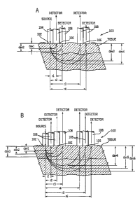
~s Referring now to FIG. 1, the apparatus of this invention comprises a means
for introducing light into tissue at a defined light introduction site. At
small distances
from the light introduction site are located a plurality of light collection
sites, each
light collection site being in contact with a light collecting element, which
collects the
light re-emitted from tissue. The intensity of the re-emitted light collected
at this site
2o will be measured by a detector. The source of light for providing light at
the light
introduction site can be a focused beam of light, a collimated beam of light,
or a
surface-mounted light emitting diode or a laser diode in contact with the
skin. Other
sources of light can also be used. In addition, the source of light can be
remote from
the light introduction site, in which case an optical fiber can be used to
carry light
2s from the remote source of light to the light introduction site. The re-
emitted light is
collected at each of multiple light collection sites located at specific
distances, r~, r2,
..., and r", from the light introduction site. The light collected is directed
towards the
detector that measures the intensity of the collected light. Re-emitted light
can be
collected by any of several means. Representative examples of these means of
3o collecting scattered light include, but are not limited to, fibers that are
in contact with
the skin and a mask with holes at predetermined distances from the light
introduction
site. The light thus collected can be imaged into a charge coupled device
(CCD)
CA 02380243 2002-O1-23
WO 01/09589 PCT/US00/20730
22
camera, a series of photodiodes in contact with the skin, a one-dimensional or
a two-
dimensional photodiode array, or any other suitable type of detector.
Although the previous discussion has focused primarily upon a single light
introduction site and a plurality of light collection sites comprising light
collecting
s elements, in an alternative embodiment, a plurality of light introduction
sites and a
single light collection site can be used. A single light collection site
replaces the light
introduction site, and a plurality of light introduction sites replaces the
light collection
sites at distances r~, r2, ..., and r~.
The apparatus of the present invention requires that the sites for introducing
to light and for collecting light be closely spaced. Thus, the apparatus is
useful for
monitoring analyte effects on the top skin layers, such as epidermis and
dermis. The
short sampling distances allowed for in this invention are in contrast with
those
disclosed in the prior art. As an example, Kumar et al. recommend that the
separation between the light introduction site and the light collection site
be greater
is than 4 mm, in order to avoid the structural effects of the surface of the
skin. See G.
Kumar, J. M. Schmitt, "Optical probe geometry for near-infrared spectroscopy
of
biological tissue", Applied Optics, Vol. 36, 1997, pages 2286-2293.
Another feature of this invention is that it provides a method and apparatus
for
selecting the optimal distance of separation between the light introduction
site and
2o the light collection site for the determination of an analyte. For analytes
that
significantly affect the scattering properties of the epidermis and the dermis
layers by
virtue of their effect on the refractive indexes, and hence the scattering
coefficients of
these layers, their concentrations can be determined at pre-selected short
sampling
distances. Thus, a distance in the range of 0.4 mm to 1.2 mm is appropriate
for such
2s a measurement. For these analytes, such as glucose, one can first generate
a
calibration relationship between their concentrations determined in vitro and
the
reflectance signals measured from the epidermis and the upper dermis. One can
then use the calibration relationship thus generated to predict the
concentrations of
the analyte based on subsequent reflectance measurements.
3o On the other hand, analytes that affect deeper layers in the skin, such as
hemoglobin, which is carried by blood flow into the upper and lower plexuses
within
dermis and subcutaneous tissue, can be determined from measurements at greater
sampling distances. Thus, hemoglobin concentration and hematocrit can be
better
CA 02380243 2002-O1-23
WO 01/09589 PCT/US00/20730
23
measured at a longer sampling distance, e.g., greater than 1.4 mm. This longer
distance corresponds to light re-emitted from skin layers deeper than those
encountered for the determination of glucose and other analytes that
preferentially
affect the optical properties of the upper layers of the skin. This invention
offers a
s tunable sampling distance feature for optimizing analyte detection according
to the
nature of each analyte.
The properties of skin layers vary from one body part to another and from one
individual to another. The difference includes the thickness of each skin
layer,
pigmentation and hydration state of the skin, tissues in the subcutaneous
regions,
~o effects of age and disease condition of the individual on the skin, etc.
Thus, the
sampling depth and hence the sampling distance at which an analyte should be
optimally determined varies by the body part and the individual to be tested.
Other analytes that can be determined by the method and apparatus of this
invention include tissue hemoglobin, tissue urea and creatinine, and skin
water
~s content. These analytes can be determined individually by selecting the
optimal
sampling distance for each analyte determination or simultaneously by
measuring
light re-emitted at multiple sampling distances and correlating each analyte
at its
optimum sampling distance for maximum correlation with the reference method.
In another aspect, this invention provides a method for the establishment of a
2o calibration relationship for the in vivo measurement of an analyte. A
calibration
relationship, applicable to a given analyte, a given individual, and a given
body part,
determines the optimum sampling distance and subsequently the optimum sampling
depth in the tissue. It also provides the correlation relationship between the
concentration of a given analyte in the sample and the intensity of the re-
emitted light
2s detected at the optimum sampling distance. For each analyte and each
individual,
the method for generating a calibration relationship comprises the steps of:
(1) employing one of the non-invasive methods described herein to make
at least one measurement of the concentration of an analyte, by measuring the
3o reflectance of light at each of a plurality of sampling distances, and at
substantially
the same time, obtain the concentration of the analyte by a standard reference
method;
CA 02380243 2002-O1-23
WO 01/09589 PCT/US00/20730
24
(2) establishing the best achievable correlation relationship between the
non-invasive measurement at each of the sampling distances and the
concentration
of the analyte;
(3) comparing the results obtained at each of the plurality of sampling
distances; and
(4) selecting the sampling distance that provides the best correlation
performance.
To accomplish step (2) above, one usually needs to test multiple mathematical
io relationships by means of regression methods such as the classical least
squares
and the principal component regression with respect to their performances. The
performances are often measured by parameters such as the correlation
coefficient
and standard error of estimation in both the calibration process and the
validation
process. An optimal sampling distance should result in the best performance,
as
is indicated by optimal statistical parameters, such as the highest
correlation coefficient
and the lowest standard error of estimation. The calibration relationship
generated
can be used for the subsequent determination of the concentration of the same
analyte in the same individual, based only on a non-invasive measurement at a
single appropriate sampling distance.
2o Standard reference methods can be used with this invention in the
calibration
procedure, so long as they are commonly accepted, in terms of specificity and
sensitivity, by medical professionals, i.e., approved by the U. S. Food and
Drug
Administration, for the specified medical application. For example, commercial
clinical chemistry analyzers can be used for determination of the
concentrations of
2s total serum bilirubin, blood hemoglobin, and venous blood glucose. The
glucose
meter commercially available for diabetics' self use can be used to measure
glucose
concentration in the blood from a few microliters of capillary blood obtained,
e.g., by
lancing a finger. Microdialysis or other interstitial fluid sampling methods
in
combination with standard analytical chemistry methods may be used to
determine
3o the concentration of glucose in interstitial fluid samples. Hematocrit is
commonly
determined by centrifugation or cell sorting analyzers for venous blood
samples.
CA 02380243 2002-O1-23
WO 01/09589 PCT/US00/20730
FIG. 1 is a schematic diagram showing a light introduction site and several
light collection sites located at several sampling distances from the light
introduction
site. Different tissue layers are probed at different sampling distances. The
diffusely
reflected light is measured, at each wavelength, for a fixed distance between
the
s light introduction site and the light collection site. This configuration is
achieved by
using optical fibers in touch with the tissue surface. Selection of distance
is achieved
by interrogating the light collected at a given fiber at a given distance from
the source
fiber. This is a stationary illuminating and detecting system. The signal is
amplified
and is corrected for fluctuation of the light source and variation of the
fiber
to throughput. The corrected signal is used for correlating with the analyte
concentration to establish a calibration relationship or for the determination
of the
analyte.
Alternatively a non-stationary illumination and detection system can be used.
The detection distance can be selected by moving the light introduction site
on the
is surface of the sample using a mechanism similar to a compact disk (CD)
player read
head, to predetermined distances from a light collection site located at a
specific site
on the surface. Mechanisms for directing a light beam to predetermined
distances
include beam-stirring devices such as moving mirrors or prisms. The light beam
can
span a circular or linear path. Another method of achieving the same result
involves
2o illuminating a site on the surface of the sample using a stationary fiber
in contact with
the surface, or illuminating a point on the surface by a collimated or focused
beam of
light. Re-emitted light is then collected at selected sampling distances on
the
sample surface by moving a light collecting element on the surface. This can
be
affected by using a stylus-type (phonograph needle-type) arrangement.
2s The method of this invention is advantageous over the method disclosed
European Patent No. 0 843 986, which does not appreciate the effect of weakly
absorbing analytes, including glucose, on the scattering property of tissue
layers.
This patent does not disclose the method of determining different analytes
with the
use of different sampling distances, nor does it disclose the method of
optimizing the
3o sampling distance to accommodate differences in individuals.
The following non-limiting examples further illustrate this invention.
CA 02380243 2002-O1-23
WO 01/09589 PCT/US00/20730
26
EXAMPLES
Example 1
s
This example shows an apparatus having selectable sampling distances
through the use of a plurality of light collection fibers. FIG. 3 through FIG.
5 illustrate
an example of an apparatus for the measurement of optical properties, and
hence
the concentration of different analytes at various depths in tissue. Co-
pending U. S.
io Application Serial No. 09/198,049, filed November 23, 1998 ("Non-invasive
sensor
capable of determining optical parameters in a sample having multiple
layers"),
assigned to the assignee of this application, describes in detail many of the
components used in the apparatus of this application. The apparatus was
intended
for introducing light into the skin on forearms of human subjects and
measuring the
is light re-emitted therefrom. As shown in FIG. 3, the apparatus comprised a
light
source module 12, a human interface module 16, a signal detector module 18,
and a
branched optical fiber bundle 14 that conducted light signals among these
three
modules. Monochromatic light was generated from the light source module 12 at
six
wavelengths, i.e., 590 nm, 650 nm, 750 nm, 800 nm, 900 nm and 950 nm. The
light
2o was transported to the human interface module 16 through the source fiber
26 in the
branched optical fiber bundle 14 (FIG. 4A and 4B). The source fiber 26
received
light from its end housed in the source tip 20 in the light source module 12.
It
emitted the light into the skin of a subject's forearm from its other end,
which directly
touched the skin at a spot named the light introduction site, housed in the
common
2s tip 24 in the human interface module 16. Also touching the skin from the
common tip
24, six other fibers 28, 30, 32, 34, 36 and 38 were six independent light
collecting
elements. Each of these fibers collected light re-emitted from the skin at the
spot
where it touched the skin, i.e., a light collection site. The human interface
module
engaged the common tip to the skin. It also provided temperature and pressure
3o control mechanisms for the tip-skin contacting area. The area of skin
surrounding
the optical element engagement sites was kept at a predetermined constant
temperature throughout the measurement. In addition, the human interface
module
had a comfortable armrest (not shown) for the testing forearm.
CA 02380243 2002-O1-23
WO 01/09589 PCT/US00/20730
27
Both the source fiber and detection fibers were 400 ~,m in diameter. The
distance from any one detection fiber to the source fiber 26 at the end of the
common tip 24 defined the distance between the corresponding light collection
site
on the skin and the light introduction site also on the skin, i.e., the
sampling
s distances. These distances are indicated in FIG. 5 and listed in TABLE 1.
TABLE 1
r~ r2 r3 ra rs rs
Sampling Distance, 0.44 0.78 0.92 1.22 1.40 1.84
mm
~o The six detection fibers received the re-emitted light from the skin at the
common tip 24 and transmitted the light to the detector tip 22 housed in the
detector
module 18. The ends of all of these fibers at the detector tip 22 were in the
focal
plane of a lens for the detector (both lens and detector are not shown).
However,
only when the shutter between a particular fiber end and the detector (not
shown)
Is was opened was the light signal from that fiber detected.
Therefore, the sampling depth was determined by selecting a particular light
collection fiber and detecting the intensity of re-emitted light collected by
this fiber.
Selection of a particular light collection fiber was achieved by the use of a
2o programmable shutter that selected one of the six light collection fibers.
The shutter
was moved by rotating the shutter to a programmed number of steps or a pre-
selected detent on its mount.
2s ExamJ~le 2
This example illustrates the correlation of non-invasive measurements to
hemoglobin concentration or hematocrit. An apparatus as described in FIG. 3
through FIG. 5 was used for the in vivo determination of hemoglobin content
and
3o hematocrit for 28 subjects. Some of these subjects were diabetics and some
had
dark skin.
CA 02380243 2002-O1-23
WO 01/09589 PCT/US00/20730
28
Tests were conducted on the subjects three hours after their breakfast meal.
Non-invasive measurements were performed on the inner part of the subject's
left
forearm. Silicone oil (Poly(dimethylsiloxane), 200~ fluid, viscosity 1,000
cSt, Aldrich
Chemical Company) was applied to the skin, and the human interface module 16
s with the common tip 24 was placed in contact with the skin. The temperature
of the
testing site on the skin was allowed to equilibrate at 34 °C for two
minutes, and then
the measurement was started. Reflected light was collected and reflectance was
measured at the six sampling distances as shown in TABLE 1. Wavelengths used
in this measurement were 590, 650, 750, 800, 900, and 950 nm.
lo Venous blood samples of the subjects were obtained immediately following
the non-invasive measurement and used for determination of the reference
values of
hemoglobin concentration and hematocrit. The hematocrit value was determined
by
a standard micro-centrifuge method (described in C. E. Seiverd, Hematology for
Medical Technolo iq~ sts. Lea & Febiger, Philadelphia PA , 1983, pages 320-
330).
is Blood hemoglobin values were determined using a commercial kit and a
commercial
clinical chemistry analyzer (VisionO Analyzer, Abbott Laboratories, North
Chicago,
IL).
The relative reflectance at detection distance r, R(r) is defined as:
R(r) - I recreated (r) (4)
Ib~ciaeiu
20 where,
l~n~~de~t represents the relative intensity of the illuminating light from the
source
fiber 26 measured from the common tip 24; and,
Ireflected(r) represents the relative intensity of the re-emitted light from
the skin
collected by a light collection fiber which has distance r to the source fiber
2s 26 at the common tip 24, and measured at the detector module 18.
Reflectance data at different sampling distances and wavelengths was
correlated with the hematocrit and hemoglobin concentrations by means of the
linear
least square method. For hematocrit, the correlation coefficient was low at
the
3o shorter sampling distances (e.g., 0.44 mm and 0.78 mm) and increased
significantly
at sampling distances greater than about 0.92 mm. The reflectance at a fixed
sampling distance of 1.84 mm yielded the highest correlation coefficient and
the
CA 02380243 2002-O1-23
WO 01/09589 PCT/US00/20730
29
lowest standard error of calibration for correlation with reference hematocrit
values.
The correlation coefficient was plotted as a function of sampling distance and
the
plot is shown in Figure 6. The correlation coefficient was above 0.9 at
distances of
1.40 mm and 1.84 mm. At either of these two distances, the light penetrates
through
s the upper plexus and encounters blood capillaries. The standard error of
calibration
followed a reverse trend, being greater than 3.2% at the shorter distances and
less
than 2.0% at the two greater distances. The best regression plot is shown in
FIG. 7
and the regression equation is:
io Hematocrit (%) _ -0.347 - 39.0 ~ Log[R(590 nm)] + 61.0 ~ Log[R(650 nm)]
+ 151 ~ Log[R(900 nm)] - 178 ~ Log[R(950 nm)] (5)
where, Log[R(~,)] represents the natural logarithm of reflectance at
wavelength 7~
(nm) and at a sampling distance of 1.84 mm. The correlation coefficient is
0.911
is and the standard error of calibration is 1.84 % (hematocrit unit) for the
28 subjects.
A similar correlation was obtained with the use of absorption and scattering
coefficients deriving from reflectance values at the all six different
sampling
distances. This method was described in the prior art (e. g., U. S. Patent No.
5.075,695 and U. S. Patent No. 5,551,422). However, this example demonstrated
2o the correlation with diffuse reflectance data at much shorter sampling
distances
(instead of greater than 5 mm in the prior art) and using a temperature
controlled
detection device. The regression equation thus obtained is:
Hematocrit (%) = 55.8 + 11.4 ~ ~,a(590 nm) - 26.1 ~ ~a(650 nm) -
2s 5.72 ~ ~S'(590 nm) + 6.14 ~ ~S'(650 nm) (6)
The correlation coefficient was 0.87 for the 28 subjects as a group and the
standard
error of calibration was 2.2% (hematocrit unit).
Thus, the use of reflectance data from a specific sampling depth, collected at
3o a single optimized sampling distance yielded a better correlation and
smaller
standard error of calibration with respect to the reference values of the
hematocrit
than does the use of the fitted absorption and scattering coefficient values.
CA 02380243 2002-O1-23
WO 01/09589 PCT/US00/20730
Furthermore, the measurement does not require synchronizing to heart beat
pulses
or a pulsatile signal as taught by U. S. Patent Nos. 5,499,627 and 5,803,908.
From the plot of the correlation coefficient and the standard error of
calibration
as a function of sampling distance (FIG. 6), it is apparent that quality of
regression is
s no longer sensitive to the sampling distance when the distances are greater
than 1.4
mm.
Skin color was found to affect the calculation that was based on the
absorption and scattering coefficients. Thus, it is possible to improve the
correlation
with hematocrit by eliminating data points corresponding to dark-skinned
individuals.
to In this case the number of light-skinned subjects was 24 and the regression
equation
becomes:
Hematocrit (%) = 26.2 + 21.7 ~ ~a(590 nm) - 26.4 ~ ~a(650 nm) -
32.6 ~ ~a(800 nm) + 33.6 ~ ~a(950 nm) (7)
The correlation coefficient is 0.90 and the standard error of calibration is
1.8%
(hematocrit unit).
Effects of skin color were minimized in the correlation to the hematocrit,
when
the reflectance measured at a single sampling distance and generated from a
fixed
2o sampling depth in skin was used. This result is in agreement with the
layered
structure description of human skin tissue described herein, as at this
distance, light
penetrates the upper plexus of the dermis layer of the skin and encounters the
blood
capillaries. Those skilled in the art can use similar analysis and apply this
measurement method to other analytes.
Example 3
This example illustrates non-Invasive glucose correlation. The same
apparatus as described in Example 1 and a similar setup as described in
Example 2
3o were used for the in vivo determination of glucose for three subjects. For
each
individual subject, a meal tolerance test protocol was used to induce changes
in
blood glucose. Correlation between the reflectance measurement and the
reference
in vitro blood test was carried out.
CA 02380243 2002-O1-23
WO 01/09589 PCT/US00/20730
31
For each subject, the reflectance measurement was performed repetitively
with one set of readings (at six sampling distances as shown in TABLE 1 and at
three wavelengths - 590 nm, 800 nm and 950 nm) for every 100 seconds. Tests
were conducted under controlled skin temperature of 22 °C. Blood
samples were
s taken from the subject by means of a finger-stick every 5 to 15 minutes and
tested
by means of a commercially available glucose meter. The measurement started
when the subject was in a fasting condition. After 10 to 20 minutes, the
subject
ingested a high sugar drink (commercially available fruit juices, 680-mL
liquid and
100 to 120 gram sugars). The total measurement required 90 minutes to 120
to minutes.
A plot of the glucose values vs. the time during the meal tolerance test on
one
of the subjects is shown in FIG. 8. The circles represent the result of the
reference
glucose test using finger-stick capillary blood and a home glucose meter
(Glucometer Elite~, Bayer Corp., Elkhart, IN). The smooth line passing through
is these circles shows the fit values of reference glucose concentration
resulting from
cubic spline smoothing of the finger-stick capillary blood glucose values.
Interpolated data points represent the in vitro blood glucose test results at
points in
time that do not coincide with the points in time at which the tests were
actually
performed. Classical linear regression was employed to correlate a model
2o comprising reflectance measurement at each single sampling distance at
three
wavelengths with the fit values of reference glucose concentrations. In most
cases,
reflectance measurements at r3 (r = 0.92 mm) at three wavelengths yielded a
linear
model and fit to the reference glucose values. In FIG. 8, the crosses
represent the
values of glucose concentration calculated by such a model, i. e.,
Glucose (mg/dL) _ -2898 + 536 ~ Log[R(590 nm)] - 1523 ~ Log(R(800 nm)] +
2043 ~ Log[R(950 nm)] (8)
where, Log[R(~,)] represents the natural logarithm of reflectance at
wavelength ~,
(nm). The models yielded a correlation coefficient of 0.98 and a standard
error of
calibration of 8.9 mg/dL.
Two of the three subjects were non-diabetics, and the third was diagnosed as
a diabetic in less than one year. The meal tolerance test was performed on one
of
CA 02380243 2002-O1-23
WO 01/09589 PCT/US00/20730
32
the non-diabetic subjects twice in two days. For each of four meal tolerance
tests,
the reflectance measurement at single distance (0.92 mm) was used to correlate
with the reference blood glucose concentration. The calibration results of the
four
tests are compiled in a Clark error grid presentation, as shown in FIG. 9,
where the
s calculated glucose values are plotted against the reference glucose values.
The
total number of data points is 250. As seen from the plot, 96% of the data
points are
in Zone A, the rest are in Zone B, and none is in the zones C, D or E. While
data in
Zone A and Zone B are considered "acceptable" performance, data in Zones C, D
and E may cause serious adverse effects in clinical applications, as they may
lead to
to the wrong types of medical intervention.
Thus reflectance measurement at a single sampling distance that targets the
epidermis and the upper dermal layers (sampling distances shorter than 0.92
mm)
leads to a good correlation with the concentration of glucose during a meal
tolerance
test. Such a measurement is simpler than the use of spatially resolved
is measurement as taught by U. S. Patent Nos. 5,075,695 and 5,551,442 where
signals at multiple sampling distances are needed. Those who are skilled in
the art
can use some data sets as calibration sets and predict the others using prior
art
chemometric methods.
Because measurements can be carried out at wavelengths ranging from 400
2o nm to 2500 nm, the method of this invention avoids the limitations of the
method
described in EP 0 843 986. In EP 0 843 986, a light beam having a wavelength
ranging from 1300 nm to 2500 nm is projected into the skin and the re-emitted
light is
detected at distances of 0.1-2 mm from the source of light. The spectrum of
skin in
the 1300-2500 nm range is dominated by water absorption. The path length in
the
2s tissue is limited because of strong water absorption. Collecting the signal
at the
short distances will not allow a significant absorption change due to the
weakly
absorbing glucose to be measured.
It is important to mention that the determination of hematocrit and hemoglobin
(i.e., Example 2), as well as the determination of glucose were performed with
the
3o same instrument and the same optical sensor, by programming to use a
particular
sampling distance for either glucose or hematocrit. One of ordinary skill in
the art
can configure other distances for other analytes and optimize the measurement
for a
CA 02380243 2002-O1-23
WO 01/09589 PCT/US00/20730
33
particular body part that has different thickness of skin layer, a particular
individual,
or a group of individuals.
Selected distances r2 (0.78 mm) and r3 (0.92 mm) provided the best
correlation for glucose determination, depending on testing temperature used.
In
s another set of experiments, selected distance r~ (0.44 mm) provided the best
correlation for measurement at 38 °C. Thus, distances of less than 1 mm
led to a
better correlation with the glucose concentrations for the individuals and the
body
part tested.
At short sampling distances, measurement of signals from shallower sampling
io depth and with greater contribution from scattering properties yields good
correlation
with weakly absorbing analytes such as glucose. Glucose would be expected to
affect the refractive index of the dermis and the epidermis and change their
scattering properties. At large distances from the light introduction site, re-
emitted
light has greater contribution from absorption and is originated from deeper
layers.
~s Blood capillaries distributed in the upper and lower plexus regions of the
skin would
be expected to affect signals from these regions. Thus, the measurement gives
a
better correlation with hemoglobin and hematocrit, as demonstrated in Example
2.
Various modifications and alterations of this invention will become apparent
2o to those skilled in the art without departing from the scope and spirit of
this invention,
and it should be understood that this invention is not to be unduly limited to
the
illustrative embodiments set forth herein.