Note: Descriptions are shown in the official language in which they were submitted.
CA 02380487 2008-05-14
METHODS FOR SIMULTANEOUSLY DETECTING BOTH
MEMBERS OF A BINDING PAIR
Technical Field
The invention relates to methods for simultaneously detecting both members
of a binding pair in a biological sample.
Background of the Invention
Blood products used for transfusion and transfer of blood components must be
routinely screened for the presence of infectious agents such as human
immunodeficiency virus (HIV), hepatitis viruses, human T-lymphocytotropic
virus,
and cytomegalovirus. Such agents typically are detected by either
identification of
viral antigens or by detection of an inunune response to the virus (i.e., host-
derived
anti-viral antibodies) using enzyme immunoassay analysis (EIA) or
radioimmunoassays (RIA). Immunoassay techniques are limited in their ability
to
detect the presence of viral contaminants in early stages of infection, with
the window
period between infection with a virus and detection by immunoassay techniques
varying from two to four weeks for HIV and up to about 10 weeks for hepatitis
C
virus (HCV). Techniques such as reverse-transcriptase polymerase chain
reaction
(RT-PCR) or branched chain DNA analysis can shorten the time period between
infection and detection, but are cost prohibitive for use on an individual
donor basis
and do not eliminate the window period.
Summary of the Invention
The invention is based on a rapid and sensitive method for simultaneously
detecting both members of a binding pair, such as a ligand and receptor or an
antigen
and host antibody, from a biological sample. Methods of the invention can, for
example, enhance the ability to detect infections at an early stage, leading
to earlier
treatment of the infection.
-i-
CA 02380487 2008-05-14
x" = '
Various embodiments to this invention provide a method for simultaneously
measuring both members A and B of a binding pair complex in a biological
sample, said
method comprising: a) providing a solid phase reagent, said solid phase
reagent
comprising a particle coated with capture antibodies having specific binding
affinities for
said member A of said binding pair complex, and wherein said particle does not
comprise
a capture antibody having specific binding affinity for said member B of said
binding pair
complex; b) contacting said biological sample with said solid phase reagent
under
conditions in which said member A, if present, becomes bound to said particle;
c)
contacting said solid phase reagent obtained from step (b) with first
antibodies having
specific binding affinities for said meinber A, wherein said first antibodies
are labeled
with a first label, and with second antibodies having specific binding
affinities for said
member B of said binding pair complex, wherein said second antibodies are
labeled with a
second label, wherein said first label and said second label are different,
and d) measuring
said first and second labels on said solid phase reagent obtained from step
(c).
Other embodiments to this invention provide a kit for simultaneously measuring
both members A and B of a binding pair complex in a biological sample, said
kit
comprising: a) a solid phase reagent, said solid phase reagent comprising a
particle coated
with capture antibodies having specific binding affinities for said member A
of said
binding pair complex,- wherein said capture antibodies are oriented on said
particle such
that the antigen binding regions of said capture antibodies are available for
binding said
member A of said binding pair complex; b) first antibodies having specific
binding
affinities for said member A of said binding pair complex, wherein said first
antibodies are
labeled with a first label; and c) second antibodies having specific binding
affinities for
said meinber B of said binding pair complex, wherein said second antibodies
are labeled
with a second label, and wherein said first label and said second label are
different.
The invention features a method for simultaneously measuring both meinbers A
and B of a binding pair in a biological sample. The biological sample is
selected from the
group consisting of blood, plasma, serum, urine, cerebrospinal fluid, sputum,
tears,
a.inniotic fluid, vitreous humor, saliva, and tissue culture supernatants. The
-la-
CA 02380487 2002-01-29
WO 01/09608 PCT/US00/20769
method includes providing a solid phase reagent, which includes a particle
coated
with capture antibodies having specific binding affinities for member A of the
binding
pair, and contacting a biological sample with the solid phase reagent under
conditions
in which member A, if present, becomes bound to the particle, to form a first
reacted
particle. The capture antibodies can be monoclonal. The first reacted particle
is
contacted with first antibodies having specific binding affinities for member
A,
wherein the first antibodies are labeled with a first label, and with second
antibodies
having specific binding affinities for member B of the binding pair, wherein
the
second antibodies are labeled with a second label, to form a second reacted
particle.
The first and second antibodies can be monoclonal. First and second labels
(e.g.,
fluorophores) are measured on the second reacted particle using flow
cytometry.
In certain embodiments, substantially all capture antibodies are oriented on
the
particle such that the antigen binding regions of the capture antibodies are
available
for binding member A of the binding pair.
Member A of the binding pair can be, for example, an antigen and member B
can be a host antibody. The antigen can be a viral antigen such as a hepatitis
C
antigen, a hepatitis B antigen, or a human immunodeficiency virus antigen, or
an
autoantigen such as glutamic acid decarboxylase. Member A of the binding pair
also
can be a ligand, such as a cytokine, and member B can be a receptor, such as a
cytokine receptor. In addition, member A can be an enzyme and member B can be
a
substrate. For example, the enzyme can be caspase-3 or caspase-1 and the
substrate
can be poly(ADP-ribose) polymerase or proInterleukin-1, respectively.
The invention also features a kit for simultaneously measuring both members
A and B of a binding pair in a biological sample. The kit includes a solid
phase
reagent, which includes a particle coated with capture antibodies having
specific
binding affinities for member A of the binding pair; first antibodies having
specific
binding affinities for member A of the binding pair, wherein the first
antibodies are
labeled with a first label; and second antibodies having specific binding
affinities for
member B of the binding pair, wherein the second antibodies are labeled with a
second label. Substantially all the capture antibodies are oriented on the
particle such
that the antigen binding regions of the capture antibodies are available for
binding
member A of the binding pair. The kit further can include a label or package
insert,
which indicates that the solid phase reagent, the labeled first antibodies,
and the
-2-
CA 02380487 2008-05-14
labeled second antibodies can be used for simultaneously measuring both
members A
and B of a binding pair in a biological sample by flow cytometry.
Unless otherwise defined, all technical and scientific terms used herein have
the same meaning as commonly understood by one of ordinary skill in the art to
which this invention belongs. Although methods and materials similar or
equivalent
to those described herein can be used to practice the invention, suitable
methods and
materials are described below.
In
case of conflict, the present specification, including definitions, will
control. In
addition, the materials, methods, and examples are illustrative only and not
intended
to be limiting.
Other features and advantages of the invention will be apparent from the
following detailed description, and from the claims.
Brief Description of the Drawings
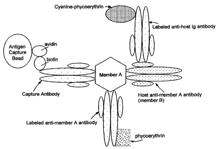
Figure 1 is a schematic representation of an assay for detecting member A and
host anti-member A antibody (member B).
Figures 2A-2H are scattergrams that indicate simultaneous detection of
hepatitis B virus (HBV) surface antigen, anti-HBV host antibody, HCV core
antigen,
and anti-HCV host antibody by flow cytometry. Figure 2A is HBV antigen and
antibody in a normal sample. Figure 2B is HCV antigen and antibody in a normal
sample. Figure 2C is HBV antigen and antibody in an HBV positive sample.
Figure
2D is HCV antigen and antibody in an HBV positive sample. Figure 2E is HBV
antigen and antibody in an HCV positive sample. Figure 2F is HCV antigen and
antibody in an HCV positive sample. Figure 2G is HBV antigen and antibody in
an
HBV positive/HCV positive sample. Figure 2H is HCV antigen and antibody in an
HBV positive/HCV positive sample.
Detailed Description
Immunoassay Format
In general, the invention uses a sandwich immunoassay method for
simultaneously detecting both members of a binding pair in a biological
sample.
Binding pairs include any combination of molecules which forms a complex,
-3-
CA 02380487 2002-01-29
WO 01/09608 PCT/US00/20769
including pairs composed of nucleic acids, proteins, or a small molecule and a
protein.
Nucleic acid pairs can be DNA:RNA pairs or DNA:DNA pairs. For example, a
DNA/RNA binding pair such as a single stranded (ss) DNA and an mRNA can be
used as a PCR product detection system. A DNA/DNA binding pair such as a ssDNA
and a viral DNA can be used in a competition assay for quantitation of virus
per
amplified DNA.
Non-limiting examples of protein, or small molecule and protein, binding pairs
include a hormone, a cytokine, a peptide, a drug, a viral protein, or other
antigen and a
cognate receptor or host antibody. Viral protein/receptor binding pairs can
be, for
example, HIV gp 120 and soluble CD4. Drug and drug receptor binding pairs can
be,
for example, cocaine and a dopamine receptor. Peptide and peptide receptor
binding
pairs can be, for example, acetylcholine and a muscarinic receptor or dopamine
and a
dopamine receptor. Hormone and hormone receptor binding pairs can be, for
example, insulin and insulin receptor. Cytokine and cytokine receptor binding
pairs
can be, for example, tumor necrosis factor (TNF) and a TNF Type I or Type 2
receptor or interleukin 2 (IL-2) and IL-2 receptor. Antigen and antibody pairs
can be,
for example, a viral protein and host anti-viral protein antibody or an
autoantigen and
a host anti-autoantigen antibody. HIV p24/human anti-HIV antibody, HIV
gp120/human anti-HIV gp120 antibody, HBV surface antigen/human anti-HBV
surface antigen, and HCV core protein/human anti-HCV core antibody are
examples
of viral protein and host antibody binding pairs. An autoantigen and host anti-
autoantigen antibody binding pair can be, for example, glutamic acid
decarboxylase
(GAD) and host anti-GAD antibody.
Other protein binding pairs that can be detected are enzyme and enzyme
substrate binding pairs. For example, the enzyme/substrate pair can be caspase-
3/poly
(ADP-ribose) polymerase or caspase-1/prolnterleukin-1.
Member A, which can be either member of the binding pair, is captured with a
solid phase reagent that is a particle coated with capture antibodies having
specific
binding affinities for member A. For example, if the binding pair to be
simultaneously detected is HIV gp120/host anti-HIV gp120 antibody, the
particle can
be coated with antibodies having specific binding affinities for HIV gp 120 or
anti-
host immunoglobulin (Ig).
-4-
CA 02380487 2002-01-29
WO 01/09608 PCT/US00/20769
Member A is captured by contacting a biological sample with the particle
coated with capture antibodies. As used herein, suitable biological samples
contain
cells or cellular material, and include, for example, blood, plasma, serum,
urine,
saliva, sputum, tears, amniotic fluid, vitreous humor, and cerebrospinal
fluid. Other
samples can include in vitro tissue culture medium/supernatants. Biological
samples
can be treated with a non-ionic detergent such as 0.5% Triton-X 100 or Nonidet
P40
(Sigma Chemical Company, St. Louis, MO) to expose core antigens from
pathogens.
The solid phase reagent and biological sample are contacted under conditions
that facilitate binding of member A, if present, to the particle, to form a
first reacted
particle. Such conditions can include use of buffer containing 1% fetal bovine
serum
(FBS) and 0.1% sodium azide in phosphate-buffered saline (PBS) at room
temperature, or use of any biological fluid, under physiologic pH conditions.
The first
reacted particle then is contacted with two sets of labeled antibodies (i.e.,
reporter
antibodies) to form a second reacted particle. The first antibodies have
specific
binding affinities for member A and are labeled with a first label. First
antibodies are
capable of binding to member A while member A is bound to capture antibodies.
Thus, the capture antibodies and first antibodies must work as a pair. Second
antibodies have specific binding affinities for member B of the binding pair
and are
labeled with a second label. Fluorescently labeled antibodies are particularly
useful in
this method.
Figure 1 provides a schematic of an assay for detecting member A and host
anti-member A antibody. In this embodiment, biotinylated capture antibodies
have
specific binding affinities for member A and are coupled to antigen capture
beads via
avidin. First antibodies have specific binding affinities for member A and are
labeled
with phycoerythrin (first label). Second antibodies are labeled with cyanine-
phycoerythrin (second label) and have specific binding affinities for host Ig
(member
B). The first reacted particle includes member A, host anti-member A
antibodies,
capture antibodies, and the solid phase reagent (e.g, antigen capture beads),
wherein
the second reacted particle includes the first reacted particle and the two
labeled
antibodies.
Flow cytometry can be used to measure the amount of label on the second
reacted particle. As used herein, the term "measure" refers to qualitative and
quantitative measurements. In other words, the term "measure" includes
reporting the
-5-
CA 02380487 2002-01-29
WO 01/09608 PCT/US00/20769
presence or absence of label on the second reacted particle, as well as
determining the
amount of label present. Flow cytometers are able to measure at least three
discrete
fluorescence emission wavelength ranges by using optical filters to split the
fluorescent emission and separate photomultiplier tubes to amplify the
individual
emission signals. The intensity of fluorescent emission associated with the
particles is
directly proportional to the concentration of analyte present in the
biological sample.
Thus, the use of different dyes with different emission spectra, wherein each
dye is
coupled to a different antibody, allows analysis of multiple analytes per
population of
particles. The flow cytometer also can distinguish particles of different
sizes such that
a particle, for example approximately 7 m in diameter, can be differentiated
from a
particle approximately 10 m in diameter. Therefore, additional components can
be
detected by using a combination of multiple fluorescent dyes and two or three
populations of particles of different average diameters.
Production ofAntibodies
Antibodies having specific binding affinities for member A or member B can
be produced through standard methods. Alternatively, antibodies may be
commercially available, for example, from BiosPacific (Emeryville, CA),
Coulter
(Hialeah, FL), Maine Biotechnology Service (Portland, ME), or Biodesign
International (Kennebunk, ME). As used herein, the terms "antibody" or
"antibodies"
include intact molecules as well as fragments thereof which are capable of
binding to
an epitopic determinant in member A or member B. The term "epitope" refers to
an
antigenic determinant on an antigen to which the paratope of an antibody
binds.
Epitopic determinants usually consist of chemically active surface groupings
of
molecules such as amino acids or sugar side chains, and typically have
specific three
dimensional structural characteristics, as well as specific charge
characteristics.
Epitopes generally have at least five contiguous amino acids. Thus, the terms
"antibody" and "antibodies" include polyclonal antibodies, monoclonal
antibodies,
humanized or chimeric antibodies, single chain Fv antibody fragments, Fab
fragments, and F(ab)2 fragments. Monoclonal antibodies are particularly
useful.
In general, a protein of interest is produced recombinantly, by chemical
synthesis, or by purification of the native protein, and then used to immunize
animals.
Various host animals including, for example, rabbits, chickens, mice, guinea
pigs, and
-6-
CA 02380487 2002-01-29
WO 01/09608 PCT/US00/20769
rats, can be immunized by injection of the protein of interest. Adjuvants can
be used
to increase the immunological response depending on the host species and
include
Freund's adjuvant (complete and incomplete), mineral gels such as aluminum
hydroxide, surface active substances such as lysolecithin, pluronic polyols,
polyanions, peptides, oil emulsions, keyhole limpet hemocyanin (KLH), and
dinitrophenol. Polyclonal antibodies are heterogenous populations of antibody
molecules that are specific for a particular antigen, which are contained in
the sera of
the immunized animals. Monoclonal antibodies, which are homogeneous
populations
of antibodies to a particular epitope contained within an antigen, can be
prepared
using standard hybridoma technology. In particular, monoclonal antibodies can
be
obtained by any technique that provides for the production of antibody
molecules by
continuous cell lines in culture such as described by Kohler, G. et al.,
Nature, 1975,
256:495, the human B-cell hybridoma technique (Kosbor et al., Immunology
Today,
1983, 4:72; Cole et al., Proc. Natl. Acad. Sci. USA, 1983, 80:2026), and the
EBV-
hybridoma technique (Cole et al., "Monoclonal Antibodies and Cancer Therapy",
Alan R. Liss, Inc., 1983, pp. 77-96). Such antibodies can be of any
immunoglobulin
class including IgG, IgM, IgE, IgA, IgD, and any subclass thereof. The
hybridoma
producing the monoclonal antibodies of the invention can be cultivated in
vitro or in
vivo.
A chimeric antibody is a molecule in which different portions are derived from
different animal species, such as those having a variable region derived from
a murine
monoclonal antibody and a human immunoglobulin constant region. Chimeric
antibodies can be produced through standard techniques.
Antibody fragments that have specific binding affinity for member A or B can
be generated by known techniques. For example, such fragments include, but are
not
limited to, F(ab')2 fragments that can be produced by pepsin digestion of the
antibody
molecule, and Fab fragments that can be generated by reducing the disulfide
bridges
of F(ab')2 fragments. Alternatively, Fab expression libraries can be
constructed. See,
for example, Huse et al., 1989, Science, 246:1275. Single chain Fv antibody
fragments are formed by linking the heavy and light chain fragments of the Fv
region
via an amino acid bridge (e.g., 15 to 18 amino acids), resulting in a single
chain
polypeptide. Single chain Fv antibody fragments can be produced through
standard
techniques. See, for example, U.S. Patent No. 4,946,778.
-7-
CA 02380487 2002-01-29
WO 01/09608 PCT/US00/20769
Once produced, antibodies or fragments thereof are tested for recognition of
member A or member B by standard immunoassay methods including, for example,
ELISA techniques or RIA. See, Short Protocols in Molecular Biology, Chapter
11,
Green Publishing Associates and John Wiley & Sons, Edited by Ausubel, F.M et
al.,
1992. Suitable antibodies preferably have equal binding affinities for
recombinant
and native proteins.
Alternatively, antibodies can be assessed for their ability to form binding
pairs
in a fluorescent sandwich assay in the following manner. Beads can be coated
with
biotinylated antibodies, for example anti-viral protein antibodies, then
incubated for
approximately 30 minutes with 2 ng/ml of the appropriate protein, e.g.,
recombinant
viral protein, in a 100 l volume. After washing the beads twice with 2 ml of
buffer
containing 1% FBS and 0.1% sodium azide in PBS, the beads are incubated with
approximately 0.5 g of phycoerythrin-labeled antibody. Pairs of antibodies
producing a strong fluorescent signal are suitable for use in assays of the
invention.
Solid Phase Reagents
Suitable particles (e.g, beads) have an average diameter of about 2 m to 15
m and can be polystyrene, ferromagnetic, or paramagnetic. For example, the
particles can have an average diameter of about 4 m to about 11 m. Typical
average particle diameters are about 4-5 m, 7-8 m, and 10-11 m. Particles
are
available commercially, for example, from Spherotech Inc., Libertyville, IL.
Particles
can be coated with capture antibodies by known techniques. For example, avidin-
or
streptavidin-coated paramagnetic beads can be coated with biotinylated capture
antibodies. In general, avidin- or streptavidin-coated beads are resuspended
in a
saline solution, such as PBS, mixed with biotinylated antibodies at saturating
conditions (approximately 40 g of protein per 3.9 x 10⁷ 7 m beads), and
incubated
at room temperature. After binding is complete, the beads are washed and
blocked
with, for example, buffer containing 1% FBS and 0.1% sodium azide in PBS.
Avidin- or streptavidin-coated beads can be coupled to biotinylated nucleic
acids when nucleic acid binding pairs are being measured. Nucleic acids can be
labeled with biotin by incorporation of biotin-11-dUTP in a standard nick
translation
reactions.
-8-
CA 02380487 2002-01-29
WO 01/09608 PCT/US00/20769
In particular embodiments, substantially all of the capture antibodies are
oriented on the particle such that the antigen binding regions are available
for binding
member A, increasing overall sensitivity of the assay. The term "substantially
all"
indicates that at least 80%, and preferably at least 90%, (e.g., 95% or 99%)
of the
antibodies are oriented in this fashion. Percent orientation can be estimated
qualitatively by measuring fluorescence associated with binding of
phycoerythrin-
labeled goat anti-mouse antibody, and comparing with standardized fluorescent
particles. Antigen binding regions of antibodies are available for binding
member A
when the antibody is biotinylated at amino acid residues primarily outside of
the
antigen binding region. Thus, during biotinylation of antibodies, a molar
ratio of
biotin:antibody of about 5:1 to about 10:1 and other standard reaction
conditions are
used. For example, biotin N-hydroxysuccinimidyl ester or biotin succinimidyl
ester
can be used at a pH of about 8.1. Alternatively, biotin hydrazide can be used
at a pH
of 4.5-5Ø
Assay sensitivity also is increased because capture of member A from a
biological sample is not limited to reaction volumes of 200 1 or less, as in
traditional
assays. Particles are easy to collect from large volumes of biological sample
by either
magnetic separation or centrifugation. Furthermore, each particle contains, on
average, approximately 180,000 to 240,000 antibody binding sites, and
approximately
300,000 to 350,000 biotinylated antigen binding sites per particle. Thus, each
particle
has a large binding capacity and a large effective range of analysis for
antigen
concentration.
Detectable Labels
Each labeled antibody can be distinctly visualized by labeling with a
fluorophore that emits light of a color that contrasts with other
fluorophores. For
example, a combination of the following fluorophores may be used: 7-amino-4-
methylcoumarin-3-acetic acid (AMCA), Texas RedTM (Molecular Probes, Inc.,
Eugene, OR), 5-(and-6)-carboxy-X-rhodamine, lissamine rhodamine B, 5-(and-6)-
carboxyfluorescein, fluorescein- 5 -isothiocyanate (FITC), 7-
diethylaminocoumarin-3-
carboxylic acid, tetramethylrhodamine-5-(and-6)-isothiocyanate, 5-(and-6)-
carboxytetramethylrhodamine, 7-hydroxycoumarin-3-carboxylic acid, 6-
[fluorescein
5-(and-6)-carboxamido]hexanoic acid, N-(4,4-difluoro-5,7-dimethyl-4-bora-3a,4a
-9-
CA 02380487 2002-01-29
WO 01/09608 PCT/USOO/20769
diaza-3-indacenepropionic acid, eosin-5-isothiocyanate, erythrosin-5-
isothiocyanate,
phycoerythrin (B-, R-, or cyanine-), allophycocyanin, Oregon GreenTM, and
CascadeTM blue acetylazide (Molecular Probes, Inc., Eugene, OR).
Antibodies also can be labeled with semiconductor nanocrystals. Water
soluble nanocrystals are composed of different sizes of cadmium-
selenium/cadmium-
sulfur core-shell nanocrystals enclosed in a silica shell or cadmium-
selenium/zinc-
sulfur nanocrystals solubilized in mercaptoacetic acid. Such water soluble
nanocrystals have a narrow, tunable, symmetric emission spectrum and are
photometrically stable. See, Bruchez Jr. et al., Science, 1998, 281:2013-2016;
and
Chan et al., Science, 1998, 281:2016-2018.
Detection of Multiple Antigens and Host Antibody
A combination of labels, such as Oregon GreenTM (Molecular Probes, Inc.,
Eugene, OR), phycoerythrin, and cyanine-phycoerythrin, can be used to detect,
inter
alia, two antigens and a host antibody. For example, HCV, HBV surface antigen,
host anti-HCV antibody, and host anti-HBV surface antigen antibody can be
simultaneously detected using phycoerythrin-labeled antibodies having specific
binding affinities for HCV, Oregon GreenTM-labeled antibodies having specific
binding affinities for HBV surface antigen, and cyanine-phycoerythrin-labeled
anti-
host Ig antibodies. Using three different labels and two populations of
particles
having different sizes (e.g., average diameters of 7-8 m and 10-11 m) allows
up to
6 different viral antigens and host antibodies to be detected simultaneously.
Use of a
third population of particles of a different average diameter allows up to 9
different
viral antigens and host antibodies to be detected.
Viral antigens can be difficult to detect in plasma samples once an individual
has seroconverted (i.e, has developed host antibodies) because the binding
sites for
the capture or reporter antibodies on the viral particle have been blocked by
the host
antibody. The present invention overcomes this difficulty due to the improved
sensitivity of the assay over traditional immunoassay formats. Thus, viral
antigen can
be detected using the present methods in situations in which traditional
immunoassay
formats cannot do so. As described herein, viral antigens can be captured
using
particles coated with monoclonal antibodies having specific binding affinities
for the
viral protein, and their presence detected with reporter monoclonal antibodies
directed
-10-
CA 02380487 2002-01-29
WO 01/09608 PCT/US00/20769
against the viral protein in seropositive individuals. Host antibody directed
against
the viral protein can be simultaneously detected through labeled goat anti-
human Ig.
Detection of viral protein without host antibody indicates the host was
recently
infected and has not seroconverted, while detection of viral protein and host
antibody
indicates the presence of infection as well as seroconversion. In certain
samples, host
antibody may be detected but viral protein is not when, for example, binding
sites for
the first antibody are not available. Although viral proteins are not directly
measured
in this instance, viral proteins are still present in the sample, as the
antibody-coated
capture bead directed against the viral protein captures the immune complex of
viral
antigen and host antibody.
The invention will be further described in the following examples, which do
not limit the scope of the invention described in the claims.
Examples
Example 1 - Biotinylation of Proteins: Antibodies were conjugated at a
concentration of 5 mg/ml; viral antigens were conjugated at a concentration of
1
mg/ml. To biotin label anti-viral protein antibodies and viral antigens, the
proteins
were exchanged into 100 mM KH2CO3 buffer (pH 8.3) using an appropriate size
Centricon (Amicon) filter.
Biotin N-hydroxysuccinimidyl ester (Molecular Probes, Eugene, OR) in
DMSO (10 mg/ml, Sigma Chemical Co., St. Louis, MO, Cat. #D8779) was prepared
immediately prior to use, and added to the protein to be biotinylated in a 5:1
or 10:1
molar ratio. Reactions were performed by vortexing the protein solution
lightly, and
adding the biotin/DMSO to the protein solution and mixing thoroughly. Protein
and
biotin ester were reacted for one hour at room temperature in the dark.
Conjugated
protein was separated from free biotin by separation on a 10 ml Sephadex-25
gel
column or spin column using 1X PBS to elute. Individual 1 ml fractions were
collected and absorbance at A280 nm was measured. Fractions representing the
initial peak of A280 were collected and pooled, while remaining fractions,
including
those representing the second A280 peak, were discarded.
When spin columns were used, the reaction mixture was distributed equally
between four spin columns, and spun using a Serofuge centrifuge on high speed
for 2
mins. Material passing through the column was collected and the columns were
- 11 -
CA 02380487 2002-01-29
WO 01/09608 PCT/USOO/20769
washed by filling the column with 1X PBS and spinning at high speed in a
Serofuge
for 2 min and repeating five times. Collected material was redistributed
equally
among four columns, then spun using the Serofuge centrifuge on high speed for
2
min. Material passing through the column was collected, pooled, and re-
analyzed for
A280 and the concentration was determined. Conjugated protein was stored at 4
C.
Example 2 - Production of Analyte Capture Beads: Analyte capture beads
were prepared by completely resuspending avidin-coated paramagnetic beads (7
m,
Spherotech, VM-60-100) by mixing well. Beads (typically 3.8 x 10⁶ beads) were
placed in a 50 ml centrifuge tube and mixed with 30 ml of 1X PBS. After
retaining
beads on the side of the tube with magnets, all PBS was removed. The beads
were
washed two more times with PBS.
After the final PBS wash, the required volume of biotinylated antibody
(typically 40 g) and 2 ml of 1X PBS were added to the beads. The beads were
resuspended by vortexing continuously for a minimum of 3 hours, or by
vortexing for
one hour and then storing overnight at 4 C. Beads stored overnight were
vortexed for
an additional 2 hours the next morning. Approximately 30 ml of buffer
containing
1% FBS and 0.1% NaN3 in PBS were used to wash the conjugated beads three
times.
Beads were resuspended in 19.25 ml of the same buffer and stored at 4 C until
use.
To label antibodies with the fluorescein derivative Oregon GreenTM
(Molecular Probes, Eugene, OR), antibodies were exchanged into 100 mM KH2CO3
buffer (pH 9.0) at a concentration of 5 mg/ml. Oregon GreenTM (10 mg/ml in
dimethylformamide, DMF) was added to the antibody at a 25:1 molar ratio and
incubated for 1 hour at room temperature, in the dark. Free Oregon GreenTM was
separated from the antibody on a G-25 Sephadex column. R-phycoerythrin (PE,
Intergen BioDiagnostics, Purchase, NY) and cyanine-phycoerythrin (Cy5PE)
conjugates were produced using 2-iminothiolane (Pierce Chemical Co., Rockford,
IL)
in a 1625:1 molar ratio to modify the fluorochrome and sulfo-SMCC (Pierce) in
a
20:1 molar ratio to modify the antibody. Modified fluorochrome and antibody
were
incubated together for 1 hour at room temperature in the dark. Free
fluorochrome and
antibody were separated from fluorochrome-conjugated antibody on Sephacryl S-
300-
HR columns (Sigma Chemical Co., St. Louis, MO). Goat F(ab')2 anti-human Ig
antiserum (heavy and light chain specific) affinity-purified and absorbed
against
-12-
CA 02380487 2002-01-29
WO 01/09608 PCTIUSOO/20769
mouse, equine, bovine, rat, and rabbit antibodies was labeled with PE or Cy5-
PE.
Alterations in the ratio of fluorochrome to protein can be made to optimize
the
fluorescent signal for a particular antibody or viral antigen.
Example 3 - Detection of Viral Antigens and Host Antibody: Plasma
samples from normal individuals and from individuals positive for HCV, HIV, or
HBV were obtained from New York Biologicals (Southampton, NY), Scantibodies
Laboratory (Santee, CA), or Intergen BioDiagnostics (Purchase, NY). Plasma
samples were treated with Triton-X 100 detergent to a final concentration of
0.5% to
lyse viral membranes and expose core particles prior to testing. E. coli
derived
recombinant viral antigens, including surface and core antigens, were obtained
from
BiosPacific (Emeryville, CA) or Intergen BioDiagnostics. Antigens were added
to
normal non-pathologic serum samples for development of a reference standard
curve
and for use in spike and recovery analysis.
Flow cytometric analysis was performed on a Coulter EPICS Profile II, a
Coulter XL, or a Partec PAS flow cytometer using linear forward vs. side light
scatter
to gate the bead population. Fluorescence signal was amplified
logarithmically.
Fluorescence emissions were segregated into discrete colors by optical
filters. A 525
nm bandpass filter was used to collect the green fluorescence (Oregon Green
and
FITC), a 565 nm bandpass filter to collect the orange fluorescence (PE), and a
630 nm
long pass filter to collect the red fluorescence (Cy5PE).
Samples were incubated with PE- and Cy5PE-labeled F(ab')2 goat anti-human
Ig (heavy and light chain specific) antibody. In each case, host anti-viral
antibodies
were detected on beads with captured antigen and not on beads from normal
samples
or an incorrect virus. HBV surface antigen and anti-HBV antibody, as well as
HCV
core antigen and anti-HCV antibody, were detected using a PE labeled antibody
having specific binding affinity for HCV core antigen, an Oregon Green labeled
antibody having specific binding affinity for HBV surface antigen, and a Cy5PE
labeled goat anti-human Ig antibody. As indicated in Figures 2A-2H, individual
and
simultaneous detection of HCV, HBV, and host antibody were possible.
-13-
CA 02380487 2002-01-29
WO 01/09608 PCT/US00/20769
Other Embodiments
It is to be understood that while the invention has been described in
conjunction with the detailed description thereof, the foregoing description
is intended
to illustrate and not limit the scope of the invention, which is defined by
the scope of
the appended claims. Other aspects, advantages, and modifications are within
the
scope of the following claims.
-14-