Note: Descriptions are shown in the official language in which they were submitted.
CA 02380953 2002-02-18
WO 01/12236 PCT/LIS00/22843
Methods, Compositions and Kits for Promoting Recovery from Dame to the Central
Nervous S stem
This application claims priority to U.S. Provisional Application No.
60/149,561, filed August 18, 1999,
incorporated herein reference in its entirety.
1. Background
The central nervous system (CNS) is particularly vulnerable to insults that
result in cell
death or damage in part because cells of the CNS have a limited capacity for
repair. As a result,
disorders of the CNS often result in debilitating and largely irreversible
degradation of a patient's
cognitive and sensorimotor functions. Conditions that result in nerve cell
death and damage
range from degenerative disorders, such as Alzheimer's disease, to ischemic
episodes, such as
stroke, to trauma.
Injury to the central nervous system (CNS) is an important cause of death and
disability
worldwide. For example, stroke is the third leading cause of death and
disability in the U.S., with
an estimated incidence of 700,000 cases annually (Furie et al. (1998)
"Cerebrovascular Disease"
in The Atlas of Clinical Neurology, R.N. Rosenberg, Ed. Current Medicine:
Philadelphia). Two-
thirds of stroke patients survive the first year following stroke, for an
average of seven years,
leading to more than 4.8 million stroke survivors currently in the U.S. Stroke
costs the U.S.
economy in excess of $30 billion per year in terms of medical costs and lost
wages.
After several hours, little can be done to prevent the direct damage to the
CNS caused by
CNS disorders. For example, stroke treatments must typically be administered
within six hours
of onset. Depending on where the injury occurs in the brain, patients may be
paralyzed on one
side, may lose the ability to speak or see, and may have difficulty walking,
among other
symptoms. Gradual recovery of these functions is common, although recovery may
be
incomplete, and depends on the size and location of injury, among other
factors.
Since damaged brain tissue does not regenerate, recovery must come from the
remaining
intact brain, which reorganizes itself, or rewires, in order to compensate for
some of the function
lost by the damage. Indeed, studies in animals and humans provide ample
evidence of such
reorganization of brain function following stroke. In particular, remaining
neurons in both the
-1-
CA 02380953 2002-02-18
WO 01/12236 PCT/US00/22843
damaged hemisphere and in the opposite intact hemisphere grow new processes
(both axons and
dendrites) and form new connections (synapses), which most likely contribute
to recovery
(Kawamata et al. (1997) Proc. Natl. Acad. Sci. USA, 94: 8179-8184; Jones et
al. (1994) J.
Neurosci., 14: 2140-2152; Stroemer et al. (1998) Stroke, 29: 2381-2395; Cramer
et al. (1997)
Stroke, 28: 2518-2527).
As an example, stroke treatment has focused on limiting the extent of damage
within the
first few hours. Stroke is generally due to a blockage of an artery leading to
the brain, resulting
in the death of brain cells supplied by that artery. Current treatments for
stroke have centered on
treatments to prevent arterial blockages (control of blood pressure, lipids,
heart disease, etc.), and
treatments to prevent brain damage once the blockage has occurred. These
latter treatments
include "thrombolytic agents" ("clot busters" such as tPA) to break up
arterial clots, and
"neuroprotective agents," designed to protect brain tissue at risk for stroke.
Such thrombolytic
and neuroprotective agents must be administered within hours after the onset
of stroke in order to
be effective.
Currently there are only a few available methods of promoting recovery in
patients after
cell death and injury has already occurred. Methods of treating stroke after
the initial phase of
damage are mechanistically different from methods used in the first few hours.
Treatments to
promote recovery typically focus on encouraging neuronal growth and rewiring.
Direct application of neurotrophic growth factors to the brain can enhance
spontaneous
functional recovery occurring in animal models of stroke (Kawamata et al.
(1997) Proc. Natl.
Acad. Sci. USA, 94: 8179-8184; Kawamata et al. (1996) J. Cereb. Blood Flow
Metab., 16: 542-
547; Kawamata et al. (1999) Exp. Neurol. 158: 89-96; Alps et al., U.S. Patent
No. 5,733,871,
Fisher et al. (1995) J. Cereb. Blood Flow Metab., 15: 953-959; Jiang et al.
(1996) J. Neurol. Sci.,
139: 173-179). For example, basic fibroblast growth factor (bFGF) is a protein
that supports
survival and axonal outgrowth from neurons. When bFGF is administered starting
a day or more
after stroke, animals recover more quickly and to a greater extent on tests of
sensorimotor
function of the impaired limbs (opposite to the side of the stroke). This
recovery is not due to a
decrease in magnitude of the original brain damage. Instead, data suggests
that this enhancement
of recovery may be due to enhancement of new neuronal sprouting and synapse
formation in the
intact remaining brain tissue. Such remodeling appears to occur in both the
damaged and
undamaged hemispheres. Other mechanisms of recovery may include stimulation of
endogenous
-2-
CA 02380953 2002-02-18
WO 01/12236 PCT/US00/22843
neural stem cells within the brain that then differentiate into neurons,
replacing to some extent
neurons lost by stroke.
Another potential approach to a treatment for stroke recovery includes the use
of neural
stem cells. These are pluripotential cells already present in the developing
and mature
mammalian brain that, given the appropriate stimulation, can differentiate
into brain neurons
and/or glial cells. Several investigators have been successful in separating
and cloning out such
neural stem cell lines from both the murine and human brain (Snyder et al.
(1997) Proc. Natl.
Acad. Sci. USA, 94: 11663-11668; Gage et al. (1995) Proc. Natl. Acad. Sci.
USA, 92: 11879-
11883; Kuhn et al. (1997) J. Neurosci., 17: 5820-5829; McKay et al., U.S.
Patent No. 5,270,191;
Johe, K., U.S. Patent No. 5,753,506; Carpenter, M., U.S. Patent No. 5,968,829;
Weiss et al., U.S.
Patent No. 5,750,376). When such stem cells are reintroduced into the
developing or mature
brain, they can divide, migrate, grow processes, and assume neural phenotypes,
including the
expression of neurotransmitters and growth factors normally elaborated by
neurons. Thus, use of
neural stem cells may be advantageous for stroke recovery in at least two
ways: (1) by the stem
cells partially repopulating dead areas and re- establishing neural
connections lost by stroke, and
(2) by secretion of important neurotransmitters and growth factors required by
the brain to rewire
after stroke. Efforts to promote recovery from brain injury in animals using
neural stem cells
have been described (Park et al. (1999) J. Neurotrauma 16: 675-687; Park et
al. (1995) Soc.
Neurosci. Abs. 21: 2027; Stroemer et al. (1999) Soc. Neuroscience Abs. 25:
1310). Efforts using
a line of teratocarcinoma-derived cells have also been described in animals
(Borlongan et al.
(1993) Int. J. Devl. Neuroscience 11: 555-568) and humans (Kokaia et al.
(1998) Eur. J.
Neurosci., 10: 2026-36).
Methods currently available for promoting recovery from CNS damage allow only
partial recovery of neurological functions. In patients suffering from
debilitating neurological
deficits, incremental improvements in function may have a significant effect
on quality of life.
Given the large number of affected patients and the limitations of current
methods, there is an
urgent need for additional and improved methods to promote recovery from
damage to the
nervous system. The modes of treatment presented herein promote a greater
degree of recovery
from CNS damage than is currently available with other known treatment
methods.
-3-
CA 02380953 2002-02-18
WO 01/12236 PCT/US00/22843
2. Summary of the Invention
One aspect of the present application relates to methods for improving a
subject's
recovery from CNS injury or damage. In one aspect, the invention comprises
administering to a
subject cells, preferably stem cells, and a neural stimulant in sufficient
amounts to improve the
subject's sensorimotor or cognitive abilities, e.g., improved limb movement
and control or
improved speech capability.
In another aspect, the invention provides kits for the treatment of CNS
damage. In
certain embodiments, kits of the invention comprise stem cells and a neural
stimulant. In other
embodiments, the kits of the invention comprise a neural stimulant and a
device for obtaining a
stem cell-containing sample from a subject. In preferred embodiments, the kits
comprise a
polypeptide growth factor, and more preferably a polypeptide at least 30%
identical but most
preferably at least 40%, 50%, 60%, 70%, 80%, 90%, 95%, 98%, 99% or 100%
identical to one
of the polypeptides of SEQ. ID. Nos. 1-3.
In a further aspect, the invention provides pharmaceutical preparations
comprising stem
cells, a neural stimulant and one or more pharmaceutically acceptable
reagents.
In preferred embodiments, stem cells for use in the invention are cells
capable of giving
rise to brain cells, eg. neurons, oligodendroglia or astroglia. In
particularly preferred
embodiments, stem cells are neural stem cells, hematopoietic stem cells,
teratocarcinoma-
derived cells or embryonic stem cells. In other preferred embodiments, stem
cells are obtained
from the subject, and optionally cultured or enriched in vitro prior to
administration.
In other embodiments, stem cells of the invention may be induced to
proliferate in vitro
by transfection with a gene encoding one or more proliferation promoting
factors, such as vmyc,
SV40 T antigen, polyoma virus large T antigen, the neu oncogene or the ras
oncogene. In
preferred embodiments, the gene is strongly expressed in vitro, promoting
proliferation, and
poorly expressed after the cell has entered the central nervous system, such
that the cell does not
proliferate rapidly in vivo.
In a further embodiment, the neural stimulant is a polypeptide growth factor.
Preferred
polypeptide growth factors comprise a polypeptide that is chosen from among
the following
polypeptide families: fibroblast growth factor family members, neurotrophin
family members,
insulin-like growth factor family, ciliary neurotrophic growth factor family
members; EGF
-4-
CA 02380953 2002-02-18
WO 01/12236 PCT/US00/22843
family members, TGF(3 family members, leukemia inhibitory factor (LIF);
oncostatin M,
interleukin-11; interleukin-6; members of the platelet-derived growth factor
family, and VEGF
family members. It is contemplated that, in certain embodiments, combinations
of factors may
be used. Preferred polypeptides comprise a polypeptide with a sequence that is
at least 30%,
40%, 50%, 60%, 70%, 80%, 90%, 95%, 98%, 99% or 100% percent identical to an
amino acid
sequence shown in any of SEQ ID Nos. 1-3.
In still other embodiments, the neural stimulant is a modulator of
neurotransmitter
activity (eg. an agonist or antagonist). In preferred embodiments, the neural
stimulant is an
antidepressant, such as Prozac, an amphetamine, Ritalin, a tricyclic
antidepressant such as Elavil,
or combinations thereof. In another embodiment, the neural stimulant is a
promoter of neuronal
differentiation such as retinoic acid. In yet another embodiment, the neural
stimulant is a so-
called guidance molecule such as a netrin, a semaphorin, a neuropilin or an
ephrin. In yet an
additional embodiment, the neural stimulant may be transcranial magnetic
stimulation.
In another aspect, the invention comprises conjoint administration of cells
with a
bioactive compound that is not a neural stimulant. Preferred bioactive
compounds include
immunosuppressants such as immunophilins (eg. cyclosporin, FK506, and
thalidomide) and
antibiotics, such as tetracycline.
A range of techniques for administering the cells and neural stimulants of the
invention
are contemplated. Cells and neural stimulants do not need to be administered
in the same way or
at the same time, but they are preferably administered such that their effects
overlap. In
preferred embodiments, administration is carned out at least 6, 10, 12 or 24
hours after the injury
has occurred.
3. Brief Description of the Figures
Figure 1 represents data from Example 1 in graphical form.
Figure 1A is a graph that depicts the results of forelimb placing tests in a
rat stroke model. Rats
were treated with bFGF alone, stem cells alone, bFGF and stem cells, or
control vehicle only.
Figure 1B is a graph that illustrates the results of hindlimb placing tests in
a rat stroke model.
-5-
CA 02380953 2002-02-18
WO 01/12236 PCT/US00/22843
Figure 1 C is a graph that illustrates the results of bodyswing tests in a rat
stroke model.
Figure 1D is a graph showing the results of spontaneous limb use tests in a
rat stroke model.
Figure 2 represents in data from Example 2 graphical form.
Figure 2A is a graph that depicts the results of forelimb placing tests in a
rat stroke model. Rats
were treated with bFGF alone, stem cells alone, bFGF and stem cells, or
control vehicle only.
Figure 2B is a graph that illustrates the results of hindlimb placing tests in
a rat stroke model.
Figure 2C is a graph that illustrates the results of bodyswing tests in a rat
stroke model.
Figure 2D is a graph showing the results of spontaneous limb use tests in a
rat stroke model.
Figure 3 is a graph depicting the results of paw reaching tests in a rat
stroke model.
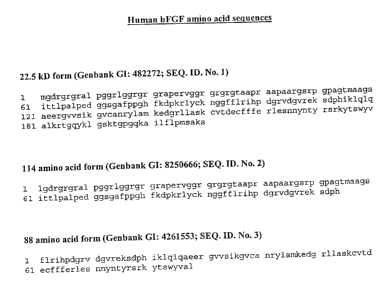
Figure 4 presents amino acid sequences for variants of bFGF (SEQ. ID. Nos.l-
3).
4 Detailed Description of the Invention
4.1 Definitions:
The term "brain cells" as used herein refers to cells comprising the brain,
including
neurons, astroglia, oligodendroglia, and microglia. Many specific cell types
belong to each
category. For example, neurons include dopaminergic, cholinergic and
glutaminergic neurons,
to name only a few.
"Bioactive compounds" is intended to include compounds with a desirable effect
when
used within the context of the invention. Bioactive compounds include many
neural stimulants
-6-
CA 02380953 2002-02-18
WO 01/12236 PCT/US00/22843
(see below) as well as many compounds that are not considered neural
stimulants but that also
have desirable effects. For example, immunosuppressants such as the
immunophilins (eg.
FK506), can exhibit the dual action of preventing rejection of the
transplanted cells and
providing a neuroprotective activity (Bavetta et al. (1999) Exp. Neurol. 158:
382-393).
Antibiotics, and particularly tetracyclines, can suppress possible infections
and also have
beneficial effects on neural cells (Yrjanheikki et al. (1998) PNAS 95: 15769-
74).
"Cell culture" refers generically to any composition of cells whether actively
growing,
differentiating, or static. Cell cultures can take on a variety of formats.
For instance, a
"suspension culture" refers to a culture in which cells are suspended in a
suitable medium. A
"continuous flow culture" refers to the cultivation of cells in a continuous
flow of fresh medium
to maintain cell growth or viability. "Continuous expansion" is a method of
growing cells by
continuous flow culture.
The "central nervous system" (CNS) as used herein, refers to any component of
the
central nervous system including the brain and spinal cord, the cells and
extracellular materials
and fluids.
"Conjoint administration" is used herein in reference to the administration of
cells and a
neural stimulant or bioactive compound to subjects. The term "conjoint
administration" is not
meant to indicate that the cells and the neural stimulant must be administered
at the same time.
The components of the conjoint administration may be delivered at different
times, at different
time intervals and by different means. The administrations should, however,
overlap in
therapeutic effects.
The term "culture medium" is recognized in the art, and refers generally to
any substance
or preparation used for the cultivation of living cells.
The term "developmental regulator" is used herein to refer to molecules that
modulate
development in brain cells or stem cells with the capacity to become brain
cells.
_7_
CA 02380953 2002-02-18
WO 01/12236 PCT/US00/22843
By "focal cerebral ischemia" as used herein in reference to the central
nervous system, is
meant the condition that results from the blockage of a single artery that
supplies blood to the
brain or spinal cord, resulting in the death of cellular elements in the
terntory supplied by that
artery.
"Global cerebral ischemia" is the diminution of blood flow to the entire
brain, often
caused by cardiac arrest or hypotension, for example. In global cerebral
ischemia, cells that are
particularly vulnerable to ischemia tend to die or become injured, resulting
in patches of damage
distributed around the brain. This differs from the type of damage that occurs
in focal cerebral
ischemia.
"Guidance molecules" are a class of proteins, normally found in the
extracellular matrix,
that function to guide cells or cellular processes (axons) to locations
required for proper
functioning. Examples are the semaphorins, the netrins, the neuropilins, and
the ephrins. (Perris
et al. (2000) Mech. Dev. 95: 3-21; Wilkinson (2000) Int. Rev. Cytol. 196: 177-
244; Van Vactor et
al. (1999) Curr. Biol. 9: 8201-4).
"Hematopoietic stem cells" (HSCs) as used herein are stem cells that can give
rise to
cells of at least one of the major hematopoietic lineages in addition to
producing daughter cells
of equivalent potential. Three major lineages of blood cells include the
lymphoid lineage, eg. B-
cells and T-cells, the myeloid lineage, eg. monocytes, granulocytes and
megakaryocytes, and the
erythroid lineage, eg. red blood cells. Certain HSCs are capable of giving
rise to many other cell
types including brain cells. "Multipotent" or "pluripotent" HSCs are HSCs that
can give rise to at
least three of the major hematopoietic lineages.
"Homology" and "identity" each refer to sequence similarity between two
polypeptide
sequences, with identity being a more strict comparison. Homology and identity
can each be
determined by comparing a position in each sequence which may be aligned for
purposes of
comparison. When a position in the compared sequence is occupied by the same
amino acid
residue, then the polypeptides can be referred to as identical at that
position; when the equivalent
_g_
CA 02380953 2002-02-18
WO 01/12236 PCT/US00/22843
site is occupied by the same amino acid (e.g., identical) or a similar amino
acid (e.g., similar in
steric and/or electronic nature), then the molecules can be referred to as
homologous at that
position. A percentage of homology or identity between sequences is a function
of the number
of matching or homologous positions shared by the sequences. An "unrelated" or
"non-
homologous" sequence shares less than 40 percent identity, though preferably
less than 25
percent identity, with the polypeptide sequence of a bioactive polypeptide of
the present
invention.
The term "ischemic episode" is used to mean any circumstance that results in a
deficient
supply of blood to a tissue. Cerebral ischemic episodes result from a
deficiency in the blood
supply to the brain. The spinal cord, which is also a part of the central
nervous system, is equally
susceptible to ischemia resulting from diminished blood flow. An ischemic
episode may be
caused by a constriction or obstruction of a blood vessel, as occurs in the
case of a thrombus or
embolus. Alternatively, the ischemic episode can result from any form of
compromised cardiac
function, including cardiac arrest.
The term "neural stimulant" refers to a treatment that affects neural function
or activity.
Such treatments are typically polypeptide growth factors, for example
neurotrophins or fibroblast
growth factors. Such treatments also include guidance molecules and non-
polypeptide molecules
that are active in the brain, such as neurotransmitters, neurotransmitter
antagonists or agonists,
and developmental regulators. "Neural stimulants" may also be agents that
affect the same
signaling transduction pathways as those affected by the above listed agents.
For example, a
chemical that activates bFGF receptor signaling could be used as a neural
stimulant. A "neural
stimulant" can also include other chemical or electromagnetic treatments that
alter the production
of molecules that affect neural function or activity (eg. transcranial
magnetic stimulation).
"Neural stem cell" (NSC) is used to describe a cell derived from tissue of the
central
nervous system, or the developing nervous system, that can give rise to at
least one of the
following fundamental neural lineages: neurons, oligodendroglia and astroglia.
Additionally, a
neural stem cell must also be able to give rise to new NSCs with similar
potential. "Multipotent"
-9-
CA 02380953 2002-02-18
WO 01/12236 PCT/US00/22843
or "pluripotent" NSCs are NSCs that are capable of giving rise to all of the
above neural lineages
as well as cells of equivalent developmental potential.
"Neuronal function" is used to refer generally to all the functions of the
nervous system,
eg. sensorimotor function and cognitive function.
"Neuroepithelial stem cells" are stem cell populations isolated from fetal
neuroepithelial
tissue. Such cells may be considered a subset of neural stem cells, as used
herein.
"Neuroeptihelial cells" tend to be multipotent.
"Neurotransmitters" are small molecules released from an axon for action
within a
synapse. Exemplary neurotransmitters include catecholamines (eg. epinephrine,
norepinephrin
and dopamine), serotonin, acetylcholine, glutamate and GABA.
A "patient" or "subject" to be treated by the subject method is a mammal,
including a
human.
As used herein, both "protein" and "polypeptide" mean any chain of amino acid
residues,
regardless of length or post-translational modification (e.g., glycosylation
or phosphorylation). A
"bioactive polypeptide", as used herein is a polypeptide that has activity as
a neural stimulant.
Examples are polypeptide growth factors and guidance molecules. "Bioactive
polypeptides" also
include active fragments and analogues of the bioactive polypeptides, which
possess one or more
the biological functions of those factors.
"Polypeptide growth factors" are generally secreted polypeptides, or active
fragments
thereof, that stimulate cell growth or growth of cell processes (eg. axons,
dendrites etc.) in at
least on cell type.
"Active fragment" as used in reference to bioactive polypeptides, indicates
any portion of
a polypeptide that has at least one activity of the full-length polypeptide.
Many polypeptides
have several different activities and it may be desirable to use an active
fragment that has only
one or a subset of these activities. The active fragment will produce at least
20%, preferably at
least 50%, more preferably at least 70%, and most preferably at least 90%
(including up to
-10-
CA 02380953 2002-02-18
WO 01/12236 PCT/US00/22843
100%) of the activity of the full-length polypeptide. An example is bFGF,
which can be
polymorphic, with observed molecular weights of 17.8, 22.5, 23.1, and 24.2
kDa; all of these
forms are biologically active and can be used in the invention.
The terms "recombinant protein", "heterologous protein" and "exogenous
protein" are
used interchangeably throughout the specification and refer to a polypeptide
which is produced
by recombinant DNA techniques, wherein generally, DNA encoding the polypeptide
is inserted
into a suitable expression construct which is in turn used to transform a host
cell to produce the
heterologous protein. That is, the polypeptide is expressed from a
heterologous nucleic acid.
A "stroke" is a sudden loss of function caused by an abnormality in the blood
supply to
the brain. Stroke presents with different levels of severity ranging from
"transient ischemic
attack" or "TIA" (no permanent disability), to "partial nonprogressing stroke"
(persistent but no
calamitous damage), to "complete stroke" (permanent, calamitous neurological
deficit). Ischemia
(diminished or stopped blood flow) and infarction (cell damage and death
within the zone of
ischemia) are the pathologic processes in stroke that lead to neurologic
deficits. "Ischemic
stroke" is caused by an obstruction of blood vessels supplying the brain. The
primary
subcategories of ischemic stroke are thrombotic stroke, embolic stroke and
lacunar infarctions.
"Hemorrhagic stroke" is caused by the rupture of blood vessels supplying the
brain. The primary
subcategories of hemorrhagic stroke are subarachnoid hemorrhage (SAH) and
intracerebral
hemorrhage (ICH).
A "therapeutically effective amount" of, eg. cells or neural stimulant, with
respect to the
subject method, refers to an amount of the therapeutic (in a preparation)
which when applied as
part of a desired dosage regimen causes an improvement in neuronal function
according to
clinically acceptable standards.
"Transcranial magnetic stimulation" (TMS) is a method for the stimulation of
neurons by
briefly generating magnetic fields with typical field strengths between 2 and
4T using coils close
to the head (currents in TMS coils can be has high as 8000A). TMS often
involves pulses of
-11-
CA 02380953 2002-02-18
WO 01/12236 PCT/US00/22843
stimulation with varying pulse and delay times. TMS is known to upregulate
monoamines in the
brain.
4.2 Overview
The present invention is based in part on the surprising finding that the
conjoint
administration of cells and neural stimulants promotes greater recovery from
CNS damage than
either treatment alone. In certain aspects, the invention provides improved
methods,
compositions and kits for stimulating recovery of damaged brain tissue,
whether damage is
localized or global. In preferred embodiments, the invention pertains to
recovery from ischemia,
hypoxia and trauma. In certain aspects, the methods of the invention comprise
the conjoint
administration of stem cells and a neural stimulant, eg. a polypeptide growth
factor or other
molecule. The conjoint treatment gives a greater degree of recovery than has
been possible with
either treatment alone. The promise of this approach was recently illustrated
in a study wherein
the polypeptide growth factor BDNF was administered conjointly with bone
marrow cells to
improve recovery in a rat stroke model (Chen et al., 2000, Neuropharmacology
39: 711-716).
The debilitating effects of CNS damage are such that even incremental
improvements in
recovery can lead to major improvements in a patient's quality of life.
The subject method has wide applicability to the treatment of CNS damage. In
this
regard, the subject method is useful for, but not limited to, treatment of
injury to the brain and
spinal cord due to ischemias, hypoxia, traumas, neurodegenerative diseases,
infectious diseases,
cancers, autoimmune diseases and metabolic disorders. Examples of disorders
include stroke,
head trauma, spinal trauma, hypotension, arrested breathing, cardiac arrest,
Rey's syndrome,
cerebral thrombosis, embolism, cerebral hemorrhage, brain tumors,
encephalomyelitis,
hydroencephalitis, operative and postoperative brain injury, Alzheimer's
disease, Huntington's
disease, Creutzfeld-Jakob disease, Parkinson's disease, multiple sclerosis and
amyotrophic
lateral sclerosis.
Thrombus, embolus, and systemic hypotension are the most common causes of
cerebral
ischemic episodes. Other causes of cerebral ischemia include hypertension,
hypertensive
cerebral vascular disease, rupture of an aneurysm, an angioma, blood
dyscrasias, cardiac failure,
cardiac arrest, cardiogenic shock, septic shock, head trauma, spinal cord
trauma, seizure,
bleeding from a tumor, or other blood loss. With respect to trauma, trauma can
involve a tissue
-12-
CA 02380953 2002-02-18
WO 01/12236 PCT/US00/22843
insult such as an abrasion, incision, contusion, puncture, compression, etc.,
such as can arise
from traumatic contact of a foreign object with any locus of or appurtenant to
the head, neck, or
vertebral column. Other forms of traumatic injury can arise from constriction
or compression of
the CNS tissue by an inappropriate accumulation of fluid (for example, a
blockade or
dysfunction of normal cerebrospinal fluid or vitreous humor fluid production,
turnover, or
volume regulation, or a subdural or intracranial hematoma or edema).
Similarly, traumatic
constriction or compression can arise from the presence of a mass of abnormal
tissue, such as a
metastatic or primary tumor.
In some cases the damage caused by the above disorders is primarily located in
a single
region of the brain, eg. focal ischemia, certain traumas and Parkinson's
disease. In other cases,
damage can be more widespread or distributed across disparate regions of the
brain, eg. hypoxia
and global ischemia, and Creutzfeld-Jakob disease. Because certain cells of
the invention are
known to migrate freely throughout the brain, and because growth factors can
be provided so as
to be generally available to all brain tissues, it is anticipated that the
methods and compositions
of the invention will be useful in promoting recovery from both global and
focal brain damage.
In a general outline, a treatment protocol of the invention involves
administering a neural
stimulant and stem cells to a patient that has suffered CNS damage. In
preferred embodiments,
CNS damage was caused by ischemia, hypoxia or trauma. Treatment may include
obtaining
cells from the patient, optionally enriching for therapeutically useful cells,
and administering the
cells to the patient. In this way, the patient is not subjected to any foreign
cells, which offers the
advantage of avoiding immune responses to the cells.
The treatment regimen according to the invention is carned out, in terms of
administration mode, timing of the administration, and dosage, so that the
functional recovery of
the patient from the adverse consequences of the central nervous system injury
is improved; for
example, the patient's motor skills (e.g., posture, balance, grasp, or gait),
cognitive skills, speech,
and/or sensory perceptions (including visual ability, taste, olfaction, and
proprioception) may
improve as result of treatment according to the invention.
While not wishing to be limited to a particular mechanism of action, it is
believed that the
methods of the invention promote recovery from CNS damage by stimulation of
neuronal
sprouting and new synapse formation. In cases of stroke, essentially all
current treatments focus
-13-
CA 02380953 2002-02-18
WO 01/12236 PCT/US00/22843
on infarct reduction and prevention of damage. Therefore, the present
invention relates to
unconventional and novel methods of treating CNS damage.
4.3 Neural stimulants and other bioactive factors
Neural stimulants of the invention include treatments, chemical or otherwise,
that affect
neural function or activity. Such treatments are typically bioactive
polypeptides, but non-
polypeptide molecules and physical treatments such as transcranial magnetic
stimulation are also
contemplated.
In one set of preferred embodiments, the neural stimulant is a polypeptide
growth factor.
The polypeptide growth factor can be administered in a pharmaceutically
acceptable carrier, and
may also be administered mixed or unmixed with cells. The polypeptide growth
factor can be a
member of the fibroblast growth factor (FGF) family; the neurotrophin family;
the insulin-like
growth factor (IGF) family; the ciliary neurotrophic growth factor (CNTF)
family; the EGF
family; the TGF-beta family; the PDGF family; the VEGF family; the leukemia
inhibitory factor
(LIF) family; an interleukin (eg. IL-11; IL-6, IL-1); or an oncostatin (eg.
oncostatin M).
Characteristics and exemplary members of each of these families are given
below and in Table 2.
In preferred embodiments the polypeptide factor is a human polypeptide factor.
The FGF family contains at least 15 distinct factors that are highly conserved
across
mammalian species, although individual family members can be highly divergent
from each
other (generally 30-70% sequence identity). FGFs are secreted proteins that
share a basic
tertiary structure composed of 12 beta-strands in a beta-trefoil fold. Most
family members have
mitogenic effects on various cell types and also bind heparin. Exemplary
members of the FGF
family include: basic FGF (bFGF, FGF-2), acid FGF (aFGF, FGF-1), FGF-3 (int-
2), FGF-4
(hst/kFGF), FGF-5, FGF-6, FGF-7 (KGF), FGF-8 (AIGF), and FGF-9 (GAF). (Stauber
et al.
(2000) PNAS 97: 49-54; Wong et al. (1998) J. Biol. Chem. 273: 18617-18622;
Szebenyi et al.
(1999) Int. Rev. Cytol. 185: 45-106).
The neurotrophin family includes several related, secreted factors that exert
their effects
primarily on the nervous system. Neurotrophins are generally produced as
precursor proteins
that are highly processed to give the mature forms. Mature neurotrophins carry
a set of six
cysteines that engage in disulfide bonding in the order 1-4 (ie. the first and
fourth cysteines form
a disulfide bond), 2-5 and 3-6. Typically, neurotrophin family members have a
surface
-14-
CA 02380953 2002-02-18
WO 01/12236 PCT/US00/22843
composed of 3 antiparallel beta-strands, and dimerization occurs along this
surface. Exemplary
members of the neurotrophin family are: nerve growth factor (NGF), brain-
derived neurotrophic
factor (BDNF), neurotrophin 3 (NT3), neurotrophin 4/5 (NT4/5) and neurotrophin
6. (Lewin et
al. (1996) Annu. Rev. Neuroscience 19:289-317).
The insulin-like growth factor family includes secreted proteins with a
sequence and
structure similar to that of insulin and a molecular weight typically in the
range of 5-10 kDa.
These factors can be found in the bloodstream, usually associated with one of
six IGF binding
proteins. Exemplary members of the family include IGF-1 and IGF-2. IGF-1 and -
2 are known
to promote recovery from various insults to the CNS. (Daughaday et al. (1989)
Endocr. Rev. 10:
68-91; Rajaram et al. (1997) Endocr. Rev. 18: 801-831; Jones et al. (1995)
Endocr. Rev. 16: 3-
34).
The epidermal growth factor family is a large family of related secreted
factors.
Members of the EGF family share at least 30% sequence homology and a set of
six conserved
cysteine residues in the C-terminal end of the protein. Most such proteins
also contain an EGF-
like domain, which is a particularly well-characterized domain that is also
present in many non-
EGF family member proteins. EGF family members are normally processed from
larger
precursors. Exemplary members of the EGF family include EGF, TGF-alpha, HB-EGF
(heparin-binding EGF), amphiregulin, betacellulin, vaccinia growth factor and
neu
differentiation factor. (Aviezer et al. (1994) 91: 12173-12177; Higashyama et
al. (1992) J. Biol.
Chem. 267: 6205-6212; Pelles et al. (1992) Cell 69:205-216).
The TGF-beta superfamily is an important class of molecules involved in cell-
cell
signaling and development in a wide range of organisms and cell types. Members
of the family
are initially synthesized as larger precursor molecules with an amino-terminal
signal sequence
and a pro-domain of varying size (Kingsley, D.M. (1994) Genes Dev. 8:133-146).
The precursor
is then cleaved to release a mature carboxy-terminal segment of 110-140 amino
acids. The
active signaling moiety is comprised of hetero- or homodimers of the carboxy-
terminal segment
(Massague, J. (1990) Annu. Rev. Cell Biol. 6:597-641). The active form of the
molecule then
interacts with its receptor, which for this family of molecules is composed of
two distantly
related transmembrane serine/threonine kinases called type I and type II
receptors (Massague, J.
et al. (1992) Cell 69:1067-1070; Miyazono, K. A. et al. EMBO J. 10:1091-1101).
TGF-beta
binds directly to the type II receptor, which then recruits the type I
receptor and modifies it by
-15-
CA 02380953 2002-02-18
WO 01/12236 PCT/US00/22843
phosphorylation. The type I receptor then transducer the signal to downstream
components
(Wrana et al, (1994) Nature 370:341-347). In general, members of the TGF-beta
superfamily
have a set of nine highly conserved cysteine residues that are involved in
disulfide bonding both
within and between monomers of the mature, dimerized signaling protein
(Griffith et al. (1996)
PNAS 93: 878-883; Luo et al. (1995) PNAS 92: 11761-11765; Schlunegger et al.
(1993) J. Mol.
Biol. 231: 445-58; Daopin et al. (1993) Proteins 17: 176-92; Murray-Rust et
al. (1993) Structure
15: 153-9; Archer et al. (1993) Biochemistry 32: 1164-71; Daopin et al. (1992)
Science 257: 369-
373; Schlunegger et al. (1992) Nature 358: 430-434; Hinck et al. (1996)
Biochemistry 35: 8517-
34; Mittl et al. ( 1996) Protein Sci. 5 :1261-71 ).
The transforming growth factor beta family is a very large family of proteins
including
the TGF-beta subfamily, the bone morphogenesis protein (BMP) subfamily, the
activin
subfamily, and others. Exemplary members of the TGF-beta subfamily include TGF-
beta-l, -2, -
3, -4 and -5. Exemplary members of the BMP subfamily include osteogenic
protein 1 (OP-l,
BMP-7) and BMP-9. (Ren et al. (2000) Neuropharmacology 39: 860-865; Lopez-
Coviella et al.
(2000) Science 289: 313-316; Withers et al. (2000) Eur. J. Neurosci. 12: 106-
116).
The vascular endothelial growth factor (VEGF) family is a group of secreted
proteins that
act as potent mitogens in embryonic and somatic angiogenesis. VEGF proteins,
including VEGF
itself, bind to cell surface receptors of the kinase domain receptor family
(KDR) and fins-like
tyrosine kinase group (Flt receptors). VEGF proteins form a homodimer with a
cystine knot
structure. Platelet-derived growth factor (PDGF) shares only limited sequence
similarity with
VEGF (19%) but has substantial structural similarity. PDGF and related family
members are
also cystine knot proteins and bind to their receptors in a similar manner.
(Lobsiger et al. (2000)
Glia 30: 290-300; Sun et al. (1995) Annu. Rev. Biophys. Biomolec. Struct. 24:
269-291; Muller et
al. (1997) Structure 5: 1325-1338; Jiang et al. (2000) EMBO J. 19: 3192-3203;
Muller et al.
( 1997) PNAS 94: 7192-7197).
Interleukins are secreted polypeptide factors that mediate signaling between
immune
cells. Many interleukins are known to have effects on the brain, particularly
IL-la and (3, IL-6
and IL-11. (Van Wagoner et al. (1999) J. Neuroimmunol. 100:124-139; Ling et
al. (1998) Exp.
Neurol. 149: 411-23; Mehler et al. (1993) Nature 362: 62-5). Intriguingly, IL-
6 and IL-11 both
act in part through a receptor protein gp130 that acts as a receptor for
ciliary neurotrophic factor
(CNTF), leukemia inhibitory factor (LIF) and oncostatin M. Thus all these
factors may have
-16-
CA 02380953 2002-02-18
WO 01/12236 PCT/iJS00/22843
similar roles in modulating neuronal function and development. (Benigni et al.
(1995) Mol. Med.
1: 568-75; Benigni et al. (1996) Blood 87: 1851-4; Murphy et al. (1997) Prog.
Neurobiol. 52:
355-78).
Table 1: Polypeptide Growth Factors
Family Exemplary subfamiliesExemplary Members
FGF bFGF, aFGF, FGF-3, FGF-4, FGF-5,
FGF-6,
FGF-7, FGF-8, FGF-9
Neurotro bins NGF, BDNF, NT3, NT4/5, NT-6
IGF IGF-1, IGF-2
EGF EGF, TGF-alpha, HB-EGF, amphiregulin,
betacellulin, vaccina owth factor
and neu
TGF-beta TGF-beta TGF-beta-1, -2, -3, -4 and -S
BMP OP-1, BMP-9
Activin Inhibin A, Inhibin B and Inhibin
C
VEGF VEGF
PDGF PDGF
LIF LIF
CNTF CNTF
Interleukins IL-la, IL-1 , IL-6, IL-11
Oncostatins Oncostatin M
Furthermore, the nomenclature in the field of polypeptide factors is complex,
primarily
because many factors have been isolated independently by different groups of
researchers and,
historically, named for the type of tissue that was used as an assay in the
process of purifying the
-17-
CA 02380953 2002-02-18
WO 01/12236 PCT/US00/22843
factor. Basic FGF has been referred to in scientific publications by a number
of different names,
and has multiple family members. These include leukemic growth factor,
macrophage growth
factor, embryonic kidney-derived angiogenesis factor 2, prostatic growth
factor, astroglial
growth factor 2, endothelial growth factor, chondrosarcoma growth factor,
cartilage-derived
growth factor 1, eye-derived growth factor l, heparin-binding growth factors
class 11, myogenic
growth. factor, human placenta purified factor, uterine-derived growth factor,
embryonic
carcinoma- derived growth factor, human pituitary growth factor, adipocyte
growth factor,
prostatic osteoblastic factor, and mammary tumor-derived growth factor. Thus,
any factor
referred to by one of the aforementioned names is considered within the scope
of the invention.
Furthermore, effort has been made to use commonly accepted names for factors,
and any factor
listed here is considered within the scope of the invention regardless of
whether it is known to
others by a different name.
The invention can also employ bioactive analogues of the aforementioned growth
factors,
which possess one or more of the biological functions of those factors. An
example is bFGF,
which can be polymorphic, with observed molecular weights of 17.8, 22.5, 23.1,
and 24.2 kDa;
all of these forms are biologically active and can be used in the invention.
It is possible to
identify bioactive analogues of the aforementioned factors. Such analogues,
when designed to
retain at least one activity of a naturally occurnng form of the polypeptide,
are considered
functional equivalents. Bioactive analogues may also include molecules that
are not
polypeptides but nonetheless mimic activities of a polypeptide growth factor.
Bioactive
analogues may also have advantageous properties, such as enhanced efficacy or
more desirable
stability properties (e.g., ex vivo shelf life and resistance to proteolytic
degradation in vivo). For
example, the analogue may be rendered either more stable or less stable to
proteolytic
degradation or other processes which result in destruction of, or otherwise
inactivation of, the
factor. A short half life can give rise to more transient biological effects
can therefore allow
tighter control of protein levels within or around a particular tissue. A
longer half life can
increase the potency of the factor.
In certain embodiments, bioactive polypeptides of the invention comprise a
polypeptide
with an amino acid sequence that is at least 30% identical to the bFGF
sequence set forth in one
of SEQ. ID. Nos. 1-3. In preferred variations, such bioactive polypeptides
comprise a
-18-
CA 02380953 2002-02-18
WO 01/12236 PCT/~JS00/22843
polypeptide with an amino acid sequence that is at least 40%, 50%, 60%, 70%,
80%, 90%, 95%,
98%, 99% or 100% identical to one of SEQ. ID. Nos.l-3.
Methods for generating such bioactive analogues are well known in the art. In
general,
variations of a polypeptide factor can be generated by introducing changes
into a nucleic acid
sequence encoding the factor. The altered nucleic acid can then be expressed
to produce altered
polypeptides, and the polypeptides can be assayed for various properties.
Changes in nucleic
acid sequences can be made individually to introduce particular, desired
changes. Alternatively,
libraries of semi-randomly generated variants may be produced and screened for
activity.
There are many ways by which a library of potential bioactive analogs can be
generated.
In an illustrative embodiment, the amino acid sequences for a population of
bFGF homologs or
other related proteins are aligned, preferably to promote the highest homology
possible. Such a
population of variants can include, for example, bFGF homologs from one or
more species, e.g.
murine and chicken, or bFGF homologs from the same species but which differ
due to mutation.
Amino acids which appear at each position of the aligned sequences are
selected to create a
degenerate set of combinatorial sequences. In a preferred embodiment, the
variegated library of
bFGF variants is generated by combinatorial mutagenesis at the nucleic acid
level, and is
encoded by a variegated gene library. For instance, a mixture of synthetic
oligonucleotides can
be enzymatically ligated into gene sequences such that the degenerate set of
potential bFGF
sequences are expressible as individual polypeptides, or alternatively, as a
set of larger fusion
proteins (e.g. for phage display) containing the set of bFGF sequences
therein.
Chemical synthesis of a degenerate gene sequence can be carried out in an
automatic
DNA synthesizer, and the synthetic genes then ligated into an appropriate
expression vector.
The purpose of a degenerate set of genes is to provide, in one mixture, all of
the sequences
encoding the desired bioactive analogs. The synthesis of degenerate
oligonucleotides is well
known in the art (see for example, Narang, SA (1983) Tetrahedron 39:3; Itakura
et al. (1981)
Recombinant DNA, Proc 3rd Cleveland Sympos. Macromolecules, ed. AG Walton,
Amsterdam:
Elsevier pp273-289; Itakura et al. (1984) Annu. Rev. Biochem. 53:323; Itakura
et al. (1984)
Science 198:1056; Ike et al. (1983) Nucleic Acid Res. 11:477. Such techniques
have been
employed in the directed evolution of other proteins (see, for example, Scott
et al. (1990) Science
249:386-390; Roberts et al. (1992) PNAS 89:2429-2433; Devlin et al. (1990)
Science 249: 404-
-19-
CA 02380953 2002-02-18
WO 01/12236 PCT/US00/22843
406; Cwirla et al. (1990) PNAS 87: 6378-6382; as well as U.S. Patents Nos.
5,223,409,
5,198,346, and 5,096,815).
Alternatives to the above combinatorial mutagenesis also exist. For example,
bFGF
analogs can be generated, for example, alanine scanning mutagenesis and the
like (Ruf et al.
(1994) Biochemistry 33:1565-1572; Wang et al. (1994) J. Biol. Chem. 269:3095-
3099; Balint et
al. (1993) Gene 137:109-118; Grodberg et al. (1993) Eur. J. Biochem. 218:597-
601; Nagashima
et al. (1993) J. Biol. Chem. 268:2888-2892; Lowman et al. (1991) Biochemistry
30:10832-
10838; and Cunningham et al. (1989) Science 244:1081-1085), by linker scanning
mutagenesis
(Gustin et al. (1993) Virology 193:653-660; Brown et al. (1992) Mol. Cell
Biol. 12:2644-2652;
McKnight et al. (1982) Science 232:316); by saturation mutagenesis (Meyers et
al. (1986)
Science 232:613); by PCR mutagenesis (Leung et al. (1989) Method Cell Mol Biol
1:11-19); or
by random mutagenesis (Miller et al. (1992) A Short Course in Bacterial
Genetics, CSHL Press,
Cold Spring Harbor, NY; and Greener et al. (1994) Strategies in Mol Biol 7:32-
34).
The above methods may be generalized to other polypeptide factors in addition
to bFGF.
Having generated one or more variants of a bioactive factor, various methods
may be
used to identify variants with the desired properties. Whether one or more
changes in the amino
acid sequence of a peptide results in a bioactive analog can be readily
determined by assessing
the ability of the variant peptide to produce a response in cells in a fashion
similar to the wild-
type peptide or competitively inhibit such a response. In addition, the
ability of such a
polypeptide to bind to its receptor can also be determined. For example, bFGF
normally binds to
the receptors FGFR1 and FGFR2. This binding is also stimulated by heparin
binding. These
properties could be checked to verify that a bFGF variant is active.
A wide range of techniques are known in the art for screening gene products of
combinatorial libraries, and for screening cDNA libraries for gene products
having a certain
property. The most widely used techniques for screening large gene libraries
typically comprise
cloning the gene library into replicable expression vectors, transforming
appropriate cells with
the resulting library of vectors, and expressing the combinatorial genes under
conditions in
which detection of a desired activity facilitates relatively easy isolation of
the vector encoding
the gene whose product was detected. Each of the illustrative assays described
below are
amenable to high through-put analysis as necessary to screen large numbers of
variant sequences
created by combinatorial mutagenesis techniques.
-20-
CA 02380953 2002-02-18
WO 01/12236 PCT/US00/22843
In one possible screening assay, the gene library is expressed as a fusion
protein on the
surface of a viral particle. For instance, in the filamentous phage system,
foreign peptide
sequences can be expressed on the surface of infectious phage. These phage can
be applied to
affinity matrices at very high concentrations, allowing screening of a large
number of phage
simultaneously. If a particular phage is recovered from an affinity matrix in
low yield, the phage
can be amplified by another round of infection in a suitable host, such as E.
coli. The group of
almost identical E. coli filamentous phages M13, fd., and fl are most often
used in phage display
libraries, as either of the phage gIII or gVIII coat proteins can be used to
generate fusion proteins
without disrupting the ultimate packaging of the viral particle (Ladner et al.
PCT publication WO
90/02909; Garrard et al., PCT publication WO 92/09690; Marks et al. (1992) J.
Biol. Chem.
267:16007-16010; Griffiths et al. (1993) EMBO J 12:725-734; Clackson et al.
(1991) Nature
352:624-628; and Barbas et al. (1992) PNAS 89:4457-4461).
In another embodiment, the combinatorial library is designed to be
extracellularly
presented (e.g. as it occurs naturally) or optionally, secreted (e.g. the
polypeptides of the library
all include a signal sequence). The library can be transfected into a
eukaryotic cell that can be
co-cultured with cells which express a functional receptor for the desired
bioactive fragment.
For example, one might use cells expressing a bFGF receptor to identify
bioactive variants of
bFGF. Bioactive analogs secreted by the cells expressing the combinatorial
library will diffuse
to neighboring receptor positive cells and induce a phenotypic change.
Phenotypic changes may
be detected using, for example, antibodies directed to epitopes that are
either created or
destroyed in response to factor treatment.
Each of these analogs can subsequently be screened for further biological
activities. For
example, receptor-binding analogs isolated from the combinatorial library can
be tested for their
effect on cellular proliferation relative to the wild-type form of the
protein. Alternatively, one
could screen the analogs for stability in vitro or in vivo. The activity of
such analogs can also be
assessed in animal models. For example, the ability of an analog to improve
neural function in a
a rat stroke model could be assessed to verify that an analog has the
appropriate bioactivity.
Many different types of mutations can give rise to bioactive analogs. For
example,
conservative changes in the amino acid sequence can be expected to give rise
to analogues that
retain one or more bioactivity. It is reasonable to expect that an isolated
replacement of a leucine
with an isoleucine or valine, an aspartate with a glutamate, a threonine with
a serine, or a similar
-21-
CA 02380953 2002-02-18
WO 01/12236 PCT/US00/22843
replacement of an amino acid with a structurally related amino acid (i.e.
conservative mutations)
will not have a major effect on the biological activity of the resulting
molecule. Conservative
replacements are those that take place within a family of amino acids that are
related in their side
chains. Genetically encoded amino acids are can be divided into four families:
(1) acidic =
aspartate, glutamate; (2) basic = lysine, arginine, histidine; (3) nonpolar =
alanine, valine,
leucine, isoleucine, proline, phenylalanine, methionine, tryptophan; and (4)
uncharged polar =
glycine, asparagine, glutamine, cysteine, serine, threonine, tyrosine.
Phenylalanine, tryptophan,
and tyrosine are sometimes classified jointly as aromatic amino acids. In
similar fashion, the
amino acid repertoire can be grouped as (1) acidic = aspartate, glutamate; (2)
basic = lysine,
arginine histidine, (3) aliphatic = glycine, alanine, valine, leucine,
isoleucine, serine, threonine,
with serine and threonine optionally be grouped separately as aliphatic-
hydroxyl; (4) aromatic =
phenylalanine, tyrosine, tryptophan; (5) amide = asparagine, glutamine; and
(6) sulfur -
containing = cysteine and methionine. (see, for example, Biochemistry, 2nd
ed., Ed. by L.
Stryer, WH Freeman and Co.: 1981).
In other embodiments, chemically modified bioactive factors are contemplated.
A
polypeptide may be chemically modified to create derivatives by forming
covalent or
aggregrative conjugates with other chemical moieties, such as glycosyl groups,
lipids, phosphate,
acetyl groups and the like. Covalent derivatives may be prepared by linking
the chemical
moieties to functional groups on amino acid side chains or at the N-terminus
or at the C-terminus
of the polypeptide. For instance, a bioactive factor can be generated which
includes a moiety,
other than sequences naturally associated with the protein, that binds a
component of the
extracellular matrix and enhances localization of the analog to cell surfaces.
For example,
sequences derived from the fibronectin "type-III repeat", such as a
tetrapeptide sequence R-G-D-
S (Pierschbacher et al. (1984) Nature 309:30-3; and Kornblihtt et al. (1985)
EMBO 4:1755-9)
can be added to a polypeptide factor to support attachment of the chimeric
molecule to a cell
through binding ECM components (Ruoslahti et al. (1987) Science 238:491-497;
Pierschbacheret
al. (1987) J. Biol. Chem. 262:17294-8.; Hynes (1987) Cell 48:549-54; and Hynes
(1992) Cell
69:11-25).
Alternatively, polypeptide growth factors useful in the invention can consist
of active
fragments of the factors. The activity of any given fragment can be readily
determined in by
methods such as those described above. For example, a fragment of bFGF that,
when
-22-
CA 02380953 2002-02-18
WO 01/12236 PCT/US00/22843
administered according to the methods of the invention described herein, is
shown to improve
performance in functional tests that is comparable to the performance that is
produced by
administration of the full-length bFGF polypeptide, would be an "active
fragment" of bFGF.
Such active fragments are described, e.g., in Baird and Gage (1997) Proc.
Natl. Acad. Sci.
U.S.A., 94 (13): 7047-52. It is well within the abilities of skilled artisans
to determine whether a
polypeptide growth factor, regardless of size, retains the functional activity
of a full length, wild-
type polypeptide growth factor.
The polypeptide factors useful in the invention are preferably substantially
purified from
their source material, be it cell culture, tissue sample, biological fluid,
etc. Substantially purified
means that the purified material is at least 60% by weight (dry weight) the
polypeptide of
interest, e.g., a bFGF polypeptide. Preferably, the polypeptide composition is
at least 75%, more
preferably at least 90%, and most preferably at least 99%, by weight, the
polypeptide of interest.
Purity can be measured by any appropriate standard method, e.g., column
chromatography,
polyacrylamide gel electrophoresis, or HPLC analysis. Substantially purified
polypeptides can
then be combined with other desired components, such as Garners or cells, to
give a composition
that is less than 60% composed of polypeptide, so long as the polypeptide is
at sufficient
concentration to be effective when administered to a patient.
The polypeptide factors useful in the invention can be naturally occurring,
synthetic, or
recombinant molecules consisting of a hybrid or chimeric polypeptide with one
portion, for
example, being bFGF, and a second portion being a distinct polypeptide. These
factors can be
purified from a biological sample, chemically synthesized, or produced
recombinantly by
standard techniques (see. e.g., Ausubel et al., Current Protocols in Molecular
Biology, New
York, John Wiley and Sons, 1993; Pouwels et al., Cloning Vectors: A Laboratory
Manual, 1985,
Suppl. 1987).
Although polypeptide growth factors are currently most preferred for use in
combination
with the cells according to the invention, other treatment modalities are
considered neural
stimulants that can be combined with cells according to the invention as well.
For example,
transcranial magnetic stimulation upregulates monoamines in the brain and is
therefore expected
to have beneficial effects in conjoint administration with cells.
-23-
CA 02380953 2002-02-18
WO 01/12236 PCT/US00/22843
One group of non-polypeptide neural stimulants that can be used as neural
stimulants are
neurotransmitter agonists or antagonists. Examples are antidepressants such as
Prozac,
amphetamines, Ritalin, and tricyclic antidepressants such as Elavil.
Other useful molecules are differentiation factors such as retinoic acid which
are capable
of priming cells to differentiate into functioning neurons.
Another class of molecules is the so-called guidance molecules, which are a
class of
proteins, normally found in the extracellular matrix, that function to guide
cells or cellular
processes (axons) to locations required for proper functioning. Examples are
the semaphorins,
the netrins, the neuropilins, and the ephrins.
In addition to the above neural stimulants, all of which have well-established
effects on
the brain, it is anticipated that other bioactive compounds that are not
considered neural
stimulants might be useful in combination with cells. These alternative
compounds are generally
compounds with well-known effects on other parts of the body with more
recently discovered
effects on cells of the CNS.
One group of alternative compounds includes immunosuppressant molecules that
are
currently used to inhibit rejection of allografts. A preferred class of such
molecules are the
immunophilins, such cyclosporin, FK506, and thalidomide. These molecules can
exhibit dual
action of preventing rejection of the transplanted cells and providing
neuroprotective function.
Another group of alternative stimulants is the tetracyclines, classically
known for their antibiotic
effects, but also possessing desirable neuroprotective effects.
4.4 Cells
Many different cell types, or mixtures thereof, may be administered to a
subject. While
not wishing to be limited by theory, it is postulated that administered cells
may affect the brain in
multiple ways. Cells may themselves become established in the brain and form
functional
connections with neurons. Additionally or alternatively, cells may produce
factors that stimulate
the endogenous nerve cells to form new processes and connections. Finally,
cells might act to
scavenge or otherwise remove or inactivate compounds that inhibit recovery
from CNS damage.
In view of these possibilities, it is understood that essentially any cell
possessing one of the
-24-
CA 02380953 2002-02-18
WO 01/12236 PCT/US00/22843
above qualities, and particularly stem cells but potentially even terminally
differentiated cells,
might have beneficial effects on brain function. Examples of terminally
differentiated cell types
that are known to have beneficial scavenging capabilities are activated
lymphocytes and
macrophages.
In certain embodiments, the cells of the invention are preferably stem cells
that have the
capability of giving rise to brain cells in vivo. Particularly preferred cells
are multipotential stem
cells. Such cells can be grown in vitro for clinical use. In preferred
embodiments, stem cell
types that can be used in the invention include neural stem cells,
hematopoietic stem cells,
embryonic stem cells, teratocarcinoma cell lines, and other stem cell types.
The term "stem cell" as used herein refers to cells with the capacity for
unlimited or
prolonged self renewal that can give rise to more than one type of more
differentiated
descendant. Preferred stem cells can undergo at least 10 cell divisions (under
appropriate
conditions) and still maintain stem cell characteristics. Particularly
preferred stem cells can
undergo at least 25, 50 or 100 rounds of division without losing stem cell
characteristics. With
respect to cells, the terms "give rise to" and "produce" are used to mean not
just the immediate
daughter cells, but all the cells that can eventually trace ancestry to that
cell. "Give rise to" and
"produce" also refer to changes in cell type that might occur without a cell
division event. Some
differentiated cells also have the capacity to give rise to cells of greater
developmental potential.
Such capacity may be natural under particular circumstances, or may be induced
artificially upon
treatment with various factors. In either case, the cells may be considered a
type of stem cell for
the purposes of the invention. Such stem cells may be referred to as "induced
stem cells" or
"differentiated stem cells". "Processed stem cells" refers to stem cells that
have been in any way
disturbed from their natural cellular environment. This includes
centrifugation, dissociation,
dispersion or other processing. The stem cells contained in an unprocessed
tissue sample are not
considered "processed stem cells".
Stem cells are usually rare cell types mixed with other, more differentiated
cells. For the
purposes of the invention, it is possible to use cell suspensions that
comprise only a minority of
stem cells. Such an approach is particularly useful with cells derived from a
stem cell rich tissue,
eg. bone marrow. In preferred embodiments, stem cells are enriched such that
they are at least
50% pure, meaning that at least 50% of the cells are stem cells at the time of
administration to a
-25-
CA 02380953 2002-02-18
WO 01/12236 PCT/US00/22843
subject. In particularly preferred embodiments, stem cells are at least 60%,
70%, 80% or 90%
pure.
4.4.1 General methods for stem cell culture and propagation
Various techniques may be employed to isolate the stem cells of the invention.
Typically, stem cells will be obtained from a tissue sample (eg. blood, bone
marrow, fetal or
adult brain tissue, etc.) wherein the desired stem cells constitute a small
percentage of the cells
present. In preferred embodiments, the tissue sample is dissociated into a
cell suspension and
optionally, various methods are used to enrich for stem cells. Preferred
procedures for
dissociation of the tissue sample are ones that result in as little cell death
as possible. For
example, stem cells can be dissociated from tissue samples by mechanical
means, e.g.,
mechanically sheared off with a pipette. In other instances, it will be
possible to dissociate the
stem cells from the surrounding tissue by enzymatic digestion. Fluid tissue
samples, such as
blood, can be fractionated by centrifugation and resuspension of certain
fractions, if appropriate.
Separation of different cell types and extracellular materials may also be
achieved by
centrifugation or settling in a density gradient of, for example Ficoll. Stem
cell populations may
be enriched based on their tendency for continued cell growth as well as
specific cellular
markers, e.g., using affinity separation techniques or fluorescence activated
cell sorting (FACS).
There are a large number of culture media that exist for culturing cells from
animals.
Some of these are complex and some are simple. While it is expected that stem
cells may grow
in complex media, it will generally be preferred that the explants be
maintained in a simple
medium, such as Dulbecco's Minimal Essential Media (DMEM), in order to allow
more precise
control over the activation of certain cell populations in a tissue sample.
The cultures may be
maintained in any suitable culture vessel, such as a 12 or 24 well microplate,
and may be
maintained under typical culture conditions for cells isolated from the same
animal, e.g., such as
37°C in 5% C02. The cultures may be shaken for improved aeration, the
speed of shaking being,
for example, 12 rpm.
In general, stem cells can be enriched by detecting and sorting based on
identifying
characteristics of the desired cells. For example, monoclonal antibodies are
particularly useful
for identifying markers (surface membrane proteins, e.g., receptors)
associated with particular
cell lineages and/or stages of differentiation. Procedures for separation of
the subject progenitor
-26-
CA 02380953 2002-02-18
WO 01/12236 PCT/US00/22843
cell may include magnetic separation, using antibody coated magnetic beads,
affinity
chromatography, and "panning" with antibody attached to a solid matrix, e.g.,
plate, or other
convenient technique. Techniques providing accurate separation include
fluorescence activated
cell sorting, which can have varying degrees of sophistication, e.g., a
plurality of color channels,
low angle and obtuse light scattering detecting channels, impedance channels,
etc.
Antibodies may be conjugated with markers, such as magnetic beads, which allow
for
direct separation, biotin, which can be removed with avidin or streptavidin
bound to a support,
fluorochromes, which can be used with a fluorescence activated cell sorter, or
the like, to allow
for ease of separation of the particular cell type. Any technique may be
employed which is not
unduly detrimental to the viability of the cells.
In addition to using antibodies, it is possible to use other proteins that
bind to the surface
of desired cells. For example, if a desired cell specifically expresses the
EGF receptor, then
labeled EGF could be used to detect those cells in much the same way as
described for the
antibodies above. Certain dyes also stain particular cell populations and can
be used as part of a
method for obtaining the desired cells. Stem cells also typically have a
distinctive morphology.
Stem cells usually have a large nucleus with a relatively small amount of
cytoplasm.
The selection methods described above may be combined with the use of
selective
growth conditions to provide further enrichment. For example, natural and
recombinantly
engineered cells can be provided as feeder layers to the instant cultures.
Such cells can also
produce an extracellular matrix that can be used as a substrate for selection
methods.
It is also possible to contact cell mixtures with an agent that causes
proliferation of one or
more populations of cells. For instance, a mitogen, e.g., a substance that
induces mitosis and cell
transformation of a particular stem cell type can be used to cause the
amplification of that
population. In this way, cells that are not responsive to the particular
factor tend not to divide
while those that are responsive divide and become a greater proportion of the
cell population.
After enrichment it is important to verify that cells obtained have the
appropriate
characteristics. Cells of the present invention can be characterized based on
responsiveness to
growth factors, specific gene expression, antigenic markers on the surface of
such cells, dye
staining and/or basic morphology. It is also valuable to determine the types
of cells that a
particular stem cell population can give rise to.
-27-
CA 02380953 2002-02-18
WO 01/12236 PCT/US00/22843
Stem cells can be induced to differentiate into various cell types by changing
the
environmental conditions. For example, the subject progenitor cells can be
recombined with the
corresponding embryonic tissue to see if the embryonic tissue can instruct the
adult cells to
codevelop and codifferentiate. Stem cells can be implanted into one of a
number of regeneration
models used in the art, e.g., neural stem cells will colonize and
differentiate in the brain of a rat
that has been lesioned (Gage et al. (1995) Proc. Natl. Acad. Sci. USA, 92:
11879-11883; Flax et
al. (1998) Nature Biotechnology 16: 1033-1039). Stem cells may be genetically
labeled by
transfection with a piece of foreign DNA. This labeling allows identification
of stem cell
descendants from among the host cells. Alternatively, the progenitor cells can
be contacted with
one or more growth or differentiation factors which can induce differentiation
of the cells.
Differentiated cell types can be identified using the same general methods
used to identify stem
cells, eg. cell surface marker, dye staining etc.
In certain situations it is desirable to measure cell proliferation. Such
methods most
commonly include determining DNA synthesis characteristic of cell replication.
There are
numerous methods in the art for measuring DNA synthesis, any of which may be
used according
to the invention. In an embodiment of the invention, DNA synthesis has been
determined using
a radioactive label (3H-thymidine) or labeled nucleotide analogues (BrdU) for
detection by
immunofluorescence.
Growth factors may also be provided in the medium to selectively expand
certain cell
populations or to encourage the production of differentiated cell types.
Cells can be sorted by positive and negative selection. For example, positive
or negative
selection may be achieved by using one or more biotinylated antibodies,
specific for factors on
the surface of the target cells. The biotinylated antibodies are introduced
into the cell culture.
After a specified incubation time any biotinylated antibodies which have not
formed a complex
with the target cells are rinsed away. Immobilized avidin matrix is then added
to the cell
suspension. The immobilized avidin matrix binds to the biotinylated
antibody/antigen complex.
This suspension can then be centrifuged to separate the avidin matrix.
Alternatively, the avidin
may be coupled to magnetic beads such that the cells bound to the antibody are
magnetically
separated from unbound cells. If the selection is positive, cells bound to the
antibody are
resuspended in nutrient medium for continued growth. If the selection is
negative, bound cells
may be disposed of, while the remaining unbound cells are resuspended for
further growth.
-28-
CA 02380953 2002-02-18
WO 01/12236 PCT/US00/22843
Clearly, many other techniques may be utilized for both positive and negative
selection,
as long as the desired affinity is provided by the selection element.
Hematopoietic stem cells
Mammalian blood cells provide for an extraordinarily diverse range of cell
types. Three
major lineages of blood cells include the lymphoid lineage, eg. B-cells and T-
cells, the myeloid
lineage, eg. monocytes, granulocytes and megakaryocytes, and the erythroid
lineage, eg. red
blood cells. Hematopoietic stem cells (HSCs) are cells that can give rise to
cells of at least two
of the above lineages in addition to producing daughter cells of equivalent
multipotency. In
preferred embodiments, the HSCs can give rise to three major blood cell
lineages. In addition to
giving rise to blood cells, HSCs are capable of differentiating into many
other cell types,
including brain cells (Eglitis and Mezey (1997) Proc. Natl. Acad. Sci. USA,
94: 4080-4085).
HSCs can be isolated from a variety of tissue types. Bone marrow cells are a
good source
of HSCs. Bone marrow cells may be obtained from a source of bone marrow, e.g.,
iliac crests,
tibiae, femora, spine, or other bone cavities. Other sources of human
hematopoietic stem cells
include embryonic yolk sac, fetal liver, fetal and adult spleen and blood,
including adult
peripheral blood.
HSCs can be identified both by the types of cells they give rise to and by
various
cytological markers. HSCs often extrude certain dyes, such as Hoechst 33324
and Rhodamine
123 (Bhatia et al. (1998) Nature Med. 4:1038). Such dye staining properties
can be used to
identify HSCs among other cells of the circulatory system. Antibodies that
react with certain cell
markers can also be used to identify and purify HSCs. For example, mAb AC133
is thought to
specifically bind to HSCs (Miraglia et al. (1997) Blood 90:5013). The Thy-1
molecule is a highly
conserved protein present in the brain and hematopoietic system of rat, mouse
and man. The
Thy-1 molecule has been identified on rat, mouse and human HSCs and can be
useful in
identifying HSCs (U.S. Patent No. 5,914,108). Many HSCs are CD34+ and/or CD38+
as well
(U.S. Patent No. 5,840,580). A population of HSCs will often have some
variation in cell
surface markers and a positive identification may be made on the basis of the
presence of at least
two of the above cytological markers.
HSCs can also be distinguished from other more differentiated cell types by
the absence
of certain markers. CD3, CD7, CDB, CD10, CD14, CD15, CD19, CD20 and CD33 are
all
-29-
CA 02380953 2002-02-18
WO 01/12236 PCT/US00/22843
typically absent from HSCs. The absence of several of the above markers adds
confidence to the
identification of HSCs. Morphology may also help distinguish an HSC, as
described above.
It is understood that HSCs may be identified by an aggregation of multiple
traits, such as
morphology, the presence of certain markers, the absence of other markers, and
the types of cells
that the putative HSCs can give rise to. A positive identification does not
typically require
detection of all of the above markers.
The culturing of HSCs to give rise to differentiated stem cells can be
achieved in many
ways. For example, cells may be cultured in a defined, enriched medium such as
Iscove's
Modified Dulbecco's Medium (IMDM), generally composed of salts, amino acids,
vitamins,
antibiotics and fetal calf serum. Cultures supplemented with hydrocortisone
tend to give rise to
myeloid cells, while cultures lacking cortisone tend to give rise to B
lymphocytes. To
demonstrate that HSCs can develop in cells of the erythroid lineage, various
conventional
methods can be used. For example culturing on methylcellulose culture can
stimulate formation
of erythroid cells. (U.S. Patent Nos. 5,840,580 and 5,914,108; Metcalf (1977)
In: Recent Results
in Cancer Research 61. Springer-Verlag Berlin, pp. 1-227).
Neural Stem Cells
Neural stem cells are cells derived from tissue of the adult or developing
nervous system
that can differentiate into at least one of the following fundamental neural
lineages: neurons,
oligodendroglia and astroglia. Additionally, neural stem cells can also give
rise to new NSCs
with similar potential. In preferred embodiments, neural stem cells are
multipotential and give
rise to cells of most or all of the fundamental neural lineages.
Each of the fundamental neural lineages can be distinguished by detecting
lineage-
specific proteins, as well as by morphology. Neurons can be recognized by
detecting, for
example, microtubule-associated protein 2 (MAP2), tau, certain beta-tubulins
(eg. TuJl, beta-
tubulin type III), certain neurofilament proteins (eg. neurofilament L or M),
neuron-specific
enolase, or NeuN. Oligodendrocytes can be recognized by detecting
galactocerebrosidase
(GaIC), CNPase, myelin basic protein, or 04 protein. Astrocytes can be
recognized by the
presence of glial fibrillary acid protein (GFAP). Certain NSCs can themselves
be recognized by
the presence of vimentin or nestin. Typically detection is done by standard
immunostaining
techniques using antibodies that recognize the desired proteins (Villa et al.
(2000) Exp.
-30-
CA 02380953 2002-02-18
WO 01/12236 PCT/US00/22843
Neurology 161: 67-84). Antibodies for each of the above markers are available
from one or
more of the following companies: Chemicon, Sigma-Aldrich, Boehringer-Mannheim,
Santa Cruz
Biotechnology, Dakopatts AB (Sweden). The expression of genes encoding lineage-
specific
proteins may also be used to distinguish cells of different lineage. Detection
of gene expression
can also be measured by a variety of well-known techniques including in-situ
hybridization,
fluorescent in-situ hybridization, quantitative rtPCR, Northern blot.
Preferred methods for isolating and propagating NSCs are described in the
following
publications: Snyder et al., U.S. Patent No. 5,958,767; McKay et al., U.S.
Patent No. 5,270,191;
Johe, K., U.S. Patent No. 5,753,506; Carpenter, M., U.S. Patent No. 5,968,829,
Weiss et al. U.5.
Patent No. 5,750,376. All of these are herein incorporated by reference.
In general, neural stem cells are maintained in a proliferative,
undifferentiated state in the
presence of one or more growth factors, for example: bFGF, EGF, TGF-alpha,
LIF, or aFGF.
Preferred factors are bFGF or EGF. Withdrawal of such factors allows
differentiation into cells
of distinct lineage. The lineages formed depend on the environment. For
example, certain NSCs
introduced into the brain can form all of the different brain cell types
depending on the particular
environment each cell finds itself in. In culture, the developmental pathway
can be influenced
by many factors. For example, CNTF can induce differentiation into astrocytes,
PDGF can
induce formation of neurons, and thyroid hormone (T3) can induce formation of
oligodendroglial
cells.
In preferred embodiments, neural stem cells are obtained as described in U.S.
Patent No.
5,958,767. This method is described here in brief as an example of a specific
method for
preparing NSCs. It is understood that many such methods exist and that the
details of this
method can be modified to give similar results. In brief, a suspension of
primary dissociated
neural cells is prepared from the telencephalon of a 15 week gestational
fetus. The suspension is
plated on uncoated tissue culture dishes with Dulbecco's Modified Eagle Medium
(DMEM) plus
F12 medium (1:1) supplemented with N2 medium (Gibco) to which bFGF and heparin
or EGF is
added. Cell aggregates are dissociated when they grow to a size larger than 10
cell diameters in
size. Dissociation is performed with trypsin and the NSC cell suspension is
resuspended in
growth medium. Dissociated stem cells can be plated on poly-L-lysine coated
slides in DMEM
+ fetal bovine serum to encourage differentiation. Astrocyte differentiation
can be stimulated by
co-culturing with primary dissociated cultures of newborn CD-1 mouse brain.
Cells may be
-31-
CA 02380953 2002-02-18
WO 01/12236 PCT/US00/22843
transfected so as to express a gene that promotes cell division, allowing cell
proliferation in vitro
without added growth factors. Processes for generating transfected cells are
well known in the
art. In preferred embodiments, the cells are transfected with an amphotrophic
replication-
incompetent retroviral vector, and the mitogenic gene is expressed from the
viral LTR region.
Preferably, the gene that promotes cell division does not encode a neural
stimulant. Preferred
genes to be expressed are vmyc, SV-40 T antigen, ras oncogene, polyoma large T
antigen, neu
oncogene or combinations thereof. Preferably, such proliferation-promoting
genes and proteins
are expressed or active in vitro but poorly expressed or inactive in vivo. The
vmyc gene appears
to be self regulating in this manner. Alternatively, inducible promoters that
require a factor,
provided in vitro, to stimulate gene expression may be used.
Other stem cells
Certain embryonal tumors contain many multipotent cell types. In certain
embodiments,
cell lines established from these tumors may be used as part of a method for
treating CNS
injuries. Useful cell lines derived from embryonal tumors have been described.
For example,
cells of the NT2/Tera cell line are capable of differentiating into all of the
major neural lineages
(U.S. Patent No. 5,175,103).
Such cells may be isolated from embryonal tumors by any of the general methods
described above and in U.S. Patent No. 5,175,103 and in Andrews (1984) Dev.
Biol. 103: 285-
293. In brief, a human teratocarcinoma cell line (Ntera 2/C1.DI or NT2 cells)
can be grown on
retinoic acid to form a dense, mufti-layered culture. These dense cultures are
replated. Small,
dense NT2-N cells are loosely associated with an underlying layer of cells.
These can be easily
dislodged and enriched, yielding a culture of small, round phase bright cells
with some flat
contaminating cells. NT2-N cells can be further enriched by culturing with a
combination of
mitotic inhibitors, such as cytosine arabinoside. The desired round cells are
resistant to this
treatment, while the flat cells do not proliferate. Enrichment of NT2-N cells
tending towards a
neural developmental pathway stain with an anti-NF-L antibody (low molecular
weight
neurofilament protein), while undifferentiated NT2 cells (flat cells) stain
with Cam5.2 which
reacts with keratins 8 and 18.
Non-cancerous embryonic tissue is also a source for stem cells. Early
embryonic cells
are totipotent, being capable of giving rise to the entire adult organism. As
a result, such cells
-32-
CA 02380953 2002-02-18
WO 01/12236 PCT/US00/22843
may be cultured to give totipotent or highly multipotent stem cells. Embryonic
stem cells may
be used as part of an inventive method for treating CNS injuries. Depending on
culture
conditions, these cells may eventually give rise to more committed cell types
and certain
terminally differentiated cell types. Embryonic stem cells may be obtained and
cultured as
described in Thomson et al. (1998) Science 282:1145-1147; Evans et al. (1981)
Nature 292:154;
Martin, G. (1981) Proc. Natl. Acad. Sci. USA 78:7634.
4.5 Administration
Administration of cells and other treatments may be carned out by various
methods, and
the methods need not be the same for each component. Generally, when the
treatment is a
chemical compound, the molecule can be administered by any known route of
administration,
including intravenously, orally, or intracerebrally (e.g., intraventricularly,
intrathecally, or
intracisternally, or directly into the brain). The dose may vary depending on
the method of
administration (see Table 2). Doses determined in rats are typically scaled up
for human
treatments. The scaling to be used depends upon the method of delivery. If the
stimulant is to be
delivered systemically (eg. orally or intravenously) then the scaling is by
body weight, where a
typical rat weighs 300 grams and a typical human weighs 70 kg. If the compound
is to be
delivered to the cerebrospinal fluid (eg. intracisternal, intraventricular),
scaling is by brain
surface area. A typical rat brain has a surface area of 1 cm2, and a typical
human brain has a
surface area of 1000 - 10,000 cmz, depending upon whether all of the various
folds buried in
convolutions are counted or not. If the compound is to be delivered to the
brain tissue, scaling is
done by brain mass. A typical rat has a 2 g brain, while the typical human
brain is 2 kg. Thus, if
a single treatment of 0.5 ~g given intracisternally is effective in a rat, it
would be expected that
an intracisternal injection of 0.5 mg would be effective in a human patient.
Of course exact
dosages can be adjusted according to the weight of the patient and other
criteria. It is anticipated
that effective dosage for all three general routes of administration may range
from 0.001 - 1000
mg total for administration to spinal fluid or brain tissue. In preferred
embodiments, the dosage
may range from 0.01 - 100 mg, 0.1 - 10 mg or 0.5 - S mg.
Table 2: Scaling for dosages of cells and stimulants
-33-
CA 02380953 2002-02-18
WO 01/12236 PCT/US00/22843
Subiect Method of Administration
Systemic (scale To Spinal Fluid To Brain Tissue
by (scale (scale
body weight) by brain surface by brain weight)
area)
Rat 300 grams 1 cm 2 grams
Human 70 kg 1000-10,000 cm' 2 kg
Compounds may be administered in a single dose or they may be distributed in a
series of
smaller doses. For example, intracisternal administration can consist of a
single injection given,
for example, six hours after an injury, a pair of injections, given, for
example, 24 and 48 hours
after an injury, or, if necessary, a series of injections of, for example, 0.1
mg/injection, or a 1 mg
injection, given biweekly (for example, every 3-4 days) in a treatment regimen
that occurs at
least six hours following the ischemic episode. The treatment regimen may last
a number of
weeks.
In certain embodiments, the cells are preferably administered directly into
the stroke
cavity, the spinal fluid, e.g., intraventricularly, intrathecally, or
intracisternally. The cells are
carned in a pharmaceutically acceptable liquid medium, which can contain the
bioactive
molecule as well. As an alternative, the cells (alone or mixed with the
stimulant) can be
administered to the stroke cavity or into the spinal fluid bathing the brain
(e.g., intrathecal or
intracisternal administration). Cells may also be injected into the region of
the brain surrounding
the areas) of damage, and cells may be given systemically, given the ability
of certain stem cells
to migrate to the appropriate position in the brain. If the cells are to be
injected into the stroke
cavity, the ventricles of the brain, or into the brain tissue, the patient's
head is immobilized in a
standard stereotactic frame, and the site of administration of the cells is
located by standard CT
or MRI scan. A small-bore hole is drilled in the skull, and the cells are
injected into the desired
location using a syringe. Cells are scaled according to method of
administration as detailed in
table 2. Generally, between 106 and 1012 cells are administered in total,
preferably between 10'
and 1011 and more preferably between 10g and 101°. Multiple cell
administrations can be used,
generally at least 2-7 days apart.
-34-
CA 02380953 2002-02-18
WO 01/12236 PCT/US00/22843
Administration of cells and treatments will be preferably carned out anywhere
from
several hours or several days following the injury to several weeks or even
months following the
stroke. In preferred embodiments, administration is carried out at least 6,
10, 12 or 24 hours after
the injury has occurred. It is anticipated that exact dosages for both cells
and neural stimulants
may be adjusted by the medical practitioner in response to the particular
needs and
characteristics of the patient. In general it is expected that the optimal
dosage is high enough to
be effective but low enough to avoid provoking excessive inflammatory
response, which can be
counter-productive. By determining the level of inflammatory response, one
could determine
whether a particular dosage rate is too high to give optimal effectiveness.
The methods of
administration presented herein are preferred because they permit precise
control and modulation
of dose levels and because the area to which cells and stimulants are applied
can be carefully
controlled. In preferred embodiments, the neural stimulant is not produced
from a transgene
contained within one or more of the administered cells.
Other desirable compounds may be administered with the cells and neural
stimulants.
For example, immunosuppressants and antibiotics are useful for preventing
graft rejection and
infection, respectively. Furthermore, as discussed above, these types of
compounds may have
additional beneficial effects.
Common methods of administering the cells and bioactive factors of the present
invention to subjects, particularly human subjects, which are described in
detail herein, include
injection or implantation of the cells and/or neural stimulants into target
sites in the subjects.
The cells and factors of the invention can be inserted into a delivery device
which facilitates
introduction by injection or implantation into the subjects. Such delivery
devices include tubes,
e.g., catheters, for injecting cells and fluids into the body of a recipient
subject. In a preferred
embodiment, the tubes additionally have a needle, e.g., a syringe, through
which the cells of the
invention can be introduced into the subject at a desired location. The cells
and factors of the
invention can be inserted into such a delivery device, e.g., a syringe, in
different forms. For
example, the cells or factors can be suspended in a solution or embedded in a
support matrix
when contained in such a delivery device. As used herein, the term "solution"
includes a
pharmaceutically acceptable Garner or diluent in which the cells of the
invention remain viable.
Pharmaceutically acceptable Garners and diluents include saline, aqueous
buffer solutions,
solvents and/or dispersion media. The use of such carriers and diluents is
well known in the art.
-35-
CA 02380953 2002-02-18
WO 01/12236 PCT/CTS00/22843
The solution is preferably sterile and fluid. Preferably, the solution is
stable under the conditions
of manufacture and storage and preserved against the contaminating action of
microorganisms
such as bacteria and fungi through the use of, for example, parabens,
chlorobutanol, phenol,
ascorbic acid, thimerosal, and the like. Solutions of the invention can be
prepared by
incorporating progenitor cells as described herein in a pharmaceutically
acceptable carrier or
diluent and, as required, other ingredients enumerated above, followed by
filtered sterilization.
Optionally, cells may be administered on support matrices. Support matrices in
which
cells can be incorporated or embedded include matrices which are recipient-
compatible and
which degrade into products which are not harmful to the recipient. Natural
and/or synthetic
biodegradable matrices are examples of such matrices. Natural biodegradable
matrices include
plasma clots, e.g., derived from a mammal, and collagen matrices. Synthetic
biodegradable
matrices include synthetic polymers such as polyanhydrides, polyorthoesters,
and polylactic acid.
Other examples of synthetic polymers and methods of incorporating or embedding
cells into
these matrices are known in the art. See e.g., U.S. Patent No. 4,298,002 and
U.S. Patent No.
5,308,701. These matrices provide support and protection for the cells in
vivo.
Cells and neural stimulants of the invention may be administered together in a
pharmaceutical composition. Appropriate compositions may include all
compositions usually
employed for systemically or locally administering drugs. The pharmaceutically
acceptable
Garner should be substantially inert, so as not to act with the active
components or interfere with
cell viability. Suitable inert carriers include water, alcohol polyethylene
glycol, propylene glycol
and the like.
To prepare the pharmaceutical compositions of this invention, an effective
amount of the
particular neural stimulant and cells as active ingredients are combined with
a pharmaceutically
acceptable carrier, which Garner may take a wide variety of forms depending on
the form of
preparation desired for administration. These pharmaceutical compositions are
desirable in
unitary dosage form suitable, particularly, for administration percutaneously,
or by parenteral
injection. Any of the usual pharmaceutical media may be employed such as, for
example, water,
glycols, oils, alcohols and the like in the case of oral liquid preparations
such as suspensions,
syrups, elixirs and solutions; or solid carriers such as starches, sugars,
kaolin, lubricants, binders,
disintegrating agents and the like in the case of powders, pills, capsules,
and tablets. For
parenteral compositions, the Garner will usually comprise sterile water, at
least in large part,
-36-
CA 02380953 2002-02-18
WO 01/12236 PCT/US00/22843
though other ingredients, for example, to aid solubility and cell viability,
may be included. Other
ingredients may include antioxidants, viscosity stabilizers, chelating agents,
buffers,
preservatives. If desired, further ingredients may be incorporated in the
compositions, e.g. anti-
inflammatory agents, antibacterials, antifungals, disinfectants, vitamins,
antibiotics.
Examples of antioxidants comprise butylated hydroxytoluene, butylated
hydroxyanisole,
propyl gallate, citric acid and ethoxyquin; examples of chelating agents
include disodium edetate
and ethanehydroxy diphosphate; examples of buffers comprise citric acid,
sodium citrate, boric
acid, borax, and disodium hydrogen phosphate; and examples of preservatives
are methyl
parahydroxybenzoate, ethyl parahydroxybenzoate, dehydroacetic acid, salicylic
acid and benzoic
acid. Injectable solutions, for example, may be prepared in which the Garner
comprises saline
solution, glucose solution or a mixture of saline and glucose solution.
Injectable suspensions may
also be prepared in which case appropriate liquid carriers, suspending agents
and the like may be
employed. Also included are solid form preparations which are intended to be
converted, shortly
before use, to liquid form preparations. In the compositions suitable for
percutaneous
administration, the carrier optionally comprises a penetration enhancing agent
and/or a suitable
wetting agent, optionally combined with suitable additives of any nature in
minor proportions,
which additives do not introduce a significant deleterious effect on the skin.
It is especially advantageous to formulate the subject compositions in dosage
unit form
for ease of administration and uniformity of dosage. Dosage unit form as used
in the
specification and claims herein refers to physically discrete units suitable
as unitary dosages,
each unit containing a predetermined quantity of active ingredient calculated
to produce the
desired therapeutic effect in association with the required pharmaceutical
carrier. Examples of
such dosage unit forms are capsules, injectable solutions or suspensions,
teaspoonfuls,
tablespoonfuls and the like, and segregated multiples thereof.
Particular compositions for use in the method of the present invention are
those wherein
the neural stimulant is formulated in liposome-containing compositions.
Liposomes are artificial
vesicles formed by amphiphatic molecules such as polar lipids, for example,
phosphatidyl
cholines, ethanolamines and serines, sphingomyelins, cardiolipins,
plasmalogens, phosphatidic
acids and cerebiosides. Liposomes are formed when suitable amphiphathic
molecules are
allowed to swell in water or aqueous solutions to form liquid crystals usually
of multilayer
-3 7-
CA 02380953 2002-02-18
WO 01/12236 PCT/US00/22843
structure comprised of many bilayers separated from each other by aqueous
material (also
referred to as coarse liposomes). Another type of liposome known to be
consisting of a single
bilayer encapsulating aqueous material is referred to as a unilamellar
vesicle. If water-soluble
materials are included in the aqueous phase during the swelling of the lipids
they become
entrapped in the aqueous layer between the lipid bilayers.
Water-soluble active ingredients are encapsulated in the aqueous spaces
between the
molecular layers. A lipid soluble active ingredient of a neural stimulant,
such as an organic
mimetic, is predominantly incorporated into the lipid layers, although polar
head groups may
protrude from the layer into the aqueous space. The encapsulation of these
compounds can be
achieved by a number of methods. The method most commonly used involves
casting a thin film
of phospholipid onto the walls of a flask by evaporation from an organic
solvent. When this film
is dispersed in a suitable aqueous medium, multilamellar liposomes are formed.
Upon suitable
sonication, the coarse liposomes form smaller similarly closed vesicles.
Water-soluble active ingredients are usually incorporated by dispersing the
cast film with
an aqueous solution of the compound. The unencapsulated compound is then
removed by
centrifugation, chromatography, dialysis or other art-known suitable
procedures. The lipid-
soluble active ingredient is usually incorporated by dissolving it in the
organic solvent with the
phospholipid prior to casting the film. If the solubility of the material in
the lipid phase is not
exceeded or the amount present is not in excess of that which can be bound to
the lipid,
liposomes prepared by the above method usually contain most of the material
bound in the lipid
bilayers; separation of the liposomes from unencapsulated material is not
required.
A particularly convenient method for preparing liposome formulated forms of
neural
stimulants is the method described in EP-A-253,619, incorporated herein by
reference. In this
method, single bilayered liposomes containing encapsulated active ingredients
are prepared by
dissolving the lipid component in an organic medium, injecting the organic
solution of the lipid
component under pressure into an aqueous component while simultaneously mixing
the organic
and aqueous components with a high speed homogenizer or mixing means,
whereupon the
liposomes are formed spontaneously.
The single bilayered liposomes containing the encapsulated neural stimulant
can be
mixed with cells and then employed directly or they can be employed in a
suitable
-38-
CA 02380953 2002-02-18
WO 01/12236 PCT/US00/22843
pharmaceutically acceptable carrier for localized administration. The
viscosity of the liposomes
can be increased by the addition of one or more suitable thickening agents
such as, for example
xanthan gum, hydroxypropyl cellulose, hydroxypropyl methylcellulose and
mixtures thereof.
The aqueous component may consist of water alone or it may contain
electrolytes, buffered
systems and other ingredients, such as, for example, preservatives. Suitable
electrolytes which
can be employed include metal salts such as alkali metal and alkaline earth
metal salts. The
preferred metal salts are calcium chloride, sodium chloride and potassium
chloride. The
concentration of the electrolyte may vary from zero to 260 mM, preferably from
5 mM to 160
mM. The aqueous component is placed in a suitable vessel which can be adapted
to effect
homogenization by effecting great turbulence during the injection of the
organic component.
Homogenization of the two components can be accomplished within the vessel,
or, alternatively,
the aqueous and organic components may be injected separately into a mixing
means which is
located outside the vessel. In the latter case, the liposomes are formed in
the mixing means and
then transferred to another vessel for collection purpose.
The organic component consists of a suitable non-toxic, pharmaceutically
acceptable
solvent such as, for example ethanol, glycerol, propylene glycol and
polyethylene glycol, and a
suitable phospholipid which is soluble in the solvent. Suitable phospholipids
which can be
employed include lecithin, phosphatidylcholine, phosphatydylserine,
phosphatidylethanol-
amine, phosphatidylinositol, lysophosphatidylcholine and phospha-tidyl
glycerol, for example.
Other lipophilic additives may be employed in order to selectively modify the
characteristics of
the liposomes. Examples of such other additives include stearylamine,
phosphatidic acid,
tocopherol, cholesterol and lanolin extracts.
In addition, other ingredients which can prevent oxidation of the
phospholipids may be
added to the organic component. Examples of such other ingredients include
tocopherol,
butylated hydroxyanisole, butylated hydroxytoluene, ascorbyl palmitate and
ascorbyl oleate.
Preservatives such a benzoic acid, methyl paraben and propyl paraben may also
be added.
Methods of introduction may also be provided by rechargeable or biodegradable
devices.
Various slow release polymeric devices have been developed and tested in vivo
in recent years
for the controlled delivery of drugs, including proteinacious
biopharmaceuticals. A variety of
biocompatible polymers (including hydrogels), including both biodegradable and
non-degradable
-39-
CA 02380953 2002-02-18
WO 01/12236 PCT/US00/22843
polymers, can be used to form an implant for the sustained release of a
bioactive factor at a
particular target site.
An essential feature of certain embodiments of the implant can be the linear
release of the
therapeutic, which can be achieved through the manipulation of the polymer
composition and
form. By choice of monomer composition or polymerization technique, the amount
of water,
porosity and consequent permeability characteristics can be controlled. The
selection of the
shape, size, polymer, and method for implantation can be determined on an
individual basis
according to the disorder to be treated and the individual patient response.
The generation of
such implants is generally known in the art. See, for example, Concise
Encylopedia of Medical
& Dental Materials, ed. by David Williams (MIT Press: Cambridge, MA, 1990);
and the Sabel
et al. U.S. Patent No. 4,883,666.
In another embodiment of an implant cells are encapsulated in implantable
hollow fibers
or the like. Such fibers can be pre-spun and subsequently loaded with the cell
source (Aebischer
et al. U.5. Patent No. 4,892,538; Aebischer et al. U.S. Patent No. 5,106,627;
Hoffinan et al.
(1990) Expt. Neurobiol. 110:39-44; Jaeger et al. (1990) Prog. Brain Res. 82:41-
46; and
Aebischer et al. (1991) J. Biomech. Eng. 113:178-183), or can be co-extruded
with a polymer
which acts to form a polymeric coat about the cells (Lim U.S. Patent No.
4,391,909; Sefton U.S.
Patent No. 4,353,888; Sugamori et al. (1989) Trans. Am. Artif. Intern. Organs
35:791-799;
Sefton et al. (1987) Biotehnol. Bioeng. 29:1135-1143; and Aebischer et al.
(1991) Biomaterials
12:50-55). Such encapsulated cells can then be combined with a neural
stimulant.
It is anticipated that, for convenience, it would be desirable for neural
stimulants and cells
to be packaged together into kits. Kits may include dose-size-specific ampules
or aliquots of
cells and/or neural stimulants. Kits may also contain devices to be used in
administering the
components of the conjoint administration. Such devices have been described
above. In certain
embodiments, wherein the cells are to be obtained from the patient, cultured,
and readministered
to the patient, the kit may comprise a device for obtaining a cell sample from
the patient from
which stem cells will be cultured.
In certain aspects, practitioners of the present invention may employ, unless
otherwise
indicated, conventional techniques of cell biology, cell culture, molecular
biology, transgenic
-40-
CA 02380953 2002-02-18
WO 01/12236 PCT/US00/22843
biology, microbiology, recombinant DNA, and immunology, which are within the
skill of the art.
Such techniques are described in the literature. See, for example, Molecular
Cloning A
Laboratory Manual, 2nd Ed., ed. by Sambrook, Fritsch and Maniatis (Cold Spring
Harbor
Laboratory Press: 1989); DNA Cloning, Volumes I and II (D. N. Glover ed.,
1985);
Oligonucleotide Synthesis (M. J. Gait ed., 1984); Mullis et al. U.S. Patent
No: 4,683,195; Nucleic
Acid Hybridization (B. D. Hames & S. J. Higgins eds. 1984); Transcription And
Translation (B.
D. Hames & S. J. Higgins eds. 1984); Culture Of Animal Cells (R. I. Freshney,
Alan R. Liss,
Inc., 1987); Immobilized Cells And Enzymes (IRL Press, 1986); B. Perbal, A
Practical Guide To
Molecular Cloning (1984); the treatise, Methods In Enzymology (Academic Press,
Inc., N.Y.);
Gene Transfer Vectors For Mammalian Cells (J. H. Miller and M. P. Calos eds.,
1987, Cold
Spring Harbor Laboratory); Methods In Enzymology, Vols. 154 and 1 SS (Wu et
al. eds.),
Immunochemical Methods In Cell And Molecular Biology (Mayer and Walker, eds.,
Academic
Press, London, 1987); Handbook Of Experimental Immunology, Volumes I-IV (D. M.
Weir and
C. C. Blackwell, eds., 1986); Manipulating the Mouse Embryo, (Cold Spring
Harbor Laboratory
Press, Cold Spring Harbor, N.Y., 1986).
The invention now being generally described, it will be more readily
understood by
reference to the following examples which are included merely for purposes of
illustration of
certain aspects and embodiments of the present invention, and are not intended
to limit the
invention.
5. Examples
Example l: Intracisternal neural stem cells (NSC) and growth factors enhance
stoke
recovery.
In this example, fetal mouse neural stem cells (NSC) with or without basic
fibroblast
growth factor (bFGF) were administered intracisternally in a model of stroke
recovery in rats.
Male Sprague-Dawley rats, 300-350 grams, were handled for one week before
surgery. They
received an antibiotic, cefazolin sodium (40mg/kg, i.p.), one day before
stroke surgery. On the
day of stroke surgery, animals were anesthetized by 2% halothane in a nitric
oxide/oxygen
mixture (2:1). Focal cerebral infarction, (stroke) was performed by proximal
electrocoagulation
of the middle cerebral artery, as described previously (Kawamata et al. (1999)
Exp. Neurol. 158,
-41-
CA 02380953 2002-02-18
WO 01/12236 PCT/US00/22843
89-96; Tamura et al. (1981) J. Cereb. Blood Flow Metab. 1, 53-60).
Specifically, the artery was
occluded from just proximal to the olfactory tract to the inferior cerebral
vein, without removing
the zygomatic arch or transsection of the facial nerve. This technique
produces a robust and
reproducible infarct, or region of cell death, in the dorsolateral cerebral
cortex and underlying
striatum. Animals received another injection of cefazolin sodium (40 mg/kg,
i.p.) immediately
after surgery. They were then allowed to awaken from anesthesia.
Twenty-four hours after stroke surgery, animals received an intracisternal
injection of
either: (1) vehicle, (2) NSC (106 cells), (3) bFGF (0.5 fig), or (4) NSC +
bFGF. Intracisternal
injection in 50 ~l total volume was done through percutaneous injection into
the cisterna magna
under halothane anesthesia. This same procedure was repeated two days later so
that animals
received treatment on days 1 and 3 following stroke. Cyclosporin, an
immunosuppressant, was
administered at 10 mg/kg, i.p. for the duration of the experiment.
The cerebral infarcts produced by the procedure cause sensorimotor dysfunction
of the
contralateral hindlimb and forelimb. For the next month following stroke, a
number of
neurological tests were done to assess sensorimotor function of the
contralateral limbs. These
tests include both the forelimb and hindlimb placing tests which test the
animal's ability to place
the limb on a tabletop in response to visual, tactile, proprioceptive, and
whisker stimulation. In
addition, a body swing test was done that measures the side to side
preferences of the animal as
he is held suspended by his tail above a tabletop. Finally, the spontaneous
limb use test is done
which measures the animal's propensity to use each forelimb spontaneously as
he rears up to
explore the inside of a narrow glass cylinder. The forelimb and hindlimb
placing test, as well as
the spontaneous limb use test reflect both cortical and striatal function. The
body swing test is
mainly a measure of striatal function.
The results of these tests are shown in Figure 1. Panels (A) and (B) show
placing activity
of the affected forelimb and hindlimb (contralateral to the side of the stroke
in the brain). Panel
(C) shows the body swing test, and panel (D) shows the spontaneous limb use
test. In each
instance, normal behavior is indicated by the data obtained on the day before
surgery (-1 day).
In each case, animals showed markedly abnormal behavior on the day following
surgery. There
was then a slow spontaneous recovery that was incomplete. Figure 1 shows that
on the limb
placing tests all three treatments: NSC, bFGF and the combination,
significantly enhanced
-42-
CA 02380953 2002-02-18
WO 01/12236 PCT/LJS00/22843
recovery compared to placebo. There was a similar trend in the spontaneous
limb use test. No
differences among treatments compared to placebo were seen on the body swing
test. In
addition, although this was nonsignificant, a trend toward superior
enhancement of function was
seen in the combination group compared to the NSC and bFGF groups alone.
At one month following stroke, animals were sacrificed, brains were removed
and
sectioned and stained with H & E. Infarct volume was determined via image
analysis, as
described previously (Kawamata et al. (1996) J. Cereb. Blood Flow Metab. 16,
542-547;
Kawamata et al. (1997) Proc. Nat. Acad. Sci. 94, 8179-8184). No significant
differences were
seen in infarct volume among groups, although there was a trend toward
slightly smaller infarct
volume in the groups receiving NSC. The stem cells that were transplanted
contain the IacZ
reporter gene and express (3-galactosidase. X-gal histochemistry was done to
examine the
location of these cells post-transplant. Indeed, the cells had migrated from
their site of
installation in the cisterna magna to positions surrounding the focal stroke
in the right
hemisphere.
In summary, this experiment showed that NSC and/or bFGF administered
intracisternally
starting one day after stroke can significantly enhance sensorimotor recovery
of the contralateral
limbs. This improvement was largely confined to tests reflecting cortical
function. No
significant differences were seen in infarct volume among the groups,
suggesting that NSC and
bFGF produced recovery-promoting effects through other mechanisms than the
prevention of
cell death. These mechanisms may include establishing new connections in
undamaged parts of
brain. Moreover in this first experiment, the combination of NSC and bFGF
appeared to be
slightly superior to either treatment alone.
Example 2: Direct intracerebral administration of NSC and intracisternal
administration of bFGF enhance recovery in rat stroke model.
In a second experiment, NSC were injected directly into the brain into tissue
surrounding
focal strokes. bFGF was injected intracisternally, as before. In this
experiment only one
administration of NSC or bFGF was performed at one day after stroke. Under
these conditions,
we clearly observed the superiority of NSC + bFGF compared to either treatment
alone.
-43-
CA 02380953 2002-02-18
WO 01/12236 PCT/US00/22843
In this experiment, animals were handled for one week before surgery. In
addition, they
were trained on an additional test, the paw reaching test (see below) for 10
days before surgery.
As before, they received cefazolin sodium (40 mg/kg, i.p.) before surgery. On
the day of
surgery, electrocoagulation of the proximal middle cerebral artery was done,
as described
previously (Kawamata et al. (1996) ,I. Cereb. Blood Flow Metab. 16, 542-547;
Kawamata et al.
(1997) Proc. Nat. Acad. Sci. 94, 8179-8184; Kawamata et al. (1999) Exp.
Neurol. 158, 89-96).
They received another injection of cefazolin sodium, 40 mg/kg, i.p. after
surgery.
At one day after stroke, animals received either: ( 1 ) vehicle inj ection
into periinfarct
tissue, and vehicle injection into the cisterns magna, (2) NSC (106 cells)
into periinfarct tissue
and vehicle into the cisterns magna, (3) vehicle into periinfarct tissue and
bFGF (0.5 pg) into the
cisterns magna or (4) the combination NSC (106 cells) into periinfarct tissue
and bFGF (0.5 ~tg)
into the cisterns magna.
These injections were done with a volume of 25 ~1 each under 2% halothane
anesthesia.
NSC was injected into striatal tissue at the margins of focal infarcts. bFGF
was injected
percutaneously into the cisterns magna (intracisternal injection) as described
previously
(Kawamata et al. (1996) J. Cereb. Blood Flow Metab. 16, 542-547; Kawamata et
al. (1997)
Proc. Nat. Acad. Sci. 94, 8179-8184; Kawamata et al. (1999) Exp. Neurol. 158,
89-96). Rats
also received cyclosporin, an immunosuppressant (10 mg/kg, i.p. per day),
throughout the
duration of the experiment.
As before, a number of behavioral tests were done for the next month following
stroke.
These tests included the forelimb and hindlimb placing tests, the body swing
test, and the
spontaneous limb use test, as described in Example 1. In addition, another
test was done, the
paw reaching test. Animals were trained on this test before stroke surgery,
and then were tested
once at the end of the experiment. This test examines the animal's ability to
reach through the
bars of his cage to grab and eat food pellets with the impaired
(contralateral) forepaw. Normally,
animals have about 100% accuracy in performing this task. Following stroke, it
drops down to
about 10%.
The results of these behavioral tests are shown in Figures 2 and 3. Again, all
three
treated groups: NSC, bFGF, and the combination of NSC + bFGF, showed
superiority in
-44-
CA 02380953 2002-02-18
WO 01/12236 PCT/US00/22843
recovery on the forelimb and hindlimb placing tests compared to placebo.
Again, there was a
trend towards best recovery in the combination group. In the body swing test,
NSC treatment
alone did not show advantage over placebo, but both the bFGF and combination
groups did. In
the spontaneous limb use test, only the combination group showed a trend
toward improved
outcome. Finally, in the paw reaching test, the combination group appeared to
show superiority
compared to either treatment alone. Histological evaluation of these brains is
still pending.
In summary, in this experiment NSC was injected directly into tissue bordering
focal
strokes. bFGF was administered intracisternally. Each of these treatments,
when delivered
alone, improved behavioral outcome on some tests. For each test, the
combination treatment
appeared to be better than either treatment alone. This was particularly
apparent on the
spontaneous limb use and paw reaching tests. This experiment supports the
notion that the
combination of stem cell and growth factor treatment is superior to either
treatment alone in
enhancing stroke recovery. Both of the examples above were done using only one
dose of NSC
and growth factor. Further studies are underway to define the dose response
characteristics of
this interaction.
Equivalents
Those skilled in the art will recognize, or be able to ascertain using no more
than routine
experimentation, many equivalents to the specific embodiments of the invention
described
herein. Such equivalents are intended to be encompassed by the following
claims.
-45-