Note: Descriptions are shown in the official language in which they were submitted.
CA 02381653 2002-02-08
WO 01/11548 PCT/USOO/21883
A METHOD AND COMPUTER-IMPLEMENTED PROCEDURE FOR CREATING
ELECTRONIC, MULTIMEDIA REPORTS
Field Of The Invention
The present invention relates generally to an image reporting method
and system and more particularly to a method and computer-implemented
procedure for creating electronic, multimedia reports based on a new reporting
paradigm.
Background Of The Invention
Image reporting as currently practiced suffers from a lack of
standardization, consistency, accountability, and efficiency. A root cause of
these problems is the manner in which reports are generated, beginning with
the
lack of a standardized report format, particularly in the medical field of
radiology.
Radiologists generally review images of a body structure and dictate
narrative descriptions of their image findings followed by summary statements.
Transcriptionists then transcribe the dictated statements and either print
-1-
CA 02381653 2002-02-08
WO 01/11548 PCTIUSOO/21883
applicable reports or enter such information into a computerized radiology
information system (RIS). As a result, the content and format of radiology
reports often vary greatly depending on the differing preferences and styles
of
individual radiologists. This inconsistency among the radiologists' reporting
styles often hinders the correlation of the reported findings with the actual
images by the recipients of the reports. Variability in the reporting styles
also
impedes on-going monitoring of specific findings from different examinations
on the same patient, a task that is critical for patient care and time-
consuming
for radiologists. Further, traditional radiology reporting practices do not
support
data mining, a powerful tool which is useful in clinical trials, epidemiology
studies, and outcomes analyses.
In addition, conventional reporting practices often provide no
mechanism to allow the radiologist to account for the effective communication
of critical report information to the recipient. Frequently, radiologists
mistakenly assume that when a report is approved and sent to a referring
medical professional, their responsibility ends. To the contrary, however,
radiologists are often held accountable for ensuring that proper action is
taken
on significant findings and are held liable for malpractice when proper action
is
not taken.
Clinicians are the typical end-users of reports from radiologists. A
major complaint of such clinicians against radiologists and their reporting
practices involves point of service. This problem is illustrated by the
following
scenario: a patient receives emergency room x-rays for an injury during the
night; a radiologist interprets the x-ray images the next morning; and,
following
transcription, a report is finally delivered to the emergency room physician,
but
-2-
CA 02381653 2002-02-08
WO 01/11548 PCT/US00/21883
typically only after the patient has been treated and released. Clinicians are
now
demanding that radiologists issue reports immediately after an imaging study
has been performed.
Hence, there is a pressing need to provide a reporting system which
offers a standardized report format, enables consistency among reports,
accounts for effective information flow, provides for quick turnaround of
information to the end-user, and supports data mining for public health
statistics. In addition, these needs extend beyond the field of radiology, and
include other medical fields such as pathology, cardiology, dermatology, as
well
as other image analysis fields such as satellite imagery and photography.
Summary of the Invention
The present invention relates to a new reporting method and system
for reporting the findings of an expert's analysis of image data and, more
specifically, to a computer system and computer-implemented method for
reporting an expert's findings relative to an analysis of image data. The
method
and system are based on a new reporting paradigm. The paradigm forms the
basis of a radiology practice management system that can efficiently and
systematically generate radiology reports, facilitate data entry into
searchable
databases, support clinical trials and outcomes analyses, and expedite
hospital
billing and collections. One fundamental aspect of this paradigm is that an
expert, e.g. a radiologist, identifies a diagnostically significant feature on
an
image and attaches a location: description code, or in the case of radiology
an
anatomical: pathological code, to the location of that feature in order to
create a
finding, or in the case of radiology a diagnostic finding. The
-3-
CA 02381653 2002-02-08
WO 01/11548 PCT/US00/21883
anatomical: pathological code includes the anatomical location followed by a
pathological description. Optionally, further attributes of that finding, such
as
dimensional measurements (e.g., length, area, and volume), audio descriptions,
3D rendered snapshots, etc., may be automatically appended to the diagnostic
finding as secondary attributes of the diagnostic finding. All of this
information
is automatically captured in an intuitive workflow scheme transparent to the
expert, and stored in a database. The expert may continue to identify
additional
diagnostically significant features and create diagnostic findings in any
order.
At the end of the expert's evaluation of the image(s), the system sorts the
diagnostic findings by selected or predetermined categories. In a medical
field,
these predetermined categories may be anatomical categories. The diagnostic
findings are further prioritized by the severity of the diagnosis in order to
alert
the report recipient, e.g., a clinician. The expert can edit and approve a
multimedia report, which may be delivered to an Internet server for immediate
access, sent to a database, sent by automated voice, fax or e-mail to the
clinician, or any combination thereof. The radiologist can sign the report by
electronic or voice signature. The final report presentation may be further
customized to satisfy the needs of the clinician.
The reporting system of the present invention is applicable to several
other image-based fields including pathology, cardiology, dermatology,
satellite
imagery, and photography.
Brief Description Of The Drawings
The foregoing summary and the following detailed description of the
preferred embodiments of the present invention will be best understood when
-4-
CA 02381653 2002-02-08
WO 01/11548 PCTIUSOO/21883
read in conjunction with the appended drawings, in which:
Figure 1 illustrates a flowchart representing a general method in
accordance with the present invention for creating an image report;
Figure 2 illustrates a block diagram of a computer system used in the
computer-implemented method of the present invention;
Figure 3 illustrates a flowchart representing the steps of the process for
creating an image report;
Figure 4 illustrates a flowchart representing steps of operation of the
method of the present invention;
Figures 5A and 5B illustrate the steps of annotating findings;
Figures 6 illustrates the user-interface of the present invention in which
Figure 6A shows a 2D viewer and Figure 6B shows a 3D viewer; and
Figures 7A-7C illustrate a selected report of the present invention.
Detailed Description Of The Invention
A method and system are provided for generating and communicating
reports containing an expert's analysis of image data as generally depicted in
Figs.1 and 2. In addition, a computer-implemented method and a computer
system function to create a database of the expert's findings from which a
report
is generated and from which data mining and other analyses may be conducted.
The database can be a computer searchable database and may be a relational
computer database.
The method and system of the present invention are applicable to any
field which relates to an expert's analysis of images. In particular, however,
the
method and system of the present invention are well-suited to image analysis
-5-
CA 02381653 2002-02-08
WO 01/11548 PCTIUSOO/21883
found in medical applications. As such, the method and system of the present
invention are illustrated in the accompanying figures and description in terms
of
the medical field of radiology.
The method and system are particularly well-suited to the analysis of
digital images. However, the method and system may also be adapted for use
with analog images such as conventional x-ray films. For example, the system
can utilize a digital camera to load a digital representation of an analog
image
into computer memory for further processing.
The computerized reporting system 10 is designed to interface with
existing information systems such as a Hospital Information System (HIS) 36, a
Radiology Information System (RIS) 34, and a Picture Archiving and
Communication System (PACS) 32. The reporting system 10 includes an
examination image storage 22, a computer console 24, a computer 26, display(s)
28, and an input device 27. For illustration purposes the input device 27 is a
three-button computer mouse, where the left and middle-mouse buttons (LMB,
MMB) are used, for example, to manipulate image data, and the right-mouse
button (RMB) is used, for example, to identify a new diagnostically
significant
feature and to start a database recording process. Other known input devices
including LCD graphics tablets and touch-screens may be used as well as other
custom devices. For example a intelligent view box and digital camera device
can be used with conventional x-rays.
Bidirectional communication between the reporting system 10 and the
information systems 32, 34, 36 allows the reporting system 10 to retrieve data
from the such information systems 32, 34, 36 and to update information in
these
systems to provide the desired report generated by the reporting system 10.
For
-6-
CA 02381653 2002-02-08
WO 01/11548 PCT/USOO/21883
example, the reporting system 10 may download image data corresponding to
radiological examinations of patients from the PACS 32. The PACS 32 stores
information according to existing standards such as "Digital Imaging and
Communications in Medicine" (DICOM). The data from the PACS 32 is stored
in the examination image storage 22 where it can be accessed via the computer
console 24 and computer 26 for display on the display 28. Alternately, the
reporting system 10 can directly access the PACS images without the need for
an intermediate storage device, such as image storage 22. Additionally, the
reporting system 10 may be linked to communication systems such as the
Internet, e-mail systems, fax, telephone, wireless communications systems such
as pagers and cellphones, and other communication systems.
Referring now to Figs. 1 and 3 which illustrate the general method and
detailed process steps of the present invention, respectively, preparation of
a
report begins with the loading of patient data, including billing,
demographics,
and image data, step 100. A file loader from computer 26 searches the
examination storage 22 for examination data files available for analysis and
displays the corresponding names of patients in a user-interface at step 300.
Upon selection of a particular patient by the radiologist, the file loader
displays
all of the associated unread examination files for that patient. The
radiologist
selects a particular examination file, and the file loader loads the
corresponding
data into computer memory at step 302. The file loader searches through the
image data in the selected examination and organizes the images by DICOM
series (or any additional subdivisions),at step 304, prior to display in 2D,
and
optional 3D, viewers.
The file loader also displays the Current Procedural Terminology (CPT)
-7-
CA 02381653 2002-02-08
WO 01/11548 PCT/US00/21883
and International Classification of Diseases (ICD) codes assigned to the
selected
examination and determines if they correlate at steps 102 and 306. (CPT codes
describe the type of radiologic examination, and ICD codes indicate the
reasons
for performing a particular examination.) Proper matching of these codes are
often essential for reimbursement by health care insurers. The file loader
compares the ICD and CPT codes and displays an alert if the codes are
incompatible. The radiologist verifies the codes and enters any necessary
changes. Correct assignment of these codes at the beginning of an examination
is effected by the reporting system 10 to intelligently guide the presentation
of
diagnostic code menus during the annotation process described below. Prior to
the review process, an anatomical-location menu and a pathology-description
menu are initialized using the CPT codes at step 308. Likewise, a series menu
is
initialized to list all of the DICOM series available in the selected
examination
file at step 308. In addition, the file loader retrieves existing "new
paradigm"
reports, i.e., those created using the present invention, from the patient's
previous examinations and makes them available for review during the current
study.
After initialization of the menus, the first available image from the
sorted images is displayed in a user-interface by a 2D viewer 610 as shown in
Fig. 6A from which the radiologist may begin analysis of the first image, at
steps 104 and 310. Alternately, the radiologist is free to select a different
DICOM series for evaluation from the series menu. For example, a CT or MRI
examination often consists of multiple series, whereas a chest x-ray may
contain
only one series. Two or more series may also be displayed simultaneously
(e.g.,
supine and prone series of a virtual colonoscopy study). A window/level menu,
-8-
CA 02381653 2002-02-08
WO 01/11548 PCT/US00/21883
W/L, is available as part of the user-interface which lists preset window and
level settings (i.e., grayscale settings) for the 2D viewer. The preset
settings can
be specified in an options menu.
The step of displaying and rendering images, step 310, includes altering
the display of the images in response to commands from the radiologist. For
example, the radiologist can pan through a number of images in the 2D viewer
as the mouse is moved and the LMB is pressed, provided that more than one
image is contained in the series. Similarly, the 2D viewer can translate
(i.e.,
move) the image up/down and sideways when the mouse is moved and the
MMB is pressed. The 2D viewer can also zoom the image display when the
mouse is moved and the LMB and MMB are pressed simultaneously. An
overview button is provided in the user-interface to re-center the image in
case
the scene is moved out of sight. However, re-centering may be unnecessary if
the ability to move or zoom an image is restricted.
A 3D viewer is also provided in the user-interface, as shown in Fig. 6B
to render images in step 310. A 2D/3D toggle button is also included in the
user-interface to allow the radiologist to toggle between the 2D and 3D
viewers
at step 310. In the 3D viewer, the mouse operations are similar to those of
the
2D viewer except that pressing the LMB while moving the mouse causes the 3D
rendered scene to rotate in space. The LMB can also be used to control a "fly-
through" mode as used in virtual endoscopy as disclosed in U.S. Patent
5,782,762.
The 3D viewer incorporates techniques including render around a point
and hybrid rendering (i.e., combined volume rendering, surface rendering, and
multiplanar [MPR] display). These techniques are the subjects of previous U.S.
-9-
CA 02381653 2009-12-31
Patents 5,782,762 and 5,920,319. When surface rendering and MPR are utilized,
identification of new diagnostically significant features, discussed below,
within the 3D environment works in the same fashion, with a RMB click. When
the 3D viewer is activated after a diagnostic finding has been created, the
volume-rendered image, e. g., a cube of CT data, (or surface-rendered or MPR
image (s)) is centered around the coordinates of the diagnostic finding.
A render-box-size menu is also provided in the user-interface to control
the size of the volume (i. e., cube of digital data) rendered in the 3D
viewer.
When changing the volume size, the 3D display automatically adjusts
the scene to fill the screen. An opacity-map menu, Op, in the 3D viewer
permits
the radiologist to control the level of transparency and grayscale/color scale
of a
3D volume rendering.
As a further aspect of the display step 310, an orientation button is
provided in the user-interface to allow the radiologist to properly set the
orientation of the image data prior to 3D rendering. For example, it is
assumed
that the 2D first image in a CT series is the most superior (i. e., highest)
image,
the patient's left is on the radiologist's right, and the patient's anterior
surface is
facing up. If the series needs to be reoriented, the radiologist can pan
through
the collection of images to locate the most superior image (or close to it).
The
radiologist then toggles the orientation button, at which time the 2D viewer
goes
into an orientation mode. The radiologist freely rotates the image plane by
pressing the LMB and moving the mouse until the proper anterior/posterior and
left/right orientation is achieved. Finally, the radiologist toggles the
orientation
button again to set the proper orientation. The 3D viewer then automatically
CA 02381653 2002-02-08
WO 01/11548 PCTIUSOO/21883
adjusts the image plane so that it is orthogonal to the radiologist's
viewpoint.
The 3D scene can also be automatically annotated with labeled 3D axes to
assist
in the visual orientation by the radiologist.
The volume-rendered image can be manipulated in various ways (i.e.,
using opacity maps, cutting planes, rotation, and fly-throughs). A second
method for switching between the 2D and 3D viewers is to click on a 2D
thumbnail image representation of a diagnostic finding (or its appended
secondary 2D and 3D images) shown in an intermediate report display, thereby
recalling the last state of the 2D or 3D viewer associated with the newly
activated finding.
When transitioning between 2D and 3D viewers, the last state of each
viewer is stored. For example, the proper grayscales (or color scales) and
opacity maps are applied according to the last recalled W/L or Op settings,
respectively. Similarly, when jumping to a previous finding by clicking on its
thumbnail image representation, the last W/L and/or Op settings for that
finding
are recalled depending on whether the thumbnail represents a 2D or 3D image.
A previous button, Pr, allows the radiologist to toggle between the two most
recent W/L settings or Op settings in the 2D and 3D viewers, respectively.
Alternatively, the user can press on the LMB followed by a click of the RMB to
activate the Pr function.
During review of an image using the viewers as described above, the
radiologist searches for any diagnostically significant image features. When
the
radiologist locates a diagnostically significant feature, the radiologist
begins the
process of recording a diagnostic finding at steps 106 and 312. The process of
recording a diagnostic finding begins with positioning the cursor over the
-11-
CA 02381653 2002-02-08
WO 01/11548 PCTIUSOO/21883
location of the feature on the digital image and clicking the RMB at step 312.
Alternatively, when applying the invention to conventional x-rays or images, a
digital camera device can be pointed at an image finding, and a representative
digital image can be recorded. Alternatively, the radiologist may point at the
feature by using an intelligent view box. Clicking on the RMB stores the image
coordinates, for example DICOM coordinates, and an image number
corresponding to the cursor location in a database. To complete the definition
of a diagnostic finding, an anatomical: pathological code and, optionally,
secondary attributes are assigned to the image coordinates and automatically
stored in the database. The anatomical code identifies the anatomical location
within the body, and the pathological code describes the pathology of the
identified feature. The anatomical:pathological codes may be derived from a
predefined lexicon, such as the American College of Radiology (ACR) Index of
Radiological Diagnoses or Systematized Nomenclature of Medicine
(SNOMED). The secondary attributes provide additional descriptions of the
finding and include, for example distance, area and volume measurements,
characteristics and status of the finding, as well as multimedia information
such
as audio descriptions, 3D snapshots, and 3D illustrated movies.
In response to the RMB click the reporting system can automatically
display the anatomical-location menu at step 314. The anatomical-location
menu may consist of a cascading list of anatomical location codes that have
been customized based on the previously verified CPT and ICD codes; i.e., the
anatomical-location menu presents only the anatomical organs associated with a
particular radiologic examination. The cascading anatomical-location menu
provides greater levels of detail of the finding's anatomical location with
each
-12-
CA 02381653 2002-02-08
WO 01/11548 PCT/US00/21883
cascading level presented. For example, a first level might specify
"Gastrointestinal System", a second level "Colon", and a third level "Sigmoid
Colon". Upon selection of an anatomical code, the reporting system displays a
cascading pathology-code menu, at step 316, which displays a cascading list of
pathology codes that correspond to the selected anatomical location. For
example, a first level of the pathology-code menu might specify "Neoplasm",
the second "Benign Neoplasm", and the third "Polyp". An
anatomical:pathological code must be assigned to any unlabeled findings prior
to final report approval; otherwise, these findings are labeled with the
default
"unknown location: unknown pathology" or any combination thereof. When a
diagnostic finding has an indeterminate etiology, the radiologist my assign a
list
of diagnostic possibilities, representing a differential diagnosis, as
secondary
attributes of that finding. Alternately, the reporting system 10 can
incorporate
voice activated control and natural language processing in conjunction with or
instead of the annotation menus, i.e. the anatomical-location and pathological-
description menus. The radiologist could speak "Sigmoid Colon Polyp" to
achieve the same result as using the annotation menus.
As each diagnostic finding is created, a representative thumbnail image
620, as shown in Fig. 6, may be displayed on the right side of the 2D and 3D
viewers (or on an independent display monitor) for immediate presentation and
recall, and the thumbnail images later may be incorporated into the final
report.
Alternately, the report can be displayed on a second monitor as it is being
created. The above method for entering an anatomical: pathological code is
denoted "click and label". Two alternative methods are also possible for
performing steps 314 and 316.
-13-
CA 02381653 2002-02-08
WO 01/11548 PCT/US00/21883
The first alternative method, "click-pause-label", allows the radiologist
to postpone assignment of an anatomical: pathological code until sometime
later
during the analysis of the finding. In this case, the radiologist must
deliberately
press anatomy-location and/or pathology-description button, An and Pa, on the
2D or 3D viewer, as shown in Fig. 6, to subsequently activate the
corresponding
annotation menu. The second alternative method, "click-click-click and
label-label-label", allows the radiologist to annotate the diagnostic findings
during final report editing. A more detailed description of these two methods
is
discussed below in conjunction with the method of operation of the reporting
system.
The method of entering and annotating diagnostic findings is not limited
to computer pull-down menus containing preselected terminology. Keyboard,
voice recognition, macros, and natural language processing are available to
enter diagnostic findings and secondary attributes.
After assignment of the anatomical:pathological codes, secondary
attributes may added at step 318 to embellish or support the diagnosis. As
shown in Fig. 6, the user-interface 600 of the reporting system 10 includes
various options for adding secondary attributes. A chart of the symbols used
on
Fig. 6 are set forth in the following chart:
An Annotation menu listing ACR Dx codes
Vo Volume measurement button
Ch Characteristic button
Di Distance measurement button
Ar Area measurement button
Au Audio record button
Pt Priority button
Rm Recommendation button
-14-
CA 02381653 2002-02-08
WO 01/11548 PCTIUSOO/21883
Sn Snapshot button
Mo Movie button
W/L ,. Window/Level presets menu
Orientation button
2 Overview button
Pr Previous window/level setting toggle
button
2D/3D 2D/3D viewer toggle button
Cr Cursor on/off toggle button
Series Series menu
MPR Multi-planar button
Surf Surface rendering button
Op .( Opacity map presets menu
Render box size menu
6l
Opaque cutting plane toggle button
For example, a characteristics button, Ch, is included to activate a menu
of descriptive attributes that enhance a specific diagnostic code set, (i.e.,
anatomy: pathology code combination). For example, "liver: metastatic
neoplasm from colon" (ACR diagnostic code 761.3375) can be further
characterized with the number of lesions (i.e., single or multiple).
A distance-measurement button, Di, is included in the user-interface of
the reporting system 10 to permit the radiologist to measure a finding in the
2D
or 3D viewer with any number of diameters. Similarly, an area-measurement
button, Ar, allows the radiologist to define a region-of-interest (ROI) from
which the cross-sectional area, mean voxel value, and standard deviation of
voxel values in that region can be calculated. Measurements automatically
-15-
CA 02381653 2009-12-31
become secondary attributes of the active diagnostic finding and are stored in
the database associated with the diagnostic finding. Additionally, a volume-
measurement button, Vo, is provided to permit the radiologist to define a
volume-of-interest VOL The reporting system 10 can create the VOI by 3D
segmentation means, as disclosed in U. S. Patents 5,782,762, 5,920,319, and
6,083,162. A volume measurement calculated from the VOI may be added as a
secondary attribute.
The reporting system also permits the assignment of both priority levels
and recommendations to a finding. A priority button, Pt, permits the
radiologist
to add a certain level of significance to a diagnostic finding as a secondary
attribute. A recommendation button, Rm, can be used to label a "leaking aortic
aneurysm" diagnostic code with "High Priority-Requires immediate attention."
By default, the reporting system 10 does not assign any particular priority or
recommendation to a diagnostic finding; however, certain diagnostic codes may
automatically receive priority and recommendation codes.
An audio button, Au, is included in the user-interface to allow the
radiologist to dictate a verbal description of a diagnostic finding, and that
audio
file becomes a secondary attribute of the finding. The audio file can be saved
in
the final report unchanged, or it can be transcribed to text by a typist or a
voice
recognition system.
A snapshot button, Sn, in the user-interface allows the radiologist to
record any number of additional 2D and 3D images as secondary attributes of a
diagnostic finding. For example, a "colon:polyp" diagnostic finding could be
supported by additional 3D snapshots of the polyp. In the case of
"spine:arthritis" which is seen over a large portion of the skeleton, a single
16
CA 02381653 2002-02-08
WO 01/11548 PCTIUSOO/21883
diagnostic finding can be created to establish the diagnosis, and additional
snapshots of other sites of the disease can support the diagnosis.
Alternatively,
creating multiple individual diagnostic findings documenting arthritis could
achieve the same result. Additionally, the recording system provides the
ability
to place a marking symbol in the 2D or 3D images indicating the location of
the
selected feature. The snapshot function also records the location of the
marking
symbol visible within the 2D or 3D viewer, as well as the state of the 2D or
3D
viewer at which time the Sn button was pressed.
A movie button, Mo, functions in a similar manner by appending cine
clips of moving 2D or 3D images, including active annotations and voice
descriptions. The active annotations can take the form of freehand notations
"drawn" over the 2D or 3D images during recording of the cine clip. The drawn
freehand notations can be similar to "chalkboard-style" markings used by
television commentators to diagram and analyze football plays.
To assist radiologists in establishing a diagnosis, the annotation menus
may also provide links to reference materials and example images related to
each potential diagnostic finding. The annotation menus may include options to
undo accidental RMB clicks. The reporting system 10 also permits the
radiologist to recall the annotation menus to reassign a diagnostic code to a
particular finding if the diagnosis is revised during the evaluation process.
The reporting system 10 may also perform computerized diagnoses at
step 320. For example, computer-assisted polyp detection (CAPD), as disclosed
in U.S. Patent 5,920,319, can be integrated with the system so that
CAPD-identified polyps can be automatically correlated with
radiologist-defined polyps by correlating the proximity (i.e., Euclidean
-17-
CA 02381653 2002-02-08
WO 01/11548 PCT/US00/21883
distances) of image finding coordinates. The identified diagnostic findings
can
be used to support advanced applications, such as the creation of "polyp maps"
for subsequent endoscopic or surgical guidance. A polyp map consists of a 3D-
rendered colon with highlighted polyp locations.
Another example of an advanced application that this reporting system
supports is a Transbronchial Needle Aspiration (TBNA) targeting scheme. The
TBNA application uses the stored data in the reporting system 10 to
automatically construct airway models and lymph node targets (i.e.,
surface-rendered models of the anatomy generated using the respective finding
coordinates). TBNA is a bronchoscopy technique that permits a needle biopsy
of suspicious mediastinal lymph nodes. The tracheobronchial tree and lymph
nodes are defined by their diagnostic finding coordinates, respectively, and
are
assigned secondary attributes by the radiologist to indicate the TBNA lymph
nodes as targets. Further refinement of the lymph node targets (i.e., modeling
lymph nodes as spherical or ellipsoid objects) can use the distance, area, and
volume measurements that are generated as secondary attributes of those lymph
nodes.
After the review of the image(s) is deemed complete, the report display
is presented for the radiologist to review at step 332. The report display is
invoked by pressing a report button in the user-interface to activate the
report
display. Alternately, when using a two-monitor system or a wide monitor
display, the report can be shown simultaneously as it is being generated. The
reporting system 10 sorts the diagnostic findings according to anatomical
categories, with high priority findings placed at the top of each category.
The
reporting system 10 can also order the findings by priority levels,
irrespective of
-18-
CA 02381653 2002-02-08
WO 01/11548 PCTIUSOO/21883
anatomical categories. The reporting system 10 highlights each high-priority
finding with color-enhanced text. The radiologist edits the final report as
necessary, including linking redundant findings at step 324.
A powerful feature of the paradigm's report format and database
structure is the ability to link and track diagnostic findings within the same
examination (i.e., vertical linking) and across serial examinations (i.e.,
horizontal linking). For example, a CT examination generally consists of a
hierarchy of series/acquisitions/images. A diagnostic finding identified on an
image within one series may also be seen in another series of the same
examination. The reporting system 10 provides the ability to vertically link
(i.e., combine) such diagnostic findings within its database. In one
implementation, the radiologist "drags and drops" a finding onto a matching
finding in the report display to achieve linking, and the "dropped" finding
becomes a subset of the primary finding. Alternatively, the reporting system
10
could perform linking via a command-line interface or voice-activated control.
The purpose of vertical linking is to manage redundancy of report information.
Similarly, the reporting system 10 provides horizontal linking as a
means to track and monitor a diagnostic finding over time and across various
imaging modalities. In horizontal linking, diagnostic findings can be "dragged
and dropped" across new paradigm reports. In this case, the diagnostic
findings
exist independently in their respective reports and do not necessarily become
subsets of other findings. Horizontal linking provides a means to efficiently
analyze a particular diagnostic finding over time.
An extension of "linking" is "compositing." A group of image findings
(e.g., pleura:pleural effusion, heart:cardiomegaly, lung:pulmonary edema) can
-19-
CA 02381653 2002-02-08
WO 01/11548 PCT/US00/21883
be linked (or composited) by the radiologist or by an artificial intelligence
(AI)
program to yield a cumulative diagnosis of "congestive heart failure."
Similarly, the radiologist or an Al program can link or composite other
clinical
information (e.g., laboratory values or pathology reports) to support and make
a
diagnosis.
The reporting system 10 also allows for the automatic incorporation of
repetitive findings from previous reports into a new report (e.g., evidence of
prior gallbladder surgery). If a previous report contains a "trackable"
finding
(e.g., index lymph node measurement), that previous finding is brought to the
attention of the radiologist. In this case, the trackable finding can be
linked
horizontally across reports, and the temporal progression of this finding can
be
observed in a specialized viewer.
The report display also includes a suspend-resume button for suspending
or resuming an examination in case the radiologist is interrupted during the
review. Upon completion of the report, the reporting system 10 stores and
sends the final report, as shown in Figs. 7A-C, at step 326. The reporting
system 10 may issue the report by any combination of telephone, fax, pager, or
e-mail and may include return receipt verification. The automated sending and
receipt verification allows the radiologist to quickly communicate his or her
findings and track this communication. Along with the prioritized and
highlighted presentation of the most significant findings, the automated
sending
feature of the reporting system 10 helps to fulfill the radiologist's duty for
timely communication of results and follow-up on the findings.
The reporting system also supports "real-time dynamic radiology." Each
diagnostic finding is annotated with a timestamp. After an initial report is
-20-
CA 02381653 2002-02-08
WO 01/11548 PCT/US00/21883
"signed off," any future changes to the report can be recorded as a history of
the
report. Any subsequent significant changes can be automatically communicated
to a clinician and verified upon their receipt.
The reporting system 10 monitors how the radiologist reviews an
examination. The final report can also indicate how much time a radiologist
spends reviewing an exam, number of findings, and average time per finding.
Statistics, including total review time, time per finding, number of findings,
and
diagnostic accuracy, are compiled during a review session and are reported as
needed. This feature creates a utilization management and quality assurance
measure that is appealing to the Health Care Financing Administration (HCFA)
and health maintenance organizations (HMOs).
The final report can also be automatically translated into a foreign
language using the standardized lexicon of anatomical:pathological codes and
simple lookup tables.
Healthcare organizations further benefit from the automation and
efficiency of the system. In particular, billing speed and accuracy are
increased.
Billing requires matching of ICD and CPT codes, a task that currently requires
highly-trained personnel to decipher radiology reports and verify proper code
assignments. Incorrect coding results in denied or delayed reimbursement by
insurers. However, the present reporting system automate the process and
allows radiologists to assume responsibility for coding.
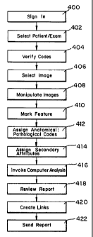
The method of operation is best illustrated by its application in the field
of radiology as shown in Fig. 4. Upon starting the software program, the
radiologist signs in, with either a password or voice signature or any other
security measure, to begin the evaluation at step 400. Secure sign-in protects
-21-
CA 02381653 2002-02-08
WO 01/11548 PCTIUSOO/21883
access to the database and validates the identity of the radiologist
generating the
report. The file loader displays a work list of patients whose examination
studies are accessible. The radiologist selects the name of a patient at step
402,
and the file loader displays all of the associated unread examination files.
The
radiologist selects a particular examination file, and that examination file
is
loaded into computer memory.
The file loader displays the CPT and ICD codes assigned to a particular
examination. This information can be obtained from the HIS 36 or entered
manually. The radiologist verifies the CPT and ICD codes and makes any
necessary changes at step 404. Correct assignment of the CPT and ICD codes
by the radiologist is essential for electronic billing and expedited
reimbursement
by insurers.
After validation of the CPT and ICD codes, the radiologist begins
analysis of the first image presented in the 2D viewer or selects an alternate
image, at step 406, from the series menu which lists all of the images or sets
of
images (i.e., series) in a patient exam available for review. The radiologist
may
change the displayed image in order to locate diagnostically significant
features
in other images at step 408. For example, the radiologist may press the LMB
while moving the mouse to pan through multiple images in the 2D viewer
(provided that more than one image is contained in the series). The
radiologist
may also translate the displayed image up, down, and sideways by pressing the
MMB while moving the mouse. The radiologist may also zoom the displayed
image by pressing the LMB and MMB simultaneously while moving the mouse.
In the 3D viewer, the mouse operations are similar except that pressing the
LMB while moving the mouse causes the 3D rendered scene to rotate in space
-22-
CA 02381653 2002-02-08
WO 01/11548 PCT/US00/21883
or to guide a "fly-through." Alternatively, multiple images or series can be
displayed simultaneously in separate windows in the viewer.
To aid in the identification of diagnostically significant features, the
radiologist may toggle between 2D and 3D viewers by pressing the 2D/3D
toggle button as shown in Fig. 6. When the 3D viewer is initially activated, a
volume-rendered image centered around the coordinates of the identified
feature
is created (i.e., a cube of CT data is volume-rendered). The radiologist may
adjust the size of the volume (i.e., cube of digital data) that is rendered in
the 3D
viewer via the render-box-size menu. The radiologist may further adjust the
volume-rendered image in various ways, such as using opacity maps, cut planes,
and rotation. MPR and surface rendering can also be activated in the 3D
viewer.
When the radiologist toggles between 2D and 3D viewers, the last state
of each viewer is recalled. The radiologist may also toggle between the 3D and
2D viewers by clicking on a primary 2D thumbnail image representation of a
diagnostic finding (or its supporting secondary 2D and 3D thumbnails), thereby
recalling the last state of the 2D or 3D viewer associated with the activated
finding. The cursor position and location of any marking symbols in the
display
are recalled as part of the last state of the viewer. The 2D or 3D viewer then
enters an edit mode, during which the radiologist can append additional
secondary attributes to the activated diagnostic finding, and these are
subsequently stored in proper locations within the database.
The radiologist can also set the orientation of the image data prior to
image analysis. If an image or image series needs to be reoriented, the
radiologist pans through the volume of images to locate the most superior
image
-23-
CA 02381653 2002-02-08
WO 01/11548 PCT/US00/21883
(or close to it). Then, the radiologist toggles the orientation button, at
which
time the viewer goes into an orientation mode. The radiologist rotates the
image plane by pressing the LMB and moving the mouse until the proper
anterior/posterior and left/right orientation is achieved. Finally, the
radiologist
toggles the orientation button again to set the proper orientation. The viewer
automatically adjusts the 2D image plane so that it is orthogonal to the
radiologist's viewpoint.
The radiologist has further control over the display of the images such as
grayscale (or color scale) and 3D opacity maps settings. The radiologist may
toggle between the two most recent W/L settings or Op settings in the 2D and
3D viewers by pressing the previous button, Pr, as shown in Fig. 6, or
simultaneously pressing the LMB and RMB. Additionally, the radiologist may
toggle a visible cursor on and off by pressing a cursor-toggle button, Cr, as
shown in Fig. 6, to indicate the location of a finding in both the 2D and 3D
viewers. By pressing the overview button, the radiologist re-centers a 2D or
3D
volume-rendered image in case the scene is moved out of sight.
When the radiologist locates a diagnostically significant feature, the
radiologist positions the cursor over the location of the feature on the
digital
image and clicks the RMB to mark the feature at step 410. Clicking on the
RMB stores the image coordinates and image number corresponding to the
cursor location in database. To complete the definition of a diagnostic
finding,
the radiologist annotates the point (location) by assigning an
anatomical: pathological code and optionally assigning secondary attributes at
steps 412 and 414.
The radiologist selects an anatomical: pathological code from a
-24-
CA 02381653 2002-02-08
WO 01/11548 PCTIUSOO/21883
predefined lexicon, such as the ACR Index of Radiological Diagnoses or
SNOMED or a custom designed lexicon, to create a diagnostic finding. As each
diagnostic finding is created, a representative thumbnail image 620 may be
displayed on the right side of the 2D and 3D viewers, or in a separate
display,
for immediate review and recall, and the thumbnail images later may be
incorporated into the final report as shown in Figs 7B and 7C.
The radiologist enters the anatomical:pathological code by one of several
modes. In a first mode, "click and label", cascading pop-up annotation menus
are presented to the radiologist immediately after a feature is marked by an
RMB click at step 500 of Fig. 5A. The radiologist selects an appropriate
anatomical location description from the anatomical-location menu at step 502.
For example, the radiologist may select Gastrointestinal System: Colon:
Sigmoidal Colon. After the selection, the radiologist selects the pathological
description from the pathology-description menu at step 502. For example, the
radiologist may select Neoplasm:Benign Neoplasm:Polyp. A secondary
attribute may then be assigned at step 504.
In a second mode, "click-click-click and label-label-label", the
radiologist identifies all the diagnostically significant features first and
subsequently annotates the features with labels and secondary attributes. As
shown in Fig. 513, the radiologist marks a designated feature at step 550 and
then
proceeds to mark successive features by repeating step 550. After all desired
features are marked, the radiologist assigns a diagnostic code to each marked
feature by assigning an anatomical code at step 552 and a pathological code at
step 554. Secondary attributes are assigned at step 556 either following the
marking of a feature at step 550 or the assigning of anatomical and
-25-
CA 02381653 2002-02-08
WO 01/11548 PCTIUSOO/21883
pathological codes at steps 552 and 554. The radiologist must assign a
diagnostic code to any unlabeled findings prior to final report approval;
otherwise, these findings may be labeled with a default "unknown
location: unknown pathology." Additionally, the radiologist may recall the
annotation menus to reassign an anatomical: pathological code to a particular
finding if the diagnosis needs to be revised during the evaluation process.
The radiologist may also assign secondary attributes to embellish or
support a diagnostic finding at step 414, but secondary attributes are not
essential for establishing a diagnostic finding. The radiologist may enter
descriptive characteristics, dimensional measurements, audio descriptions, and
specific snapshots of particular views of the identified finding as secondary
attributes. For example, the radiologist may add descriptive characteristics
that
enhance a specific diagnostic code set from a characteristics menu of
descriptive
characteristics.
The radiologist may measure one or more dimensions of a finding, for
example, a diameter of an identified feature in the 2D or 3D image. The
radiologist activates the distance measuring function by pressing the distance-
measurement button, Di, as shown in Fig. 6. The radiologist measures the
distance by clicking on first and second object points which span the
characteristic length. Similarly, the radiologist may measure the area of an
identified feature by pressing the area-measurement button, Ar, as shown in
Fig.
6 and defining a region-of-interest (ROI) using the input device 27. The
cross-sectional area, mean voxel value, and standard deviation of voxel values
in the ROI can be calculated. The radiologist may also add a volume-
measurement as a secondary attribute by pressing the volume-measurement
-26-
CA 02381653 2002-02-08
WO 01/11548 PCTIUSOO/21883
button, Vo, as shown in Fig. 6.
As part of step 414, the radiologist may also add a priority level and
recommendation to the diagnostic finding by pressing the priority button, Pt,
or
recommendation button, Rm, respectively, as shown in Fig. 6. In addition, the
radiologist may append a verbal description of the diagnostic finding in the
form
of an audio file. To add a verbal description the radiologist presses the
audio
button, Au, as shown in Fig. 6 to initiate recording and then dictates a
verbal
description of the diagnostic finding. The radiologist presses the audio
button
again to stop recording, and an audio file of the verbal description is stored
in
the database attached to the finding. Audio files can be attached to the
"global"
finding or attached to individual snapshot images or movies.
Additionally, the radiologist may record snapshots of any of the
displayed 2D and 3D images as a secondary attribute by pressing the snapshot
button, Sn, as shown in Fig. 6. For example, the radiologist may record any
number of additional images showing differing views of a particular
diagnostically significant feature. For example, a "colon:polyp" diagnostic
finding could be supported by additional 3D snapshots of the polyp. The
radiologist may also append cine clips of moving 2D or 3D images (including
audio and active annotations) as a secondary attributes in a manner similar to
recording snapshots by pressing the movie button, Mo, as shown in Fig. 6.
Pressing of the movie button starts and stops the recording of the cine clip.
Prior to final report review, the radiologist may also invoke computer-
aided location and analysis of diagnostically significant features, at step
416,
whereby the system automatically identifies and diagnoses suspicious features.
For example, the radiologist can review polyps found by the CAPD that were
-27-
CA 02381653 2002-02-08
WO 01/11548 PCTIUSOO/21883
not previously identified by the radiologist.
After the radiologist's review is deemed complete, the radiologist clicks
a report button on the bottom of either the 2D or 3D viewer as shown in Figs.
6
to activate the report display at step 418. Alternately, the report can be
generated and simultaneously displayed on a second monitor while the
diagnostically significant findings are being located and coded. The
diagnostic
findings are sorted according to anatomical categories and priorities, with
high
priority findings being placed at the top of each category. Each high-priority
finding is highlighted with color-enhanced text. The sorting and highlighting
of
the diagnostic findings alerts the end-user to the most significant diagnostic
findings.
The radiologist edits the final report as necessary, including linking
redundant findings at step 420. The step of creating links, step 420, may be
performed before or after the step of reviewing the report, step 418, as
depicted
in Fig. 1, where the step of creating the links, step 110, occurs prior to the
step
of reviewing the report, step 112. In one implementation of vertical linking,
the
radiologist "drags and drops" a finding onto a matching finding in the same
report display, and the "dropped" finding becomes a subset of the primary
finding. Alternatively, the radiologist can form links via a command-line
interface or voice-activated commands (control). Similarly, the radiologist
may
assign horizontal linking to track and monitor a diagnostic finding over time
and
across various imaging modalities. In horizontal linking, diagnostic findings
can be "dragged and dropped" across new paradigm reports in a similar
fashion.
The radiologist may also composite a group of image findings to yield a
-28-
CA 02381653 2002-02-08
WO 01/11548 PCTIUSOO/21883
diagnosis as illustrated above for "congestive heart failure." In this
process, the
radiologist or an Al program can link (or composite) additional clinical
information (e.g., laboratory and pathology report values) to support a
diagnosis.
The radiologist further reviews any repetitive diagnostic findings from
previous reports which are brought to the attention of the radiologist by the
system. If a previous report contains a repetitive diagnostic finding (e.g.,
evidence of prior gallbladder surgery), that finding is presented to the
radiologist for automatic incorporation into the new report. If a previous
report
contains a "trackable" diagnostic finding (e.g., index lymph node
measurement),
the radiologist can link the trackable diagnostic findings horizontally across
reports, and the temporal progression of this diagnostic finding can be
observed
in a specialized viewer.
The radiologist can suspend an examination for later resumption by
pressing the suspend-resume button during the review. Upon completion of the
report the radiologist instructs the system to send the report to the end-
users
(e.g., clinicians) at step 422. Additionally, the end-user can access the
report
via a Web server after the report has been posted. As noted above, the report
may be sent by a combination of telephone, fax, pager, or e-mail and may
include return receipt verification. The automated sending and receipt
verification allows the radiologist to quickly communicate his or her findings
and verify this communication.
End-users receiving the radiologist's report can customize the display of
the information to best suit their needs. For example, the clinician can click
on
a thumbnail image in the final report to access the original PACS image data.
-29-
CA 02381653 2002-02-08
WO 01/11548 PCTIUSOO/21883
Additionally, the reporting system can automatically translate the
radiologist's
report into a different language for the end-user. The standardized lexicon of
diagnostic findings supports rapid translation of reports to foreign languages
by
employing translation look-up tables.
The reporting system of the present invention has further application
beyond the preparation and delivery of reports. The ability of the reporting
system to enter diagnostic findings into searchable databases readily supports
data mining for clinical trials, epidemiology studies, and outcomes analyses.
Additionally, the reporting paradigm supports radiologic training. For
example, a radiology resident can issue a preliminary report indicating his or
her
findings, and the preliminary report can later be modified by an attending
radiologist to indicate any corrections. In the latter case, the system
automatically informs the referring clinician of any significant changes. The
history of report changes can be recorded with each finding (or changed
finding) having a timestamp. The reporting scheme also supports standardized
testing (e.g., replacement of the American Board of Radiology's Oral Board
examination) by objectively measuring a student's performance. Such an
objective performance measure could also assist in comparing a radiologist's
performance to that of a non-radiologist.
These and other advantages of the present invention will be apparent to
those skilled in the art from the foregoing specification. Accordingly, it
will be
recognized by those skilled in the art that changes or modifications may be
made to the above-described embodiments without departing from the broad
inventive concepts of the invention. For example, while the above invention
has been illustrated in terms of its application to the field of radiology,
the
-30-
CA 02381653 2002-02-08
WO 01/11548 PCTIUSOO/21883
invention is equally applicable to other fields of medicine as well as other
image
analysis fields such as satellite imagery and photography. It should therefore
be
understood that this invention is not limited to the particular embodiments
described herein, but is intended to include all changes and modifications
that
are within the scope and spirit of the invention as set forth in the claims.
-31-