Note: Descriptions are shown in the official language in which they were submitted.
CA 02381706 2002-02-12
WO 01/12674
PCT/US00/21574
1
OVARIAN CANCER CELL AND MYELOMA CELL SURFACE
GLYCOPROTEINS, ANTIBODIES THERETO, AND USES THEREOF
FIELD OF THE INVENTION
The application is related to new surface glycoproteins of human myeloma
cells and human ovarian tumor cells, monoclonal antibodies thereto, and
methods of
diagnosis and treatment of myeloma and ovarian cancer based thereon.
BACKGROUND OF THE INVENTION
Multiple myeloma (MM) embodies a plasma cell disorder characterized by
neoplastic proliferation of a single clone of plasma cells engaged in the
production
of a monoclonal immunoglobulin, usually monoclonal IgG or IgA. MM accounts
for 1% of all malignant disease and slightly more than 10% of all hematologic
malignancies. The annual incidence of multiple myeloma is 4 per 100,000. The
annual incidence is linked to aging population. The median age of patients at
the
time of diagnosis is 61 years. MM is most common in men, and in individuals of
African ancestry.
MM remains a disease for which a cure is a rarity. Most patients succumb to
their disease within 36-48 months from the time of diagnosis. The limitations
of
effective therapy for MM are primarily associated with a low cell
proliferation rate
and multi-drug resistance. Therapy for multiple myeloma includes induction,
maintenance, and supportive aspects. The induction portion of the treatment
aims at
reducing the tumor volume and achieving a plateau phase. Different drugs and
treatment modalities, such as bone marrow transplantation, have been
entertained,
and used without a significant impact on the disease or the overall survival.
Supportive care in multiple myeloma has advanced significantly over the
past few years. Growth factor support with erythropoietin replacement and GM-
CSF for stimulating the white blood cell (WBC) population are safe and
effective
methods of decreasing or preventing the occurrence or the severity of
neutropenia.
Also, high dose chemotherapy followed by autologous bone marrow or peripheral
blood progenitor cell (PBMC) transplantation has recently increased the
complete
CA 02381706 2002-02-12
WO 01/12674
PCT/US00/21574
2
remission rate and remission duration. However, overall survival has only been
slightly prolonged, and no evidence for a cure has been obtained. All patients
ultimately relapse, even under maintenance therapy with interferon-a (IFN-a)
alone
or in combination with steroids. Adoptive immunotherapy rather than active
vaccination may prove to be a more effective therapy for MM patients. There
are
relatively few known surface antigens on plasma cells that are suitable for
antibody-
directed treatment. Possible molecules include HM1.24, CD38, ICAM-1 (CD54),
CD40, CD45, CD20, and syndecan 1. To date, there are no exclusive markers
reported for MM. CD20, CD38, CD56 and CD130 are all markers that are
expressed on normal B-cells, T-cells, or natural killer (NK) cells.
Ovarian cancer is the fifth leading cause of cancer deaths among U.S.
women and has the highest mortality of any of the gynecologic cancers. It
accounted
for an estimated 26,600 new cases and 14,500 deaths in 1995. The overall 5-
year
survival rate is at least 75%, if the cancer is confined to the ovaries, and
decreases
to17% in women diagnosed with distant metastases. Symptoms usually do not
become apparent until the tumor compresses or invades adjacent structures, or
ascites
develops, or metastases become clinically evident. As a result, two thirds of
women
with ovarian cancer have advanced (Stage III or IV) disease at the time of
diagnosis.
Carcinoma of the ovary is most common in women over age 60. Other important
risk factors include low parity and a family history of ovarian cancer. Less
than
0.1% of women are affected by hereditary ovarian cancer syndrome, but these
women may face a 40% lifetime risk of developing ovarian cancer.
Potential screening tests for ovarian cancer include the bimanual pelvic
examination, the Papanicolaou (Pap) smear, tumor markers, and ultrasound
imaging. The pelvic examination, which can detect a variety of gynecologic
disorders, is of unknown sensitivity in detecting ovarian cancer. Although
pelvic
examinations can occasionally detect ovarian cancer, small, early-stage
ovarian
tumors are often not detected by palpation due to the deep anatomic location
of the
ovary. Thus, ovarian cancers detected by pelvic examination are generally
advanced and associated with poor survival. The pelvic examination may also
CA 02381706 2002-02-12
WO 01/12674
PCT/US00/21574
3
produce false positives when benign adnexal masses (e.g., functional cysts)
are
found. The Pap smear may occasionally reveal malignant ovarian cells, but it
is not
considered to be a valid screening test for ovarian carcinoma. Ultrasound
imaging
has also been evaluated as a screening test for ovarian cancer, since it is
able to
estimate ovarian size, detect masses as small as 1 cm, and distinguish solid
lesions
from cysts.
Serum tumor markers are often elevated in women with ovarian cancer.
Examples of these markers include carcinoembryonic antigen, ovarian
cystadenocarcinoma antigen, lipid-associated sialic acid, NB/70K, TAG 72.3,
CA15-3, and CA-125, respectively. Evidence is limited on whether tumor markers
become elevated early enough in the natural history of occult ovarian cancer
to
provide adequate sensitivity for screening. Tumor markers may have limited
specificity. It has been reported that CA-125 is elevated in 1% of healthy
women,
6-40% of women with benign masses (e.g., uterine fibroids, endometriosis,
pancreatic pseudocyst, pulmonary hamartoma) and 29% of women with
nongynecologic cancers (e.g., pancreas, stomach, colon, breast). Prospective
studies
involving asymptomatic women are needed, however, to provide definitive data
on
the performance characteristics of serum tests when used as screening tests.
SUMMARY OF THE INVENTION
In its broadest aspect, the present invention is directed to a monoclonal
antibody, or binding fragment thereof, which specifically binds to antigens
sharing a
common epitope present on the surface of human myeloma cells and ovarian
cancer
cells. The antigen on multiple myeloma cells is a single glycosylated
polypeptide
with a molecular weight of about 78 kDa to about 120 kDa, as determined by SDS-
PAGE under reducing conditions. The antigen on ovarian cancer cells is a
single
glycosylated polypeptide with a molecular weight of about 76 kDa to about 213
kDa, as determined by SDS-PAGE under reducing conditions. The antigens are
absent from human peripheral blood mononuclear cells, absent from human B
cells,
and absent from human chronic myelongenic leukemia cells. Further, the
antigens
are not present on cells from a breast cancer tumor, not present on a prostate
cancer
CA 02381706 2002-02-12
WO 01/12674
PCT/US00/21574
4
cell line, not present on a neuroblastoma cell line, and not present on a
cervical
cancer cell line. The antigens are also not found on an Epstein-Barr virus-
transformed B cell tumor.
A non-limiting example of the monoclonal antibody is that produced by the
hybridoma cell line deposited at the American Type Culture Collection (ATCC),
10801 University Boulevard, Manassas, VA 20110-2209 on August 3, 1999, and
having ATCC Accession No. PTA-450. The monoclonal antibody produced by the
deposited hybridoma cell line having ATCC Accession No. PTA-450 is termed both
MoAb 69 and VAC69 herein.
The present invention is further directed to antibodies that are capable of
binding to the same antigenic determinant as does the monoclonal antibody
produced by the hybridoma cell line deposited at the American Type Culture
Collection having ATCC Accession No. PTA-450; binding fragments of the
hybridoma cell line deposited at the American Type Culture Collection having
ATCC Accession No. PTA-450; and to binding fragments of a monoclonal antibody
capable of binding to the same antigenic determinant as does the monoclonal
antibody produced by the hybridoma cell line deposited at the American Type
Culture Collection having ATCC Accession No. PTA-450.
Such monoclonal antibodies, or antibody fragments, may be human, or they
may be derived from other mammalian species, such as rodent, hybrids thereof,
chimeric antibodies, and the like. Binding fragments of the monoclonal
antibodies
of the present invention include, but are not limited to, F(abI)2, Fab', Fv,
Fd', or Fd
fragments.
In another aspect, the present invention is directed to a cell line produced
by
a hybridoma technique, which produces a monoclonal antibody which specifically
binds to surface antigens of human myeloma cells and of ovarian cancer cells.
The
antigen on multiple myeloma cells is a single glycosylated polypeptide with a
molecular weight of about 78 kDa to about 120 kDa as determined by SDS PAGE
under reducing conditions. The antigen on ovarian cancer cells is a single
glycosylated polypeptide with a molecular weight of about 76 kDa to about 213
kDa
CA 02381706 2002-02-12
WO 01/12674
PCT/US00/21574
,as determined by SDS-PAGE under reducing conditions. The antigens are absent
from human peripheral blood mononuclear cells, absent from human B cells, and
absent from human chronic myelongenic leukemia cells. A non-limiting example
of
a monoclonal antibody according to the present invention is that produced by
the
5 hybridoma cell line deposited at the American Type Culture Collection
having
accession No. PTA-450. Furthermore, the antigens are not present on cells from
a
breast cancer tumor, not present on a prostate cancer cell line, not present
on a
neuroblastoma cell line, and not present on a cervical cancer cell line. The
antigens
are also not found on an Epstein-Barr virus-transformed B cell tumor.
A further aspect of the present invention is the hybridoma cell line deposited
at the American Type Culture Collection (ATCC), 10801 University Boulevard,
Manassas, VA 20110-2209 on August 3, 1999, and having ATCC Accession No.
PTA-450.
In another broad aspect of the present invention, an isolated surface antigen
of human multiple myeloma cells is described, the antigen being a single
glycosylated polypeptide with a molecular weight of about 78 IcDa to about 120
IcDa, as determined by SDS-PAGE under reducing conditions; the antigen being
absent from human peripheral blood mononuclear cells, absent from human B
cells,
and absent from human acute myelogenic leukemia cells. The antigen is not
present
on cells from a breast cancer tumor, not present on a prostate cancer cell
line, not
present on a neuroblastoma cell line, and not present on a cervical cancer
cell line.
It is also not found on an Epstein-Barr virus-transformed B cell tumor. The
isolated
multiple myeloma surface antigen binds to a monoclonal antibody produced by
the
hybridoma cell line deposited at the American Type Culture Collection having
ATCC Accession No. PTA-450.
In another broad aspect of the present invention, an isolated surface antigen
of human ovarian cancer cells is described, the antigen being a single
glycosylated
polypeptide with a molecular weight of about 76 IcDa to about 213 IcDa, as
determined by SDS-PAGE under reducing conditions; the antigen being absent
from
human peripheral blood mononuclear cells, absent from human B cells, and
absent
CA 02381706 2002-02-12
WO 01/12674
PCT/US00/21574
6
from human acute myelogenic leukemia cells. The antigen is not present on
cells
from a breast cancer tumor, not present on a prostate cancer cell line, not
present on
a neuroblastoma cell line, and not present on a cervical cancer cell line. It
is also
not found on an Epstein-Barr virus-transformed B cell tumor. The isolated
ovarian
cancer surface antigen binds to a monoclonal antibody produced by the
hybridoma
cell line deposited at the American Type Culture Collection having ATCC
Accession No. PTA-450.
The present invention is also directed to methods of inhibiting the growth of,
or killing, myeloma cells in a patient by administering the monoclonal
antibody, or
a binding fragment as described above, under conditions sufficient for the
binding
of the monoclonal antibody, or the binding fragment, to the myeloma cells to
cause
inhibiting or killing of the cancer cells by the immune cells of the patient.
In
another aspect, a method for inhibiting or killing myeloma cells in a patient
is
provided by administering the monoclonal antibody, or binding fragment as
described above, which is conjugated with a cytotoxic moiety, under conditions
sufficient for the binding of the monoclonal antibody, or binding fragment, to
the
cancer cells to inhibit the growth of, or to kill, the cells. The cytotoxic
moiety may
be, by way of non-limiting example, a chemotherapeutic agent, a photo-
activated
toxin, or a radioactive agent.
In still another aspect of the invention, the above-mentioned conjugate of the
monoclonal antibody, or binding fragment, described herein and a cytotoxic
moiety
may be used in vitro to inhibit growth of, or kill, myeloma cells in a
cellular sample,
such as a bone marrow sample.
The invention is also directed to anti-idiotypic antibodies which mirror the
binding site of the monoclonal antibody of the invention, and are specific to
the
myeloma and ovarian cancer conformational epitope recognized by the antibody
of
the invention. The invention is further directed to the use of the
aforementioned
anti-idiotypic antibodies for the treatment of MM or ovarian cancer by active
immunization.
In a further aspect of the invention, a method is provided for removing
CA 02381706 2002-02-12
WO 01/12674
PCT/US00/21574
7
myeloma cells from an isolated cellular sample, such as, but not limited to,
bone
marrow cells, by exposing the cellular sample to a solid matrix on which the
monoclonal antibody, or binding fragment, described above is bound under
conditions wherein the myeloma cells adhere to the monoclonal antibody, or
binding fragment, and isolating a cellular fraction of said cellular sample
which
does not bind to the matrix. This method may be used, for example, in the
removal
of myeloma cells from a bone marrow sample for autologous bone marrow
transplant.
The invention is also directed to the monoclonal antibody, or binding
fragment, as described above bound to a solid support.
In yet another aspect of the invention, a method is provided for localizing
myeloma cells or tumor cells, or ovarian cancer cells or tumor cells, in a
patient by
administering the monoclonal antibody, or binding fragment, as described
above,
allowing the monoclonal antibody, or binding fragment thereof, to bind to the
cancer cells within said patient, and determining the location of the
monoclonal
antibody, or binding fragment thereof, within the patient. In another related
aspect,
the monoclonal antibody, or binding fragment, is detectably labeled, for
example,
with a radionuclide.
The present invention is further directed to methods of inhibiting the growth
of, or killing, ovarian cancer cells in a patient by administering the
monoclonal
antibody, or binding fragment, as described above under conditions sufficient
for
the binding of the monoclonal antibody, or binding fragment, to the ovarian
cancer
cells to cause growth inhibition or killing of the ovarian cancer cells by
immune
cells of the patient. In another aspect, a method for inhibiting or killing
ovarian
cancer cells in a patient is provided by administering the monoclonal
antibody, or
binding fragment, as described above which is conjugated with a cytotoxic
moiety,
under conditions sufficient for the binding of the monoclonal antibody, or
binding
fragment, to ovarian cancer cells to cause growth inhibition or killing of the
ovarian
cancer cells. The cytotoxic moiety may be, by way of non-limiting example, a
chemotherapeutic agent, a photo-activated toxin, or a radioactive agent.
CA 02381706 2013-07-15
7a
According to another aspect of the present invention, there is provided a
monoclonal antibody or fragment thereof selected from the group consisting of
(i) the
monoclonal antibody produced by the hybridoma cell line deposited at the
American
Type Culture Collection having Accession No. PTA-450; (ii) monoclonal antibody
that is capable of binding to the same antigenic determinant as does the
monoclonal
antibody produced by the hybridoma cell line deposited at the American Type
Culture
Collection having ATCC Accession No. PTA-450; (iii) binding fragments of a
monoclonal antibody produced by the hybridoma cell line deposited at the
American
Type Culture Collection having ATCC Accession No. PTA-450; and (iv) binding
fragments of a monoclonal antibody capable of binding to the same antigenic
determinant as does the monoclonal antibody produced by the hybridoma cell
line
deposited at the American Type Culture Collection having ATCC Accession No.
PTA-450.
According to still another aspect of the present invention, there is provided
a
cell line produced by a hybridoma technique which produces a monoclonal
antibody
which specifically binds to the same antigenic determinant as a monoclonal
antibody
produced by the hybridoma cell line deposited at the American Type Culture
Collection having ATCC Accession No. PTA-450.
According to yet another aspect of the present invention, there is provided an
isolated surface antigen of human myeloma cells, said antigen being
characterized in
that (a) it is a single polypeptide with a molecular weight of 78 kDa to 120
kDa as
determined by SDS PAGE under reducing conditions; (b) it is absent from human
peripheral blood mononuclear cells, absent from human B cells, and absent from
human B cell myelogenic leukemia cells; (c) it is glycosylated; and (d) it
binds to a
monoclonal antibody produced by the hybridoma cell line deposited at the
American
Type Culture Collection having ATCC Accession No. PTA-450.
According to a further aspect of the present invention, there is provided an
isolated surface antigen of human ovarian cancer cells, said antigen being
characterized in that (a) it is a single polypeptide with a molecular weight
of 76 kDa
to 213 kDa as determined by SDS PAGE under reducing conditions; (b) it is
absent
CA 02381706 2013-07-15
7b
from human peripheral blood mononuclear cells, absent from human B cells, and
absent from human B cell myelogenic leukemia cells; (c) it is glycosylated;
and (d) it
is specifically bound by a monoclonal antibody produced by the hybridoma cell
line
deposited at the American Type Culture Collection having ATCC Accession No.
PTA-450.
CA 02381706 2002-02-12
WO 01/12674
PCT/US00/21574
8
In yet another aspect of the invention, a method is provided for localizing
ovarian cancer cells in a patient by administering the monoclonal antibody, or
binding fragment, described above, allowing the monoclonal antibody, or
binding
fragment thereof, to bind to ovarian cancer cells within said patient, and
determining
the location of said monoclonal antibody, or binding fragment thereof, within
said
patient. In another related aspect, the monoclonal antibody, or binding
fragment, is
detectably labeled, for example, with a radionuclide.
It is a further aspect of the invention to permit the detection of the cell
surface glycoproteins described herein in a sample of bodily fluid, to aid in
the
diagnosis of multiple myeloma, ovarian cancer, or other cancer cells
expressing a
glycoprotein with the epitope recognized by the antibodies herein, by the
detection
of the glycoprotein antigen shed from cancer cells into the bodily fluid, such
as
blood. Furthermore, the stage of the disease may be monitored and the
effectiveness of anti-cancer therapies can be monitored by determining the
level or
changes over time of the level of shed surface glycoprotein in a bodily fluid
such as
blood.
In still yet another aspect, the invention is directed to pharmaceutical
compositions comprising a monoclonal antibody, or binding fragment, as
described
above and a pharmaceutically-acceptable carrier, diluent, or excipient.
In another aspect, the present invention is directed to a monoclonal antibody,
or binding fragment, as described above labeled with a detectable moiety, such
as,
by way of non-limiting examples, a fluorophore, a chromophore, a radionuclide,
or
an enzyme.
These and other aspects of the present invention will be better appreciated by
reference to the following drawings and Detailed Description.
BRIEF DESCRIPTION OF THE DRAWINGS
Figure 1 depicts a first screen of B cell hybridomas generated from mice
immunized with a pool of three human plasmacytoma cells compared with their
binding to human myelogenic leukemia cell line (K562), which serves as a
control.
Figure 2 presents the results of selected hybridomas for the second screen.
CA 02381706 2002-02-12
WO 01/12674
PCT/US00/21574
9
Figure 3 depicts the net binding values obtained for the first screen
compared with the second screen.
Figure 4 presents the results of cell surface staining using a panel of
monoclonal antibodies and analyzed by flow cytometry.
Figure 5 represents further evaluation of the selected monoclonal antibodies
using Western blot method, using membrane proteins extracted from five human
myeloma cell lines tested individually, and controls, fractionated on SDS-
PAGE.
Figure 6 presents results similar to those described for Figure 5, using a
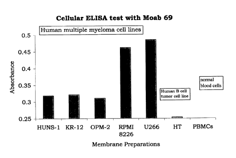
cellular ELISA method.
Figure 7 shows an SDS-PAGE gel of concentrated culture fluid from
multiple myeloma cells grown in serum-free medium for 5 days, blotted and
probed
with an antibody of the present invention.
Figure 8 depicts SDS-PAGE results of cell lysates from three ovarian cancer
tumor cells from 3 patient. The tumor cells were digested with trypsin and
homogenized. The gel was blotted and probed with an antibody of the present
invention.
Figure 9 shows the results of a Western Blot in which cellular lysates
prepared from a prostate cancer cell line (LnCaP), breast cancer (fresh
tumor),
ovarian cancer (fresh tumor), lung cancer (fresh tumor), neuroblastoma (cell
line),
normal PBMC and a pool of human MM cell lines (RPMI-8226, U266, OPM-2,
KR-12 and Huns-1) were fractionated by SDS-PAGE (8% gel). The gel was blotted
onto nitrocellulose and incubated with VAC69 antibody. Note that what may
appear as curved bands in lanes D (#5 on Blot) and E (# 6 on Blot) are in fact
a tear
in the gel.
Figure 10 presents the results of an in vitro cytotoxicity assay performed
using a human MM cell line (RPMI-8226), human lymphoma cell line (Namalwa)
and chronic myelogenic leukemia cell line (K562). The tumor cells were
incubated
with VAC69 antibody at a concentration of 10[1g/m1 or 1 i.tg/m1 in the
presence of
human complement as described in Example 10. To assess the background level of
cytotoxicity, the tumor cells were incubated with medium alone (untreated),
CA 02381706 2002-02-12
WO 01/12674
PCT/US00/21574
complement alone, or with VAC69 alone. The cultures were pulsed with 3H-
Thymidine and incubated for an additional 16 hours. The % killing of cancer
cells
was calculated from the values of3H-Thymidine incorporation recorded for the
test
samples compared with the medium control (untreated).
5 DETAILED DESCRIPTION OF THE INVENTION
Identification of unique cancer antigens enables the design of selective
immunotherapy for neoplastic diseases. The capacity to utilize a determinant
exclusively expressed by cancer cells, and which is devoid in normal tissues,
ensures the targeting and elimination of the neoplastic cells while insulating
the
10 function of normal cells. Although the last decades have witnessed great
activity
and significant success in the search for novel cancer antigens for various
neoplastic
diseases, cancer-specific antigens have not yet been defined for many
malignancies.
The majority of cancer antigens are self-antigens that are derived from and
expressed by normal counterpart cells. Frequently, the cancer antigen is
identical to
the normal antigen even though it is expressed at higher levels, or endowed
with a
negligible mutation insufficient for its distinction from the self-antigen.
One of the
escape mechanisms of malignant cells from the immune system is their
similarity to
their normal counterpart cells, thus resulting in low visibility of the
malignant cells
by the immune system.
New surface glycoprotein antigens that are present on human myeloma cells
and human ovarian cancer tumor cells, but absent from normal cells and from
leukemic cells, are provided by the present invention. Such antigens present a
target
for therapeutic intervention in myeloma and ovarian cancer, as well as for
diagnostic and cell purification purposes. These antigens share at least one
common
epitope.
A technique known as contrasting or differential immunization was
employed for obtaining monoclonal antibodies to the antigen and for the
identification of the novel cancer antigens described herein. As described in
the
examples below, two divergent immunogens provided at different locations were
used. The dual immunization polarizes the migration of the distinct
populations of
CA 02381706 2002-02-12
WO 01/12674
PCT/US00/21574
11
immune cells to discrete draining lymph nodes. In an example herein, a mixture
of
human myeloma cells was used as the immunogen to obtain murine monoclonal
antibodies to a myeloma cell surface antigen. Control cells in this example,
i.e., a
related, myelogenic leukemic cell line, were used to polarize the immune
response
to effectively delete undesired cells from the lymph nodes near the site of
immunization with the desired antigen. The immune cells extracted from the
draining lymph nodes close to the immunization site with the desired neoplasms
were immortalized by fusion with murine myeloma cells. The antipodal draining
lymph nodes were populated with immune cells specific to the undesired
(control)
immunogens.
By use of the foregoing protocol, a series of monoclonal antibodies were
prepared which were found to bind specifically to antigens on the surfaces of
human
myeloma cells and on ovarian cancer cells. These antigens share at least one
epitope. The antigens are further characterized in that the antigen on
multiple
myeloma cells is a single glycosylated polypeptide with a molecular weight of
about
78 kDa to about 120 kDa, as determined by SDS-PAGE under reducing conditions;
and it is absent from human peripheral blood mononuclear cells, absent from
human
B cells, and absent from human B cell myelogenic leukemia cells.
The antigen on ovarian cancer cells is a single glycosylated polypeptide with
a molecular weight of about 76 kDa to about 213 kDa, as determined by SDS-
PAGE under reducing conditions; and it is absent from human peripheral blood
mononuclear cells, absent from human B cells, and absent from human B cell
myelogenic leukemia cells. An antigen recognized by the antibody of the
invention
is also present on liver cancer cells; thus, the liver cancer cell surface
antigen has at
least one epitope in common with the myeloma and ovarian cancer surface
glycoprotein. An example of a hybridoma cell line that produces a monoclonal
antibody which recognizes these antigens has been deposited at the American
Type
Culture Collection (ATCC), 10801 University Boulevard, Manassas, VA 20110-
2209, on August 3, 1999, and accorded ATCC Accession No. PTA-450.
The aforementioned antigens were found not to be present on cells from a
CA 02381706 2002-02-12
WO 01/12674
PCT/US00/21574
12
breast cancer tumor, and were not present on a prostate cancer cell line, or
on a
neuroblastoma cell line, or on a cervical cancer cell line. They were also not
found
on an Epstein-Barr virus-transformed B cell tumor.
As further exemplified herein, VAC69, the monoclonal antibody produced
by the hybridoma deposited under ATCC Accession No. PTA-450 was shown to
react with a single chain cell surface glycoprotein with a Mr of about 78 kDa
to
about 120 kDa. The VAC69 MoAb did not react with an array of human cancers
such as lung, prostate, breast, cervical, neuroblastoma, lymphoma and
leukemia. In
addition, the antigen was not detected in human normal tissues such as those
derived
from breast, ovary, prostate, colon, or lung. Interestingly, VAC69, when
tested on a
panel of human malignancies reacted with ovarian cancer. VAC69-reactive
antigen
expressed by ovarian cancer cells appears to be distinct from that expressed
by MM
by its pattern of expression, i.e., being either a single high Mr glycoprotein
(¨ 200
kDa) or a set of glycoproteins with a Mr of about 76 kDa to about 213 kDa. One
ovarian tumor expressed a single high Mr band, thereby indicating that the
existence
of a multiple subunit antigen is unlikely. Accordingly, the lower Mr bands may
represent degradation products of the larger glycoprotein. The increase in Mr
of the
glycoprotein expressed by ovarian cancer compared with that expressed by the
MM
antigen may imply that VAC69 recognizes a communal epitope on two distinct
antigens.
The present invention is directed to monoclonal antibodies, and binding
fragments thereof, which recognize the aforementioned myeloma cell and ovarian
cancer cell surface glycoproteins. Thus, the present invention embraces the
deposited monoclonal antibody described above and monoclonal antibodies and
their binding fragments having binding specificity for the aforementioned
antigens.
Such antibody fragments capable of binding the aforementioned antigens,
include,
but are not limited to, F(ab')2 fragments, Fab' fragments, Fv fragments, Fd'
fragments, or Fd fragments. Antibodies may be human, mammalian, such as
mouse, and hybrid or chimeric antibodies. The antibody fragments and means for
preparing then from antibodies are known to one of skill in the art.
CA 02381706 2002-02-12
WO 01/12674
PCT/US00/21574
13
The monoclonal antibodies and antibody binding fragments may be
characterized as those which are 1) produced from the hybridoma cell line
deposited
at the American Type Culture Collection and having ATCC Accession No. PTA-
450; 2) capable of binding to the same antigenic determinant as does the
monoclonal antibody produced by the hybridoma cell line deposited at the
American Type Culture Collection having ATCC Accession No. PTA-450; 3)
binding fragments of the hybridoma cell line deposited at the American Type
Culture Collection having ATCC Accession No. PTA-450; or 4) binding fragments
of a monoclonal antibody capable of binding to the same antigenic determinant
as
does the monoclonal antibody produced by the hybridoma cell line deposited at
the
American Type Culture Collection having ATCC Accession No. PTA-450.
Accordingly, the aforementioned monoclonal antibodies and binding
fragments recognize a common epitope of cell surface glycoproteins present on
human myeloma cells and on human ovarian cancer cells, but absent from human
peripheral blood mononuclear cells, absent from human B cells, and absent from
human B cell myelogenic leukemia cells. Further, the cell surface glycoprotein
on
myeloma cells is a single polypeptide with a molecular weight of about 78 kDa
to
about 120 kDa, as determined by SDS-PAGE under reducing conditions. The cell
surface glycoprotein on ovarian cancer ells is a single polypeptide with a
molecular
weight of about 76 kDa to about 213 kDa, as determined by SDS-PAGE under
reducing conditions. As the myeloma, ovarian cancer, and liver cancer cell
surface
glycoproteins share a common epitope recognized by the antibodies of the
invention, such antibodies may be used therapeutically and diagnostically for
these
conditions. As mentioned above, the antigen is also present on the surface of
liver
cancer cells, but is not present on cells from a breast cancer tumor, not
present on a
prostate cancer cell line, not present on a neuroblastoma cells line, and not
present
on a cervical cancer cell line. It is also not found on an Epstein-Barr virus-
transformed B cell tumor.
The present invention is also directed to hybridoma cell lines which produce
a monoclonal antibody which specifically binds to the surface antigens of
human
CA 02381706 2002-02-12
WO 01/12674
PCT/US00/21574
14
myeloma cells and ovarian cancer cells as described and characterized herein.
These antigens have a shared region or an epitope contained in the cell
surface
glycoproteins of these neoplasms. The methods for the preparation of such
hydridomas are known to the skilled artisan. The contrasting immunization
procedure described herein is but one example of various means for obtaining
the
desired antibodies. For preparation of monoclonal antibodies directed toward
the
surface glycoprotein antigens described herein, any technique that provides
for the
production of antibody molecules by continuous cell lines in culture may be
used.
For example, such techniques include, but are not limited to, the hybridoma
technique originally developed by Kohler and Milstein (Nature, 256:495-497,
1975), as well as the trioma technique, the human B-cell hybridoma technique
(Kozbor et al., Immunology Today, 4:72, 1983; Cote et al., Proc. Natl. Acad.
Sci.
U.S.A., 80:2026-2030, 1983), and the EBV-hybridoma technique to produce human
monoclonal antibodies (Cole et al., in Monoclonal Antibodies and Cancer
Therapy,
Alan R. Liss, Inc., pp. 770-96, 1985).
In another embodiment of the present invention, monoclonal antibodies can
be produced in germ-free animals utilizing the technology described in
international
application number WO 98/02545. Also, according to the invention, techniques
developed for the production of "chimeric antibodies" are suitable for use.
Preferred are human or humanized chimeric antibodies for use in therapy of
human
diseases or disorders as described infra, since the human or humanized
antibodies
themselves are much less likely than xenogenic antibodies to induce an immune
response, particularly an allergic response.
According to the present invention, techniques described for the production
of single chain antibodies (U.S. Patent Nos. 5,476,786 and 5,132,405 to
Huston; and
U.S. Patent No. 4,946,778) can be adapted to produce myeloma surface antigen-
specific single chain antibodies. An additional embodiment of the invention
utilizes
the techniques described for the construction of Fab expression libraries
(Huse et al.,
Science, 246:1275-1281, 1989) to allow the rapid and easy identification of
monoclonal Fab fragments with the desired specificity, or fragment
derivatives, or
CA 02381706 2002-02-12
WO 01/12674
PCT/US00/21574
analogs.
Antibody fragments which contain the idiotype of the antibody molecule can
also be generated by known techniques. For example, such fragments include,
but
are not limited to, the F(ab')2 fragment which can be produced by pepsin
digestion
5 of the antibody molecule; the Fab' fragments which can be generated by
reducing
the disulfide bridges of the F(abI)2 fragment, and the Fab fragments which can
be
generated by treating the antibody molecule with papain and a reducing agent.
As mentioned above, the present invention is also directed to the isolated
surface antigens of human myeloma cells and human ovarian cancer cells,
wherein
10 the human myeloma cell-expressed antigen is characterized as being a
single
glycosylated polypeptide with a molecular weight of about 78 kDa to about 120
kDa, as determined by SDS-PAGE under reducing conditions and the human
ovarian cancer cell-expressed antigen is characterized as having a molecular
weight
of about 76 kDa to about 213 kDa. These antigens are absent from human
15 peripheral blood mononuclear cells, absent from human B cells, and
absent from
human B cell myelogenic leukemia cells. The antigens are also absent from
breast
cancer cells, as determined using fresh tumor tissue; absent from prostate
cancer
cells, determined using a prostate cancer cell line; absent from neuroblastoma
cells,
as determined using a neuroblastoma cell line, and absent from cervical cancer
cells
as determined by using a cervical cancer cell line. The glycoproteins have
been
found to be present on the surfaces of cells from a freshly-isolated liver
cancer
tumor. Thus, the above-described methods are also applicable to the therapy
and
diagnosis of liver cancer. The isolated surface antigens are further
characterized in
that they bind to the monoclonal antibody produced by the hybridoma cell line
deposited at the American Type Culture Collection having ATCC Accession No.
PTA-450.
The monoclonal antibody MA69 reacts consistently with a single chain
glycoprotein with a molecular weight of 78-120 kDa on multiple myeloma (MM)
cells. On ovarian carcinoma cells, however, MA69 recognizes one or more
glycoproteins ranging in size from 76 to 213 kDa. These results imply that
MA69
CA 02381706 2002-02-12
WO 01/12674
PCT/US00/21574
16
reacts with two distinct molecules expressed on MM and ovarian cancer cells
through the recognition of a shared region or an epitope contained in the cell
surface
glycoproteins of these neoplasms. This epitope is uniquely expressed on cells
of
ovarian and MM malignancies and was not found on the cell surfaces of a panel
of
human tumors, such as lung cancer, cervical cancer, neuroblastoma, breast
cancer,
prostate cancer, leukemia and lymphomas. Thus, the present invention is also
generally directed to cell surface glycoproteins which comprise an epitope
recognized by the antibodies of the invention. As noted above, cell surface
glycoproteins comprising this epitope are absent from the various normal and
cancer
cells tested and listed above.
The present invention is also directed to therapeutic methods for the
treatment of myeloma and related dysproliferative diseases in humans,
including
multiple myeloma, as well as ovarian cancer, using the antibodies of the
present
invention. The therapeutic and diagnostic uses described herein embrace
primary
tumors as well as metastases. For example, a method for inhibiting or killing
myeloma cells or ovarian cancer cells in a patient may be carried out by
administering to the patient, in a single dose or in successive doses, the
monoclonal
antibody, or antibody binding fragment as described above, under conditions
sufficient for the binding of the monoclonal antibody, or binding fragment, to
tumor
cells in the patient. Binding of antibodies to the tumor cells induces the
growth
inhibition and/or killing of the tumor cells by immune cells in the patient.
The aforementioned therapy may be accompanied by other treatments
directed at the tumor cells, such as chemotherapy, radiation, etc., as well as
by
adjunctive therapies to enhance the immune system's attack on the opsonized
tumor
cells following the procedure described above. For example, a growth factor
such
as erythropoietin and/or GM-CSF can be co-administered to the patient for
stimulating the white blood cells and supporting the immunocompetence status
of
the patient.
Further, chimeric or other recombinant antibodies of the invention may be
used therapeutically. For example, a fusion protein comprising at least the
antigen-
CA 02381706 2002-02-12
WO 01/12674
PCT/US00/21574
17
binding region of the antibody of the invention joined to at least a
functionally
active portion of a second protein having anti-tumor effects, e.g., a
lymphokine or
oncostatin, may be used to treat a human tumor in vivo. In addition, a
chimeric
antibody, wherein the antigen-binding site is joined to human Fc region, e.g.,
IgGI,
may be used to promote antibody-dependent mediated cytotoxicity or complement-
mediated cytotoxicity. In addition, recombinant techniques known in the art
can be
used to construct bispecific antibodies wherein one of the binding
specificities is
that of the antibody of the invention (See, e.g., U.S. Patent No. 4,474,893).
It will be appreciated by the skilled practitioner that other dysproliferative
diseases in which the glycoprotein antigens of the invention are present on
the cell
surface are treatable by the methods described herein.
The above-described methods utilize the antibodies or binding fragments
without modification, relying on the binding of the antibodies or fragments to
the
surface antigen(s) of the myeloma or ovarian cancer cells in situ to stimulate
an
immune attack thereon. In another aspect of the therapeutic methods, the
aforementioned method may be carried out using the monoclonal antibodies or
binding fragments to which a cytotoxic agent is bound. Binding of the
cytotoxic
antibodies, or antibody binding fragments, to the tumor cells inhibits the
growth of
or kills the cells. By way of non-limiting example, suitable cytotoxic agents
may be
a chemotherapeutic agent, a photo-activated toxin or radioactive agent. For
example, cytotoxic agents such as ricin A chain, abrin A chain, modeccin A
chain,
gelonin, melphalan, bleomycin, adriamycin, daunomycin, or pokeweed antiviral
proteins (PAP, PAPII, PAP-S).
Those skilled in the art will realize that there are numerous radioisotopes
and
chemocytotoxic agents that can be coupled to tumor specific antibodies by well
known techniques, and delivered to specifically destroy tumor tissue. See,
e.g., U.S.
Patent No.4,542,225 to Blattler et al. Examples of photo-activated toxins
include
dihydropyridine- and omega-conotoxin (Schmidt et al., J Biol. Chem.,1991,
266(27):18025-33). Examples of imaging and cytotoxic reagents that can be used
include 1251, "In, 123 1, 99n'Tc, 32P, 3H, and 14C; fluorescent labels such as
CA 02381706 2002-02-12
WO 01/12674
PCT/US00/21574
18
fluorescein and rhodamine, and chemiluminescers such as luciferin. The
antibody
can be labeled with such reagents using techniques known in the art. For
example,
see Wenzel and Meares, Radioimmunoimaging and Radioimmunotherapy, Elsevier,
New York (1983) for techniques relating to the radiolabeling of antibodies
(see also,
Coleer et al., "Use of Monoclonal Antibodies As Radiopharmaceuticals For The
Localization Of Human Carcinoma Xenografts In Nude Mice", Methods Enzymol.,
121:802-16, 1986: "Order, Analysis, Results and Future Prospective of the
Therapeutic Use of Radiolabeled Antibody in Cancer Therapy", in Monoclonal
Antibodies for Cancer Detection and Therapy, Baldwin et al. (eds), pp. 303-16
(Academic Press 1985).
Other covalent and non-covalent modifications of the antibodies or antibody
fragments of the present invention are embraced herein, including agents which
are
co-administered or administered after the antibody or fragments, to induce
growth
inhibition or killing of the cells to which the antibody or fragment has
previously
bound.
Anti-idiotypic monoclonal antibodies to the antibodies of the invention may
also be used therapeutically in active tumor immunization and tumor therapy
(see,
e.g., Hellstrom et al., "Anti Idiotypes" in Covalently Modified Antigens and
Antibodies in Diagnosis and Therapy, supra at pp. 35-41).
In the area of multiple myeloma, the antibodies or antibody fragments of the
present invention have further utility in the preparation of cellular samples
from
which myeloma cells have been removed. This use is particularly important in
autologous bone marrow transplants, wherein a sample of bone marrow is
harvested
from a cancer patient prior to the patient's undergoing high-dose
chemotherapy.
The goal of the high dose chemotherapy is to destroy the cancer cells, which
also
results in the depletion of bone marrow cells. Following such treatment, the
harvested bone marrow cells are reintroduced into the patient.
In myeloma and related diseases, the harvested bone marrow is
contaminated with myeloma cells; thus, reintroduction of untreated bone marrow
will simply reintroduce the disease. Previous methods to prevent
reintroduction of
CA 02381706 2002-02-12
WO 01/12674
PCT/US00/21574
19
cancer cells have included treatment of the bone marrow sample with
chemotherapeutic agents and other anti-neoplastic agents in vitro. Other
methods
include purging the sample of cancer cells.
In a further practice of the present invention, the monoclonal antibodies and
fragments described herein may be used to remove myeloma cells from a
patient's
bone marrow sample before reintroduction into the patient. In one nonlimiting
example, the monoclonal antibodies, or binding fragments, are attached to a
matrix,
such as beads. This may be accomplished by any of several well-known methods
for preparing an affinity matrix comprising antibodies or their binding
fragments.
The bone marrow sample is then exposed to the matrix, such as by passage of
the
cells over a column containing the matrix, under conditions to promote the
binding
of the myeloma cells in the sample through antigen/antibody interactions with
the
antibodies or binding fragments attached to the matrix. The myeloma cells in
the
sample adhere to the matrix; while the column effluent, i.e., the non-adherent
cellular population, is depleted of myeloma cells. The effectiveness of the
procedure may be monitored by examining the cells for residual myeloma cells,
such as by using a detectably-labeled antibody as described below. The
procedure
may be repeated or modified to increase effectiveness.
This purging procedure (see, e.g., Ramsay et al., J. Clin. Immunol., 8(2):81-
88, 1988) may be performed together with other methods for removing or killing
cancer cells, including, but not limited to, exposing the purified bone marrow
cells
to chemotherapeutic agents. Such chemotherapeutic agents include the use of
the
antibodies or antibody binding fragments of the present invention conjugated
to a
cytotoxic agent, as those described above for in vivo therapeutic treatment.
Accordingly, conjugates of the antibodies or antibody fragments of the present
invention with cytotoxic agents may be used for the ex vivo killing of tumor
cells in
a cellular sample. The methods may additionally include exposing the cells to
cytokines (e.g., GM-CSF, IL-6), cytokine receptors (e.g., IL-6-receptor),
mitogens
(e.g., poke weed mitogen (PWM)), or adhesion molecules (e.g., CD40 ligand) in
order to stimulate the myeloma cells to rapidly differentiate and thereby
upregulate
CA 02381706 2002-02-12
WO 01/12674
PCT/US00/21574
expression of cancer-specific antigens on their cell surface. These treatment
modalities are intended to render the myeloma cells vulnerable to the in vitro-
mediated cytotoxicity achieved by incubation with the monoclonal antibody, or
fragments thereof, according to the present invention.
5 In a related aspect of the present invention, the monoclonal antibodies
according to this invention can be used for immunotherapy, either unlabeled or
labeled with a therapeutic agent. These therapeutic agents can be coupled
either
directly or indirectly to the described monoclonal antibodies, using
techniques
routinely practiced in the art. One example of indirect coupling is by the use
of a
10 spacer moiety. Spacer moieties, in turn, can be either insoluble or
soluble (Dieher et
al., 1986, Science, 231:148) and can be selected to enable drug release from
the
monoclonal antibody molecule at the target site. Examples of therapeutic
agents
which can be coupled to the monoclonal antibodies of the invention for anti-
cancer
immunotherapy are drugs, radioisotopes, lectins, and toxins.
15 The drugs with which can be conjugated to the monoclonal antibodies of
the
present invention include non-proteinaceous as well as proteinaceous
compounds.
The term "non-proteinaceous drugs" encompasses compounds which are classically
referred to as drugs, for example, mitomycin C, daunorubicin, and vinblastine.
The
proteinaceous drugs with which the monoclonal antibodies of the invention can
be
20 labeled include immunomodulators and other biological response
modifiers.
The term "biological response modifiers" is meant to encompass substances
that are involved in modifying the immune response in such manner as to
enhance
the destruction of the antigen-bearing tumor for which the monoclonal
antibodies of
the invention is specific. Examples of immune response modifiers include such
compounds as lymphokines. Lymphokines include tumor necrosis factor,
interleulcins, e.g., IL 1 through IL15, lymphotoxin, macrophage activating
factor
(MAF), migration inhibition factor (MIF), colony stimulating factor (CSF), and
interferon. Interferons with which the monoclonal antibodies of the invention
can
be labeled include alpha-interferon, beta-interferon and gamma-interferon and
their
subtypes.
CA 02381706 2002-02-12
WO 01/12674
PCT/US00/21574
21
In using radioisotopically conjugated monoclonal antibodies of the invention
for immunotherapy, certain isotopes may be more preferable than others
depending
on such factors as leukocyte distribution as well as isotope stability and
emission. If
desired, the tumor cell distribution can be evaluated by the in vivo
diagnostic
techniques described above. Depending on the malignancy, some emitters may be
preferable to others. In general, alpha and beta particle-emitting
radioisotopes are
preferred in immunotherapy. For example, if an animal has solid tumor foci, as
in a
carcinoma, a high energy beta emitter capable of penetrating several
millimeters of
tissue, such as 90 Y, may be preferable. On the other hand, if the
malignancy
consists of simple target cells, as in the case of leukemia, a shorter range,
high
energy alpha emitter, such as 212 Bi, may be preferable. Examples of
radioisotopes which can be bound to the monoclonal antibodies of the invention
for
therapeutic purposes are 251 I, 131 I, 90 Y, 67 Cu,
212 Bi,
211 At, 212 Pb, 47 Sc, 109 Pd, and 188 Re.
Lectins are proteins, usually isolated from plant material, which bind to
specific sugar moieties. Many lectins are also able to agglutinate cells and
stimulate
lymphocytes. Ricin is a toxic lectin that has been used immunotherapeutically.
This is preferably accomplished by binding the alpha-peptide chain of ricin,
which
is responsible for toxicity, to the antibody molecule to enable site specific
delivery
of the toxic effect.
Toxins are poisonous substances produced by plants, animals, or
microorganisms that, in sufficient dose, are often lethal. Diphtheria toxin
(DT), a
substance produced by Corynebacterium diphtheria, can be used therapeutically.
DT consists of an alpha and beta subunit which under proper conditions can be
separated. The toxic alpha component can be bound to an antibody and used for
site
specific delivery to a cell bearing an antigen for which the monoclonal
antibodies of
the invention are specific. Other therapeutic agents which can be coupled to
the
monoclonal antibodies of the invention are known, or can be easily
ascertained, by
those of ordinary skill in the art.
The labeled or unlabeled monoclonal antibodies of the present invention can
CA 02381706 2002-02-12
WO 01/12674
PCT/US00/21574
22
also be used in combination with therapeutic agents such as those described
above.
Especially preferred are therapeutic combinations comprising the monoclonal
antibody of the invention and immunomodulators and other biological response
modifiers. Thus, for example, the monoclonal antibodies of the invention can
be
used in combination with alpha-interferon. This treatment modality enhances
monoclonal antibody targeting of carcinomas by increasing the expression of
monoclonal antibody reactive antigen by the carcinoma cells (Greiner et al.,
1987,
Science, 235:895). Alternatively, the monoclonal antibodies of this invention
may
be used, for example, in combination with gamma-interferon to activate and
increase the expression of Fc receptors by effector cells, which, in turn,
results in an
enhanced binding of the monoclonal antibody to the effector cell and killing
of
target tumor cells. Those of skill in the art will be able to select from the
various
biological response modifiers to create a desired effector function which
enhances
the efficacy of the monoclonal antibodies of the invention.
When the monoclonal antibodies of the present invention are used in
combination with various therapeutic agents, such as those described herein,
the
administration of the monoclonal antibody and the therapeutic agent usually
occurs
substantially contemporaneously. The term "substantially contemporaneously"
means hat the monoclonal antibody and the therapeutic agent are administered
reasonably close together with respect to time. Usually, it is preferred to
administer
the therapeutic agent before the monoclonal antibody. For example, the
therapeutic
agent can be administered 1 to 6 days before the monoclonal antibody. The
administration of the therapeutic agent can be daily, or at any other
interval,
depending upon such factors, for example, as the nature of the tumor, the
condition
of the patient and the half-life of the agent.
Using the monoclonal antibodies of the present invention, it is possible to
design therapies combining all of the characteristics described herein. In a
given
situation, it may be desirable to administer a therapeutic agent, or agents,
prior to
the administration of the monoclonal antibodies of the invention, in
combination
with effector cells and the same, or different, therapeutic agent or agents.
For
CA 02381706 2002-02-12
WO 01/12674
PCT/US00/21574
23
example, it may be desirable to treat patients with malignant disease by first
administering gamma-interferon and interleukin-2 daily for 3 to 5 days, and on
day
administer the monoclonal antibody of the invention in combination with
effector
cells, as well as gamma-interferon, and interleukin-2.
5 It is also possible to utilize liposomes with the monoclonal
antibodies of the
present invention in their membranes to specifically deliver the liposome to
the area
of the tumor expressing SCLC-specific antigens. These liposomes can be
produced
such that they contain, in addition to monoclonal antibody, immunotherapeutic
agents, such as those described above, which would then be released at the
tumor
site (e.g., Wolff et al., 1984, Biochem. et Biophys. Acta, 802:259).
The dosage ranges for the administration of the monoclonal antibodies of the
invention are those large enough to produce the desired effect in which the
symptoms of the malignant disease are ameliorated. The dosage should not be so
large as to cause adverse side effects, such as unwanted cross-reactions,
anaphylactic reactions, and the like. Generally, the dosage will vary with the
age,
condition, sex and extent of disease of the patient and can be determined by
one of
skill in the art. The dosage can be adjusted by the individual physician in
the event
of any complication. Dosage can vary from about 0.1 mg/kg to about 2000 mg/kg,
preferably about 0.1 mg/kg to about 500 mg/kg, in one or more dose
administrations
daily, for one or several days.
Generally, when the monoclonal antibodies of the present invention are
administered conjugated with therapeutic agents, lower dosages, comparable to
those used for in vivo immunodiagnostic imaging, can be used. The monoclonal
antibodies of the invention can be administered parenterally by injection or
by
gradual perfusion over time. The monoclonal antibodies of the invention can be
administered intravenously, intraperitoneally, intramuscularly,
subcutaneously,
intracavity, or transdermally, alone or in combination with effector cells.
Preparations for parenteral administration include sterile aqueous or non-
aqueous
solutions, suspensions, and emulsions. Examples of non-aqueous solvents are
propylene glycol, polyethylene glycol, vegetable oils such as olive oil, and
CA 02381706 2002-02-12
WO 01/12674
PCT/US00/21574
24
injectable organic esters such as ethyl oleate. Aqueous carriers include
water,
alcoholic/aqueous solutions, emulsions or suspensions, including saline and
buffered media. Parenteral vehicles include sodium chloride solution, Ringer's
dextrose, dextrose and sodium chloride, lactated Ringer's, or fixed oils.
Intravenous
vehicles include fluid and nutrient replenishers, electrolyte replenishers
(such as
those based on Ringer's dextrose), and the like. Preservatives and other
additives
may also be present such as, for example, antimicrobials, anti-oxidants,
chelating
agents, and inert gases and the like.
In another aspect of the therapeutic methods of the present invention, the
antibodies, or binding fragments thereof, conjugated with cytotoxic agents,
such as
chemotherapeutic agents, a photo-activatable toxin, or a radionuclide, may be
used
in vitro or ex vivo to inhibit or kill myeloma cells from a bone marrow
sample, in
the absence of the purging technique described above. The treatment of a
sample
with the cytotoxic antibodies, or antibody fragments, may be combined with
other
methods to kill cancer cells to increase the effectiveness of a bone marrow
transplant, particularly an autologous bone marrow transplant, by removing
cells
from the tissue to be transplanted. These methods may include additionally
exposing the cells to cytokines, etc. Thus, a method is described herein for
removing myeloma cells from a isolated cellular sample comprising the steps of
exposing the cellular sample to a solid matrix on which a monoclonal antibody,
or
antibody binding fragment as described herein, is bound under conditions in
which
the myeloma cells adhere to the monoclonal antibody, or binding fragment
thereof,
and isolating a cellular fraction of the cellular sample which does not bind
to the
matrix. By way of non-limiting example, bone marrow cells are used,
particularly
for a transplant, and preferably, an autologous bone marrow transplant.
In a further aspect of the present invention, compositions are provided which
comprise the monoclonal antibody, or antibody binding fragment as described
herein, bound to a solid support. A solid support for use in the present
invention
will be inert to the reaction conditions for binding. A solid phase support
for use in
the present invention must have reactive groups or activated groups in order
to
CA 02381706 2002-02-12
WO 01/12674
PCT/US00/21574
attach the monoclonal antibody or its binding partner thereto. In another
embodiment, the solid phase support may be a useful chromatographic support,
such
as the carbohydrate polymers SEPHAROSEO, SEPHADEX , or agarose. As used
herein, a solid phase support is not limited to a specific type of support.
Rather, a
5 large number of supports are available and are known to one of ordinary
skill in the
art. Solid phase supports include, for example, silica gels, resins,
derivatized plastic
films, glass beads, cotton, plastic beads, alumina gels, magnetic beads,
membranes
(including, but not limited to, nitrocellulose, cellulose, nylon, and glass
wool),
plastic and glass dishes or wells, etc.
10 The present invention is also directed to diagnostic and imaging
methods for
multiple myeloma and ovarian cancer using the monoclonal antibodies and
binding
fragments thereof as described hereinabove. Other cancers bearing the surface
antigens of the invention are also amenable to these diagnostic procedures.
The
method involves administration or infusion of monoclonal antibodies or binding
15 fragments as described herein, with or without conjugation to a
detectable moiety,
such as a radionuclide. After administration or infusion, the antibody, or
antibody
fragment, binds to the tumor cells, after which the location of the
antibodies, or
fragments, is detected. For detectably-labeled antibodies or their binding
fragments,
such as those labeled with a radionuclide, imaging instrumentation may be used
to
20 identify the location of the agent within the body. For use of unlabeled
antibodies
or fragments, a second, detectable reagent may be administered which locates
the
antibodies or antibody fragments, and thus may be suitably detected. These
methods have been used for other antibodies, and the skilled artisan will be
amply
aware of these various methods for imaging the location of antibodies or
fragments
25 within the body.
In the case of ovarian cancer, as well as other cancers expressing the
antigens described herein, the present invention is further directed to the
diagnosis
of cancer by the identification and measurement of shed cell surface
glycoprotein in
bodily fluids, such as blood, serum, or plasma. As ovarian cancer is a
particularly
difficult cancer to diagnose in its early stages, thus thwarting the
opportunity for
CA 02381706 2002-02-12
WO 01/12674
PCT/US00/21574
26
early treatment, methods for early diagnosis are particularly needed.
Measurement
of shed surface glycoprotein in a whole blood sample, for example, by use of
an
antibody, or fragment thereof, of the invention provides such early diagnosis
and the
opportunity for treatment. Such treatment may comprise the foregoing antibody-
based therapy, in combination with other agents, or the use of such agents in
the
absence of the antibodies of the invention.
Furthermore, the level of shed ovarian cancer antigen measured in blood or
other bodily fluids provides a means for monitoring the course of ovarian
cancer
therapy, including surgery, chemotherapy, radiation therapy, and the
therapeutic
methods of the present invention. By correlating the level of shed antigen
with the
severity of disease, the level of shed antigen can be used to indicate
successful
removal of the primary tumor and/or metastases, and the effectiveness of other
therapies over time. A decrease in the level over time indicates a reduced
tumor
burden in the patient. In contrast, no change, or an increase in level,
indicates
ineffectiveness of therapy or the continued growth of the tumor.
The present invention is also directed to pharmaceutical compositions
comprising a monoclonal antibody, or binding fragment thereof, which
specifically
binds to an antigen on the surface of a human myeloma cell, the antigen being
further characterized as described hereinabove, together with a
pharmaceutically-
acceptable carrier or diluent. The invention is further directed to
pharmaceutical
compositions comprising a monoclonal antibody, or binding fragment thereof,
including the monoclonal antibody produced from the hybridoma cell line
deposited
at the American Type Culture Collection having ATCC Accession No. PTA-450;
antibodies that are capable of binding to the same antigenic determinant as
does the
monoclonal antibody produced by the hybridoma cell line deposited at the
American Type Culture Collection having ATCC Accession No. PTA-450; binding
fragments of the hybridoma cell line deposited at the American Type Culture
Collection having ATCC Accession No. PTA-450; and binding fragments of a
monoclonal antibody capable of binding to the same antigenic determinant as
does
the monoclonal antibody produced by the hybridoma cell line deposited at the
CA 02381706 2002-02-12
WO 01/12674
PCT/US00/21574
27
American Type Culture Collection having ATCC Accession No. PTA-450; and a
pharmaceutically-acceptable carrier, excipient, or diluent. Antibody fragments
include but are not limited to F(ab1)2 fragments, Fab' fragments, Fv
fragments, Fd'
fragments, or Fd fragments.
A pharmaceutical composition includes a pharmaceutically acceptable
carrier, excipient, or diluent. Preferably, the antibodies or binding
fragments thereof
are delivered parenterally, such as by intravenous administration. Alternative
modes of administration include, but are not limited to, subcutaneous,
intraperitoneal, oral, intranasal, intrathecal, rectal, of intramuscular
administration,
and the like. Suitable buffers, carriers, and other components known to those
in the
art are used in formulating a composition comprising the antibody, or
fragments
thereof, for suitable shelf-life and compatibility with administration. These
substances may include ancillary agents such as buffering agents and protein
stabilizing agents (e.g., polysaccharides).
The antibodies of the present invention are also useful for diagnostic
applications, both in vitro and in vivo, for the detection of human multiple
myeloma
and ovarian cancer cells that possess the antigen for which the antibodies are
specific. In vitro diagnostic methods include immunohistological detection of
tumor cells (e.g., on human tissue cells for excised tumor specimens), or
serological
detection of tumor-associated antigens (e.g., in blood samples or other
biological
fluids). Immunohistochemical techniques involve staining a biological specimen
such as tissue specimen with the antibody of the invention and then detecting
the
presence of antibody complexed to its antigen as an antigen-antibody complex.
The
formation of such antibody-antigen complexes with the specimen indicates the
presence of multiple myeloma cells in the tissue. Detection of the antibody on
the
specimen can be accomplished using techniques known in the art such as
immunoenzymatic techniques, e.g., immunoperoxidase staining technique, or the
avidin-biotin technique, or immunofluorescence techniques (see, e.g., Ciocca
et al.,
"Immunohistochemical Techniques Using Monoclonal Antibodies", Methods
Enzymol, 121:562-79, 1986 and Kimball, (ed), Introduction to Immunology (2nd
Ed),
CA 02381706 2002-02-12
WO 01/12674
PCT/US00/21574
28
pp. 113-117 (Macmillan Pub. Co., 1986).
Serologic diagnostic techniques involve the detection and quantification of
tumor-associated antigens that have been secreted or "shed" into the serum or
other
biological fluids of patients thought to be suffering from multiple myeloma.
Such
antigens can be detected in the body fluids using techniques known in the art
such
as radioimmunoassay (RIA) or enzyme-linked immunoabsorbant assays (ELISA)
wherein antibody reactive with the "shed" antigen is used to detect the
presence of
the antigen in a fluid sample (see, e.g., Uotila et al., "Two-Site Sandwich
ELISA
With Monoclonal Antibodies to Human AFP", Immunol. Methods, 42:11, 1981
and Allum et al., supra, at pp 48-51). Detection of the shed ovarian cancer
antigen
can be performed as described above.
Also as mentioned above, the antibodies of the present invention are useful
for the measurement of shed ovarian cancer cell antigen in bodily fluids such
as
whole blood, serum, or plasma, for the diagnosis of ovarian cancer and the
monitoring of the effectiveness of therapies.
In yet a further aspect of the present invention, monoclonal antibodies, or
binding fragments thereof, having specificity for myeloma surface glycoprotein
and
ovarian cancer glycoprotein, as described, are labeled with a detectable
moiety so
that they can be used to diagnose or identify cells having the aforementioned
antigens. Non-limiting examples of such labels include fluorophores, such as
fluorescein isothiocyanate; chromophores, radionuclides, or enzymes. Such
labeled
antibodies or binding fragments may be used for the histological localization
of the
antigens, for ELISA, for cell sorting, and for other immunological techniques
to
detect and/or quantify the antigens, and cells bearing the antigens, for
example. As
noted above, a particular use of such labeled antibodies, or fragments
thereof, is in
determining the effectiveness of myeloma cell depletion from bone marrow
tissue
prior to transplant, particularly autologous bone marrow transplant.
EXAMPLES
The present invention may be better understood by reference to the
following non-limiting Examples, which are provided as exemplary of the
CA 02381706 2002-02-12
WO 01/12674
PCT/US00/21574
29
invention. The following examples are presented in order to more fully
illustrate the
preferred embodiments of the invention and should in no way be construed to
limit
the broad scope of the invention.
EXAMPLE 1
Preparation and Screening of Hybridomas
1. Sources of cells Human myeloma cell lines (U266, OPM, RPMI1860,
KR12 and NCI H929), and chronic myelogenic leukemic cell line (K562) were
purchased from the American Type Culture Collection (ATCC). Fresh human
ovarian cancer, breast cancer, and liver cancer specimens were used. Cell
lines of
prostate cancer, LnCap (ATCC); neuroblastoma cell line, NCI H2106 (ATCC); and
a cervical cancer, Caski (ATCC) were also evaluated, as well as an EBV-
transformed B cell tumor, Namalwa (ATCC).
2. Immunization Mice were immunized with a pool of plasmacytoma cells,
U266, RPMI1860 and OPM (5x10⁶ total in 50 p.1 containing Ribi adjuvant, 50%),
in
the left footpad, and with K562 cells (5x10⁶ total in 50 pl containing Ribi
adjuvant,
50%) in the right footpad. The immunization was repeated after 14 days. The
left
popliteal lymph node was removed and the extracted cells were fused 3 days
after
the second immunization.
3. Generation of B cell hybridomas Monoclonal antibodies specific to
multiple myeloma cells were produced by conventional methods. Popliteal (left)
lymph node cells from immunized mice were fused with a mouse myeloma cell line
(Sp2/0) in the presence of polyethylene glycol (PEG) to form hybridomas which
were capable of producing monoclonal antibodies that specifically bound to
human
plasmacytoma cells.
4. Cellular ELISA - Flow cytometry analysis Various human tumor cell
lines grown in in vitro culture were washed and stained with a panel of
monoclonal
antibodies selected on the basis of cellular ELISA screen. After 30 minutes of
incubation on ice, the cells were washed and incubated with Rabbit anti-mouse
IgG
monoclonal antibody conjugated with fluorescein isothiocyanate (FITC). The
mean
CA 02381706 2002-02-12
WO 01/12674
PCT/US00/21574
intensity of the fluorescence was determined by flow cytometry using the
FACScaliber (Becton and Dickinson). Histograms plotting the intensity of the
staining correlated with cell count demonstrated the specificity of monoclonal
antibody 69 (MA69) to human plasmacytoma cells.
5 5. Western blot analysis SDS-PAGE gels were prepared from stock
solutions
of 30% acylamide/0.8% bisacrylamide. TRIS-HC1/SDS, pH 8.8, sterile distilled
H20, 10% (w/v) ammonium persulfate and TEMED were added, following standard
procedures. A stacking gel was included if the samples were greater than 10
p.1 in
volume. Surface membrane proteins from cells were prepared for electrophoresis
10 by the following protocol: Cells from in vitro cultures were collected
and washed.
The cells were lysed following 3 repeated cycles of freeze-thaw (-80 C and 37
C).
The lysates were stored at -20 C until use. Membranes were prepared from cell
lysates following a 30 minute centrifugation at 2500 rpm. The supernatant
consisting of cytosolic protein and membranes was further separated by
15 centrifugation at 40,000 rpm using an ultracentrifuge. The pellet
containing the
membrane fraction was collected and stored at ¨20 C.
Proteins were separated at 150 V for about 1.5 hours at 4 C. After
separation, the proteins were transferred onto nitrocellulose in a Transfer
box at 22
V run overnight at 4 C. The nitrocellulose was blocked using BLOTTO A for 45
20 minutes at room temperature, reacted first with primary antibody for 45
minutes at
room temperature, followed by washing and reaction with the appropriate
secondary
antibody conjugated to horseradish peroxidase. After washing, Amersham ECL
reagents were used for detection.
The results of a first screen of B cell hybridomas generated from mice
25 immunized as described above are shown in Figure 1. The method of the
screen
was cellular ELISA which tested the binding of the supernatants removed from B
cell hybridoma cultures to a pool of human plasmacytoma cells in one well,
compared with their binding to human myelogenic leukemia cell line (K562),
which
served as a control. Net binding was calculated as the absorbance recorded for
30 binding to the pool of human plasmacytoma cells after subtraction of
binding to the
CA 02381706 2002-02-12
WO 01/12674
PCT/US00/21574
31
K562 control cell line. In the initial screen, a pool of 4 hybridomas was
tested in
each well. Hybridoma pools that recorded high levels of differential binding
were
selected and then each hybridoma was tested individually.
No binding to cells from a fresh breast cancer tumor, a prostate cancer cell
line, a neuroblastoma cell line, or a cervical cancer cell line was detected.
In
addition, no binding to an EBV-transformed B cell tumor was detected. Figure 2
presents the results of selected hybridomas for the second screen. Hybridoma
numbers 69, 75, and 194, i.e, MoAb 69, 75 and 194, showed specificity,
compared
with K562 cells.
In a further analysis of the above data, the net binding values (0.D.)
obtained for the first screen compared with the second screen are shown in
Figure
3.
EXAMPLE 2
Specificity Assessment of Monoclonal Antibodies
Cell surface staining using a panel of monoclonal antibodies 7/16/99
analyzed by flow cytometry is depicted in Figure 4. A strong staining of
plasmacytoma cells by MA69 (also called VAC69 herein) is demonstrated in panel
F, while negative staining was demonstrated for the control cell lines,
including
human B cell tumor lines IM9 and HT (IM9 with isotype control, panel A; IM9
with
MA69, panel B; HT with isotype control, panel C; HT with MA69, panel D) and
myeloma cell line U266 with an isotype control monoclonal antibody (panel E).
Furthermore, peripheral blood cells (PBMC) from normal individuals showed
insignificant binding to the antibodies.
Hybridoma cell line IMM002.69.47.4 which produces monoclonal antibody
MA69 was deposited on August 3, 1999, with the American Type Culture
Collection, 10801 University Boulevard, Manassas VA 20110-2209, and has been
assigned Accession No. PTA-450.
EXAMPLE 3
Binding of Monoclonal Antibody to Cell Surface Glycoprotein
B cell hybridoma culture designated MA69 (also VAC69 herein) was shown
CA 02381706 2002-02-12
WO 01/12674
PCT/US00/21574
32
to detect a distinct band on myeloma membranes from five human multiple
myeloma cell lines (RPMI1860, U266, KR-12, OPM-1 and NCI H929) using
Western blots. Four of the five myeloma cell lines showed binding to a cell
surface
glycoprotein with an approximate molecular weight of between about 78 and
about
120 IdDa. The PBMC serving as a negative control did not show binding to the
antibody. In addition, no staining of two human B cell tumors (HT and IM9) was
observed (Figure 5).
Figure 6 graphically presents the results of a repeat of the experiment
described in Figure 5 using a cellular ELISA method. In this experiment, the
MA69
detected a distinct band in 5 out of 5 myeloma cell lines with varying
intensities.
The control membrane preparations consisting of normal PBMC and a human B cell
tumor (HT) were not stained by the antibody.
EXAMPLE 4
Detection of Shed Surface Glycoprotein from Cultured Myeloma Cells
Human myeloma cells were grown in AIM V serum-free medium for 5 days.
The medium was collected and concentrated ten-fold using a Centricon device
(Amersham). As a control, a cell lysate was prepared from MM cultured in vitro
and fractionated by SDS-PAGE. Concentrated growth medium was fractionated
coincidently as a control. Blotting and probing with MA69 demonstrated the
presence of the surface glycoprotein in the medium (Figure 7, left lane).
These results show that VAC69-reactive glycoprotein was detectable in the
growth medium of cultured MM. In addition, further experiments have
demonstrated similar results for ovarian cancers. More specifically, VAC69-
reactive glycoprotein was detectable in the growth medium of ovarian cancer
cells
and, more importantly, in the serum of ovarian cancer patients. Thus, VAC69
may
be employed to detect the cancer-specific glycoprotein(s) in serum. An early
screen
for detection of ovarian cancer does not currently exist and would be useful
in
reducing the toll of fatalities inflicted by this malignancy.
CA 02381706 2002-02-12
WO 01/12674
PCT/US00/21574
33
EXAMPLE 5
Surface Glymprotein Present on the Surface of Human Ovarian Cancer Cells
Three ovarian cancer tumors from three patients were digested with trypsin
and homogenized. The cell lysates were fractionated by SDS-PAGE. The gel was
blotted onto nitrocellulose (Western Blot) and probed with MA69 monoclonal
antibody. As shown in Figure 8, the surface glycoprotein of the invention is
expressed on these cells. The antigen on ovarian cancer cells is a single
glycosylated polypeptide with a molecular weight of about 76 lcDa to about 213
kDa, as determined by SDS-PAGE under reducing conditions. The ovarian cancer
cell antigen shares at least one epitope with the aforementioned multiple
myeloma
surface antigen.
EXAMPLE 6
In vivo Adoptive Immunotherapy with the VAC69 Monoclonal Antibody of the
Present Invention Induced The Obliteration of Established Tumors in SCID
mice
The therapeutic potential of the VAC69 monoclonal antibody to treat
ovarian carcinoma and MM was evaluated in SCID mice transplanted with human
tumors, i.e., the human MM cell line (U266) or a primary culture established
from a
fresh ovarian tumor, (injected i.p. at 1 x 10⁶ cells per mouse). The tumor
cells were
allowed to proliferate and induce gross malignant ascites for seven days prior
to
initiation of treatment.
The mice were treated with 200 pg of the VAC69 monoclonal antibody, or
an isotype-matched (IgG1) control, which were administered a total of nine
times (3
times per week) over a three-week period. The control groups treated with
isotype
control developed large peritoneal tumors which incapacitated their movement
and
ability to feed; thus, these animals were consequently euthenized 14-16 days
after
the initiation of treatment. In others of the mice, tumor growth and survival
rate
were monitored. The experiment was terminated after 35 days.
The results of the adoptive immunotherapy experiments are presented in
Tables 1-4 below. Tables 1 and 2 show the results of SCID mice that had
received
CA 02381706 2002-02-12
WO 01/12674
PCT/US00/21574
34
multiple myeloma cells and were subsequently treated with the VAC69 monoclonal
antibody, or an isotype-matched control antibody, respectively, according to
the
present invention. Tables 3 and 4 show the results of SCID mice that had
received
ovarian tumor cells and were subsequently treated with the VAC69 monoclonal
antibody, or an isotype-matched control antibody, respectively, according to
the
present invention. The results presented in Tables 1-4 indicate that 100% of
the
mice in the MM and ovarian carcinoma experimental groups had objective
responses to treatment with VAC69. In the experimental groups, complete
responses were noted in 60% (3/5) of mice that had received ovarian carcinoma
cells (Table 3), and in 40% (2/5) of mice that had received MM cells (Table
1).
These results indicate that VAC69 is cytotoxic to the tumor cells and is
capable of
curbing a rampant cancer, and, in some instances, completely eradicating an
already
established malignant tumor.
TABLE 1
MULTIPLE MYELOMA
ANTIBODY 1 Mouse # Ascites L Survival Response
VAC69 1 >35 CR
VAC69 2 >35 CR
VAC69 I 3 >35 PR
VAC69 4 -H- >35 PR
VAC69 5 >35 PR
Objective Response, Table X: 100% (5/5); Partial Response (PR): 60% (3/5);
Complete Response (CR): 40% (2/5)
CA 02381706 2002-02-12
WO 01/12674
PCT/US00/21574
TABLE 2
MULTIPLE MYELOMA ¨ ISOTYPE-MATCHED CONTROL
ANTIBODY Mouse # Ascites Survival Response
Isotype-matched 1 , +++ [ 14 ,
NR
control
Isotype-matched 2 +++ ' 15 1 NR
control
Isotype-matched 3 +++ 16 NR
control
Isotype-matched 4 +++ I 14 NR
control
Isotype-matched 5 +++ 14 NR
control
5 Objective Response: 0% (0/5); No response (NR).
TABLE 3
OVARIAN CARCINOMA
ANTIBODY ' Mouse # ' Ascites Survival Response
VAC69 1 >35 CR
VAC69 2 >35 CR
VAC69 3 >35 CR
VAC69 4 ++ >35 PR
VAC69 5 >35 PR
Objective Response: 100% (5/5); Partial Response (PR) : 40% (2/5); Complete
Response (CR): 60% (3/5).
CA 02381706 2002-02-12
WO 01/12674
PCT/US00/21574
36
TABLE 4
OVARIAN CARCINOMA ¨ ISOTYPE-MATCHED CONTROL
ANTIBODY Mouse # Ascites Survival Response I
Isotype-matched 1 +++ 14 NR
control
Isotype-matched 2 -H-+ 15 NR
control
Isotype-matched 3
+-H- 16 NR
control
Isotype-matched 4
+-H-
14 NR
control
Isotype-matched 5
+++ i 14 NR
control
Objective Response: 0% (0/5); No Response (NR).
EXAMPLE 7
Analysis of the Performance of the Monoclonal Antibody of the Present
Invention in Normal Human Tissues and Tumor Specimens
Materials and Methods
A. Source of Tissues
Histologically normal human tissues and tumors were obtained from surgical
and autopsy specimens. Fresh tissues were embedded in OCT compound (Miles
Laboratories, Inc., Naperville, IL) in cryomolds, and snap-frozen in
isopentane
cooled by liquid nitrogen. Specimens were stored frozen at -80 C until needed.
At
the time of analysis, the specimens were cut at 5 microns, placed on
positively-
charged slides, and air-dried.
Positive and negative control cell lines were used. U266, a human multiple
myeloma cell line, (ATCC Accession No. TIB-196), served as a positive control,
CA 02381706 2002-02-12
WO 01/12674
PCT/US00/21574
37
and IM9 (ATCC Accession No. CCL-159) and HT (ATCC Accession No. CRL-
2260), two B cell lines, were used as negative controls.
B. Reagents
The VAC69 murine monoclonal antibody was characterized as described
hereinabove. The species and antibody isotype were murine IgGI/2a at a
concentration of 1.15 mg/ml. VAC69 was stored at -80 C until needed. Once
defrosted, it was stored at 2-8 C. The negative reagent control, murine IgGI,
was
purchased from DAKO Corporation, Carpinteria, CA, was supplied at a
concentration of 0.1 mg/ml, and was stored at 2-8 C. The antibodies were
diluted to
working concentrations (as dictated by titration analyses) with Primary
Antibody
Diluent (Research Genetics, Huntsville, AL). The murine IgGI control was
diluted
to the same working concentration as was the VAC69 MoAb.
The reagent, cell line and specimen information is summarized in Table 5
below.
TABLE 5
Test Article: Murine IgG 1/2A, VAC69
Monoclonal Antibody
Test Article Titer: 5 1.1g/m1
Isotype Control: Murine IgGI (DAKO Corp.)
Positive Specimen Control: Human multiple myeloma cell
line, U266, (ATCC Accession No.
TIB-196)
Negative Specimen Controls: Human B cell lines, IM9 and HT
Table 5: All cells were provided in flasks with 50% viable cells per flask.
The results described represent five repeat exp,criments.
C. Immunohistochemistry
Irnmunohistochemical studies were performed using an indirect peroxidase-
conjugated immunohistochemical detection technique, called the DAKO
Envision+TM System (DAKO Corporation, Carpinteria, CA), according to the
manufacturer's instructions. Cryostat-cut sections were removed from the -80 C
freezer, and air-dried for 30 minutes. The slides were fixed in acetone for 5
minutes
CA 02381706 2009-03-13
38
at 4 C and then were washed in Phosphate Buffered Saline (PBS; Amrescomi,
Solon,
OH) at pH 7.2. Endogenous peroxidase activity was blocked with a 5-minute
hydrogen peroxide solution, provided with the Envision+TM kit, followed by PBS
washes. The slides were incubated with the VAC69 antibody or the isotype-
matched control antibody for 30 minutes at room temperature, followed by PBS
washes. Then, the slides were incubated with an anti-mouse antibody conjugated
to
a peroxidase-labeled dextran polymer, provided in the Envision+TM kit, for 30
minutes at room temperature. The slides were then washed in PBS. The
peroxidase
reaction was visualized by incubating for 5 1 minutes with 3,3'-
diaminobenzidine-
tetrahydrochloride substrate solution. The slides were thoroughly washed with
tap
water, counterstained with a modified Harris hematoxylin (American Master
Tech.
Scientific Inc., Lodi, CA), dipped in 0.25% acid alcohol, blued in 0.2%
ammonia,
dehydrated through graded alcohols, cleared in xylene, and coverslipped.
D. Controls
The positive control sections were derived from a frozen cell block prepared
from the U266 cell line. The negative control sections were derived from
frozen
cell blocks prepared from the IM9 and HT cell lines described above.
The negative reagent control comprised the substitution of the primary
antibody with an isotype-matched control antibody at the same antibody
concentration as the test article. The negative control section refers to the
tissue
section to which the isotype control antibody was applied.
E. Interpretation of Slides
Interpretation of stained slides was performed by microscopic examination.
In general, a morphologic review of the tissue on the slide determined whether
an
adequate amount of tissue was present, and whether the designated tissue was
appropriately represented. Samples failing to meet the above standards were
rejected from the analysis.
The scoring system included an analysis of staining intensity. The staining
intensity of the test article was judged relative to the intensity of a
control slide
containing an adjacent section stained with an irrelevant, negative control
antibody
CA 02381706 2002-02-12
WO 01/12674
PCT/US00/21574
39
that was species- and isotype-matched to the test article. Staining of the
section
labeled with the negative reagent control was considered "background." A 0
indicated no staining relative to background, 1+ indicated minimal staining or
a
blush of stain, 2+ indicated moderately heavy staining, and 3+ indicated
intensely
heavy staining. In keeping with standard pathology practice, staining
intensity was
reported at the highest level of intensity observed.
F. Fixation Analyses
The rationale underlying fixation analysis is to select a fixative that allows
for the highest percentage of positively staining cells, greatest staining
intensity, and
best morphological preservation.
A fixation analysis was performed using the positive and negative control
cell lines, U266 and IM9, respectively. Of the fixatives evaluated (unfixed,
acetone,
ethanol, methanol/acetone, and 10% neutral buffered formalin), acetone for 5
minutes at 4 C gave the best combination of morphological preservation and
staining intensity.
G. Titration Analyses
The purpose of a titration analysis is to select the highest titer of VAC69
monoclonal antibody in order to detect antigen in tissues that may express low
levels, but one that minimizes nonspecific binding. In addition, the test
article titer
is selected to ensure the highest combination of staining intensity and
percentage of
positively staining cells. Using acetone as the fixative, serial antibody
dilutions (20
g/ml to 1.25 g/m') were tested on the positive and negative target controls,
U266
and IM9 cells, respectively. A concentration of 5 g/ml of VAC69 gave the best
results in terms of greatest percent of U266 cells staining, with the greatest
staining
intensity and without significant background staining. At this same
concentration,
no staining was observed in the IM9 cells.
The objective of the experiments described in this Example was to examine
the expression of VAC69, a monoclonal antibody according to the present
invention
in a panel of normal tissues and tumors using indirect immunohistochemistry.
This
study was performed using Envision+TM, (DAKO Corporation, Carpinteria, CA),
CA 02381706 2002-02-12
WO 01/12674
PCT/US00/21574
which is a sensitive, biotin-free detection system. This system employs a
dextran
molecule to which a large number of peroxidase enzyme molecules and secondary
antibodies are bound. The resulting polymeric conjugate offers increased
sensitivity
and minimized non-specific background staining. The sensitivity of this
detection
5 system allows the use of lower antibody titers and increases the signal
to noise ratio.
Fixation and titration analysis indicated that a 5-minute acetone fixation at
4 C and a titer of 5 n/m1 provided the optimum results. U266 and IM9 cells
were
used as the positive and negative cell line controls, respectively. As
expected, the
U266 cells exhibited strong (3+) staining and the IM9 cells stained negatively
in all
10 cases.
A selection of normal human tissues was stained using the optimal fixation
and titration (Table 6). Normal breast, colon, lung, lymph node, ovary, and
prostate
tissue samples did not exhibit significant staining, although one specimen of
lymph
node had weak (1+) staining of plasma cells. Moderate (2+) to strong (3+)
staining
15 of rare histiocytes and mononuclear cells was observed in some
specimens. No
staining was recorded for normal endothelium, smooth muscles, fibroblasts,
stroma
or nerve cells (see Table 6).
In addition to the normal tissues, four specimens of breast carcinoma, four
specimens of colon carcinoma, three specimens of non-small cell lung
carcinoma,
20 three specimens of myeloma, six specimens of ovarian carcinoma, and
three
specimens of prostatic carcinoma were also tested (see Table 7). As observed
in
Table 7, no staining was observed in the tumor cell component of any of the
breast
or prostatic carcinoma specimens. Of the colon carcinoma specimens examined,
one of four contained 50% moderately staining tumor cells, while the remaining
25 specimens were negative. Of the non-small cell lung carcinoma cases
examined,
one of three contained 20% weakly staining tumor cells, a second case
contained
40% moderately-staining tumor cells, while the third case was negative. Of the
three myeloma specimens examined, all were positive, with 80% of the tumor
cells
staining with a strong intensity. Of the six ovarian carcinoma specimens
examined,
30 one contained less than 10% moderately staining tumor cells, a second
contained
CA 02381706 2002-02-12
WO 01/12674
PCT/US00/21574
41
about 20% moderately-staining tumor cells, and a third contained less than
about
10% weakly-staining tumor cells; the remaining ovarian carcinoma specimens
were
negative. As with the normal tissue, moderate (2+) to strong (3+) staining of
rare
histiocytes and mononuclear cells was observed in some specimens.
TABLE 6
IMMUNOHISTOCHEMISTRY RESULTS OF VAC69 STAINING OF
NORMAL HUMAN TISSUES
TISSUE TYPE Incidence % Positive Staining Comment
____________________________________________ Intensity I _______
Occasional
Breast 0/3 0 0 Staining of
' Rare
Histiocytes or
Mononuclear
_______________________________________________________ Cells
Occasional
Colon 0/3 0 0 Staining of
Rare
Histiocytes or
Mononuclear
Cells
Occasional
Lung 0/3 0 0 Staining of
Rare
Histiocytes or
Mononuclear
_______________________________________________________ Cells
Occasional
Lymph Node 2/3 <10 3+ cm Staining of
Rare
(plasma cells) Histiocytes or
Mononuclear
_______________________________________________________ Cells
Occasional
Ovary 0/3 0 0 Staining of
Rare
Histiocytes or
Mononuclear
_______________________________________________________ Cells
Occasional
Prostate 0/3 0 0 Staining of
Rare
Histiocytes or
Mononuclear
Cells
=
CA 02381706 2002-02-12
WO 01/12674
PCT/US00/21574
42
TABLE 7
IMMUNOHISTOCHEMISTRY RESULTS OF VAC69 STAINING OF
HUMAN TUMORS
TISSUE TYPE Incidence I % Positive I Staining I Comment
____________________________________________________ Intensity
Occasional
Multiple Myeloma3/3 80 3+ cm Staining of
I
Rare
Histiocytes or
Mononuclear
_______________________________________________________ Cells
Occasional
Ovarian 3/6 10-20 1-2+ CM Staining of
Rare
Histiocytes or
Mononuclear
Cells
Occasional
Prostate 0/3 0 0 Staining of
Rare
Histiocytes or
Mononuclear
Cells
Occasional
Breast 0/4 1 0 0 Staining of
Rare
Histiocytes or
Mononuclear
_______________________________________________________ Cells
Occasional
Colon 1/4 50 2+ cm Staining of
Rare
Histiocytes or
Mononuclear
Cells
Occasional
Non-SCLS -27 20-40 1-2+ CM
Staining of
Rare
Histiocytes or
Mononuclear
Cells
In summary, monoclonal antibody VAC69 recognizes an antigen present in
some colon, non-small cell lung and ovarian carcinoma samples tested, and in
all
CA 02381706 2002-02-12
WO 01/12674
PCT/US00/21574
43
myeloma samples tested. The antibody also recognizes plasma cells, histiocytes
and
mononuclear cells, which may reflect the presence of the same or a cross-
reactive
epitope on these cell types. That the antibody moderately stained about 20% of
one
ovarian carcinoma specimen and weakly stained about 10% of the cells of
another is
due to the presence of only a small number (%) of tumor cells in many cancer
tissues, which are by-and-large composed of normal cells and sporadic numbers
of
tumor cells. In contrast, for MM, the population is monoclonal and accumulates
in
the bone marrow. Also, as will be appreciated by the skilled practitioner,
ovarian
cancer can be derived from diverse origins, e.g., fully differentiated or
poorly- to
moderately-differentiated, and also, from peritoneal tumors versus ovarian
tumors.
In addition, the VAC69 epitope for ovarian cancers may be sensitive to the
fixation
method used prior to staining the cells. Indeed, by Western Blot analysis, all
four
ovarian cancers tested expressed high levels of the VAC69 antigen. The results
of
Example 7 point to significant and specific staining of the specified tumor
cell types
by the VAC69 monoclonal antibody.
EXAMPLE 8
VAC69 Cross-Reacts with an Antigen found in the Cell Membranes
of Ovarian Carcinoma
To further examine the cancer-specificity or tissue distribution of the
VAC69 monoclonal antibody of the present invention, extracts from human
prostate
cancer, breast cancer, ovarian cancer, lung cancer, neuroblastoma, normal
peripheral
blood mononuclear cells (PBMC) and a pool of multiple myeloma (MM) cell lines
were subjected to SDS-PAGE. The Western Blot scans presented in Figure 9 show
that the VAC69 MoAb failed to react with prostate cancer, breast cancer, lung
cancer, or neuroblastoma extracts. In addition, immunohistochemical studies
indicated the absence antibody binding to normal human tissues such as
ovaries,
prostate, lung, and colon (see Example 7), thus confirming the cancer-
specificity of
VA69. Moreover, a single chain molecule with a Mr in the range of 78-120 kDa
was detected in the lanes containing MM and ovarian cancer. The band
visualized
in ovarian cancer appears to be of a higher Mõ implying that this band may
CA 02381706 2002-02-12
WO 01/12674
PCT/US00/21574
44
represent a distinct molecule expressing a communal epitope with the MM
antigen.
EXAMPLE 9
VAC69-Reactive Glycoprotein is present on the surface of Human Ovarian
Fresh Tumors
To determine the expression of the VAC69 monoclonal antibody in human
ovarian cancer, three freshly excised ovarian carcinomas were digested with
trypsin
and homogenized. The resultant cell lysates were fractionated by SDS-PAGE. The
gel was blotted and probed with VAC69 MoAb. As shown in Figure 8 VAC69
reacted with all three ovarian tumors. The antigen expressed by ovarian cancer
cells
appears as a single high Mr glycoprotein (around 200 kDa) or a set of
glycoprotein
bands with Mr ranging from 76 kDa to 213 kDa. Since one ovarian tumor specimen
expressed only the single high Mr band, it is unlikely that the antigen
consists of
multiple subunits. Therefore, the lower Mr bands may represent degradation
products of the larger glycoprotein.
EXAMPLE 10
VAC69 Induces In Vitro Cancer-Specific Cytotoxicity
To elucidate the ability of the VAC69 monoclonal antibody to kill cancer
cells and to define the specificity of its cytotoxicity, cultures of human MM,
chronic
myelongenic leukemia (CML) cells, (K562), and B cell lymphoma cells (Namalwa)
were incubated for one hour with VAC69 and complement. Figure 10 shows that
treatment with 10 g/ml and 1 tg/m1 of VAC69 resulted in the killing of 100%
and
70%, respectively, of MM cells. Human lymphoma and leukemia cells were used as
controls and were unaffected. VAC69 was also tested for its ability to induce
tumor-specific killing of ovarian carcinoma cells. Incubation of ovarian
carcinoma
cells with complement and 10 g/ml of VAC69 induced the killing of 50% of the
cells following a 1 hour incubation with complement. Cytotoxicity controls
displayed approximately 20% killing for complement alone and 0% killing for
VAC69 alone. Thus, VAC69 was demonstrated to trigger cancer-specific killing
of
both ovarian cancer cells and multiple myeloma cells, results which parallel
those of
CA 02381706 2012-06-27
binding specificity.
The therapeutic effectiveness of VAC69 was demonstrated both in vitro
(present Example) and in vivo (Example 6 above). Treatment with the VAC69
MoAb abrogated tumor growth and prolonged the survival of treated mice.
5 Furthermore, treatment with VAC69 induced complete regression of large
peritoneal tumors in 40 % and 60% of SCID mice bearing human MM and ovarian
cancer, respectively. Thus, VAC69 is cytotoxic to MM and ovarian cancer in its
naked form without the aid of radioisotypes or toxins. The fact that VAC69 was
effective in eradicating cancer in an animal model with advanced disease
implies its
10 potential for therapeutic intervention in human MM and ovarian cancers.
Various modifications of the invention in
15 addition to those described herein will become apparent to those skilled
in the art
from the foregoing description and the accompanying figures. Such
modifications
are intended to fall within the scope of the appended claims.