Note: Descriptions are shown in the official language in which they were submitted.
CA 02381790 2007-10-03
-1-
INSERTION/DEPLOYMENT CATHETER SYSTEM FOR
INTRAFALLOPIAN CONTRACEPTION
10
BACKGROUND OF THE INVENTION
The present invention generally relates to contraception and/or
sterilization, and more particularly to temporary or permanent intrafallopian
contraceptive
devices, delivery systems, and non-surgical methods for their deployment.
While the theoretical effectiveness of existing non-surgical contraceptive
techniques, including banier methods and hormonal therapies, is well
established, the
actual effectiveness of most known methods is disappointing. One reason for
these
disappointing results is that many of the presently available methods for
inhibiting
pregnancy without surgery depend upon significant user involvement. Non-
compliance
typically results in quite high rates of failure, and overcoming user non-
compliance to
improve overall efficacy has proven quite difficult.
One form of long tenn contraception which is less susceptible to user non-
compliance is the intrauterine device (IUD). IUDs have been found to have
higher rates
of reliability, and are effective for a longer period of time, then most other
commercially
available contraceptives. Unfortunately, IUDs are also associated with serious
infectious
complications. For this reason, the use of IUDs within the United States has
decreased
dramatically. Additionally, IUDs are subject to unplanned expulsion, and are
removed
due to excessive pain or bleeding in a significant percentage of cases,
further reducing
acceptance of the IUD as a method of inhibiting pregnancy.
Commercially available options for permanent sterilization include
fallopian tube ligation and vasectomy. These methods are surgical and are not
available
CA 02381790 2007-06-29
-2-
to many people in the world. It is common knowledge that fertilization occurs
in the
fallopian tubes where the sperm and ovum meet. Tubal ligation avoids this by
surgical
and complete occlusion of the fallopian tubes.
In work done in connection with the present invention, it has previously
been proposed to transcervically introduce a resilient coil into a fallopian
tube so as to
inhibit conception. PCT Patent Application No. 99/15116, assigned to the
present
assignee describes
devices which are transcervically inserted into a tubal ostium and
mechanically anchored
within the fallopian tube. The described devices may promote a tissue ingrowth
network
to provide long term conception and/or pennanent sterilization without the
need for
surgical procedures, and should avoid the risks of increased bleeding, pain,
and infection
associated with intrauterine devices.
While the recently proposed intrafallopian contraceptive devices represent
a significant advancement in the art, still further improvements would be
desirable. In
general, it would be desirable to provide improved non-surgical devices,
systems, and
methods for inhibiting pregnancy. It would be beneficial if these improved
techniques
increased the ease with which these contraceptive devices could be deployed,
and if the
improvements further enhanced the long term retention of the contraceptive
device once it
has been deployed. It would be further beneficial if these improved access and
deployment techniques were suitable for a wide variety of physiological
geometries,
ideally without having to tailor the device, deployment system, or deployment
method for
specific individuals. Some or all of these advantages are provided by the
devices and
methods described hereinbelow.
SUMMARY OF THE IIWENTION
The present invention generally provides improved contraceptive and/or
sterilization methods, systems, and devices. The invention generally improves
the ease,
speed, and reliability with which a contraceptive device can be deployed
transcervically
into an ostium of a fallopian tube. In many embodiments, a distal portion of
the
contraceptive device will function as a guidewire, facilitating advancement of
the device
(and the deployment system) into the tubal ostium. Typically, a proximal
portion of the
device will remain covered by a deployment sheath until the device is in
position.
CA 02381790 2002-02-12
WO 01/13832 PCT/US00/23012
-3-
Thereafter, the sheath can be withdrawn proximally, exposing a surface which
is well
adapted for retaining the device within the tube and/or uterotubal junction
(but which
would not be ideal for facilitating advancement of the device if left
unsheathed during
positioning). In the exemplary embodiment, the proximal portion remains in a
small
profile configuration while the sheath is withdrawn proximally, and is
thereafter
expanded to a large profile configuration engaging the surrounding tissues.
Actuation
may be affected after withdrawal of the sheath by a variety of mechanisms,
ideally by
restraining a helical coil of the proximal portion using first and second
elongate bodies.
Releasing one of the bodies relative to the other can release the exposed
helical coil to
expand resiliently. The released helical coil can safely engage and anchor the
contraceptive device within a wide variety of physiological tissue geometries.
Using the
distal end of the contraceptive device as a guidewire avoids the complexity of
multiple
step deployments (which might otherwise involve separate guidewire access,
catheter
access, and advancement of the device), while still providing a smooth, easily
advanced
outer system profile.
In a first aspect, the invention provides a contraceptive method comprising
guiding a contraceptive device distally into an ostium of a fallopian tube
with an exposed
distal portion of the contraceptive device while a sheath covers a proximal
portion of the
contraceptive device. The proximal portion of the guided contraceptive device
is
uncovered by withdrawing a sheath proximally from the proximal portion. The
uncovered contraceptive device is released so that the contraceptive device
inhibits
conception.
Typically, the contraceptive device comprises an axially elongate flexible
structure. Advantageously, the proximal portion of this flexible structure can
be
supported by the surrounding sheath while the distal portion is acting as a
guidewire.
Often times, at least a portion of the exposed distal portion can be supported
with a core
support (for example, a removable core wire) disposed within an axially
oriented lumen
of the contraceptive device. Preferably, the distal portion will flex
laterally to track
through the uterotubal junction so that the contraceptive device is positioned
across the
muscular lumen narrowing adjacent of the uterotubal junction. A distal ball
tip having a
diameter in a range from about .020 inches to .050 inches can help avoid
perforation and
facilitate tubal navigation.
CA 02381790 2002-02-12
WO 01/13832 PCTIUSOO/23012
-4-
In another aspect, the invention provides a contraceptive method
comprising inserting a contraceptive device distally into an ostium of a
fallopian tube. A
proximal portion of the inserted contraceptive device is uncovered by
withdrawing a
sheath from around the proximal portion. An expandable structure of the
proximal
portion is maintained in a small profile configuration during the uncovering
step so as to
avoid restricting movement of the sheath while the sheath is withdrawn. The
uncovered
expandable structure is radially expanded to a large profile configuration so
as to affix the
contraceptive device within the ostium. The uncovered contraceptive device is
released
so that the contraceptive device inhibits conception.
Preferably, the expandable portion is maintained in the small profile
configuration using a restraining force or torque. This restraint can be
transmitted
proximally using a first elongate body and a second elongate body. Typically,
the first
and second elongate bodies sustain a wind-down torque on the expandable
structure. The
expandable structure can be expanded by actuating the proximal handle so as to
rotationally and/or axially release a proximal end of the elongate bodies
relative to each
other.
In another aspect, the invention provides a contraceptive system
comprising an intrafallopian contraceptive device having a proximal portion
adjacent a
proximal end and a distal portion adjacent a distal end. The distal portion
has a flexibility
suitable to function as a guidewire. A sheath is releasably secured over the
proximal
portion of the contraceptive device so that the distal portion of the
contraceptive device
remains exposed when the contraceptive device and sheath are inserted
transcervically
into an ostium of the fallopian tube. A first elongate body extends from a
proximal end
distally into detachable engagement with the contraceptive device for
withdrawing the
sheath from around the inserted contraceptive device.
In yet another aspect, the invention provides a contraceptive kit
comprising a contraceptive device and instructions for deploying the
contraceptive
device. The instructions describe the method steps of guiding the
contraceptive device
into an ostium of a fallopian tube with a distal portion of the contraceptive
device. The
instructions also describe uncovering a proximal portion of the contraceptive
device so
that the proximal portion can restrain the contraceptive device within the
ostium.
CA 02381790 2002-02-12
WO 01/13832 PCT/USOO/23012
-5-
BRIEF DESCRIPTION OF THE DRAWINGS
Fig. 1 illustrates the uterine and tubal anatomy for deployment of the
contraceptive devices of the present invention.
Fig. IA schematically illustrates method steps for an exemplary
contraceptive device deployment method.
Fig. 1B is a partial cut-away side view of a contraceptive system according
to the principles of the present invention.
Fig. 2 is a side view of a removable core wire of the contraceptive system
of Fig. 1B.
Fig. 3 is a contraceptive device of the contraceptive system of Fig. 1B, in
which an outer helical coil is in a large profile configuration.
Fig. 3A is an end view of the contraceptive device of Fig. 3.
Fig. 3B illustrates a contraceptive device having a tubular band for
smoothly disengaging a release pin of a release catheter.
Fig. 4 is a side cross-section of a distal end of a delivery catheter of the
contraceptive system of Fig. 1B.
Fig. 4A is an axial cross-sectional view of the delivery catheter of Fig. 4.
Fig. 5 is an axial cross-sectional view of an outer sheath of the delivery
system of Fig. 1B.
Figs. 5A through 5F illustrate sheaths having positioning surfaces for
axially positioning the contraceptive device relative to the tubal ostium.
Fig. 6 is a partial cut-away view showing engagement between the outer
helical coil of the contraceptive device and the release catheter so as to
maintain the
wind-down torque on the outer helical coil.
Fig. 7 schematically illustrates a contraceptive kit according to the
principles of the present invention.
Figs. 8, 8A, 8A1, 8A2, 8B, 8B1, 8B2, 8C, 8C1, and 8D are illustrations
schematically showing a method for deploying a contraceptive device using the
system of
Fig. 1A.
Fig. 9 illustrates an alternative deployment method using an alternative
imaging system.
CA 02381790 2002-02-12
WO 01/13832 PCT/US00/23012
-6-
Fig. 10 schematically illustrates a side view of alternative distal
components for a contraceptive system.
Figs. 11A and 11B illustrate alternative coupling structures at a proximal
end of an outer helical coil, the coil couplers adapted to releasably maintain
torque on the
coil in cooperation with a release catheter.
Fig. 12 is a partial cut-away view of a proximal end of a primary coil
showing an alternative threaded connector for coupling the primary coil to a
core wire.
Fig. 13 is a schematic illustration of an alternative core wire structure
having a threaded connector suitable for engagement with the primary coil
connector of
Fig. 12.
Fig. 14 schematically illustrates a release catheter suitable for releasably
maintaining torque in cooperation with the connectors of Figs. 1 1A and 11B.
Fig. 15 schematically illustrates a separate positioning catheter slidably
disposed over the sheath for axially positioning the contraceptive device.
Figs. 16A and 16B are end views of alternative embodiments of an
integrated release catheter/sheath for both maintaining a wind-down torque on,
and being
slidably disposed over, an expandable outer coil.
Fig. 17 schematically illustrates a tool and method for loading a radially
expandable contraceptive device into a combination release catheter/sheath.
Fig. 18 illustrates a method for using a positioning surface of a sheath or
positioning catheter.
Fig. 19 illustrates an alternative outer sheath structure.
Fig. 20 schematically illustrates an optional proximal handle to facilitate
coordinated movement of the structures of the delivery system.
DESCRIPTION OF THE SPECIFIC EMBODIMENTS
The present invention provides a contraceptive device, system, and method
which can be used to inhibit pregnancy, typically for the long-term inhibition
of
pregnancy, and often providing permanent contraception or sterilization. By
introducing
at least a portion of these contraceptive devices into an ostium of a
fallopian tube, the
risks of unplanned expulsion, pelvic pain, and infectious complications may be
significantly reduced. Although the present invention may be included within a
group of
CA 02381790 2007-06-29
-7-
contraceptive techniques generally referred to as fallopian tube occlusion
methods, the
invention need not be advanced fully into the fallopian tube, and in some
embodiments,
need not fully block the tubal lumen to effectively disrupt fertilization. As
described in
U.S. Patent Application No. 09/324,078, now U.S. Patent No. 6,634,361,
assigned to the present assignee, contraception may optionally be
provided by fully occluding the tubal lumen, and/or by sufficiently disrupting
the
fertilization process without total occlusion. In some embodiments, including
a bioactive
material such as copper may enhance the device's effectiveness.
As used herein, a structure is inserted "within a tubal ostium" whenever
the structure is advanced from the uterus into (and optionally beyond) the
tubal ostium,
the uterotubal junction, and/or the fallopian tubes.
Referring now to Fig. 1, access to uterus U will generally be gained
through cervix C. From within uterus U, fallopian tubes F are accessed via
tubal ostia 0.
Fallopian tubes F generally include three segments between ostium 0 and
the fimbria FIM. Beginning adjacent uterus U, the intramural segment INT of
fallopian
tubes F are surrounded by the muscular uterine tissues. Beginning at
uterotubal
junction UTJ, fallopian tubes F extend beyond the uterine tissues and within
the
peritoneal cavity along an isthmic segment ISC, and then along an ampullary
segment AMP.
In general, the ideal placement for the intrafallopian contraceptive devices
of the present invention is spanning the intramural INT to isthmic ISC portion
of the
fallopian tube. Where a radially expandable attachment mechanism such as an
outer coil
is included on the intrafallopian contraceptive device, that expandable or
anchoring
structure will preferably span the uterotubal junction UTJ. It should be noted
that the
uterotubal junction UTJ may be defined as the plane where the fallopian tube
meets the
peritoneal cavity. It should also be noted that the narrowest portion of the
fallopian tube
need not necessarily be disposed in the isthmic segment ISC, particularly once
the
contraceptive fallopian device (often having a radially expandable anchoring
structure) is
deployed therein. In fact, work in connection with the present invention has
shown that
the effectively narrowest portion of the tube may be at or adjacent the
uterotubal junction
UTJ.
CA 02381790 2002-02-12
WO 01/13832 PCTIUSOO/23012
-8-
Referring now to Fig. 1A, an overview of an exemplary method 2 for
deploying and using the contraceptive devices of the present invention is
helpful to
understand the selection of structures used in those devices. It should be
understood that
not all steps need be performed in every deployment. Nonetheless, reviewing
the
exemplary deployment method 2 will help to understand the structures described
hereinbelow.
Identification of the anatomy and target location 3 allows the operator to
determine the preferred placement of the contraceptive device within the
ostium, and also
to determine if any special circumstances are present for a particular device
placement
procedure. Anatomy and target location identification can be facilitated using
a variety of
known visualization modes, including hysteroscopy, sonography (ultrasound),
fluoroscopy, and the like. Hence, an exemplary contraceptive device may be
adapted to
delivery using more than one imaging modality.
The exemplary contraceptive device will also preferably be able to
accommodate a wide variety of anatomies. Two factors contribute to the
importance of
this variability: First, a wide variation may be observed between tubal
anatomies of
differing patients. Secondly, it can be quite difficult to determine and
identify the specific
tubal anatomy of a particular patient. As a result, the preferred
contraceptive device may
incorporate safeguards allowing sufficiently accurate placement (with
tolerance for
normal operator error), as well as for the variance in the length and diameter
of the
various segments of the fallopian tube.
Exemplary deployment method 2 in Fig. lA will also include positioning
of the device at the target location 4. Once again, a wide variety of
techniques might be
used to assist a healthcare professional in positioning the device in the
correct location,
including visualization techniques, providing high-contrast markers (such as
radiopaque
markers, echogenic markers, or the like), providing tactile indication of the
placement
position by including physical stops or "bumpers" (which may be adapted to
engage
reference tissues in such a tactile way as to send a signal to the healthcare
professional),
or the like. Device positioning can be significantly facilitated by providing
an appropriate
device and/or deployment system design having the proper flexibility,
navigation
characteristics, friction reduction surfaces, small delivery profile,
coatings, and the like.
CA 02381790 2002-02-12
WO 01/13832 PCTIUSOO/23012
-9-
Once again, device positioning 4 will preferably compensate for anatomical
variations,
operator error, and difficulties in visualization so as to help promote
accurate placement.
In the exemplary deployment method 2, the device is deployed and/or
expanded at the target location in the step indicated by reference numeral 5.
Optionally,
the device and/or deployment system may allow visualization and/or
confirmation of
device expansion while expansion takes place.
Generally, the contraceptive device will be detached from its deployment
system at the target location in step 6. Once again, it is helpful to provide
visualization
and/or confirmation of detachment, which may be provided visually, via
ultrasound,
fluoroscopy, or the like. It should be understood that a wide variety of
detachment
mechanisms might be used to decouple the device from the deployment system.
In the exemplary method, it should be possible to confirm the position of
the device at the target location 7. Confirmation may be provided, once again,
by
visualizing at least a portion of the device after detachment, often using the
same
visualization modality used during placement. In addition to optical
visualization
techniques, this may be provided by including radiopaque markers for
fluoroscopic
placement confirmation, sonographic markers for ultrasound placement
confirmation, or
the like. Optionally, specific marker locations may be provided along the
contraceptive
device 2, for example, to indicate the specific locations of proximal and/or
distal ends of
the device.
Exemplary method 2 further includes a step 9 for anchoring and stability
of the device at the target location. Aspects of this step include
accommodating
visualization of the device so as to monitor it's stability. Anchoring of the
device at the
target location may include anchoring on an acute basis (such as using an
expanded
helical coil that can adjust and adapt to variations in the tubal lumen, an
expanded stent-
like structure, expanded braid, or the like) and long-term (such as may be
provided by
including a fiber mesh or lattice which incites a tissue reaction such as
ingrowth, thereby
providing fibrous tissues which affix the device in place within the fallopian
tube).
Similarly, stability will preferably be provided for both a short-term and a
long-term,
typically by designing a device with the proper resiliency and shape to
accommodate
physiological movement without shifting. The device will preferably be wear-
profile
CA 02381790 2002-02-12
WO 01/13832 PCT/US00/23012
-10-
balanced to provide sufficient anchoring without inducing pain or losing its
stability due
to erosion for the life of the patient.
The final step indicated on the exemplary method 2 of Fig. 1A is efficacy.
This may be provided by incorporating a lumen/space filling design that
sufficiently alters
the function and architecture of the fallopian tube so as to inhibit
conception. This may
include the use of polyester fibers or the like to incite the desired tissue
reaction.
In general, the devices of the present invention may be adapted to incite a
reaction tissue response in the fallopian tube through the presence polyester
fibers, or the
like. Ideally, this reaction can be classified as a highly localized, benign
tissue reaction.
The reaction results in the incorporation of the contraceptive device into the
tubal lumen
tissues, so that the device is firmly embedded into the surrounding tissue
structure. This
reaction can typically be characterized by the proliferation of smooth muscle
cells and
associated fibrosis. Additionally, the tubal lumen will generally exhibit an
absence of the
normal tubal architecture which is generally necessary for conception. The
tubal lumen
may also be obstructed, occluded, and/or functionally occluded by the presence
of the
device and associated fibrosis sufficiently to inhibit conception. The
reaction is a benign
one, and there appears to be no change in anatomy or structure of the outer
tubal wall
beyond approximately 5 to 10 mm radially outwardly from the outer coil of the
device.
Similarly, normal tubal architecture will often be visible about 5 mm axially
beyond the
device (typically distal of the device, as the device often extends into the
uterus), again
indicating a very localized reaction.
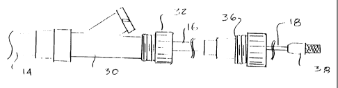
Referring now to Fig. 1B, an exemplary contraceptive system 10 generally
includes a contraceptive device 12, a sheath 14 partially surrounding the
contraceptive
device, a release catheter 16, and a core shaft 18. Contraceptive device 12
generally has a
proximal portion 20 adjacent a proximal end 22 (disposed within sheath 14),
and a distal
portion 24 adjacent a distal end 26 (which are exposed beyond the distal end
of
sheath 14). Distal portion 24 generally functions as a distal guidewire while
system 10 is
advanced within the tubal ostium. Proximal portion 20 includes a radially
expandable
structure which can be expanded after sheath 14 is withdrawn so as to affix
the
contraceptive device in the deployed position.
Sheath 14 is generally a tubular structure having a distal end 28 and
extending proximally to a proximal housing 30. Sheath 14 will generally have a
length in
CA 02381790 2002-02-12
WO 01/13832 PCT/US00/23012
-11-
a range from about 25 to about 50 cm, and will typically have an outer
diameter in a
range from about 0.020 to about 0.060 inches, the exemplary sheath having a
length of
about 39.5 cm and an outer diameter of about 0.04 inches. The inner diameter
of
sheath 14 may be in a range from about 0.02 inches to about 0.05 inches, with
the
exemplary sheath having an inner diameter of about 0.033 inches. Proximal
housing 30
includes a side arm with an injection port to allow infusion of fluids for
patency checks,
delivery of local anesthetic, or the like. Proximal housing 30 also includes a
Touhy-Borst
valve 32 releasably securing sheath 14 to release catheter 16.
Release catheter 16 generally comprises a tube having a distal end 34
which releasably engages contraceptive device 12, and a proximal end adjacent
a
proximal fitting 36. Release catheter 16 will generally be longer than sheath
14, and
fitting 36 will include another Touhy-Borst valve releasably securing release
catheter 16
to core shaft 18. The release catheter length is sufficiently longer than the
sheath 14 so
that full retraction of the sheath exposes the distal end of the release
catheter, thereby
allowing the release of the expandable structure upon movement of the release
catheter to
be hysteroscopically monitored. It should be understood that the Touhy-Borst
valve may
be replaced by any coupling structure which inhibits axial and rotational
movement
between the coupled devices, such as a key-slot arrangement or the like.
In the exemplary embodiment, core shaft 18 comprises a resilient tapering
structure extending from within distal portion 24 of contraceptive device 12
proximally
through fitting 36 of release catheter 16 to a proximal handle 38. Core shaft
18
threadably engages contraceptive device 12 proximally of distal end 28 of
sheath 14
before deployment. In the exemplary embodiment, core shaft 18 and release
catheter 16
transmit a wind-down torque onto an expandable structure of the contraceptive
device so
as to maintain the expandable structure in the small profile configuration.
Hence, release
catheter 16 relative to releasing core shaft 18 by actuating the Touhy-Borst
valve of
fitting 36 allows the expandable structure to be activated independently of
movement of
the surrounding sheath.
While exemplary contraceptive device 12 makes use of a radially
expandable helical coil to help restrain the structure during tissue ingrowth,
a wide variety
of mechanical and other restraint mechanisms might be included. For example,
alternative mechanical anchors might be attached to the device, such as
resilient coils
CA 02381790 2002-02-12
WO 01/13832 PCT/US00/23012
-12-
biased to form bends, loops, and/or other secondary shapes having enhanced
cross-
sections, slotted tubes, Malecot-type structures, radially expandable braids,
stent-like
devices, and the like. The mechanical structures may be resilient, plastically
deformable,
or the like, and suitable structures are described in more detail in, for
example, PCT
Publication No. WO 99/15116.
Still further device-restraint techniques might be employed, including
thermal, chemical, adhesive, and the like. These techniques can be used to
avoid
expulsion by increasing friction between the device and the surrounding
tissues, by
imposing limited tissue damage to promote scar tissue formation, and/or by
promoting
tissue ingrowth into the device. Thermal techniques may include, for example,
transmission of electrical or laser energy along contraceptive system 10.
Resistive
heating of contraceptive device 10 might be effected by applying an electrical
potential
across the device with conductors extending along sheath 14 and release
catheter 16, laser
energy along an optical wave guide attached to core wire 18, or the like.
Monopolar
tissue desiccation might be effected via a large return electrode patch by
energizing core
wire 18 with radiofrequency energy, or an adhesive and/or caustic agent (such
as a
cyanoacrylate or silver nitrate) might be introduced via any of the lumens of
the delivery
system, via a dedicated lumen or structure, or the like. Biodegradable plugs
and the like
might also be included, and the retained structure may optionally comprise
copper or
other bioactive agents to help inhibit conception.
Tissue reaction to the retained contraceptive device 12 can help to provide
long term contraception and/or sterilization. To promote conception inhibiting
tissue
reaction, device 12 will often include a tissue reaction material, the
material often
comprising fibers. The fibers may comprise a polyester, such as Dacron
polyesters, silk,
nylon, or the like. The fibers may be in the form of a weave, a knit, a braid,
a felt, or the
like, or may comprise stands attached to the device body.
The components of contraceptive system 10 can be further understood
with reference to Figs. 2 through 5, in which these components are illustrated
individually. Beginning with Fig. 2, core shaft 18 tapers to a gradually
increasing
diameter proximally of distal end 40 so as to provide increasing support of
distal
portion 24, proximal portion 20, and the catheter structures proximal of
contraceptive
device 12. This increasing support (and the associated increase in column
strength)
CA 02381790 2002-02-12
WO 01/13832 PCT/US00/23012
-13-
enhances the pushability of the contraceptive system while accessing the
target
deployment site. Threads 42 threadingly engage a coil of the contraceptive
device, and
are generally formed by affixing a coil with separated windings to a central
core wire at a
bond 44. A tube 43 may also be affixed at bond 44 to prevent binding and/or
jumping of
the cooperating threads, the tube ideally comprising stainless steel,
platinum, or the like.
In the exemplary device, core wire 18 comprises a high strength metallic
structure having a diameter in a range from about .003 inches to about .037
inches. The
ideal core wire has a total length of about 65 cm between distal end 40 and
proximal
handle 38, while threads 42 are separated from the distal end by a distance of
about 3 cm.
Core wire 18 tapers from a minimum diameter of about .003 inches near the
distal end to
a diameter of about 0.011 inches adjacent threads 42, and to a maximum
diameter of
about 0.029 inches proximally of the threads. The exemplary core wire
comprises nickel
titanium, while threads 42 comprise stainless steel attached to the central
wire by a
bond 44 of silver tin.
While the exemplary system uses threads to couple the core wire (or other
deployment shaft) with the contraceptive device, a variety of alternative
detachable
connections might be used, including cooperating keys/slots, BNC connectors,
or the like.
The exemplary contraceptive device 12 is illustrated in more detail in Fig. 3.
Contraceptive device 12 includes a primary coil 50 which extends from a distal
ball tip 52
to proximal threads 54, which may conveniently be formed by separating the
proximal
windings of the primary coil. The expandable structure, here in the form of a
helical
outer coil 56, has a proximal end bent to form a wind-down attachment 58, and
has a
distal end affixed to coil 50 at coil bond 60. Fiber 62 extends between the
inner and outer
coils, and is also disposed within primary coil 50 so as to promote tissue
ingrowth
throughout the cross-section of contraceptive device 12. The arrangement of
coil
attachment 58 and position of fiber 62 can be seen in the axial view of Fig.
3A. By
making use of a contraceptive device having a distal portion 24 which can act
as a
guidewire, no open lumen need be provided through the center of the
contraceptive
device (for example, for a separate guidewire), and multiple access/deployment
steps (for
example, accessing the target location with a guidewire, advancing a catheter
over the
guidewire, removing the guidewire from the positioned catheter, and then
advancing the
contraceptive device) can be avoided.
CA 02381790 2002-02-12
WO 01/13832 PCT/US00/23012
-14-
A slight variation upon the wind-down attachment is illustrated in Fig. 3B.
An alternative contraceptive device 12a includes a small tube or band 59
soldered within
a small diameter proximal section of outer coi156. Band 59 can have a
relatively large
interface area with coil 56 to facilitate bonding, avoids stress
concentrations, and presents
a smooth inner lumen which may inhibit binding of the release catheter. Band
59 may
comprise stainless steel or platinum, ideally having an inner diameter of
about
0.023 inches and an outer diameter, with the thickness of the surrounding
outer coil and
solder bond, of about 0.03 inches. A similar band 59' may be disposed within
threads 54
of coil 50 to provide a radiopaque marker, and to inhibit thread jump. Band
59' may be
similar in structure to band 59, but shorter in length. Still further
alternative attachment
mechanisms are possible. For example, a mass or knob may be formed at the
proximal
end of outer coil 56 from a simple ball of solder or coil material, bend, or
the like, which
is slidably receivable within a slot or other opening of the delivery
catheter.
In the exemplary embodiment, coil 50 is formed of a high strength resilient
material, ideally comprising stainless steel wire having a diameter of about
.005 inches,
and wound to form a coil having an outer diameter of about .022 inches. Ball
tip 52
preferably has a cross-section which is larger than the cross-section of
coi150, the ball tip
generally having a diameter in a range from about 0.020 inches to about 0.050
inches, the
exemplary ball tip having a diameter of 0.027 inches.
Helical coi156 comprises a highly elastic high strength metal which is
biased to expand from the low profile configuration illustrated in Fig. 1 to
the larger
profile configuration illustrated in Fig. 3 when released within the target
site. In the
exemplary embodiment, outer coil 56 comprises a ribbon of a superelastic or
shape
memory alloy, and has a thickness in the range from about 0.001 inches to
0.002 inches
and a width in a range from about 0.010 inches to 0.020 inches, with the
ribbon being
biased to form a helical coil having an outer diameter of about 0.080 inches
and a length
of about 3.5 cm when not otherwise restrained. Outer coil 56 is preferably
fixed to
primary coil 50 by a bond 60 of solder. Bond 60 will preferably be separated
from ball
tip 52 by a distance in a range from about 0.4 cm to about 0.7 cm.
Advantageously,
bond 60 may be aligned with the distal end 28 of sheath 14 so as to help
present an
atraumatic increase in diameter between distal portion 24 of contraceptive
device 12 and
the sheathed proximal portion 20 prior to deployment.
CA 02381790 2002-02-12
WO 01/13832 PCT/US00/23012
-15-
Fiber 62 may comprise a polyester or the like. The fiber may be loosely
woven or matted strands, with at least one end of the fibers affixed to
primary coil 50 or
outer coil 56. In the exemplary embodiment, fiber 62 comprises between about
20 and 70
filaments of textured PET fibers.
Generally, the expandable structure will help hold contraceptive device 12
in place at least until tissue ingrowth occurs sufficiently so as to
permanently retain the
contraceptive device and/or may restrain the device permanently. Hence, the
expandable
structure will often benefit from a relatively high friction outer surface.
Such an outer
surface might make it difficult to advance the contraceptive device into
position if the
device is advanced without sheath 14.
Work in connection with the present invention has shown that resiliently
expandable structures which have sufficient strength to reliably hold the
contraceptive
device within the ostium of the fallopian tube may impose significant
frictional forces
against a surrounding sheath. These frictional forces can significantly
complicate the
accurate delivery of contraceptive device. Hence, outer coil 56 is preferably
maintained
in a small profile configuration within sheath 14 by applying a wind-down
torque
between core wire 18 and release catheter 16. The core wire can transfer the
wind-down
torque to outer coil 56 through cooperating threads 42, 54, with the direction
of the wind-
down torque preferably being arranged so that the wind-down torque discourages
decoupling of the threads. In other words, rotation of core wire 18 relative
to
contraceptive device 12 in a direction opposed to the wind-down torque is used
to detach
core wire 18 from contraceptive device 12. It should be understood that a
variety of
alternative deployment/expansion mechanisms might be used with alternative
expandable
structures, such as stent-like expandable structures, braids, etc.
The distal structure of release catheter 16 is shown in Figs. 4 and 4A. The
wind-down torque is releasably transferred between outer coi156 and release
catheter 16
by cooperation between bend 58 and pin 66 at the distal end 34 of the release
catheter 16.
Release catheter 16 generally includes a tubular body 68 formed of polyimide.
Pin 66 is
disposed within a lumen of tubular body 68, and is supported within the
tubular body by a
helical support coi170 and adhesive 72. Pin 66 comprises a stainless steel bar
having a
width of about .008 inches, a thickness of about .003 inches, and a total
length of about
1 cm, and extends distally from distal end 34 by a distance of about 3 mm.
Support
CA 02381790 2007-06-29
-16-
coil 70 also comprises stainless steel, and the support coil and pin 70 are
bonded within
tubular body 68 by cyanoacrylate, with the exemplary tubular body having an
inner
diameter of about 0.030 inches and an outer diameter of about 0.033 inches.
Interestingly, these tubular body dimensions may be driven by the wind-down
torque
transferred proximally by release catheter 16. Optionally, the device and/or
delivery
system may be adapted to facilitate visualization and/or confirmation that
release is
successful. For example, the outer coil may look visibly different before and
after
deployment due to gaps in the coil winding, or the like. Similar feedback may
be
provided by fluoroscopic or sonographic image changes.
The structure of sheath 14 is illustrated in more detail in Fig. 5. Distal
end 28 (see Fig. 5A) of sheath 14 will preferably be rounded, with the distal
end ideally
cooperating with coil bond 60 of contraceptive device 12 so as to avoid
friction and
facilitate distal navigation of delivery system 16 through the uterotubal
junction and into
the fallopian tube. The rounded distal end 28 may optionally be rounded along
both the
inner and outer diameter of sheath 14, or may primarily be rounded along the
outer
diameter so as to taper inwardly distally.
Sheath 14 will preferably have a multi-layer structure, with the layers
comprising (beginning at the outside) a hydrophilic coating 76 to reduce
friction during
tracking and navigation. Such hydrophilic coatings become quite slippery when
exposed
to fluid. Below hydrophilic coating 76 is a structural layer of a polymer 78
such as
TecoflexTM' along the proximal portion of sheath 14, and a reinforcing braid
80 of a metal,
ideally of stainless steel, is disposed within a layer of polyimide below
polymer layer 78.
Along the more distal portion of sheath 14, metal braid 82 is disposed within
polymer
layer 78 of TecoflexTM, or the like, and the polyimide layer is absent so as
to provide
enhanced flexibility. The inner lumen of sheath 14 is defined by a low
friction polymer
coating 84, the low friction polymer ideally comprising a PTFE such as Teflon
. Suitable
sheaths 14 may be commercially available from a variety of vendors. Exemplary
structures may be described in more detail in published PCT patent application
WO
98/57589-
As schematically illustrated in Figs. 5A through F, alternative
sheaths 14A, B, and C, include bumpers 57, 57', and 57", respectively. Bumper
57 has an
outer surface extending radially from the outer surface of the underlying
sheath.
CA 02381790 2002-02-12
WO 01/13832 PCT/USOO/23012
-17-
Although bumper 57 may optionally provide a tactile indication that the sheath
14A is
advancing distally beyond the target deployment position, it does not
necessarily prevent
the sheath from advancing so that the bumper can enter into the tubal ostium.
Bumper 57
may also provide a visible marker that hinders pushing of the sheath so that
the bumper
moves past the ostium. Optionally, bumper 57 may comprise a colored adhesive,
or may
comprise a clear adhesive with a colored band of material disposed underneath.
Alternative bumpers 57' and 57" may comprise polymer or metallic
structures, ideally comprising a polyethylene or a super-elastic shape memory
alloy.
These radially expandable bumper structures can be collapsed for delivery
through a
working lumen of a hysteroscope, and can then expand to impede advancement of
the
sheath by engaging the uterine tissue adjacent to the tubal ostium.
Referring now to Fig. 6, the sliding engagement between pin 66 of release
catheter 16 and bend 58 of outer coi156 is more clearly illustrated. Fig. 6
also shows how
the wind-down torque imposed on the outer coil by the core shaft 18 and
release
catheter 16 help maintain the outer coil in a small profile configuration
within sheath 14,
allowing the sheath to be withdrawn easily. The wind-down torque can be
released by
sliding release catheter 16 so that pin 66 slides free of bend 58. Optionally,
the release
catheter may first be allowed to rotate relative to the core shaft to reduce
the engagement
forces between bend 58 and pin 66.
Referring now to Fig. 7, a contraceptive kit 90 generally includes
packaging 92 containing delivery system 10 and instructions for its deployment
94.
Contraceptive system 10 will generally be hermetically sealed within a sterile
pouch 96.
Alternatively, packaging 92 may hermetically seal the contraceptive system.
Instructions
for use 94 will describe method steps for deployment of the contraceptive
system, as
described herein. The instructions for use may comprise printed material,
and/or may
optionally include machine-readable code (such as a CD ROM, floppy disk, or
the like)
and/or graphical information (such as a video tape). In some embodiments, the
instructions for use may at least in part be incorporated into packaging 92 or
sterile
pouch 96.
An exemplary method for use of contraceptive system 10 can be
understood with reference to Figs. 8 through 8D. System 10 is introduced
transcervically
through uterus U, generally under optical direction. Using hysteroscope S the
physician
CA 02381790 2002-02-12
WO 01/13832 PCT/US00/23012
-18-
directs the distal end of the system toward ostium 0 of fallopian tube F.
Alternatively,
some or all of the procedure may be performed under any medical imaging
modality,
including fluoroscopy, sonography, computer tomography, or the like. Uterus U
may be
irrigated using scope S and/or a separate irrigation system. Once ostium 0 is
located and
the scope S is oriented toward the ostium, system 10 is advanced distally
through the
working lumen of the scope and through the ostium and into the fallopian tube
using
distal portion 24 of the contraceptive device as a guidewire, while the
remainder of the
contraceptive device remains covered by sheath 14.
The outer hydrophilic coating of sheath 14 minimizes friction while
advancing system 10, and the sheath also provides structural column strength
to the
system. The distal ball tip of distal portion 24 aids tracking and navigation
through
fallopian tube F, while the primary coil structure flexes laterally to track
the tortuous
bends often found within the fallopian tube. In the exemplary embodiment, core
wire 18
extends into distal portion 24 to enhance column strength of the distal
portion beyond
sheath 14, but does not extend to the ball tip. Hence, the stiffness of distal
portion 24
increases proximally, further enhancing the distal portion's ability to track
the lumen.
In the exemplary embodiment, sheath 14 includes a visual marker 98
which can be seen from the scope of hysteroscope S (see Fig. 8B). Marker 98
will
preferably be positioned partially within ostium 0 and partially within uterus
U, thereby
indicating that contraceptive device 12 is disposed at the target position, as
the sheath,
core shaft, and contraceptive device are releasably locked together during
advancement
and positioning. As described above, marker 98 may comprise a bumper, a
structure
which extends radially from the sheath to provide a tactile position
indication.
Preferred positioning of contraceptive device 12 is illustrated in Fig. 8B.
Preferably, device 12 extends along the uterotubal junction UTJ, with the
device ideally
extending both proximally and distally of the uterotubal junction. The
uterotubal
junction UTJ typically has a length in a range from about 1 to about 2 cm, and
outer
coil 56 will preferably extend proximally beyond ostium 0 into uterus U by a
distance in
a range from about 0.5 to about 1.0 cm. Outer coil 56 will preferably extend
distally of
the uterotubal junction UTJ by a distance of at least .6 cm. Ideally, outer
coil 56 will
extend both proximally and distally of the plane of the uterotubal junction
UTJ by a
distance of at least about .275 inches. Extending the expandable structure
both distally
CA 02381790 2002-02-12
WO 01/13832 PCT/US00/23012
-19-
and proximally of this effective isthmus can provide anchoring proximally and
distally of
the isthmus, thereby avoiding movement of contraceptive device 12 from the
target
position while tissue ingrowth takes place. Advantageously, positioning
accuracy with a
range of about 1 cm may be provided by limiting marker 98 to a 1 cm length.
This
provides a sufficient positional tolerance for ease of use while helping to
ensure reliable,
well-anchored deployments.
Referring now to Figs. 8A, 8A1, and 8A2, positioned contraceptive
device 12 is deployed by first withdrawing sheath 14 from over the expandable
structure.
Touhy-Borst valve 32 of proximal housing 30 is actuated to allow sliding
movement
between sheath 14 and release catheter 16, and the proximal housing slides
proximally
along the release catheter while maintaining fitting 36 of the release
catheter in a fixed
position, as illustrated in Fig. 8A2. Advantageously, core shaft 18 and
release catheter 16
remain locked together by fitting 36, so that the expandable structure does
not impede
proximal movement of the sheath. Retraction of sheath 14 from the positioned
(but as
yet unexpanded) device 12 leaves the distal end of deployment system 10 in the
configuration illustrated in Fig. 8B. Advantageously, it may still be possible
to adjust the
position of the device while viewing a proximal portion of outer coil 56.
As can be understood with reference to Figs. 8B and 8B1, once proximal
housing 30 engages fitting 36, the Touhy-Borst valve of the fitting can be
actuated so as
to allow movement between core shaft 18 and release catheter 16. The core
shaft and/or
release catheter may be allowed to rotate relative to each other to at least
partially expand
outer coil 56. The surgeon slides release catheter 16 proximally while holding
handle 38
of core shaft 18 in a fixed position, as shown in Fig. 8B2, thereby
disengaging the release
catheter from the outer coil and allowing the outer coil to expand fully and
firmly
attaching contraceptive device 12 to the surrounding tissue, as seen in Fig.
8C.
Referring now to Fig. 8C1, to fully release contraceptive device 12 from
the remaining components of delivery system 10, core shaft 18 is rotated to
disengage the
threaded coupling 42 between the core shaft and the contraceptive device. As
described
above, the direction of rotation of the core shaft for disengagement will be
opposite that
imposed by the wind-down torque, so that the wind-down torque helps maintain
the
threaded engagement prior to release of the core shaft relative to release
catheter 16.
Once core shaft 18 is unthreaded from contraceptive device 12, the core shaft
and other
CA 02381790 2007-06-29
-20-
delivery components can be withdrawn proximally into scope S, as shown in Fig.
8D.
Scope S can view outer coi156 to verify that the amount of the coil extending
proximally
of the ostium is within an acceptable range (and hence that device 12 is
disposed at the
target position) and the scope can be withdrawn after visually verifjring that
the
deployment has been successful.
Referring now to Fig. 9, a variety of alternative deployment methods
might be used to deploy the contraceptive system 10. For example, using a
simple
cervical catheter 102, deployment might be directed sonographically,
fluoroscopically,
under magnetic resonance imaging, and possibly even solely from tactile
information. In
the alternative exemplary method illustrated in Fig. 9, a balloon 104 of
cervical
catheter 102 is inflated via inflation port 106. This allows the uterus U to
be distended by
introduction of distention media through a uterine catheter 108 inserted
through the
working lumen of cervical catheter 102. Preferably, anatomy and target
location
identification, device positioning, deployment, detachment, and position
confirmation (as
outlined in method 2 with reference to Fig. 1 A) is performed under the
guidance of
ultrasound and/or fluoroscopic imaging. Relevant uterine catheter manipulation
structures and methods are described in U.S. Patent Nos. 5,346,498; and
5,389,100.
As described above, the delivery systems of the present invention will
often hold the contraceptive device in a fixed position while the
contraceptive device is
uncovered, expanded, and/or released. When moving, for example, outer sheath
14 so as
to expose the proximal portion of the contraceptive device, friction between
the outer
sheath and the surrounding hysteroscope (or other introducing structure,
surrounding
tissue, or the like) may cause inadvertent movement of the contraceptive
device. To
avoid such inadvertent movement, an outer sleeve may be slidably disposed
around outer
sheath 14. The sleeve provides a sliding interface between the sheath and
surrounding
structures. By axially coupling the sleeve and core shaft 18, friction between
the sleeve
and surrounding structures may inhibit movement of the contraceptive device.
Such a
sleeve will typically be shorter in length than sheath 14, and is more fully
described in a
concurrently filed application for a Deployment Actuation System for
Intrafallopian
Contraception.
CA 02381790 2002-02-12
WO 01/13832 PCT/US00/23012
-21-
Referring now to Figs. 10 and 11A, an alternative contraceptive system
150 includes a contraceptive device 152 having many of the components
described above,
but having an alternative wind-down outer coil connector 154 disposed at a
proximal end
of outer coil 56. An alternative release catheter 158 having a corresponding
connector
160 for engagement with connector 154 of contraceptive device 152 again allows
a wind-
down torque to be released, as described above. In this embodiment, wind-down
connector 160 of release catheter 158 comprises an opening which receives a
protrusion
162 extending radially from a tubular band 156 of connector 154. In the
exemplary
embodiment, band 156 comprises a platinum tube having a length of about 2.2
mm, and is
affixed to coil 56 using a solder bond. Protrusion 162 also comprises solder.
Referring now to Fig. 11B, an alternative wind-down connector 164 may
be affixed to a proximal end of outer coil 56 using a stainless steel ring
166, with the
outer coil welded to band 156 and the stainless steel ring welded to the band
over the
outer coil. In this embodiment, protrusion 162' is formed by welding a bent
platinum
ribbon to band 156. Band 156 may have a length of about 1.6 mm and an outer
diameter
of about 0.031", while protrusion 162' has an axial length of about 0.020",
and is formed
of a ribbon having a thickness of about 0.0015", with the ribbon being bent so
as to
extend about 0.04" radially beyond band 156. Typically, protrusions 162, 162'
will
extend radially a sufficient distance to extend into opening 160 of release
catheter 158,
with the release catheter and/or protrusion often having sufficient
flexibility to allow
disengagement of the wind-down connectors.
Referring now to Figs. 10, 12, and 13, contraceptive system 150 also uses
alternative threaded connectors 170, 172 for engagement between primary coil
50 and
core wire 18. Threaded connector 170 is affixed to primary coi150 of
contraceptive
device 152 by solder, and includes first and second interleaved coils 174,
with one of the
interleaved coils terminating 1/4 turn distally of the other to define a'/4
open-winding or
thread 176. An outer tube or stopper band 178 inhibits radial displacement of
the threads,
particularly when the threads are engaged between core wire 18 and the
stopper.
Preferably, primary coi150 comprises 0.005" diameter 316L stainless steel
wound to have
an outer diameter of 0.0125" with a 0.005" pitch and a length of about 2.9 cm.
First and
second interleaved coils 174 comprise 0.0039" x 0.008" 316L stainless steel
ribbon
wound to have an outer diameter of about 0.0205" and a 0.018" pitch. Stopper
178 may
CA 02381790 2002-02-12
WO 01/13832 PCT/US00/23012
-22-
comprise a platinum or Ptlr band having an outer diameter of about 0.026" and
a length
of about 1 mm. The stopper 178 and/or other components of at least one of the
connectors coupling inner coil 50 to corewire 18 and outer coil 56 to a
deployment
catheter will preferably provide a high contrast imaging marker.
Threaded connector 172 may similarly comprise interleaved coils 174
having differing lengths or axially positions so as to provide a'/4 turn open
winding or
thread, with the interleaved coils typically having more windings than used on
threaded
connector 170. An additional blocker coil 180 is disposed over and/or
proximally of
interleaved coils 174, with the coil being soldered to core wire 18, typically
using a SnAg
solder.
Preferably, threaded connectors 170, 172 will have less than five windings
engagement therebetween, more preferably having less than two engaged windings
and
ideally having less than a single winding of engagement. These limited engaged
windings are sufficient to maintain coupling between core wire 18 and the
contraceptive
device so long as wind-down torque is maintained, and facilitate detachment
after release
of the wind-down torque by limiting the number of rotations of core wire 18,
friction
between the core wire and the contraceptive device, and the like.
Release catheter 158 is shown in isolation in Fig. 14. The specific
configuration of connector or opening 160 may vary, for example, with the
opening being
nearer a distal end 182 of release catheter 14 when alternative protrusion
162' is used
(rather than protrusion 162 formed of solder). Still further variations are
possible,
including rectangular openings or channels having differing shapes or
extending axially
to distal end 182. In general, coupler or opening 160 will have a
circumferentially
oriented surface to releasably maintain a wind-down torque by corresponding
engagement
with an associated connector of the contraceptive device. In the exemplary
embodiment,
release catheter 158 comprises an polyimide affixed to a proximal release
catheter
housing by an adhesive such as a Lock-TiteTM 3321 adhesive. During assembly,
core
wire 18 may be inserted through release catheter 158 and coupled to the
contraceptive
device with the outer coil 56 being wound-down over the primary coil, and the
wind-
down torque maintained by coupling the proximal portions of the core wire 18
and release
catheter.
CA 02381790 2002-02-12
WO 01/13832 PCT/US00/23012
- 23 -
Referring now to Figs. 15 and 18, positioning surface 57 may optionally be
affixed to sheath 14 to help axially position contraceptive device 152 across
intermural
region INT, as described above. Engagement between radially protruding
positioning
surface 57 and the uterine tissues surrounding ostium 0 allows initial axial
positioning by
taking advantage of the axially coupling of sheath 14 to contraceptive device
152.
However, sheath 14 will be withdrawn proximally into scope S early-on during
deployment, and it is often desirable to maintain the axially positioning of
the
contraceptive device at least until proximal coil 56 begins to expand
radially.
As schematically illustrated in Fig. 15, by affixing positioning surface 57
(which may optionally comprise any of the alternative positioning surface
configurations
described hereinabove, or still further alternative structures such as
radially expandable
torroidal balloons, or the like) at a distal end of a separate positioning
catheter 184
slidably disposed over sheath 14, the axial positioning provided by the
positioning surface
may be maintained during and/or after withdrawal of sheath 14. Optionally, a
proximal
portion of release catheter 184 may be axially coupled to a proximal portion
of release
catheter 16, core wire 18, or another of the axially elongate structures so as
to maintain an
axial position of contraceptive device 152 using positioning surface 57.
Alternatively, the
positioning surface may be movable independently of these structures.
Still further structures and methods for releasably restraining the proximal,
radially expandable portion of the contraceptive device might be provided, as
can be
understood with reference to Figs. 16A, 16B, and 17. Figs. 16A and 16B each
illustrate a
distal end of an integrated sheath/release catheter 186, 188 having an axially
channel 190
defining a circumferentially oriented channel surface. Channel 190 cooperates
with a
protrusion 162 of connector 154 so as maintain wind-down torque on the
radially
expandable proximal coil of contraceptive device 152 via cooperation between
core wire
18 and the integrated release catheter/sheath. Additionally, the integrated
release
catheter/sheath slidingly surrounds the proximal, radially expandable portion
of
contraceptive device 152 so as to facilitate insertion of the device into the
fallopian tube.
As illustrated in Fig. 17, a tubular tool 192 having a lumen (which receives
the
contraceptive device) and a notch 194 (which receives protrusion 162) may
facilitate
winding-down proximal coil 56 and insertion of the proximal coil into the
integrated
CA 02381790 2007-06-29
-24-
release catheter/sheath, particularly if the tool has an outer diameter
sufficiently to allow
introduction of the tool into the lumen over the contraceptive device.
Channel 190 will generally have a length sufficient so as to allow an
integrated release catheter/sheath to slide axially from over protrusion 162
and over the
outer coi156, typically having a length of about 2.5 cms. Channel 190 may be
formed
during fabrication of the tubular sheath structure as shown in Fig. 16A, or
may be defined
by structures (such as a stainless steel, or NiTi ribbon) affixed within the
lumen using an
adhesive, a supporting coil, and/or the like.
Referring now to Fig. 19, an alternative outer sheath 214 may be used in
place of outer sheath 14 in the system of Fig. 1 B. Sheath 214 has a proximal
portion 216
with a relatively stiff, thicker-walled tubular structure, such as a PeBax
polymer tube
having an outer diameter of about 0.062", and an inner diameter of about
0.042". A distal
portion of sheath 14 includes an inner tube 218 of a low friction polymer and
an outer
tube 220 of a polymer, (such as carbothaneTM 73A) with at least one ribbon
coi1222
therebetween. Inner tube 218 may comprise a PTFE (such as a Teflon material)
with an
inner diameter of about 0.034" and a wall thickness of about 0.001" with the
outer
diameter etched, and a length of about 5.0 cm, while there are preferably two
counterwound ribbon coils 222 of a superelastic or shape memory alloy, such as
nickel
titanium (optionally with chromium) of about 0.007" by about 0.010" with a
pitch of
about 0.015" and a length of about 4.0 cm. Inner tube 218 might altematively
comprise
ETFE, gamma stable PTFE, FEP, or the like, while ribbon coils 222 may comprise
a
stainless steel or other medical grade materials. An inner diameter of the
distal portion
may be about 0.034", with the distal outer diameter of sheath 214 being about
0.041 ". An
intermediate outer tube 224 may comprise a polyurethane having a durometer of
about
55. A length of outer tube 220 may be about 1.0 cm, a length of intermediate
tube 224
may be about 5 mm, and a length of proximal portion 216 may be about 40 cm.
As can be understood with reference to Fig. 20, and as explained in detail
in co-pending application serial no. 09/644,287, now U.S. Patent No. 6,709,667
(Attorney
Docket No. 16355-003910US), a proximal handle mechanism 230 may be provided to
help
coordinate motion of the outer sheath, delivery catheter, corewire, and/or the
like. This proximal
handle may have a handle body which is axially coupled to the contraceptive
device, and any of
a wide variety of actuation mechanisms (such as syringe-like sliders,
ratcheting
CA 02381790 2002-02-12
WO 01/13832 PCT/USOO/23012
-25-
trigger handles, rack-and-pinion thumb wheels, and the like) can be used to
move, for
example, a proximal end of the outer sheath 14 and/or a proximal end of
release catheter
16 relative to a proximal end of core wire 18. Advantageously, these proximal
handle
mechanisms can be arranged to, for example, expose the proximal portion of
contraceptive device 12, then deploy the retention structure, and then detach
the deployed
device from the delivery system (as explained above), with two or more of
these steps
integrated into a continuous actuation movement at handle 230. Such actuation
handles
may greatly reduce the workload on the attending medical staff, possibly
reducing the
number of persons needed to effect deployment, and/or allowing contraceptive
device
exposure, deployment, and/or detachment to be effected with one hand on handle
230
(allowing the other hand to position a hysteroscope or the like).
Still further modifications of the contraceptive device and/or delivery
system are possible. For example, polyester fibers may be disposed both within
primary
coil 50 (ideally in the form of fiber loops) and around coil 50 (ideally in
the form of
wound Dacron layers disposed between primary coil 50 and outer coil 56) so as
to more
fully occlude the tubal lumen.
While the exemplary embodiment of the present invention has been
described in some detail, for clarity of understanding and by way of example,
a variety of
adaptations, changes, and modifications will be obvious to those who are
skilled in the
art. Hence, the scope of the present invention is limited solely by the
following claims.