Note: Descriptions are shown in the official language in which they were submitted.
CA 02381904 2007-07-04
IMPLANTS AND METHODS FOR SNORING TREATMENT
BACKGROUND
1. Field of the Invention
This invention is directed to methods and apparatuses for treating
snoring.
2. Description of the Prior Art
Snoring has received increased scientific and academic attention.
One publication estimates that up to 20% of the adult population snores
habitually.
Huang, et al.,"Biomechanics of Snoring", Endeavour, p. 96-100, Vol. 19, No. 3
(1995).
Snoring can be a serious cause of marital discord. In addition, snoring can
present a
serious health risk to the snorer. In 10% of habitual snorers, collapse of the
airway
during sleep can lead to obstructive sleep apnea syndrome. Id.
Notwithstanding numerous efforts to address snoring, effective
treatment of snoring has been elusive. Such treatment may include mouth guards
or
other appliances worn by the snorer during sleep. However, patients find such
appliances uncomfortable and frequently discontinue use (presumably adding to
marital
stress).
Electrical stimulation of the soft palate has been suggested to treat
snoring and obstructive sleep apnea. See, e.g., Schwartz, et al.,"Effects of
electrical
stimulation to the soft palate on snoring and obstructive sleep apnea", J.
Prosthetic
Dentistry, pp. 273-281 (1996). Devices to apply such stimulation are described
in U. S.
Pat. Nos. 5,284,161 and 5,792,067. Such devices are appliances requiring
patient
adherence to a regimen of use as well as subjecting the patient to discomfort
during
sleep. Electrical stimulation to treat sleep apnea is discussed in Wiltfang,
et al., "First
results on daytime submandibular electrostimulation of suprahyoidal muscles to
prevent night-time hypopharyngeal collapse in obstructive sleep apnea
syndrome",
International Journal of Oral & Maxillofacial Surgery, pp. 21-25 (1999).
1
CA 02381904 2002-03-15
WO 01/19301 PCT/US00/40830
Surgical treatments have been employed. One such treatment is
uvulopalatopharyngoplasty. In this procedure, so-called laser ablation is used
to
remove about 2 cm of the trailing edge of the soft palate thereby reducing the
soft
palate's ability to flutter between the tongue and the pharyngeal wall of the
throat.
The procedure is frequently effective to abate snoring but is painful and
frequently
results in undesirable side effects. Namely, removal of the soft palate
trailing edge
comprises the soft palate's ability to seal off nasal passages during
swallowing and
speech. In an estimated 25% of uvulopalatopharyngoplasty patients, fluid
escapes
from the mouth into the nose while drinking. Huang, et al., supra at 99.
Uvulopalatopharyngoplasty (UPPP) is also described in Harries, et al., "The
Surgical
treatment of snoring", Journal of Laryngology and Otology, pp. 1105 - 1106
(1996)
which describes removal of up to 1.5 cm of the soft palate. Assessment of
snoring
treatment is discussed in Cole, et al., "Snoring: A review and a
Reassessment",
Journal of Otolaryngology, pp. 303 - 306 (1995).
Huang, et al., supra, describe the soft palate and palatal snoring as an
oscillating system which responds to airflow over the soft palate. Resulting
flutter
of the soft palate (rapidly opening and closing air passages) is a dynamic
response
generating sounds associated with snoring. Huang, et al., propose an
alternative to
uvulopalatopharyngoplasty. The proposal includes using a surgical laser to
create
scar tissue on the surface of the soft palate. The scar is to reduce
flexibility of the
soft palate to reduce palatal flutter. Huang, et al., report initial results
of complete or
near-complete reduction in snoring and reduced side effects.
Surgical procedures such as uvulopalatopharyngoplasty and those
proposed by Huang, et al., continue to have problems. The area of surgical
treatment (i.e., removal of palatal tissue or scarring of palatal tissue) may
be more
than is necessary to treat the patient's condition. Surgical lasers are
expensive. The
proposed procedures are painful with drawn out and uncomfortable healing
periods.
The procedures have complications and side effects and variable efficacy
(e.g.,
Huang, et al., report promising results in 75% of patients suggesting a full
quarter of
patients are not effectively treated after painful surgery). The procedures
may
involve lasting discomfort. For example, scar tissue on the soft palate may
present a
continuing irritant to the patient. Importantly, the procedures are not
reversible in
the event they happen to induce adverse side effects not justified by the
benefits of
the surgery.
SUMMARY OF THE INVENTION
According to one aspect of the present invention, methods and
apparatuses are disclosed for treating snoring of a patient. The invention
includes an
2
CA 02381904 2007-07-04
implant of bio-compatible material sized to be embedded within the soft
palate.
When inserted, the implant is able to alter a dynamic response of the soft
palate to
airflow past the soft palate.
Accordingly, in one aspect of the present invention there is provided an
apparatus for use in the treatment of snoring, comprising:
an implant of biocompatible material suitable for embedding in a soft
palate of a patient;
the biocompatible material including a fibrosis-inducing material
selected to induce a fibrotic response of tissue in the soft palate following
implantation
of said implant; and
said fibrosis-inducing material provided in an amount which is sufficient
to induce a fibrotic stiffening of the soft palate following implantation of
said implant
to passively and without application of external force alter the dynamic
response of said
soft palate to airflow.
According to another aspect of the present invention there is provided an
implantation device comprising a needle and the apparatus as described above,
the
needle being suitable for implanting said implant into the soft palate.
According to yet another aspect of the present invention there is
provided a pack comprising the apparatus as described above and further
including
instructions to use the apparatus.
BRIEF DESCRIPTION OF THE DRAWINGS
Embodiments of the present invention will now be described more fully
with reference to the accompanying drawings in which:
Fig. 1 is a side sectional view of a portion of a human head showing a
soft palate in a relaxed state and in relation in adjacent anatomical
features;
Fig. 2 is a portion of the view of Fig. 1 showing the soft palate in a
flexed state;
Fig. 3 is a front view of an interior of the mouth shown in Fig. 1 and
showing an area to be ablated according to a first prior art surgical
procedure;
Fig. 4 is the view of Fig. 3 and showing an area to be scarred according
to a second prior art surgical procedure;
Fig. 5 is a schematic representation of a spring-mass system model of
the soft palate;
3
CA 02381904 2007-07-04
Fig. 6 is the view of Fig. 1 with the soft palate containing an implant
according to a first embodiment of the present invention;
Fig. 7 is the view of Fig. 3 showing the embodiment of Fig. 6;
Fig. 8 is a cross-sectional view of the implant of Fig. 6;
Fig. 9 is a first modification of the implant of Fig. 8 having a tissue in-
growth layer;
Fig. 10 is a second modification of the implant of Fig. 8 having a smooth
outer layer;
Fig. 11 is the view of Fig. 6 with the soft palate containing an iinplant
according to a second embodiment of the present invention;
Fig. 12 is the view of Fig. 7 showing the embodiment of Fig. 11;
Fig. 13 is a perspective view of the implant of Fig. 11;
Fig. 14 is a cross-sectional view of the implant of Fig. 13;
Fig. 15 is a view of the implant of Fig. 14 with the implant pre- formed
to assume the shape of a soft palate in a relaxed state;
Fig. 16 is the view of Fig. 14 with the implant constructed to have
greater flexion in a downward direction;
Fig. 17 is an exploded perspective view of first modification of the
implant of Fig. 13;
Fig. 18 is a perspective view of a modification of a housing of the
embodiment of Fig. 17;
Fig. 19 is a side section view of a second modification of the implant of
Fig. 13;
3a
CA 02381904 2002-03-15
WO 01/19301 PCT/US00/40830
Fig. 20 is a cross-sectional view of an implant that is another
embodiment of the present invention, the implant is shown in a flattened
orientation;
Fig. 21 is a cross-sectional view of the implant of Fig. 20 in an
expanded orientation;
Fig. 22 shows the implant of Fig. 20 in the flattened orientation and
implanted in the soft palate;
Fig. 23 shows the implant in Fig. 21 in the expanded orientation and
implanted in the soft palate;
Fig. 24 is a top plan view, shown partially broken away, of a still
further embodiment of the present invention;
Fig. 25 is a view taken along line 25 - 25 in Fig. 24;
Fig. 26 is a side sectional view of the implant of Fig. 24 collapsed
and placed within a delivery tool;
Fig. 27 is the view of Fig. 26 with the implant in the process of being
ejected from the delivery tool;
Fig. 28 is a view taken along line 28 - 28 in Fig. 26;
Fig. 29 is a side sectional view of the soft palate showing a palatal
muscle in the soft palate;
Fig. 30 is the view of Fig. 29 showing the delivery tool of Fig. 26
being advanced through an incision into the soft palate;
Fig. 31 is the view of Fig. 30 following delivery of the implant and
removal of the delivery tool; and
Fig. 32 is a view taken along line 32 - 32 in Fig. 31.
Fig. 33 is a perspective view of an implant according to a still further
embodiment of the present invention showing only a bio-resorbable, first
component;
Fig. 34 is a perspective view of the implant of Fig. 33 showing both a
first component and a second component;
Fig. 35 is a perspective of the implant of Fig. 33 showing only the
second component following bio-resorption of the first component;
Fig. 36 is a graph showing decrease of palatal stiffening attributable
to the first component and increase of palatal stiffening attributable to the
first
component;
Fig. 37 is a perspective view of an implant for use in the delivery
system of Figs. 38-39;
Fig. 38 is a side-sectional view of a delivery system for placing an
implant in the soft palate;
4
CA 02381904 2002-03-15
WO 01/19301 PCTIUSOO/40830
Fig. 39 is the view of Fig. 38 following delivery of the implant from
the delivery system;
Fig. 40 is a perspective view of a braided implant;
Fig. 41 is an end view of the implant of Fig. 40;
Fig. 42 is a side sectional view of an implant with an anchor;
Fig. 43 shows an implant in a perforated needle tip; and
Fig. 44 is a cross-sectional view of the implant and needle tip of Fig.
43.
DESCRIPTION OF THE PREFERRED EMBODIMENT
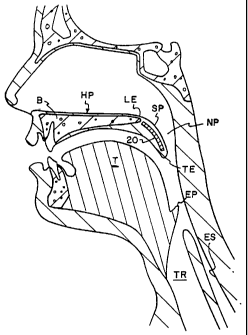
For ease of understanding the present invention, the dynamics of
snoring are explained with reference to Figs. 1- 4. The hard palate HP
overlies the
tongue T and forms the roof of the mouth M. The hard palate HP includes a bone
support B and does not materially deform during breathing. The soft palate SP
is
soft and is made up of mucous membrane, fibrous and muscle tissue extending
rearward from the hard palate HP. A leading end LE of the soft palate SP is
anchored to the trailing end of the hard palate HP. A trailing end TE of the
soft
palate SP is unattached. Since the soft palate SP is not structurally
supported by
bone or hard cartilage, the soft palate SP droops down from the plane of the
hard
palate HP in an arcuate geometry of repose.
The pharyngeal airway passes air from the mouth M and the nasal
passages N into the trachea TR. The portion of the pharyngeal airway defined
between opposing surfaces of the upper surface of the soft palate SP and the
wall of
the throat is the nasopharynx NP.
During normal breathing, the soft palate SP is in the relaxed state
shown in Fig. 1 with the nasopharynx NP unobstructed and with air free to flow
into
the trachea TR from both the mouth M and the nostrils N.
During swallowing, the soft palate SP flexes and extends (as shown
in Fig. 2) to close the nasopharynx NP thereby preventing fluid flow from the
mouth
M to the nasal passages N. Simultaneously, the epiglottis EP closes the
trachea TR
so that food and drink pass only into the esophagus ES and not the trachea TR.
The
soft palate SP is a valve to prevent regurgitation of food into the nose N.
The soft
palate SP also regulates airflow through the nose N while talking. Since the
soft
palate SP performs such important functions, prior art techniques for
surgically
altering the soft palate SP can compromise these functions.
The majority of snoring is caused by the soft palate SP flapping back
and forth. If breathing is solely through the nose N with the mouth closed,
the
trailing edge TE of the soft palate SP is sucked into the nasopharyngeal space
NP
5
CA 02381904 2002-03-15
WO 01/19301 PCT/US00/40830
obstructing the airway and subsequently falls opening the airway in a
repeating
cycle. When the mouth is open, air flows over the upper and lower surfaces of
the
soft palate SP causing the soft palate SP to flap up and down alternating in
obstructing the oral and nasal passageways M, N. The snoring sound is
generated by
impulses caused by rapid obstruction and opening of airways. Huang, et al.,
state
the airway passage opening and closing occurs 50 times per second during a
snore.
Huang, et al., utilize a spring-mass model (Fig. 5) to illustrate oscillation
of the soft
palate in response to airflow (where the soft palate is the ball B of mass
depending
by a spring S from a fixed anchor A).
Huang, et al., analogize the shortening of the soft palate SP in
uvulopalatopharyngoplasty as effectively raising the critical air flow speed
at which
soft palate flutter will occur. The shaded area SA in Fig. 3 shows the area of
the
trailing end TE of the soft palate SP to be removed during this procedure. The
alternative procedure proposed by Huang, et al., reduces the flexibility of
the soft
palate SP through surface scarring which is asserted as effecting the critical
flow
speed. The shaded area SA' in Fig. 4 shows the area to be scarred by this
alternate
procedure. In Fig. 4, dashed line L shows the demarcation between the soft and
hard
palates.
Using the spring-mass model of Fig. 5 as a convenient model of the
soft palate SP, the present invention is directed to a surgical implant into
the soft
palate SP to alter the elements of the model and thereby alter the dynamic
response
of the soft palate SP to airflow. The implant can alter the mass of the model
(the
ball B of Fig. 5), the spring constant of the spring S, the dampening of the
spring S
or any combination of these elements. Unlike the prior art surgical
techniques, the
implants that will be described are easy to insert in a small incision
resulting in
reduced patient discomfort and are not exposed to the interior of the mouth
(such as
the surface scarring of Huang, et al.) as a patient irritant. Also, as will be
described,
the degree of dynamic remodeling can be fine tuned avoiding the need for
excessive
anatomical modification and are reversible in the event of adverse
consequences.
Figs. 6 - 7 illustrate a first embodiment of the present invention
where individual units 10 of mass (in the form of implantable modular devices
such
as spheres or implants of other geometry) are imbedded in the soft palate SP
in close
proximity to the trailing end TE. With reference to the model of Fig. 5, the
spheres
add mass to the mass-spring system thereby altering dynamic response to
airflow
and adding resistance to displacement and accelerating. The placement of the
units
10 of mass also alter the location of the soft palate's center of mass further
altering
the model and dynamic response.
6
CA 02381904 2002-03-15
WO 01/19301 PCTIUSOO/40830
The embodiment of Figs. 6 - 10 is tunable to a particular patient in
that multiple modules 10 can be implanted (as illustrated in Fig. 7). This
permits the
surgeon to progressively increase the number of implanted modules 10 until the
altered dynamic response is such that snoring inducing oscillation is abated
at
normal airflow. The individual modules 10 may be placed into the soft palate
SP
through small individual incisions closed by sutures which is much less
traumatic
than the gross anatomical destruction of uvulopalatopharyngoplasty or the
large
surface area scarring proposed by Huang, et al.
Preferably, such modules 10 of mass are solid modules such as
spheres of biocompatible material which are radiopaque (or radio-marked) and
compatible with magnetic resonance imaging (MRI). Titanium is such a material.
By way of non-limiting example, the modules 10 of mass may be about 2 - 4 mm
in
diameter. In the case of pure, non-sintered titanium, each such sphere 10
would add
.15-1.22 gm of mass to the trailing end TE of the soft palate SP and
contribute to re-
modeling the mass distribution of the soft palate SP. An example of an
alternative
material is any biocompatible ceramic.
As shown in Fig. 9, the spheres (labeled 10' to distinguish from the
version 10 of Fig. 8) may be sintered throughout or otherwise provided with
tissue
growth inducing material 12 on their outer surface. Such material may be a
sintered
outer layer or a coating or covering such as a polyester fabric jacket. Such
material
permits and encourages tissue in-growth to secure the implant 10' in place.
Also,
placement of an implant 10 or 10' will induce a fibrotic response acting to
stiffen the
soft palate SP (and further alter the dynamic response and resistance to
displacement
and acceleration). A sintered or coated sphere 10' will enhance the fibrotic
response
and resulting stiffening.
While tissue in-growth and enhanced fibrotic response have the
benefits described above, such embodiments may make the implant 10' more
difficult to remove in the event reversal of the procedure is desired.
Therefore, as
shown in Fig. 10 as an alternative, the spheres (labeled 10" to distinguish
from the
implants 10, 10') may be coated with smooth coating 14 (such as parylene or
PTFE)
to reduce fibrosis.
The embodiments of Figs. 6- 10 add to and relocate the mass of the
spring-mass system of Fig. 5 to remodel the dynamic response. The amount of
mass
is selected to alter the dynamic response but not preclude the soft palate SP
being
moved to close off nasal passages N during swallowing. Through fibrotic
response
and incision healing, the spring S of the model is stiffened.
In addition to modifying the mass profile of the spring-mass system,
the spring component S of Fig. 5 can be modified (alone or in combination with
7
CA 02381904 2002-03-15
WO 01/19301 PCTIUSOO/40830
mass modification) to alter dynamic response. Fig. I 1- 16 illustrate an
implant 20
in the form of a flexible strip for placement in the soft palate. The use of
the term
"strip" herein is not intended to be limited to long, narrow implants but can
also
include plates or other geometries implanted to alter the dynamic model of the
soft
palate SP. Elongated strips are presently anticipated as a preferred geometry
to
facilitate ease of implant.
The strip 20 has a transverse dimension less than a longitudinal
dimension. By way of non-limiting example, the strip may have a length LS of
about
20-30mm,athicknessTSofabout2-4mmandawidthWSof5-10mm. As
shown in Fig. 11, the strip 20 is embedded in the soft palate SP with the
longitudinal
dimension LS extending from adjacent the hard palate HP toward the trailing
end TE
of the soft palate SP. As shown in Fig. 12, multiple strips 20 may be embedded
in
the soft palate SP extending either straight rearward or angled to the sides
while
extending rearward. The strips 20 may be formed straight (Fig. 14) or pre-
shaped
(Fig. 15) to have a rest shape approximate to the side-cross section shape of
the soft
palate in a relaxed state.
The strips 20 may be any flexible, biocompatible material and are
preferably radiopaque or radio-marked as well as MRI compatible. The strips 20
need not be elastic and having a material spring constant biasing them to
their
original shape. Such strips 20 could simply be flexible, plastically
deformable strips
which are stiffer than the soft palate SP to reinforce the soft palate SP and
assist the
soft palate SP in resisting deflection due to airflow. Such stiffening of the
soft palate
SP stiffens and dampens the spring S in the spring-mass system of Fig. 5 and
alters
the dynamic response of the soft palate SP. The strip 20 may be a spring
having a
spring constant to further resist deflection of the soft palate SP as well as
urging the
soft palate SP to the relaxed state of Fig. 5. The stiffness of the strip 20,
a spring
constant of the strip 20, and the number of strips 20, are selected to avoid
preclusion
of closure of the soft palate SP during swallowing. Examples of suitable
materials
include titanium and nitinol (a well-known nickel-titanium alloy). As with the
examples of Figs. 9 and 10, the strips 20 may be provided with tissue in-
growth
surfaces or may be coated as desired. Also, the strips may be structurally
modified
to control their flexibility. In Fig. 16, the bottom 22 of the strip 20
(facing the
tongue after placement) is provided with transverse notches 24 to enhance
downward flexion of the strip 20 relative to upward flexion of the strip 20
following
placement.
Fig. 17 provides an alternative to the strips 20 of Fig. 13. In Fig. 17,
the strip 20' includes a housing 26 having an interior space 28 with an access
opening 30. The interior space 28 extends in the longitudinal dimension of the
8
CA 02381904 2002-03-15
WO 01/19301 PCTIUSOO/40830
housing 26. The strip 20' further includes a longitudinal insert 32 sized to
be passed
through the access opening 30 and into the space 28. By way of non-limiting
example, the housing 26 could be silicone rubber (with radio-markers, not
shown, to
indicate placement) and the inserts 32 could be titanium rods or other
flexible
member. With the embodiment of Fig. 17, the housing 26 (without an insert) may
be embedded in the soft palate SP. The housing 26 acts independently as a
stiffening strip to add stiffness to the soft palate SP to alter the soft
palate's dynamic
response. In the event further stiffening or a spring action is desired, the
implant 20'
can be selectively tuned to the patient's unique dynamic model by placing the
insert
32 into the space 28 at the time of initial surgery or during a subsequent
procedure.
The embodiment of Fig. 17, permits selection of an insert 32 from a wide
variety of
materials and construction so that an insert 32 of desired characteristics
(e.g.,
stiffness and spring action) can be selected to be inserted in the space 28
and alter
the dynamic response as desired. The embodiment of Fig. 17 also permits later
removal of the insert 32 and replacement with a different insert 32 of
different
properties for post-surgery modification of the soft palate's dynamic
response.
The embodiment of Fig. 18 is similar to that of Fig. 17. The housing
26' is provided with multiple, parallel-aligned interior spaces 28' and access
openings 30'. In addition to the function and benefits of the embodiment of
Fig. 17,
the number of inserts 32 may be varied to alter and adjust the dynamic
response of
the soft palate SP.
Fig. 19 illustrates a still further embodiment of the strip implant. In
Fig. 19, the strip 20"' is a bladder having a housing 26" in the form of a
completely
sealed envelope of flexible synthetic material defining an interior space 28".
The
envelope 26" is preferably self-sealing following needle injection. Fluid is
injected
into the housing 26" (e.g., through hypodermic needle 40 injection) to stiffen
the
strip 20"'. Addition of fluid further stiffens the strip 20"' and further
alters the
dynamic response of the soft palate SP. Removal of fluid increases the
flexibility.
Unlike the embodiments of Fig. 17 (where inserts 32 are most effectively
replaced
post-operatively through incision to alter flexibility), the embodiment of
Fig. 19
permits selectively varying flexibility of the soft palate SP through needle
injection.
An alternative to Fig. 19 is to fill the space 28" with a so-called phase
change
polymer and inject a stiffening agent into the space 28" to alter the
flexibility of the
polymer.
Figs. 20 - 23 illustrate a still further embodiment of the present
invention. In the foregoing embodiments, the spring-mass system of Fig. 5 is
altered
by altering the mass of the soft palate SP or the spring characteristics of
the soft
palate SP. The dynamic response can also be altered by altering the force
acting on
9
CA 02381904 2002-03-15
WO 01/19301 PCT/US00/40830
the spring-mass system. Namely, the force acting on the soft palate SP is
generated
by airflow over the surface of the soft palate. The soft palate acts as an
airfoil which
generates lift in response to such airflow. By modifying the longitudinal
(i.e.,
anterior to posterior) cross-sectional geometry of the soft palate SP, the
aerodynamic
response and, accordingly, the dynamic response are altered.
In the embodiments of Figs. 20 - 23, the implant 30 is inserted into
the soft palate SP through an incision. The implant 30 has an oval shape to
cause
deformation of the geometry of the soft palate SP. Prior to implantation, the
implant
30 is preferably formed as a flat oval (Figs. 20 and 22) for ease of
insertion. After
implantation, the implant 30 expands to an enlarged oval (Fig. 21 and 23).
While
such expansion could be accomplished mechanically (i.e., through balloon
expansion), the implant 30 is preferably formed as a shape-memory alloy (such
as
nitinol) which expands to the enlarged shape in response to the warmth of the
body.
In addition to changing the aerodynamics of the soft palate SP, the implant 30
can be
constructed with a mass and stiffness as desired to alter the spring and mass
components of the spring-mass system of Fig. 5.
Figs. 24 - 32 illustrate an expandable implant 50 and a delivery tool
60 for placing the implant 50 in the soft palate SP through a small incision.
In Figs.
24 and 25, the implant 50 is best illustrated as a flexible rim 52 with a
fibrosis-
inducing agent in the form of a flexible material, for example polyester
fabric 54,
retained on the rim 52. The rim 52 may be titanium or other material and
resiliently
biased to a rest geometry shown as an oval in Fig. 24 having a fully expanded
width
W and a length L. An oval is illustrated as a preferred geometry but other
geometries may suffice. The geometries may include geometries selected to
alter the
shape of the soft palate SP. The polyester fabric 54 (such as Dacron or the
like)
contains interstitial spaces for fibrosis and tissue integration to impart a
stiffening to
the soft palate SP.
The soft palate SP is schematically shown in Figs. 29 - 32 with a
palatal muscle PM extending distally from the bone B of the hard palate and
surrounded by the soft tissue ST of the soft palate SP. The implant 50 is
placed by
compressing the implant 50 against the bias of the rim 52 into a compact
cylindrical
shape of length L and placing the compressed implant 50 in a distal end of a
cylindrical delivery tool 60. The distal tip 62 of tool 60 is a blunt beveled
end to
follow an incision and to separate tissue as the tip 62 is advanced. A rod 64
is
positioned proximal to the implant 50. The distal tip 62 is severable such
that
pushing rod 64 urges the implant 50 out of the distal tip 62. When removed
from
the delivery tool 60, the implant 50 springs back to an oval geometry.
CA 02381904 2002-03-15
WO 01/19301 PCTIUSOO/40830
The implant 50 is placed by forming a small incision 70 in the soft
palate. In Fig. 29, the incision is made on the lower surface of the soft
palate. The
procedure could also be performed through the upper surface of the soft
palate. The
incision is sized to pass the distal tip 62 of tool 60 which is substantially
smaller
than the full width W of the expanded implant 50.
Any suitable blunt dissecting tool may be inserted into incision 70 to
separate the soft tissue ST from the palatal muscle PM by an amount sufficient
to
receive the expanded implant 50. The distal tip 62 is placed through the
incision 70
and advanced through the soft palate SP with the distal tip 62 separating the
soft
tissue ST and the palatal muscle PM (Fig. 30). The tool 60 can be advanced by
the
physician tactilely noting position of the too160 or through any visualization
technique (e.g., an endoscope on the distal tip 62). When the distal tip 62 is
fully
advanced, the outer tube 66 of tool 60 is retracted while holding rod 64 in
place
causing the implant 50 to be expelled through the distal tip 62. After full
expulsion
of the implant 50, tool 60 is removed through incision 70. The released
implant 50
then expands into the oval shape and residing between the palatal muscle PM
and
the soft tissue ST (Figs. 31 and 32).
In place, the fabric 54 of implant 50, encourages fibrosis and
stiffening of the soft palate SP. By inserting a collapsed implant 50 through
a small
incision 70, a large surface area of fibrosis (and greater stiffening) can be
achieved
with a minimized incision 70 (resulting in reduced patient discomfort). Also,
while
the implant 50 is illustrated as being resiliently expandable, the implant 50
could
expand or swell in response to other factors such as shape memory alloys
(e.g.,
nitinol), smart polymers and balloon expandable and plastically deformable
metals.
As an alternative to the foregoing, a catheter (not shown) can be
passed through incision 70 and passed through the soft palate SP. The delivery
tool
60 can be passed through the catheter. If desired, a coring tool (not shown)
can be
passed through the catheter to remove tissue from the soft palate SP prior to
placing
the implant 50 (or any implant of the previous embodiments). Also, for small
implants, an implant can be placed through any short tube inserted into the
soft
palate through a needle poke and need not include a pre-incision.
With reference to Figs. 33-36, a still further embodiment of the
invention is described. In Figs. 33-36, an implant 80 is shown having a
cylindrical
shape. The shape is illustrative only. The implant 80 may be deployed through
a
delivery tool 60 as previously described.
The implant 80 includes two stiffening components. A first
component 82 is a base of a bio-resorbable material such as bio-resorbable
suture
formed into a woven cylindrical shape. Such material has a stiffness greater
than
11
CA 02381904 2002-03-15
WO 01/19301 PCT/USOO/40830
soft tissue and is absorbed into the body over time. An example of such
material is
synthetic absorbable suture such as polydioxanone suture sold by Ethicon, Inc.
under
the trademark PDS II. Alternative materials could include absorbable bio-
adhesives.
A first component as described provides immediate post-operative stiffening to
reduce or eliminate snoring immediately following placement of the implant 80
in
the soft palate.
The second component 84 is any fibrosis inducing material combined
with the first component 82. By way of non-limiting example, the second
component may be filaments of polyester or polyester fabric (such as Dacron )
intertwined in the interstitial spaces of the first component 82. The presence
of the
second component 84 in the soft tissue of the soft palate SP induces fibrosis
which
stiffens the soft palate to reduce or eliminate snoring. The stiffening
increases with
time following implantation until the fibrotic response is steady state. The
polyester
second component 84 is permanent and does not bio-resorb. Therefore, the
fibrosis
effect (and, hence, the snoring reducing stiffening) remains permanently
following
implantation and following complete absorption of the first component 82.
The first component 82 and the second component 84 cooperate for
the implant 80 to provide effective stiffening immediately post-operatively
and
chronically thereafter. The first component has a stiff material which
stiffens the
soft palate SP upon placement. However, over time, the first component is
absorbed
and the stiffening influence reduces and is eliminated. The second component
84 is
formed of very floppy material which does not materially stiffen the soft
palate
immediately upon implantation of implant 10. However, with time, fibrosis
induced
by the material of the second component 84 stiffens the soft palate. This
phenomena
is illustrated in the graph of Fig. 36 in which the horizontal axis represents
time and
the vertical axis represents stiffening provided by the implant 10. Line A is
stiffening attributable to the first component 82 (which decays to zero as the
first
component is absorbed). Line B represents stiffening attributable to the
second
component (which is at near zero at implantation and increases to a maximum
representing a steady-state level of fibrosis). Line C represents stiffening
of the soft
palate SP which is a sum of the stiffening of lines A and B.
Therefore, with the embodiment of implant 80, immediate post-
operative stiffening (and snoring abatement) is achieved. Chronic stiffening
is
provided by fibrotic response which is permanent. Total stiffening is
controlled
since the first component 82 is being absorbed as the fibrosis at the second
component 84 increases.
Figs. 37-39 show an alternative delivery system 100 for placing an
implant in the soft palate SP. Figs. 37-39 illustrate use of the novel
delivery system
12
CA 02381904 2002-03-15
WO 01/19301 PCT/USOO/40830
100 with a cylindrical implant 102 (such as implant 80 of Fig. 34 or implant).
However, the method and apparatus described with reference to Figs. 37-39
could
also be used with other geometries (e.g., the spherical implants of Fig. 7 or
rectangular cross-section implants of Fig. 13) as well as an expandable
implant as
such implant 50 of Fig. 24.
A needle 66' is provided having a ground beveled distal tip 61' for
piercing tissue of the soft palate. The needle 66' is hollow and carries the
implant
102 in sliding close tolerance. A rod 64' is slidably positioned in the needle
66'
proximal to the implant 102. As described above with reference to Figs. 26-32,
the
implant 102 is carried by the needle 66' to a desired implant site within the
soft
palate. At the desired site, the implant 102 is deployed by retracting the
needle 66'
while holding the rod 64' in place. Relative movement between the rod 64' and
needle 66' causes the rod 64' to dispel the implant 102 from the needle 66'
without
need for moving the implant 102 relative to the soft palate.
While advancing the needle 66' through the soft palate, tissue and
body fluids may be inclined to enter the needle 66' and later interfere with
discharge
of the implant 102 from the needle 66'. The embodiment of Figs. 26-27 avoids
such
introduction of tissue and fluids into needle 60 by use of a flap 62 on the
distal tip of
the needle 66. The embodiment of Figs. 38-39 provides an alternative technique
to
prevent admission of tissue into the needle 66'.
In Figs. 38-39, the needle 66' is provided with a plug 104 at the distal
tip 61'. Preferably, the plug 104 is a bio-resorbable material (such as the
material of
the first component 82 of the implant 80 of Fig. 34.). After placing the plug
104 in
the needle 66' at the distal tip 61', the distal tip 61' may be ground to a
final bevel
resulting in the plug 104 assuming the shape of the distal tip of 61' as shown
in
Figs. 38-39.
During discharge, the rod 64' (due to retraction of the needle 66')
urges both the plug 104 and implant 102 out of the needle 66'. Since the plug
104 is
bio-resorbable, it resorbs into the patient's body over time. The implant 102
provides the therapeutic effect described above with reference to altering the
dynamic response of the soft palate.
To avoid the plug 104 being urged proximally into the needle 66', the
needle 66' includes a first bore 66a' having a diameter approximate to that of
the rod
64' and implant 102 and a second bore 66b' at the distal tip 61'. The second
bore
66b' is coaxial with the first bore 66a' and is larger than the first bore
66a' so that an
annular retaining edge 65' is defined within the needle 66'. The plug 104
abuts the
retaining edge 65' and is restricted from being urged into the needle 66' as
the needle
66' is advanced through the tissue of the soft palate.
13
CA 02381904 2002-03-15
WO 01/19301 PCT/US00/40830
The needle 66' may be porous at the distal tip 61' so the needle with
a loaded implant 102 may be soaked for sterilization. Figs. 43 - 44 illustrate
an
implant in a perforated needle tip having through-holes 69' for perforations.
No
plug (such as plug 104) is shown in Figs. 43 - 44 to illustrate the needle 66'
can be
used with or without a plug (in which case the needle 66' has a constant
diameter
bore 67'). With the perforated needle, the implant 102 can be pre-loaded into
the
distal tip of the needle at time of assembly. This frees a physician from the
cumbersome task of loading the implant into a needle. At or shortly before the
implantation in the palate, the physician may soak the needle distal tip in a
solution
of antibiotic (such as well known antibiotics Gentamycin or Betadine). The
fluid
antibiotic flows through perforations 69' in the needle and soaks the implant
102.
As a result, a combined needle and implant can be fabricated economically with
the
combination readily treatable with antibiotic and with the needle disposable
following placement of the implant. During loading, the implant may be sized
larger
than the needle bore 67'. Therefore, the implant expands following discharge.
Fig. 40 - 41 illustrate an implant 102' formed of twisted or braided
fibers 103a, 103b. While a single type fiber could be used, the embodiment is
preferably formed of two different fibers 103a, 103b braided or twisted
together.
One fiber 103a may be provided for encouraging fibrotic response. Such a fiber
103a may be polyester or silk suture material (in which individual fibers 103a
may
be formed of braided or twisted elements). The other fiber 103b may be a bio-
resorbable fiber as in Fig. 33 (e.g., bio-resorbable suture material which may
include
natural materials such as collagen or synthetic materials such as the PDS
suture
material previously described). Alternatively, the second fiber 103b may be a
non-
resorbable material such as polypropylene suture material to provide added
stiffness
to the implant. The fibers 103a, 103b may be bonded together along the axial
length
of the implant 102' to provide added stiffness.
Referring to Fig.42 and using implant 102 of Fig. 37 as an example, a
distal end 102a of the implant 102 (i.e., the first end of the implant 102 to
be
discharged from needle 66') may be scored or otherwise provided with an anchor
103 to flair outwardly following discharge from the needle 66'. Such flaring
aids to
anchor the implant 102 in place while tissue in-growth matures. Such flaring
can
also be provided by radially extending fibers on the implant 102 which are
folded
down in the needle and which would radially project in the event the implant
were to
follow the needle 66' during needle retraction.
A braiding operation as described with reference to Figs. 40 - 41
provides enhanced design flexibility. Such braiding can incorporate many
different
types of fibers for various functions. For example, radio-opaque fibers may be
14
CA 02381904 2002-03-15
WO 01/19301 PCT/US00/40830
provided in the braid to permit visualization of the implant under
fluoroscopy. The
structure (and flexibility) of the braided implant can be varied by adding a
core
material to the braid or varying tightness of the braid. Figs. 40 and 41 show
a core
or central fiber 105. The central fiber 105 may be the same material as either
of
fibers 103a, 103b or may be a different material to add stiffness or other
mechanical
property. For example, the fibers 103a, 103b may be non-bio-resorbable while
core
105 is resorbable. Core 105 may be metal to add stiffness or be radio-opaque.
Core
105 may be a coil or spring-shape core. In the construction of the braided
implant
102', all fibers 103a, 103b and core 105 are preferably co-terminus with the
implant
102'. In other words, the ends of the fibers 103a, 103b and core 105 are
positioned
at the axial ends of the implant 102'. The ends may be heat treated or
otherwise
adhered to prevent unraveling of the braided implant 102'.
It will be appreciated that the implant should be sterile. The implant
and or the implantation device may be supplied in pre-sterilized form in a
sterile
pack, or may be subjected to a suitable sterilizing treatment before being
inserted.
Further, instructions to use the implants disclosed herein in accordance with
the
methods disclosed herein can be included with the pack.
The foregoing describes numerous embodiments of an invention for
an implant for the soft palate to alter a dynamic response of the soft palate.
The
invention is much less traumatic than prior surgical treatments. Further, the
invention permits use of reversible procedures as well as procedures which can
be
selectively tuned both during surgery and post-operatively. Having described
the
invention, alternatives and embodiments may occur to one of skill in the art.
For
example, the strips of Fig. 13 may be encased coiled springs which may be
tightened
to further stiffen the strips. Such strips may also be hinged segments. Also,
the
present invention can cover any fibrosis-inducing agent (e.g., polyester
fabric, with
or without heat application or chemical application - such as ethyl alcohol,
or such
application in a manner to create a permanent scar with the soft palate)
placed into
the soft palate to stiffen the soft palate. For example, such chemical may be
introduced through incision 70 or a heat source may be inserted through
incision 70.
The present invention need not be repeated to continue efficacy since the
stiffening
is permanent. It is intended that such modifications and equivalents shall be
included within the scope of the following claims.