Note: Descriptions are shown in the official language in which they were submitted.
CA 02381931 2002-02-13
WO 01/13989 PCT/US00/23262
TISSUE ELECTROPERFORATION FOR DRUG DELIVERY AND DIAGNOSTIC SAMPLIf~.G
FIELD OF THE INVENTION
The present invention relates to methods and devices
for the ablation of barrier membranes using electric
current in order to both enhance drug delivery for
therapeutic purposes and enable sampling of biological
substances for diagnostic purposes.
BACKGROUND OF THE INVENTION
Transdermal and topical drug dosage forms have been
widely prescribed for decades in the treatment of
systemic diseases and local conditions such as those
involved with the skin and underlying tissues. These
drugs are typically "easy-to-deliver" since they freely
permeate through the skin or mucosal membrane with a
high potency. Permeation of the drug across the skin or
mucosal membrane is a result of the chemical
concentration gradient across the membrane. Examples of
"easy-to-deliver" drugs include nitroglycerin,
scopolamine, nicotine, hydrocortisone, betamethasone,
benzocaine, and lidocaine.
Most drugs and biological active ingredients,
however, do not easily permeate membranes and,
therefore, are categorized as "difficult-to-deliver"
drugs. Examples of "difficult-to-deliver" drugs
include insulin, vasopressin, erythropoietin,
interferons, and growth hormone and its releasing
factors. Typically, "difficult-to-deliver" drugs have
high hydrophilicity and/or high molecular weight, such
as polypeptides, proteins, and polynucleotides (e. g.,
1
CA 02381931 2002-02-13
WO 01/13989 PCT/US00/23262
genes). To increase skin permeation of these drugs,
various chemical and physical permeation enhancing
methods have been employed. This process, however, is
usually only effective for drugs having relatively low
molecular weights (e. g., less than approximately 1000
daltons).
Electricity may be employed to facilitate drug
transport across the membranes barrier by applying an
electric potential gradient across the membrane to
facilitate drug transport. There are three such types of
electrically facilitated drug transport methods, namely,
iontophoresis, electro-osmosis, and electroporation. In
iontophoresis, an ionized drug is driven across the
membrane by an applied electric potential gradient. In
electro-osmosis, a non-ionic or poorly ionized drug is
carried by a fluid that is driven across the membrane by
an applied electric potential gradient. Electro-osmosis
can also be used to extract interstitial fluid out of a
body for diagnostic purposes. This process is called
"reverse iontophoresis." Electroporation is a process of
creating transient microscopic pores on a barrier
membrane, by extremely short pulses of high electric
voltage and low current. U.S. Patent Nos. 5,019,034,
5,547,467, 5,667,491, and 5,749,847 describe an
"electroporation" method of treating a tissue in order
to transiently increase the tissue's permeability to
enhance molecular transport either for drug delivery or
for sampling of interstitial fluids. All three of these
transport methods are described by Sun in "Skin
Absorption Enhancement by Physical Means: Heat,
Ultrasound, and Electricity," Transdermal and Topical
Drug Delivery Systems, Interpharm Press, Inc., 1997,
pages 327-355.
2
CA 02381931 2002-02-13
WO 01/13989 PCT/US00/23262
Although the above electrical methods can provide a
powerful driving force for transdermal drug delivery,
perforation of barrier membranes (e. g., the stratum
corneum of the human skin) is still desirable to further
facilitate drug transport. The following references
disclose the disruption of the skin barrier membranes
with mechanical means, i.e., with either small blades
(i.e., microblades) or needles (i.e., microneedles):
PCT Patent Applications WO 98/11937 and WO 97/48440;
U.S. Patent Nos. 5,250,023 and 5,843,114; and Henry et
al., "Microfabricated Microneedles: A Novel Approach to
Transdermal Drug Delivery", S. Henry, D.V. McAllister,
M.G. Allen and M.R. Prausnitz, Journal of Pharmaceutical
Sciences, Vol. 8, August 1998, pages 922-925.
As an alternative approach, U.S. Patent No.
5,885,211 describes a method of enhancing the
permeability of the skin utilizing microporation by
using a hot metal wire heated by electric current. The
disclosed "hot-wire" method for stratum corneum ablation
comprises an ohmic heating element, namely, a material
with high electric resistance that is heated up to very
high temperature when an electric current passes through
it. This "hot-wire" method described in this patent is
similar to electrocautery commonly used in surgery to
stop bleeding.
Radio Frequency ("RF") electric current has been
used in electrosurgery for various surgical procedures.
Electrosurgical machines produce high frequency
alternating currents with frequencies of 500 kHz - 4000
kHz. These frequencies are part of the low RF range and
produced by oscillating circuits. Advantages of
electrosurgery, in comparison to other surgical
3
CA 02381931 2002-02-13
WO 01/13989 PCT/US00/23262
techniques, include simplicity of the technique, high
speed, compact equipment, good safety, and applicable to
both benign and malignant lesions.
Electrosurgery is different from electrocautery.
In eletrocautery, a metal wire that becomes heated as a
result of its high resistance to the passage of direct
current electricity is used to cut the tissue. The
electric current does not pass through the tissue of a
patient under treatment, but rather only through the
high resistance wire (the ohmic element) in order to
heat it up. On the contrary, electrosurgery equipment,
capable of producing RF electric current, are used to
move or destroy tissue via a "cold" electrode, as
described by S.V. Pollack, "Electrosurgery", in
Dermatology, Ed. S.L. Moschella and H.J. Hurley, W.B.
Saunders Company, 1992, pages 2419-2431). In
electrosurgery, the RF current passes through the
patient tissue to produce intended heat to cause tissue
disruption.
Previously published information regarding use of RF
current in electrosurgery field has primarily been
focused on the cutting and removing living tissues. The
cutting depth is usually well into and often beneath the
dermal tissues in dermatological and other surgeries. In
contrast, the present invention relates to the novel use
of electric current to ablate a barrier membrane (e. g.
the stratum corneum of the human skin) to both enhance
drug delivery for therapeutic purposes and enable
sampling of biological substances for diagnostic
purposes.
4
CA 02381931 2002-02-13
WO 01/13989 PCT/US00/23262
SUNff~2ARY OF THE INVENTION
In one aspect, the present invention features a
method for transporting a molecule through a barrier
membrane of at least one layer of cells (e. g., the skin
of a mammal such as a human) comprising the steps of:
ablating the membrane (e.g., destroying the cells of the
membrane) with an electric current from a treatment
electrode; and utilizing a driving force to move the
molecule through the perforated membrane (e. g., either
moved into or out of the mammal through the membrane).
Examples of membranes include, but are not limited to,
skin, buccal, vaginal, and rectal membranes (e.g., of a
human ) .
The transport processes associated with this
invention lend themselves to use with a wide variety of
molecules including drugs and molecules of diagnostic
interest within the mammal. Molecules (e. g., compounds
such as active agents) which may be delivered by the
method and/or device of the present invention include,
but are not limited to, any material capable of exerting
a biological effect on a human body, such as therapeutic
drugs, including, but not limited to, organic and
macromolecular compounds such as polypeptides, proteins,
saccharides, polysaccharides, polynucleotides, and
nutrients.
In one embodiment the treatment electrode does not
contact the membrane and an electric current forms an
electric arc between the treatment electrode and the
membrane. In another embodiment, the method further
comprises the use of an indifferent electrode, where the
electric current passes from the treatment electrode,
through the membrane, and to the indifferent electrode.
5
CA 02381931 2002-02-13
WO 01/13989 PCT/US00/23262
Depending on the mode of an electroperforation
application, the two electrodes may or may not have
direct contact with the skin.
The electric current may be a direct current, an
alternating current, or a mixture thereof. The frequency
of the alternating current may be between about 30 Hz
to about 10,000kHz (e. g., between about 60 kHz to about
5 MHz such as between about 100 kHz to about 4 MHz).
The voltage of the current, the energy output, the
duration of the process, as well as the size, shape and
number of the electroperforation electrodes, may vary
depending on the size and depth of the ablation
required. The voltage may range from about 1 to about
2000 volts (e.g., 5 to 700 volts). The waveform of the
electric current may be a damped sine wave, modulated
sine wave, pure sine wave, damped square wave, modulated
square wave, pure square wave, direct current, or a
blend wave thereof.
Examples of driving forces include, but are not
limited to: iontophoresis, electro-osmosis, reverse
iontophoresis, and electroporation where a delivery
electrode and a return electrode are used to transport
the molecule through the membrane; phonophoresis where
an ultrasonic transducer that converts electric energy
into acoustic energy to transport the molecule; pressure
gradients where a mechanic apparatus that is capable
generating either a positive or negative pressure
gradient across the barrier membrane is used,
respectively to move molecules into or out of the
mammal; heat where the increase in temperature enhances
transport of the molecule; and concentration gradients
where the higher concentration of the molecule one side
6
CA 02381931 2002-02-13
WO 01/13989 PCT/US00/23262
of the membrane causes its transport across the
membrane.
In one embodiment, the method further comprises the
step of piercing the membrane with a member selected
from the group consisting of needles or blades. In one
embodiment, the method further comprises the step of
applying a conductive material to the membrane prior the
ablation. Examples of a conductive materials include,
but are not limited to, electrolytes, metal particles,
or carbon particles. In one embodiment, the method
further comprises the step of cooling and/or applying an
analgesic to the membrane prior to or during the
ablation. In one embodiment, the method further
comprises the step of monitoring the electrical
resistance (e.g., impedance) of the membrane in order to
determine the presence of ablation in the membrane.
In another aspect, the present invention features a
device for transporting a molecule through a barrier
membrane of a mammal comprising: a housing having a skin
contacting surface; a reservoir having an orifice in
communication with the skin contacting surface; a
current controller for making an electric current
capable of ablating the membrane; and a treatment
electrode proximate to the skin contacting surface for
delivering the current to the membrane where the
treatment electrode is in electronic communication with
the current controller; wherein upon contacting the skin
contacting surface with the membrane, the device is
capable of both ablating the membrane with the electric
current and transporting the molecule either from the
reservoir, through the membrane, and into the mammal or
from the mammal, through the membrane, and into the
7
CA 02381931 2002-02-13
WO 01/13989 PCT/US00/23262
reservoir. The treatment electrode may or may not come
into contact with the membrane.
In one embodiment, the device comprises a plurality
of treatment electrodes (e.g., between 2 and 200
treatment electrodes, such as between 2 and 50 treatment
electrodes, per square centimeter of the electrode
surface). In one embodiment, the device comprises an
indifferent electrode which is used either as an return
electrode when in contact with the membrane to complete
the electric circuit in bi-terminal electroperforation,
or, when not in contact with the membrane, to help
directing the electric energy to the barrier membrane in
the mono-terminal mode of electroperforation. See S.V.
Pollack, S.V.: "Electrosurgery", in Dermatology, Ed.
S.L. Moschella and H.J. Hurley, (W. B. Saunders Company,
1992), pages 2419-2431. In one embodiment, the device
comprises a sensor for measuring the electrical
resistance (e. g., impedance) of the membrane.
In one embodiment, the reservoir comprises an
iontophoretic electrode for drug delivery by
iontophoresis and/or electro-osmosis, or for
interstitial fluid sampling by reverse iontophoresis.
In a further embodiment, the reservoir comprises a
delivery electrode and a semipermeable membrane (e. g.,
permeable to the fluid within the reservoir, but not
permeable to the molecule being transported through the
membrane), wherein the semipermeable membrane separates
the delivery electrode and the orifice. In one
embodiment, the reservoir further comprises a sensor
selected from the group consisting of sensors for
measuring the pH, molecule or ion concentration,
electric conductivity, amperage, and potential,
8
CA 02381931 2002-02-13
WO 01/13989 PCT/US00/23262
pressure, color and temperature of the fluid in the
reservoir.
In one embodiment, the device further comprises a
power supply (e.g., a battery) for providing a source of
electric current to the current controller from which
the current controller modifies (e. g., via a circuit)
the electric parameters of the current (e.g., the
voltage, waveform, frequency, and duration) for use in
ablating the membrane. In another embodiment, the
current controller is capable of being attached to an
external power supply.
Other features and advantages of the present
invention will be apparent from the brief description of
drawings, the detailed description of the invention and
from the claims.
BRIEF DESCRIPTION OF THE DRAWINGS
FIG. 1 is a schematic representation of an example
of an apparatus of the present invention that can be used
for the electroperforation process under a "mono-
terminal" condition.
FIG. 2a is a schematic representation of an example
of an apparatus of the present invention that can be used
for the electroperforation process under a "bi-terminal"
condition, using one small treatment electrode and one
large indifferent electrode.
FIG. 2b is a schematic representation of an example
of an apparatus of the present invention that can be used
for the electroperforation process under a "bi-terminal"
condition, using two small, closely positioned electrodes
parallel to the barrier membrane.
FIG. 2c is a schematic representation of an example
of an apparatus of the present invention that can be used
9
CA 02381931 2002-02-13
WO 01/13989 PCT/US00/23262
for the electroperforation process under a "bi-terminal"
condition, using two closely positioned electrodes. The
small treatment electrode is located closer to the
membrane.
FIG. 3 is a schematic representation of an example
of an apparatus of the present invention that can be used
both for the electroperforation process and for the
transportation of a molecule through the perforated
barrier.
FIG. 4 is a schematic representation of an example
of an apparatus of the present invention with four
electroperforation electrodes that can be used for the
electroperforation process under a "mono-terminal"
condition.
FIG. 5 is a schematic representation of an example
of an apparatus that combines an electroperforation unit
with an iontophoresis unit. The electroperforation unit
has four electroperforation electrodes that can be used
for the electroperforation process under a "mono-
terminal" condition. The iontophoresis unit is used for
the transportation of a molecule through the perforated
barrier.
FIG. 6 is a schematic representation of an example
of an apparatus of the present invention with a "roller-
like" shape.
FIG. 7a is a top-view of a schematic representation
of an example of an apparatus of the present invention
having spacers.
FIG. 7b is a cross-section view of a schematic
representation of an example of an apparatus of the
present invention having spacers.
FIG. 8 is a cross-section view of a schematic
representation of some examples of electroperforation
CA 02381931 2002-02-13
WO 01/13989 PCT/US00/23262
electrode tips that can be used in the apparatus of the
present invention.
FIG. 9 shows typical microscopic biopsy results
(magnification = 220X) of pig-skin treated with
electroperforation.
FIG. 10 shows the blood glucose reduction in two
pigs as a result from transdermal insulin delivery by
iontophoresis through the skin treated with
electroperforation.
DETAILED DESCRIPTION OF THE INVENTION
It is believed that one skilled in the art can,
based upon the description herein, utilize the present
invention to its fullest extent. The following specific
embodiments are to be construed as merely illustrative,
and not limitative of the remainder of the disclosure in
any way whatsoever.
Unless defined otherwise, all technical and
scientific terms used herein have the same meaning as
commonly understood by one of ordinary skill in the art
to which the invention belongs. Also, all publications,
patent applications, patents, and other references
mentioned herein are incorporated by reference.
In one aspect, the present invention relates to a
method whereby it is possible to increase and control
the transport of molecules across barrier membranes
(e. g., tissues including mammalian skin and mucosal
membranes such as rectal, vaginal, and buccal membranes)
using an electric current to create openings (e. g.,
pores) in the membrane as transport pathways for the
molecules. This method of ablating the barrier membrane
is herein termed as "electroperforation." This ablation
11
CA 02381931 2002-02-13
WO 01/13989 PCT/US00/23262
of the membrane (e.g., the destruction of the layer of
cells) is a result of the heat generated as the electric
current passes through the membrane. As used herein, the
term "pore" refers to a disruption of the membrane
leading to an increased molecular transport. In this
context, a pore is not restricted by its size and shape.
For example, it may be a discrete hole having a
diameter, for example, of between about lam to about 5mm
(e. g., between about 10~,to about lmm), or a line having
a length, for example, up to about 10 cm (e.g., up to
about 1 cm). An electroperforation process may result in
an array of such pores, a grid of the lines, or a
mixture thereof.
Because the electroperforation process in the
present invention destroys the membrane at the point of
application, this transport enhancement method is
essentially independent of differences in membrane
properties, either between different subjects or on the
same subject but on the different anatomic sites.
Examples of such differences include the chemical
compositions of the membrane (e. g., lipid and ceramide
contents), membrane thickness, mechanic properties
(e. g., elasticity and toughness), and electric
properties (e. g., conductivity), as well as biological
characteristics (e. g., numbers and types of sweat glands
and hair follicles). These differences are known to have
a profound impact on transdermal drug delivery.
For example, stratum cornea with different lipid
contents respond differently toward the use of chemical
penetration enhancers that primarily affect lipid domain
and pathways. Stratum cornea thickness affects most
transdermal delivery relying on passive diffusion of
drugs. Mechanical properties such as skin elasticity and
12
CA 02381931 2002-02-13
WO 01/13989 PCT/US00/23262
toughness dictate the outcome of mechanical ablation of
stratum corneum utilizing methods described in PCT
Patent Applications WO 98/11937 and WO 97/48440, U.S.
Patent Nos. 5,250,023 and 5,843,114, and Henry et al.,
"Microfabricated Microneedles: A Novel Approach to
Transdermal Drug Delivery", S. Henry, D.V. McAllister,
M.G. Allen and M.R. Prausnitz, Journal of Pharmaceutical
Sciences, Vol. 8, August 1998, pages 922-925.
Additionally, sweat glands and hair follicles are known
as primary pathways in transdermal drug delivery by
iontophoresis. Since transdermal drug delivery through
electroperforation with electric current eliminate these
variables by creating new openings in the stratum
corneum as drug transport pathways, this invention
provides a superior method for transdermal and
transmucosal drug delivery over methods known in the
prior arts.
Furthermore, the pores created by electroporation
according the present invention are not transient (in
contrast to electroporation), but permanent in a sense
these pores will remain open until the new cells re-grow
over the opening. This result simplifies the drug
delivery process by eliminating the need for constant
monitoring the state of the transient microscopic
"pores" as in electroporation. Furthermore, in contrast
to the electroporation process described in U.S. Patent
No. 5,019,034, it is not necessary to have an
electrolyte solution in the electrode chamber for the
electroperforation of the present invention to take
place. In fact, a small air gap between the stratum
corneum and the electrode tip may be used for
eletrofulguration, as described below.
13
CA 02381931 2002-02-13
WO 01/13989 PCT/US00/23262
Furthermore, unlike the "hot wire" method described
in U.S. Patent No. 5,885,211 which can not be used when
the ohmic heating element is immersed in a liquid (e. g.,
a drug solution), the electroperforation process of the
present invention may be conducted in a liquid such as
drug solution. It, therefore, is possible to repeat
electric current treatment to the skin during a drug
delivery process if the pores created previously have
closed due to eventual tissue growth or other reasons.
In order to perform the electroporation process,
any number of current generating devices may be used.
Examples of suitable devices include electrosurgical
devices currently on the market (e. g., Bovie~ Specialist
and Aaron 800TM both by Aaron Medical Industries, St.
Petersburg, FL; Surgitron FFPF, Ellman International
Inc., Hewlett, NY; and Hyfrector 2000, by ConMed
Corporation, Englewood, CO). It should be noted that the
electroperforation apparatus can be fabricated into any
shapes, sizes with various physical properties to suite
various therapeutic applications. For example, as shown
in Fig. 8, it can be made in the shape of a plate, a
rod, a thin wire, a sharp needle, a blade, or a ball.
The following publications describe the circuits, for
generating electric currents for electrosurgery. These
circuits can be used in the devices to be used for the
electroporation process of the present invention: S.V.
Pollack, S.V.: "Electrosurgery", in Dermatology, Ed.
S.L. Moschella and H.J. Hurley, (W. B. Saunders Company,
1992), pages 2419-2431; K.H. Burdick in Electrosurgery
Apparatus and Their Applications in Dermatology, Charles
C. Thomas Publisher, 1966; J.A. Pearce in
Electrosurgery, John Wiley & Sons, Inc., 1986; J.A.A.
Langtry and A. Carruthers, "True Electrocautery in the
14
CA 02381931 2002-02-13
WO 01/13989 PCT/US00/23262
Treatment of Syringomas and Other Benign Cutaneous
Lesions". J Cutaneous Medicine and Surgery 1997, 2:1:60-
63; J.G. Levasseur, J.G. "Dermatologic Electrosurgery
in Patients with Implantable Cardioverter-
Defibrillators and Pacemakers", Dermatologic Surgery
1998, 24:233-240; J.E. Sebben in Cutaneous
Electrosugery, Chicago: Year Book Medical Publications,
1989; S.V. Pollack, in Electrosurgery of the Skin, New
York: Churchhill, Livingston, 1991; R. Usatine, et al.
in Skin Surgery: A Practical Guide, Mosby, 1998; B.C.
Schultz, in Office Practice of Skin Surgery, WB
Saunders, 1985; C. Lawrence in An Introduction to
Dermatological Surgery, Blackwell Science, 1996; and S.
Burge in Simple Skin Surgery, Blackwell Science, 1996.
The following patent disclosures describe the circuit
designs, electrode designs and application methods for
electrosurgery and endoscopic procedures: U.S. Patent
Nos. 5,451,224, 4,231,372, 5,282,799, 5,514,130,
5,785,705, 5,865,788, 5,545,161, 5,542,916, 5,540,681,
5,383,917, 5,125,928, 5,792,138, 4,071,028, 4,674,499,
4,805,616, 5,269,780, 5,693,052, 5,098,430, 4,979,948,
4,532,924, 5,785,705, 5,893,885, 5,906,613, and
5,897,553.
The outcome of an electroperforation process, such
as the effects on a biological tissues and pore
formation, is dependent upon the selection of the
waveform, frequency, amperage, voltage, and the
application technique of the electric current. All these
criteria depend on circuit and electrode designs.
Further, the electric current for electroperforation in
the present invention may be applied in a continuous or
a discontinuous fashion.
CA 02381931 2002-02-13
WO 01/13989 PCT/US00/23262
There are five typical waveforms (i.e.,
electrofulguration, electrodesiccation,
electrocoagulation, pure cut electrosection, and blend
electosection) used in electrosurgery as summarized in
TABLE 1), all of which are also useful for the
electroperforation in the present invention.
TABLE 1
MODE OF WAVE FORM APPLICATION TECHNIQUE AND
APPLICATION BIOLOGICAL EFFECT
(MODALITY)
Electro- Damped sine No electrode-membrane contact;
fulguration wave form arc from electrode tip to
membrane; Mono-terminal
Electro- Damped sine Electrode-membrane contact;
desiccation wave form Mono-terminal
Electro- Moderately Electrode-membrane contact;
coagulation Damped Bi-terminal
Electro- Pure sine Electrode-membrane contact;
section wave Bi-terminal
Pure Cut
Electro- Modulated Electrode-membrane contact;
section sine wave Bi-terminal
Blend
Visual diagrams of these waveforms are depicted on page
22 of Sebben, Cutaneous Electrosurgery (Year Book
Medical Publishers, 1989). The waveforms may be
generated by a spark gap circuit or an electronic
circuit (e. g., a solid state circuit). See Pollack,
"Electrosurgery," in Dermatology, eds. Moscella, et al.
(W.B. Sanders, 3d. ed. 1992). Other waveforms, such as
any symmetric, asymmetric, or irregular waveforms (e. g.,
16
CA 02381931 2002-02-13
WO 01/13989 PCT/US00/23262
square waveform, damped square waveform, combination
waveform of various waveforms and frequencies) may also
be used for electroperforation.
The terms "mono-terminal" and "bi-terminal" are
used herein to describe the method of delivery of the
current to the patient. Mono-terminal refers to the use
of a treatment electrode without an indifferent
electrode. True electrodesiccation and its variant,
electrofulguration, are considered mono-terminal
procedures. Bi-terminal denotes that both treatment and
indifferent electrodes are used, as in
electrocoagulation and electrosection. When utilizing a
bi-terminal procedure, the treatment and indifferent,
electrodes can be in a concentric relation to each
other, with the treatment electrode in the center and
the indifferent electrode positioned concentrically
around the treatment electrode. The indifferent
electrode may have a much greater membrane contacting
surface to help disperse the current. The two electrodes
may also be placed apart (e. g., on the same or opposite
sides of the membrane).
The measurement of the changes in the electric
resistance or impedance of the barrier membrane
undergoing the electroperforation process can be used to
provide an indication of the occurrence of
electroperforation with electric current, thereby
providing a basis for selecting the magnitude and
duration as well as the waveforms of the electric
current. In one embodiment of this invention, the values
and changes in values of the electrical impedance
between a pair of electrodes, either during or after
electric current treatment or treatment series, are
monitored to allow a determination of the occurrence
17
CA 02381931 2002-02-13
WO 01/13989 PCT/US00/23262
and/or extent of electroperforation for any tissue
transport situation. More specifically, by monitoring
the electrical resistance or impedance between a pair of
electrodes, e.g., using a low level alternating current
with a frequency between 100 Hz and 10,000 Hz, the mass
transport resistance associated with low molecular
weight ionic species such as sodium rations and chloride
anions, which occur at naturally high concentrations in
biological tissues, can be used to indicate the
occurrence of electroperforation.
The membrane site undergoing electroperforation may
also be pretreated to render it more electrically
conductive to facilitate the electroperforation. A
topical composition containing conductive materials such
as electrolytes or carbon and/or metal powders, in the
form of solution, suspension, gel, cream, or lotion, may
be applied to the membrane prior to the
electroperforation process. The compositions typically
contain water, and may also contain organic solvents as
vehicles. One example of such a preparation is a
solution containing about 0.5 - about 5o NaCl, about 70%
ethanol and/or isopropyl alcohol and about 29.5% -
about 25% water. Alternatively, a conductive coating
layer for the tissue, containing a film-forming polymer
or gelling agent, may also be used for this purpose. One
example of such a coating layer is a thin hydrogel or a
hydrocolloidal gel layer containing electrolyte ions
One example of such a preparation is a gel containing
about 1% hydroxypropyl cellulose, about 0.9% sodium
chloride, and about 98.1% distilled water. Suitable
gelling agents include, but are not limited to, agar,
gelatin, pectins, gums (e.g., alginates, karaya gum, gum
arabic, tragacanth gum, carrageenan gum, guar gum, gum
18
CA 02381931 2002-02-13
WO 01/13989 PCT/US00/23262
ghatti, locust bean gum, tamarind gum and xanthan gum),
and hydrophilic cellulose polymers (e. g.,
hydroxymethylcellulose, hydroxyethylcellulose,
hydroxypropylcellulose and carboxymethylcellulose),
polyacrylamide, polyethylene oxide, polyethylene
glycols, polypropylene glycols, polyvinyl alcohol,
polyvinylpyrrolidone, starch, polyacrylic acid,
polyacrylates, and derivatives, copolymers, and polymer
blends of aforementioned polymers. Other gelling agents
are listed in Hand of Water-soluble Gums and Resins,
eds. Crawford and Williams, (1980, McGraw-Hill, Inc.).
The tissue site undergoing electroperforation may
be cooled to a temperature below ambient temperature
prior to and during the electroperforation process in
order to minimize potential discomfort and living tissue
damage. The cooling process may be accomplished by
spraying a cryogenic liquid directly onto the membrane
prior to the electroperforation process. Examples of
cryogenic liquids include, but are not limited to,
fluorinated chlorinated hydrocarbons such as
tetrafluoroethane, ethyl chloride and ethyl fluoride,
dimethyl ether, propane, isobutane, liquid nitrogen, or
other liquefied gases.
The cooling may also be accomplished by contacting
the tissue with a heat sink device, which is made of a
heat conducting material (e.g., a metal) and contains a
cryogenic liquid. As the cryogenic liquid is allowed to
evaporate with a proper releasing mechanism (e. g.,
through a releasing valve), the temperature of the metal
is lowered. Alternatively, instead of using a cryogenic
liquid above, the heat sink may be cooled from
endothermic dissolution process, such as dissolving
certain materials (e. g., potassium or sodium nitrate,
19
CA 02381931 2002-02-13
WO 01/13989 PCT/US00/23262
urea) into water. The advantage of using a heat sink is
that no direct contact is necessary between the
cryogenic liquid and the tissue, thus avoiding potential
side effects of the liquid such as tissue irritation.
An advantage of the electroperforation process is
its ability to increase desired material transport across
the barrier membrane which otherwise is rather
impermeable. Thus, the present invention further
pertains to a process of utilizing a driving force to
move molecules across the regions of the membrane
undergoing, or having undergone, electroperforation with
electric current. The driving force to move molecules
across the perforated barrier membrane may be electrical
in nature, such as iontophoresis, electro-osmosis,
reverse iontophoresis, or electroporation. The driving
force may also be of acoustic energy in nature, such as
in the case when ultrasound (i.e., frequencies above 20
kHz) or an audible sound (i.e., frequencies below 20 kHz)
is used to enhance drug delivery (a process called
"phonophoresis"). The driving force may also be other
physical or chemical force such as provided by a
temperature gradient, a pressure gradient, or simply a
concentration gradient (e.g., a concentrated form of the
material to be transported is held in a reservoir
contacting the tissue surface at the site of
electroperforation). With respect to the use of a
concentration gradient, the driving forces of
concentration difference in combination with an
externally elevated hydrostatic pressure causes the
material to pass through the electroperforation-generated
pores into the underlying tissue.
Thus, an electric force, in a form of iontophoresis,
electroporation, electro-osmosis, or reverse
CA 02381931 2002-02-13
WO 01/13989 PCT/US00/23262
iontophoresis, can be used as the driving force to
transport molecules across the tissue once the pores have
been formed through electroperforation. Simultaneously
with or subsequent to the completion of
electroperforation, an electrical potential of much lower
voltage and greater duration for iontophoresis is applied
to the electroperforated skin site. Ions present in this
low voltage field will migrate toward sources of opposite
charge. Thus, if an electrode is present at another
distant site, oppositely charged drug ions will migrate
through the pores created by electroperforation into the
body. Neutral molecules can also be moved by electro-
osmosis for transdermal delivery or by reverse
iontophoresis for interstitial fluid sampling. A single
apparatus in the present invention may have the build-in
capability to operate several functions simultaneous or
in sequence. Taking gene delivery to dermal tissue as an
example, a three-step process may be conducted: (1) using
electric current to create pores on stratum corneum by
electroperforation, (2) applying iontophoresis to
transport the genes across the stratum corneum into
living epidermis and dermis tissues, and (3), applying
electroporation to increase gene uptake into the
epidermis and dermis cells by increasing cell membrane
permeability. The U.S. Patent Nos. 5,019,034, 5,547,467,
5,667,491, and 5,749,847 and PCT Patent Application WO
99/22809 describe the use of electroporation to increase
tissue permeability. Iontophoresis and electroporation in
the steps (2) and/or (3) may also be replaced by
phonophoresis.
The transport processes associated with this
invention lend themselves to use with a wide variety of
molecules including drugs and molecules of diagnostic
21
CA 02381931 2002-02-13
WO 01/13989 PCT/US00/23262
interest. Molecules (e.g., active agents) which may be
delivered by the method and/or device of the present
invention include, but are not limited to, any material
capable of exerting a biological effect on a human body,
such as therapeutic drugs, including, but not limited to,
organic and macromolecular compounds such as
polypeptides, proteins, polysaccharides, nucleic acid
materials comprising DNA, and nutrients. Examples of
polysaccharide, polypeptide and protein active agents
include, but are not limited to, heparin and fragmented
(low molecular weight) heparin, thyrotropin-releasing
hormone (TRH), vasopressin, gonadotropin-releasing
hormone (GnRH or LHRH), melanotropin-stimulating hormone
(MSH), calcitonin, growth hormone releasing factor (GRF),
insulin, erythroietin (EPO), interferon alpha, interferon
beta, oxytocin, captopril, bradykinin, atriopeptin,
cholecystokinin, endorphins, nerve growth factor,
melanocyte inhibitor-I, gastrin antagonist, somatostatin,
encephalins, cyclosporin and its derivatives (e. g.,
biologically active fragments or analogs).
Other examples of active agents include
anesthetics, analgesics, drugs for psychiatric disorders,
epilepsies, migraine, stopping drug additions and buses;
anti-inflammatory agents, drugs to treat hypertension,
cardiovascular diseases, gastric acidity and GI ulcers;
drugs for hormone replacement therapies and
contraceptives; antibiotics and other antimicrobial
agents; antineoplastic agents, immunosuppressive agents
and immunostimulants; and drugs acting on blood and the
blood forming organs including hematopoietic agents and
anticoagulants, thrombolytics, and antiplatelet drugs.
Other active agents suitable for transdermal delivery to
treat allergies are selected from the group consisting of
22
CA 02381931 2002-02-13
WO 01/13989 PCT/US00/23262
fine particles or extracts from natural substances (e. g.,
from herbs, grass seeds, pollens, and animal debris).
Also, other cationic and anionic active agents, such as
those described in M. Roberts, et al., "Solute Structure
as a Determinant of Iontophoretic Transport", Mechanisms
of Transdermal Drug Delivery, R.O. Potts and R.H. Guy,
Ed., Marcel Dekker, pages 291-349, 1997, may be delivered
with a device utilizing iontophoresis. Active agents that
are non-ionized or with a net charge equal to zero may
also be delivered with this apparatus by electro-osmosis
as described by Pikal in "The role of Electroosmotic Flow
in Transdermal Iontophoresis", Advanced Drug Delivery
Reviews, pages 210-238, Vol. 9, 1992. Other active
agents that may be used are disclosed in Mosby's Complete
Drug Reference Physician's GenRx, ed. BeDell (Mosby-Year
Book, Inc., 7th ed. 1997) and the Physicians Desk
Reference (Medical Economics, 52nd Ed, 1998).
Similarly, molecules and substances of diagnostic
interest, including both naturally occurring substances
and therapeutically introduced molecules in interstitial
fluid or blood if deeper penetration is desired, can be
extracted out of the barrier membrane by elelctro-
osmosis (reverse iontophoresis) for subsequent assaying.
These molecules and substances include, but are not
limited to, natural and therapeutically introduced
metabolites, hormones, amino acids, peptides and
proteins, polynucleotides, cells, electrolytes, metal
ions, suspected drugs of abuse, enzymes, tranquilizers,
anesthetics, analgesics, anti-inflammatory agents,
immunosuppressants, antimicrobials, muscle relaxants,
sedatives, antipsychotic agents, antidepressants,
antianxiety agents, small drug molecules, and the like.
Non-limiting representative examples of such materials
23
CA 02381931 2002-02-13
WO 01/13989 PCT/US00/23262
include glucose, cholesterol, high density lipoproteins,
low density lipoproteins, triglycerides, diglycerides,
monoglycerides, bone alkaline phosphoatase (BAP),
prostate-Specific-Antigen (PSA), antigens, lactic acid,
pyruvic acid, alcohols, fatty acids, glycols, thyroxine,
estrogen, testosterone, progesterone, theobromine,
galactose, uric acid, alpha amylase, choline, L-lysine,
sodium, potassium, copper, iron, magnesium, calcium,
zinc, citrate, morphine, morphine sulfate, heroin,
insulin, interferons, erytheopoietin, fentanyl,
cisapride, risperidone, infliximab, heparin, steroids,
neomycin, nitrofurazone, betamethasone, clonidine,
acetic acid, alkaloids, acetaminophen, and amino acids.
In one embodiment, more than one substance is sampled at
one time.
In one embodiment, the invention includes a
continuous monitoring of the levels of glucose or
glucose metabolite (e. g., lactic acid) from the body.
The method can also be used for measurement of blood
substance (glucose) levels in either a semi-continuous
or a single measurement method. The method can be
practiced by a device that provides electrodes or other
means for applying electric current to the tissue at the
collection site; one or more collection reservoirs or
sampling chambers to receive the substance (glucose);
and a substance concentration measurement system. U.S.
Patent Nos. 5,735,273, 5,827,183, 5,771,890 describe the
method of reverse iontophoresis for non-invasive
interstitial fluid sampling for diagnostic purpose.
Interstitial fluid may also be extracted from the
openings) created by electroperforation on the barrier
membrane using one of the following methods: mechanical
suction device with a structure similar to a syringe; a
24
CA 02381931 2002-02-13
WO 01/13989 PCT/US00/23262
pre-manufactured vacuum chamber with the working
mechanism similar to the Vacumtainer~ (Becton, Dickinson
and Company, Franklin Lakes, NJ); placing on the
openings) a capillary tube or an absorbent material
(e. g., gauze or non-woven pad, sponge, hydrophilic
polymers of porous structure); or combining
aforementioned methods. For example, interstitial fluid
can be extracted out of the pores) following
electroperforation using either a vacuum or an osmotic
pressure by contacting the perforated skin with a
hygroscopic material such as glycerin, urea,
polyvinylidone polymer either alone or as a concentrate
aqueous solution. The glucose and other biological
substances of interest in the extracted interstitial
fluid can be analyzed by the methods described in D.
Buerk, Biosensors - Theory and Applications (Technomic
Publishing Company, Inc., 1993), and in the U.S. Patent
Nos 5,789,255, 5,453,360, 5,563,031, 5,304,468, 5,563042,
and 5,843692.
After the interstitial fluid is driven out of the
barrier membrane (e. g., the skin) through the openings)
created by the electroperforation process by one or more
aforementioned driving forces, analysis of certain
biological substances in the interstitial fluid can be
performed with an analytical method such as a sensor
based on enzymatic reaction, antibody interaction, ion-
selective electrode, oxidation-reduction electrode;
infrared ( IR) , ultraviolet (UV) spectrophotometry, or
colorimetry.
The invention features an apparatus for performing
the electroperforation methods of the present invention.
One embodiment of an apparatus for producing the pores
in a barrier membrane via electroperforation is
CA 02381931 2002-02-13
WO 01/13989 PCT/US00/23262
represented schematically in FIG.1. In FIG.l, the
apparatus, represented generally as 100, comprises a
housing 10, a current generator 14, a current controller
12, and a treatment electrode 16 for electroperforation
in mono-terminal operation. The housing 10 may be
fabricated from a variety of materials such as metal or
plastics commonly used to fabricate the housings of
medical devices. The current generator 14 may either
comprise a power supply (e. g., a battery such as single
use batteries made of alkaline, silver, lithium or high
capacity batteries used in implantable electromedical
devices; rechargeble Ni-Cd or other types of batteries)
or can be connected to a power supply (e. g., plugged
into a wall electrical outlet). The current controller
12 comprises a circuit that establishes and/or modifies
the parameters of the electric current (e.g., the
waveform, polarity, voltage, amperage, and duration)
from the current generator 14.
In operation, the treatment electrode 16 is placed
in contact with, or at a small distance from, the
surface of the stratum corneum 52. The current generator
14 and the current controller 12, in communication with
the treatment electrode 16, provides an electric current
of a specific wave form, frequency, voltage, amperage,
and duration to the treatment electrode 16. The electric
current passes from treatment electrode 16 to the
stratum corneum 52. As a result of the passing electric
current, the stratum corneum 52 ,at the application
site, is destroyed and a small pore 50 is formed. In one
embodiment, there is no damage, or only minimal damage
inflicted to the living tissues epidermis 54 and dermis
56.
26
CA 02381931 2002-02-13
WO 01/13989 PCT/US00/23262
The waveform, frequency, voltage, amperage, and
duration of the electric current are controlled by
current controller 12. The electric current may be
applied for only a short period, such as less than 5
seconds (e.g., less than 1 second or less than 100
milliseconds), to accomplish a desired effect of
electroperforation. The electric current may be also
applied in a series of short pulses until the
electroperforation is satisfactory. At that point, the
electroperforation process is completed, and the barrier
membrane of the tissue is perforated (e. g., becoming
permeable to the molecules to be delivered during a
subsequent delivery process).
The resulting pore 50 serves as the transport
pathway for molecules of interest, such as a
pharmaceutical for therapeutic treatment or interstitial
fluid for diagnostic sampling. In the case of pore
formation for sampling interstitial fluid, there can be
a slightly more damage intentionally done by
electroperforation to the underlying living tissues so
that more interstitial fluid or even blood can be
collected through the pore 50.
In one embodiment, a second electrode (not shown),
or the same treatment electrode 16, can be used to
monitor electrical resistance or impedance through
stratum corneum 52. U.S. Patent No. 5,738,107 describes
a method for impedance measurement and an electric
circuit that can be used in this invention. Other
impedance measurement circuits commonly used in
biomedical devices are also suitable for this purpose.
The electrode for electric resistance/impedance
measurement may be operatively connected to the current
controller 12 and serve as a means for detecting the
27
CA 02381931 2002-02-13
WO 01/13989 PCT/US00/23262
electroperforation effect occurring during the electric
current application. Thus, it serves to inform the
current controller 12 of the time point at which the
electroperforation process should be terminated and/or
reinstated. Since the stratum corneum contributes to
almost all the electric resistance of the skin, prompt
detection of the elimination of the electric resistance
by electroperforation by the treatment electrode 16 or
the additional electric resistance-detecting electrode
enables the current controller 14 to shut off the
electric current in time to avoid any undesirable tissue
damage.
Another embodiment of an electroperforation
apparatus of the present invention, is represented
schematically in FIG. 2a. In FIG. 2a, the apparatus,
represented generally as 200, comprises a housing 10, an
electric current generator 14, an electric current
controller 12, a treatment electrode 16 for
electroperforation, and an indifferent electrode 20
(which may also be called "return electrode" or a
"disperse electrode"). Apparatus 200, thus, is in bi-
terminal operation. The apparatus operates much like
that of the previous embodiment in FIG. 1, except that
instead of being mono-terminal, which is suitable for
electroperforation by electrofulguration and
electrodesiccation, the apparatus 200 works in bi-
terminal operation, which is suitable for
electroperforation by electrocoagulation and
electrosection.
In operation, the treatment electrode 16 is placed
in contact with, or at a small distance from, the
surface of the stratum corneum 52. The indifferent
electrode 20 is placed in contact with the surface of
28
CA 02381931 2002-02-13
WO 01/13989 PCT/US00/23262
the stratum corneum 52. The current generator 14 and the
current controller 12, in communication with the
treatment electrode 16 and indifferent electrode 20,
provide an electric current of a specific wave form,
S frequency, voltage, amperage, and duration to the
treatment electrode 16. The electric current passes from
treatment electrode 16, through the stratum corneum 52,
and into the indifferent electrode 20. As a result of
the passing electric current, the stratum corneum 52 at
the application site is destroyed and a small pore 50 is
formed.
Another embodiment of an electroperforation
apparatus of the present invention is represented
schematically in FIG. 2b. It is a bi-terminal apparatus
with two electrodes 16 and 17, that are located very
close to, but separated from, each other. Either
electrode can serve as the indifferent electrode for the
other. The primary effect on the membrane during
electroperforation is limited to the area immediately
between the electrodes 16 and 17, thus confining the
tissue action to a very limited area and not
incorporating the person under treatment into the
general circuit, and minimizing any potential side
effects.
Another embodiment of an electroperforation
apparatus of the present invention is represented
schematically in FIG. 2c. Similar to the apparatus shown
in FIG 2b, it is also a bi-terminal apparatus with two
electrodes 16 and 18. The two electrodes share the same
supporting structure but are electrically insulated from
each other. The treatment electrode 16 is located closer
to the barrier membrane 52 than the indifferent
electrode 18. This apparatus is suitable for
29
CA 02381931 2002-02-13
WO 01/13989 PCT/US00/23262
electroperforation conducted with the electrodes
immersed in an electrically conductive solution (e. g.,
electrolyte solution or a solution containing an ionized
drug). The electric current passes from treatment
electrode 16, through the barrier membrane stratum
corneum, and returns to the indifferent electrode 18.
As a result of the passing electric current, the stratum
corneum 52 at the application site is destroyed and a
small pore 50 is formed.
These apparatuses can be used to pre-treat a
membrane by forming pores on the stratum corneum.
Subsequent drug application to the pretreated membrane
site can be any form of a pharmaceutical preparation,
including but not limiting to, a solution, cream,
lotion, ointment, gel, spray, aerosol, powder, hydrogel,
and a transdermal device in which the pharmaceutical is
driven into the skin by a driving force including, but
not limiting to, a concentration gradient, pressure
gradient, electric force, and ultrasonic energy. For
diagnostic purposes, interstitial fluid can be collected
from the mammal through the pores using means comprising
negative pressure (e. g., a vacuum), electric force
(e. g., reverse-iontophoresis), and ultrasound.
Since the subsequent transdermal pharmaceutical
delivery method, or interstitial fluid sampling, can be
accomplished using, electrical means (e. g.,
iontophoresis, electro-osmosis, reverse iontophoresis,
and electroporation), it is possible to incorporate the
components for these delivery devices into the
electroperforation apparatus.
Thus, another embodiment of a drug
delivery/diagnostic apparatus of the present invention,
is represented schematically in FIG. 3. In FIG. 3, the
CA 02381931 2002-02-13
WO 01/13989 PCT/US00/23262
apparatus, represented generally as 300, comprises a
housing 10, an electric current generator 14, an
electric current controller 12, a treatment electrode 16
for electroperforation in mono-terminal operation, and a
S sensor electrode 18 for detecting the change in electric
resistance across the stratum corneum 52 (e.g., a
decrease increase following electroperforation).
Depending on the impedance signal obtained by the sensor
18, the electroperforation process can be terminated
after the opening 50 is successfully created and the
impedance drops, or repeated until desirable results are
obtained.
In a one embodiment, apparatus 300 may be used as
a minimally invasive means for collecting interstitial
fluids for diagnostic purposes. After the
electroperforation process is finished, and the
interstitial fluids can be transported out of the tissue
into the chamber 24 by negative pressure (e. g., a vacuum
or osmotic pressure) or ultrasound (devices for
generating vacuum, osmotic pressure, or ultrasound not
shown). To create an osmotic pressure to extract the
interstitial fluid, a concentration amount of a solute
species (e. g., highly water soluble salts, carbon
hydrates including cellulose polymers and various
sugars, urea, solvents such as glycols, polyglycols and
glycerol) may be placed in the chamber 24. The
interstitial fluid can then be used in a variety of
diagnostic procedures.
In another embodiment, the chamber 24 can be used
as a drug reservoir for drug delivery into the skin
through the pore 50. A drug containing formulation
(e. g., as a solution, gel, or any other pharmaceutically
31
CA 02381931 2002-02-13
WO 01/13989 PCT/US00/23262
acceptable form) can be placed in the chamber 24 for
drug delivery purpose.
Apparatus 300 also comprises an adhesive layer 11
for affixing the device to the barrier membrane.
Suitable adhesive materials include those commonly used
with medical devices and transdermal patches. The
adhesive may be a polymeric, pressure sensitive and
nonconductive and remains adherent even after prolonged
exposure to water. Typically, the adhesive has a broad
working temperature range. Suitable adhesive materials
include, but are not limited to, silicones,
polyisobutylenes and derivatives thereof, acrylics,
natural rubbers, and combinations thereof. Suitable
silicone adhesives include, but are not limited to, Dow
Corning 355 available from Dow Corning of Midland, MI;
Dow Corning X7-2920; Dow Corning X7-2960; GE 6574
available from General Electric Company of Waterford,
NY; and silicone pressure sensitive adhesives, such as
those disclosed in U.S. Patent Nos. 2,857,356,
4,039,707, 4,655,767, 4,898,920, 4,925,671, 5,147,916,
5,162,410, and 5,232,702. Suitable acrylic adhesives
include, but are not limited to, vinyl acetate-acrylate
multipolymers, including, such as Gelva~ 7371, available
from Monsanto Company of St. Louis, MO; Gelva~ 7881;
Gelva~ 2943; I-780 medical grade adhesive available from
Avery Dennison of Painesville, OH; and acrylic pressure
sensitive adhesives, such as those disclosed in U.S.
Patent Nos. 4,994,267, 5,186,938, 5,573,778, 5,252,334,
and 5,780,050. Alternative affixing methods, such as an
elastic or Velcro~ strap may also be used. Another
embodiment of an apparatus of the present invention,
represented generally as 400 having housing 10, contains
multiple treatment electrodes 16 for electroperforation
32
CA 02381931 2002-02-13
WO 01/13989 PCT/US00/23262
as shown in FIG. 4. Such an array of electroperforation
electrodes allows a large area of skin 52 to be
perforated with multiple pores 50 in a timely manner by
the electroperforation apparatus 400. The treatment
electrodes 16 may operate either simultaneously or in
sequence, as controlled by the current generator 14 and
the electric current controller 12. Apparatus 400 also
comprises multiple sensor electrodes 18.
Since the subsequent transdermal pharmaceutical
delivery method, or interstitial fluid sampling, can be
accomplished using, electrical means (e. g.,
iontophoresis, electro-osmosis, reverse iontophoresis,
and electroporation), it is possible to incorporate the
components for these delivery devices into the
electroperforation apparatus.
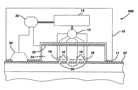
Thus, another embodiment of the apparatus of the
present invention, represented generally as 500 in FIG.
5, a transdermal iontophoresis device is incorporated
into the electroperforation apparatus. The combination
apparatus 500, capable of providing both
electroperforation and iontophoresis, comprises a housing
10, adhesive layer 11, an electric current generator 14,
an electric current controller 12, treatment electrodes
16 for electroperforation, sensor electrodes 18 for skin
resistance detection, a chamber 34 as a drug/interstitial
fluid reservoir, a delivery electrode 32 as a conductive
electrode for iontophoretic drug delivery, a return
electrode 36 to complete the circuit with iontophoretic
electrode 32 for iontophoresis operation, and an
iontophoresis control unit 30, in communication with the
current generator 14, the conductive electrode 32 for
iontophoresis, and the return electrode 36.
33
CA 02381931 2002-02-13
WO 01/13989 PCT/US00/23262
The iontophoretic drug delivery may be conducted
following, or simultaneously with, the
electroperforation process. U.S. Patent Nos. 4,301,794,
4,406,658, 4,340,047, 4,927,408, 5,042,975, and
5,224,927 describe the process of iontophoretic delivery
of a substance across tissue that can be used in the
present invention.
For delivering a drug through pores 50 in membrane
54, a drug solution may be present or absent during the
electroperforation process. In the latter case, the drug
solution may be subsequently placed into the chamber 34
(e. g., either through a septum with a syringe or through
a port on the wall of the chamber 34 from a breakable
capsule (neither shown)) after the electroperforation
process is completed.
There may be an optional semipermeable membrane to
separate the chamber 34 horizontally into two sub-
chambers (not shown). The upper sub-chamber thus created
serves as the iontophoresis electrode chamber
(containing delivery electrode 32) and the lower sub-
chamber serves as the drug reservoir that is in
communication with the membrane surface. The
semipermeable membrane has pores smaller than the drug
molecules being delivered so that the drug molecules can
not pass through the semipermeable membrane from the
drug reservoir into the iontophoresis electrode chamber
(e. g., to be deactivated by the delivery electrode 32).
The combination apparatus 500 may also contain
sensors (e.g., sensors for measuring the pH, molecule or
ion concentration, electric conductivity, amperage, and
potential, pressure, color and temperature of the fluid
in chamber 34 (not shown)) to assist in achieving
optimal iontophoresis operation. The iontophoresis
34
CA 02381931 2002-02-13
WO 01/13989 PCT/US00/23262
operation may also use a reverse polarity mode, such as
described in U.S. Patent Nos. 4,406,658, 4,301,794,
4,340,047, and 5,224,927.
In yet another embodiment of the present invention,
the electroperforation apparatus may be constructed in a
form of a "roller-like" device, represented generally as
apparatus 600 in FIG. 6. The handle 70 of the roller-
like electroperforation apparatus 600 comprises an
electric current controller and an electric current
generator. The arms 80 are built comprise the connecting
wires allowing electric communication between the
current controller and current generator in the handle
70 and the electrode array 96 on the roller 90. The body
of the roller 90 may contain both an array of treatment
electrodes for electroperforation and an array of sensor
electrodes for skin resistance detection. It may also
contain an iontophoresis unit, as described above.
The "roller-like" electroperforation apparatus 600
is used to create pores on the barrier membrane of a
patient. When the apparatus rolls over a skin area, the
electroperforation process occurs as the roller surface
comes in contact with the membrane, resulting in the
formation of numerous pores at pre-determined intervals
for a subsequent drug application. The advantages of
such an apparatus include an easy and rapid operation
over a large membrane area with complex contours.
Alternatively, an electroperforation device in FIG
6 may be fabricated into a "stamp-like" device where the
roller is replaced with a flat or nearly flat surface on
which to electrodes are located. In operation, this
"stamp-like" electroperforation device can be used to
electroperforate the membrane by pressing the surface
against the membrane.
CA 02381931 2002-02-13
WO 01/13989 PCT/US00/23262
In yet another embodiment of the electroperforation
apparatus of the present invention, the treatment
electrodes 16 may be placed within a spacers 42 as shown
in FIG. 7. The function of spacers 42 is two fold: (a)
separating the treatment electrodes 16 from each other at
a predetermined distance and (b) providing a precise
distance between the tips of the treatment electrodes 16
and the barrier membrane (e.g., the stratum corneum) 32
to be electroperforated. For example, when
electrofulguration or electrodesiccation is the mode of
action for an electroperforation process, there should be
no direct contact between the treatment electrode 16 and
the stratum corneum 32, but rather only a predetermined
small gap as controlled by the spacers 42. With other
modes of action, such as electrocoagulation and
electrosection, the treatment electrode 16 should contact
the tissue. In these cases, the spacers 42 prevent
undesirable damages to the deeper tissues 34 and 36 other
than stratum corneum 32. The open areas 40 provide the
liquid pathways for a drug solution to reach the stratum
corneum openings 50 from the drug reservoir.
It should be noted that the relative ratio of the
open areas 40 to the areas occupied by the spacers 42 and
electrodes 16 will vary depending on a particular need.
The shapes of the electrodes 16, spacers 42 and the
openings 40 may also vary significantly. For example, the
tip or the working area of the electrode 16 may be
sharply pointed, dull pointed, rounded, blade-like,
symmetric or asymmetric, flat, irregularly shaped, with
smooth or rough surface. The material used for the
electrode 16 may be pure metal, metal alloy, carbon,
ceramic, or other any other conductive materials such as
conductive composites (e. g., metal-polymer, carbon-
36
CA 02381931 2002-02-13
WO 01/13989 PCT/US00/23262
polymer, metal-glass, and metal-ceramic) suitable for
making the electrodes.
In another embodiment of the invention, the
treatment electrode may be made of a consumable material,
S which is either burned out or melted away during the
electroperforation process. For example, when current
passes through a thin carbon rod or a carbon fiber to the
barrier membrane during the electroperforation process,
the heat generated burns out the carbon electrode, thus
automatically cutting off the current. This can act as a
safety measure to prevent any excess burning which could
result from potential malfunction of the current
controller. The use of such a consumable electrode to
self-terminate the current can also serve as a means to
control the duration of electroperforation. Other
consumable electrode materials include low melting point
metal alloys and metal-polymer composites.
In another embodiment of the invention, the
electroperforation electrodes are fabricated as needles
or blades. In operation, stratum corneum is first
treated by electroperforation. Then the sharp electrodes
can be pressed against the electroperforated stratum
corneum to further disrupt it. In this case, because it
is not necessary to completely perforate the stratum
corneum with electric current, a much lower energy power
can be used to denature the barrier membrane to make it
easier to be penetrated by the needle or blade.
In another embodiment of the invention, the
electroperforation process can be conducted while the
electrodes are immersed in the drug solution, so that
the drug delivery process starts immediately following
electroperforation. The electroperforation process can
37
CA 02381931 2002-02-13
WO 01/13989 PCT/US00/23262
be repeated when necessary (e.g., as indicated by the
sensors discussed above).
In another embodiment of the invention, the
electroperforation process may be conducted
simultaneously with all the treatment electrodes (e. g.,
the electrodes in the electrode array shown in FIG. 7).
Alternatively, the electroperforation process may be
conducted using only one or a few of electrodes at a
given time, and then proceeding stepwise with the other
electrodes (e. g., in a fashion resembling a "scanning"
action). The mode of turning select electrodes on or off
may be controlled by the current controller (e. g.,
current controller 12 in FIGS 1-5). The advantage of the
"scanning" mode of action is the minimal amount of
electric energy required, thus minimizing any potential
side effects.
In another embodiment of the invention, a further
step is used to retard the closure of the pores (e. g.,
by keeping the pores occluded for drug delivery or
interstitial fluid sampling). In one embodiment, the
pores are kept in an aqueous solution that may also
contain the drug to the delivered and/or contain
compounds that retard epidermal cell differentiation or
the tissue growth leading to the closure of the pores.
Examples of such compounds include, but are not limited
to, saccharides, polysaccharides, cyclodextrins, heparin
and fragmented (low molecular weight) heparin
derivatives.
To evaluate the feasibility of using
electroperforation as a permeability enhancing method to
increase transport across a barrier membrane such as the
skin, several electroperforation experiments were
38
CA 02381931 2002-02-13
WO 01/13989 PCT/US00/23262
conducted to examine molecular transport of drugs and
water through pig skin in vivo.
Example 1. Increase in Transepidermal Water Loss (TEWL)
after Electroperforation in Pias
To evaluate the pore transport pathway created
through the stratum corneum of the skin by
electroperforation, an in vivo experiment was conducted
on the back skin of Yorkshire pigs (female, ~12 kg) using
an electrosurgery apparatus (SurgitronT"', Ellman
International, Inc., Hewlett, NY). The pigs were
immobilized with appropriate anesthetics and analgesics.
Electrofulguration current was used with a fine wire
electrode (0.26 mm in diameter) and power output setting
at between scale 3 to 10. A small pore was created on the
surface of the skin by carefully moving the electrode
towards the skin until the tip of the electrode almost
touched the skin. The electrode was quickly moved away
from the skin as soon as an electric arc appeared in the
gap between the electrode tip and the skin surface.
Typical microscopic biopsy results (magnification =
220X) of the pig skin treated with electroperforation are
shown in FIG. 9. FIG. 9a shows a pore (~64 micrometers)
created by electroperforation through the stratum corneum
10 with a minimal damage to the underlying living
epidermis 20. FIG. 9b shows a pore that perforated
through both stratum corneum 10 and living epidermis 20,
but not dermis 30. These results show the flexibility of
the electroperforation process of the present invention.
Desired depths of tissue perforation may be achieved with
the modification of the power and duration of the
electric current. For example, stratum corneum
perforation may be suitable for transdermal drug
39
CA 02381931 2002-02-13
WO 01/13989 PCT/US00/23262
delivery, while perforation through the epidermis, or
even some part of dermis, may be suitable for
interstitial fluid sampling or vaccination.
Transepidermal water loss (TEWL) was also measured
on the skin site of electroperforation with Evaporimeter~
EPl (Servomed AB, Stockholm, Sweden). Four measurements
were made for each condition. TEWL measurement is well-
known in the field of transdermal drug delivery and
cosmetic industry as a good indicator for stratum corneum
integrity. An increase in TEWL value implies disrupted
stratum corneum.
In this experiment, TEWL measurements were conducted
as a function of the pores created on the pig skin. We
found that as the number of the pores created by
electroperforation increased, the TEWL value increased
almost proportionally. This result demonstrates that the
electroperforation procedure successfully produced pores
across the stratum corneum, through which water molecules
escaped from the pig body to the outside. This result
further demonstrates that interstitial fluid may be
extracted through the pores created by
electroperforation, and analyzed for its biological
substances for diagnostic purposes. Other techniques such
as vacuum may be used to aid the interstitial fluid
extraction.
Example 2. Electroperforation Followed by Passive
Diffusion of Insulin for Transdermal Delivery
The electroperforation procedure described in
Example 1 was conducted in two pigs with a pore density
of 39 pores/cm2 of the skin and subsequently followed by
transdermal insulin delivery with passive diffusion. An
insulin-containing chamber was immediately placed onto
CA 02381931 2002-02-13
WO 01/13989 PCT/US00/23262
the electroperforation-treated skin. The chamber was made
of flexible polyethylene containing 0.5 ml of insulin
injection solution (Pork insulin, Molecular Weight - 6000
daltons, 100 U/ml, Regular Iletin~ II, Eli Lilly,
Indianapolis, IN). The contact area of the insulin
solution in the chamber to the electroperforation-treated
skin was 2.3 cm2. The chamber was affixed to the pig skin
with a veterinary silicone adhesive at the rim of the
chamber. Blood glucose of the pigs was monitored by
obtaining blood samples of the ear vein, which were
analyzed using two blood glucose analyzers separately to
assure the accuracy (One Touch~ Basic, LifeScan, Inc.,
Milpitas, CA). The blood glucose levels in both pigs
declined rather quickly from the onset of the insulin
delivery experiment. The significant blood glucose
reduction (greater than 50% of the basal level) indicates
that insulin from the drug-containing chamber indeed
passed through the pores on the stratum corneum into the
body and entered the systemic blood circulation,
resulting in the severe hypoglycemia in these pigs.
Example 3 Electroperforation followed by Iontophoresis
of Insulin for Transdermal Delivery
An electroperforation procedure was conducted in two
pigs similar with a pore density of 9 pores/cmz on the
skin and subsequently was followed by transdermal insulin
delivery. The purpose of using a lower pore density in
this experiment was to examine the effect of pore number
(e.g., the extent of the transport pathway available) to
transdermal insulin delivery. The same insulin-containing
chamber and drug application procedures were used in this
experiment as those in the Example 2. However, a steel
wire was placed in the insulin-containing chamber to
41
CA 02381931 2002-02-13
WO 01/13989 PCT/US00/23262
serve as a delivery electrode for iontophoresis. The
power source of iontophoresis was a commercial
iontophoresis apparatus (Phoresor IIT'", PM700, Motion
Control, Inc., Salt Lake City, Utah). The first 1.5 hours
S of the delivery experiment was by passive diffusion of
insulin only. Iontophoresis of insulin was conducted
twice in two 30-minute sections with 4 mA DC current at
1.5 hour and 3 hour, respectively, as indicated by the
arrows in FIG. 10. The electric polarity of the
conductive electrode was reversed every 5 minutes to
prevent pH shifting of the drug solution in the chamber.
FIG. 10 shows that the blood glucose levels in both
pigs did not decline during the first 1.5 hours of
passive diffusion. The result implies that the limited
transport pathway available with 9 small pores per cmz in
the stratum corneum might not be enough to deliver
insulin and to produce a therapeutically significant
blood glucose reduction via passive diffusion (e. g.,
merely utilizing a concentration gradient). On the other
hand, rapid blood glucose reduction during iontophoresis
indicates insulin was delivered into the pigs during
this time. This result shows that even with limited
disruption of stratum corneum, additional driving forces
such as iontophoresis can still deliver a macromolecular
drug into the skin to exert its therapeutic efficacy.
This result shows the possibility of making a very
small transdermal delivery device (e.g., smaller than 1
cm2 or even 0.1 cmz). All the transdermal drug delivery
patches currently available are much greater in size
(e. g., 10-40 cm~). Such a small size transdermal device
would be much more discrete and comfortable for a
patient to wear, and would reduce the potential of skin
42
CA 02381931 2002-02-13
WO 01/13989 PCT/US00/23262
irritation due to skin response to these adhesive-
containing devices and prolonged occlusion.
Example 4. Electroperforation followed by Passive
Diffusion of Erythropoietin for Transdermal Delivery
An electroperforation procedure was conducted in
two pigs similar to that in Example 3, followed by
passive diffusion of erythropoietin (20kU/ml, Procrit~,
Ortho Biotech, Inc., Raritan, NJ) at the treatment site.
There were 25 pores/cmz generated with electroperforation
on each pig. The drug chamber based over the
electroperforation-treated skin area contained 0.5 ml of
erythropoietin solution. Blood samples were collected
for erythropoietin analysis with an ELISA method. The
erythropoietin delivery procedure was carried out for 7
hours. The drug-containing chamber was removed at the
end of the delivery procedure, but the blood sampling
was continued for up to 30 hours following the start of
the experiment. It was found that there was a
progressive increase in plasma erythropoietin
concentration until the drug-containing chambers were
removed from the skin of the pigs. One day after the
delivery experiment, the plasma erythropoietin
concentrations in the pigs were still above the
endogenous basal level
It is understood that while the invention has been
described in conjunction with the detailed description
thereof, that the foregoing description is intended to
illustrate and not limit the scope of the invention,
which is defined by the scope of the appended claims.
Other aspects, advantages, and modifications are within
the claims.
What is claimed is:
43