Note: Descriptions are shown in the official language in which they were submitted.
CA 02381939 2002-04-02
WO 01/12261 PCT/US00/22601
M_ ICROWAVE DEVICES FOR MEDICAL HYPERTHERMIA,
THERMOTHERAPY AND DIAGNOSIS
Background of the Invention
The present invention relates to microwave devices used in medical
hyperthermia
and thermotherapy (referred to collectively herein as "heat therapies") and
diagnostics, and to
methods of using such devices.
Localized heat therapies, i.e., hyperthermia (heating to temperatures below 45
° C)
and thermotherapy (heating to temperatures above 45 ° C), have been
intensively investigated
for the last two decades for many disease processes.
However, methods of delivering heat including warm fluid, focused ultrasound,
radio frequency, and microwave approaches have been applied to abnormal tissue
with only
limited success. Because microwave energy can be applied with limited
invasiveness, this
approach is one that is currently being proposed.
For heat therapy to be applied safely, it is very important that the applied
heat be
confined to a target area alone, to avoid damaging nearby healthy tissue or
organs.
Some devices for heat therapy have utilized microwave heating, for example,
those disclosed in U.S. Patent Nos. 4,700,716 and 4,776,086, the disclosures
of which are
incorporated herein by reference. Microwave energy elevates temperature by
increasing the
molecular motion within cell structures. As the frequency decreases, tissue
penetration
increases. Small diameter microwave antenna and other probes have been
inserted into the
body through normal body passages or, on occasion, directly into diseased
tissue, using
hollow plastic catheters.
Summary of the Invention
The invention features a medical treatment system which utilizes microwave
energy to provide heat treatment and diagnostic imaging of an arbitrarily
shaped tissue mass.
The term "microwave", as used herein, refers to electromagnetic energy in the
microwave
frequency spectrum of about 300 MHZ to about 300 GHz.
In one aspect of the invention, a medical treatment system for treatment of
tissue
includes a radiating antenna system, positioned to radiate electromagnetic
energy through the
-1-
CA 02381939 2002-04-02
WO 01/12261 PCT/US00/22601
tissue, and receiving elements, each configured to be positioned within or on
the periphery of
the tissue to receive at least a portion of the radiated electromagnetic
energy from the
radiating antenna system to the tissue. Each receiving element has an interior
volume for
receiving a heat exchange fluid to change the temperature of the tissue
proximal to the
receiving element.
In another aspect of the invention, a medical treatment system for treatment
of
tissue includes a radiating element system, positioned to radiate
electromagnetic energy
through the tissue, and reflecting elements, each configured to be positioned
within or on the
periphery of the tissue to reflect at least a portion of the radiated
electromagnetic energy from
the radiating antenna system to the tissue.
The inventions have numerous advantages. The radiated energy from the
radiating antenna system is used to heat a desired area of tissue and the
receiving elements
are positioned to operate as "heat pipes", which act as a source or sink for
the heated tissue.
In addition, the individual receiving elements receive the radiated energy and
provide signals
which together provide an image and a property map of the area of tissue
defined by the
positioning of the elements. Thus, the receiving elements improve control of
the temperature
of the volume of the tissue mass being radiated by the radiating antenna
system. With this
arrangement a safer, more efficacious delivery of microwave energy is
provided. It is
important to recognize that although the receiving elements serve as "heat
pipes", in
operation, they can provide both heating as well as cooling, depending on
whether the fluid
(e.g., liquid or gas) flowing through the heat pipe structure is hot or cold.
Embodiments of these aspects of the invention may include one or more of the
following features.
At least one of the reflecting elements can include an interior volume for
receiving a heat exchange fluid to change the temperature of the tissue
proximal to the
reflecting element.
One (or more) of the receiving and/or reflecting elements has a conduit for
conveying the heat exchange fluid from a heat exchanger to a distal end of the
receiving
element. The receiving and/or reflecting element also has a transmission line
extending from
the distal end to a proximal end of the receiving and/or reflecting element.
The conduit
-2-
CA 02381939 2002-04-02
WO 01/12261 PCT/US00/22601
extends through the transmission line and forms a hollow center conductor of
the
transmission line. The transmission line also has an outer shield which is
coaxial with
respect to the conduit. The interior volume of the receiving and/or reflecting
element and the
conduit are sized to cause capillary action of fluid flowing between the
internal volume and
the conduit. The heat exchanger can include a condenser and the heat exchange
fluid can be
a coolant.
One or more of the receiving and/or reflecting elements has a temperature
detector for sensing the temperature at a location proximate to that receiving
element. In
response to the sensed temperature, the detector provides signals for
controlling the amount
of fluid delivered to the interior volume of the receiving and/or reflecting
element by the heat
exchanger.
A measurement analyzer, connected to one (or more) of the receiving and/or
reflecting elements, measures electrical characteristics associated with the
receiving and/or
reflecting element. These electrical characteristics include amplitude and
phase voltage
characteristics. The electrical characteristics can also be magnitude and
phase of Slz
scattering parameter between the radiating antenna system and the receiving
and/or reflecting
element. A processor processes the measured electrical characteristic to
generate an image of
the tissue, and a display then displays the generated image.
One (or more) of the receiving andlor reflecting elements and the antenna
system
can be configured to deliver a material to the tissue. The material can be a
chemotherapeutic
agent, a heat sensitizer, or a cyropreservative.
At least one of the receiving elements includes a reflecting structure for
reflecting
the radiated electromagnetic energy from the radiating antenna system in a
desired direction,
thereby increasing the uniformity of the radiation applied to the targeted
tissue.
The radiating antenna system has a plurality of antennas in the form of a
collinear
array. The radiating antenna system is configured to be received within the
tissue to be
treated.
A cannula is provided to receive the radiating antenna system within its inner
lumen. The radiating antenna system includes antennas, each in the form of a
collinear array.
-3-
CA 02381939 2002-04-02
WO 01/12261 PCT/US00/22601
The electromagnetic energy is radiated at a frequency in a range between 0.3
and
GHz, and at a power level in a range between about 100 mwatts and 150 watts.
In another aspect of the invention, a method of treating tissue is provided
where a
radiating antenna system is positioned within the tissue to radiate
electromagnetic energy into
a portion of the tissue desired to be heated, and receiving elements are
positioned for
receiving the radiated electromagnetic energy from the radiating antenna
system with each
receiving element positioned so that the path of received energy is through
the portion of the
tissue desired to be heated.
In still another aspect of the invention, a method of treating tissue is
provided
10 where a radiating antenna system is positioned within the tissue to radiate
electromagnetic
energy into a portion of the tissue desired to be heated, and reflecting
elements are positioned
for reflecting the radiated electromagnetic energy from the radiating antenna
system toward
the tissue to be treated.
With respect to these methods of treating tissue, the receiving and/or
reflecting
elements can be substantially positioned around a periphery of the portion of
the tissue
desired to be heated. The temperature proximate to at least one of the
receiving and/or
reflecting elements is sensed and, in response to the sensed temperature, the
amount of fluid
delivered to an interior volume of the receiving and/or reflecting elements is
controlled.
Other features and advantages of the invention will be apparent from the
drawings, the following Detailed Description, and the claims.
Brief Description of the Drawings
Fig. 1 is a schematic diagram of one embodiment of a tissue treatment and
diagnosis system, showing three receiving microwave probes and one radiating
microwave
probe.
Fig. 2 is a diagrammatic view of the positions of the receiving microwave
probes
and the radiating microwave probe relative to a tissue mass under treatment.
Fig. 3 is a cross-sectional side view of a receiving antenna of one of the
receiving
microwave probes.
-4-
CA 02381939 2002-04-02
WO 01/12261 PCT/US00/22601
Fig. 4 is a schematic diagram showing the circuitry of a diagnosis and
treatment
station of tissue treatment and diagnosis system of Fig. 1.
Figs. SA-5C are diagrammatic views of exemplary positions of the receiving
probes and the radiating probe relative to different tissue masses under
treatment.
Detailed Description
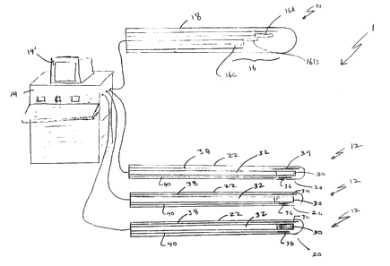
Refernng to FIG. 1, microwave treatment and diagnosis system 1 includes at
least
one radiating microwave probe 10, at least two receiving microwave probes 12,
a diagnosis
and treatment station 14, and a monitor 14'. Receiving microwave probes 12 are
configured
and operated to act as "heat pipes." Hence, each one of receiving microwave
probes 12
serves as a source or sink for thermal energy at the interface of that
receiving microwave
probe 12 and the adjacent tissue, resulting in a greater control of
temperature at the interface.
It is important to note that although receiving microwave probes 12 are said
to act as heat
pipes, receiving microwave probes 12 can cool as well as heat a targeted
tissue. This allows
for further control of temperature at the interface.
Referring to Fig. 2, diagnosis and treatment station 1 allows diagnosing and
applying heat therapy to a tissue mass 11 of arbitrary shape (here, a tumor in
a patient's
kidney 13). In particular, radiating microwave probe 10 and receiving
microwave probes 12
are inserted into kidney 13 and positioned relative to one another so that
electromagnetic
energy travels through tissue mass 11 from radiating microwave probe 10 to
receiving
microwave probes 12. Therefore, as radiating microwave probe 10 radiates
microwave
electromagnetic energy, tissue mass 11 is heated. As can be seen in Fig. 2,
radiating
microwave probe 10 and receiving microwave probes 12 each extend to radiating
probe
contacts 10a and receiving probe contacts 12a, which, in use, are attached to
the skin. Lead
wires 15 connect probe contacts 10a, 12a to diagnosis and treatment station
14.
Receiving microwave probes 12 perform multiple functions. Because of their
heat pipe structure, receiving microwave probes 12, under control of diagnosis
and treatment
station 14, act as heat sinks at the boundary of tissue mass 11. Hence,
depending on their
number and positioning relative to tissue mass 11, receiving microwave probes
12 can
substantially limit heating to tissue mass 11. In effect, based on their
positioning within the
-5-
CA 02381939 2002-04-02
WO 01/12261 PCT/US00/22601
tissue, receiving microwave probes 12 can be used to define any arbitrary area
within the
tissue, and limit heat therapy substantially to that arbitrary area.
In addition, receiving microwave probes 12 act as antennas receiving radiated
electromagnetic energy. Characteristics of the electromagnetic energy received
at receiving
microwave probes 12 depend on characteristics of tissue mass 11. Hence, for
diagnosis, the
characteristics of the received electromagnetic energy are measured by
diagnosis and
treatment station 14. Based on that measurement, diagnosis and treatment
station 14
determines the characteristics of tissue mass 11 and can also generate an
image of the tissue
mass 11.
We will now describe in detail an embodiment of microwave treatment and
diagnosis system 1. Refernng to Fig. 1, each one of receiving microwave probes
12 includes
a receiving antenna 20 deployed within a cannula 22. (The term "cannula" is
intended to
include all cannula-like structures, whether rigid or flexible, including
catheters.) Receiving
antenna 20 is configured not only to receive the microwave energy radiated by
radiating
microwave probe 10. Cannula 22 is constructed to be inserted into a portion of
the body,
typically through a body opening or passage, a small incision, or by using an
internal stylet,
as will be described in detail below. Receiving microwave probes 12 are
preferably sized to
have a diameter of 5-16 French (F.) and a length of approximately 1-18 cm.
FIG. 3 shows a detailed diagram of the structure of receiving antenna 20.
Receiving antenna 20 includes an antenna portion 30 connected via a coaxial
transmission
line 32 to diagnosis and treatment station 14.
Antenna 20 further includes an RF reflector 34 and an RF director 36, located
at
the end of dielectric members 38 and 40, respectively. RF reflector 34 and RF
director 36
are constructed by forming a metallic coating on dielectric members 38 and 40.
RF reflector
34 and RF director 36 serve to improve the gain of antenna portion 30 by
forming a three
element Yagi array. The length of RF reflector 34 is generally commensurate
with, or longer
than, the length of antenna portion 30, while the length of director 36 is
generally shorter
(e.g., 75% of antenna length).
Reflector 34, in addition to increasing the gain of antenna portion 30,
reflects
microwave electromagnetic energy from radiating microwave probe 10 back
towards the
-6-
CA 02381939 2002-04-02
WO 01/12261 PCTNS00/22601
tissue. Reflector 34 can be differently shaped, such as having semi-
cylindrical shape, to
better reflect microwave electromagnetic energy back towards the tissue.
Thus, with their reflectors and directors, each one of receiving microwave
probes
12 reflect the radiated energy back towards tissue mass 11. In combination,
all of the
receiving microwave probes 12 cause multiple reflections within tissue mass
11. The result
of the multiple reflections is similar to a result of a microwave resonant
cavity i.e., the
multiple reflections increase the uniformity of the heat applied to the tissue
mass.
Receiving antenna 20 is shown in Fig. 3 as being configured for providing heat
pipe temperature control. Receiving antenna 20 includes antenna portion 30, a
heat
exchanger 56 and a flexible RF coaxial transmission line 32 connecting antenna
portion 30 to
diagnosis and treatment station 14. Antenna portion 30 is formed by a hollow
conductive
pipe 60 and a dielectric sheath 70 extending substantially the entire length
of the conductive
pipe. Conductive pipe 60 is one part of coaxial transmission line 32 for
transmitting energy
from antenna portion 30 to diagnosis and treatment station 14. At diagnosis
and treatment
station 14, antenna portion 30 can be selectively either grounded or open
circuited.
When used as a heat pipe, conductive pipe 60 also functions as a capillary
wick
for a liquid or gas 62 passing therethrough. The capillary action is
accomplished by having a
relatively larger diameter portion 66 at antenna portion 30 to provide
evaporative cooling,
and a relatively smaller diameter "wick" portion 67 extending between portion
66 and heat
exchanger 56. Larger diameter portion 66 is approximately A/2 in length. At a
junction 71,
wick portion 67 extends beyond transmission line 32 to the heat exchanger 56
in the form of
a dielectric tube 69.
When used in applications where cooling is required, heat exchanger 56 acts as
a
condenser having a refrigerant (e.g., cryogenic fluid). A pressure mechanism
140 under the
control of diagnosis and treatment station 14 is used to control the amount
and rate at which
the fluid is delivered to antenna portion 30.
Receiving antenna 20 also includes several temperature sensors positioned at
various points within receiving antenna 20. In particular, a temperature
sensor 42 is placed
on RF reflector 34. Another temperature sensor 44 is placed on RF director 36.
Other
temperature sensors (not shown) can also be placed along the walls of
receiving antenna 20
_7_
CA 02381939 2002-04-02
WO 01/12261 PCT/US00/22601
or cannula 22. Sensors 42-44, and the other temperature sensors in receiving
antenna 20, can
be in the form of fiber optic sensors surrounded by a dielectric outer
envelope. One example
of a fiber optic sensor of this type is described in U.S. 4,700,716.
Antenna portion 30, RF reflector 34, and RF director 36 are fixed in position
by
potting them in a solid material within a tube (not shown), for example, by
placing them in a
tube and filling the tube with liquid, hardenable TEFLON polymer. The tube can
then be
easily inserted into the cannula 22 for use by a physician.
Receiving antenna 20 also includes a transformer 54 provided by the
combination
of conductive pipe 60, an outer conductive coaxial sheath 64, dielectric
sheath 70, and a
metallic cylinder 73. Outer conductive coaxial shield 64 surrounds dielectric
sheath 70 and
extends along the length of conductive pipe 60 until terminating at a point
just before larger
diameter portion 66. Metallic cylinder 73 is approximately one-quarter
wavelength in length
and covers outer conductive coaxial shield 64, thereby electrically shorting
the pair of
members at point A. This electrical short presents an effective open circuit
(high impedance)
along the transmission line one-quarter wavelength away from the short.
Transformer 54 minimizes the reflected power seen by receiving element 30.
Equally important, transformer 54 also prevents leakage of antenna currents
along the outside
structure of antenna 20. By appropriate selection of operating parameters,
transformer 54
can be designed to provide both a minimum reflection coefficient as well as
minimum
leakage within the same frequency range.
Having described receiving microwave probes 12, we will now describe in
general terms radiating microwave probe 10. A detailed description of
radiating antenna
probe 10 can be found in U.S. patent application serial no. 09/248,165, filed
February 9,
1999, incorporated herein by reference (hereinafter, referred to as "the '165
application").
Refernng back to Fig. 1, radiating microwave probe 10 includes a collinear
antenna 16
having a set of radiating antennas 16A, 16B, 16C deployed within a cannula 18.
Cannula 18
is constructed to be inserted into a portion of the body, typically through a
body opening or
passage, a small incision, or by using internal stylets. Cannula 18 is
preferably sized to have
a diameter of 1-3 mm and a length of 2-6 cm.
_g_
CA 02381939 2002-04-02
WO 01/12261 PCT/US00/22601
The amplitude and phase of the radiation from each one of radiating antennas
16A, 16B, 16C is independently controlled by diagnosis and treatment station
14, so that
their respective electromagnetic fields constructively add within, and
subtract outside, a
targeted tissue mass With this approach, a radiation pattern with desired
narrow beamwidth
and direction provides relatively high temperature and a focused heating to
the tissue mass.
Additionally, radiating antennas 16A, 16B, 16C can also have a heat pipe
structure similar to
receiving microwave probes 12, thereby improving the temperature control at
the interface of
radiating microwave probe 10 with the targeted area. Radiating antennas 16A,
16B, 16C
thereby can have the same structure as that shown in Fig. 3 for receiving
element 30, except
that antenna portion 30 would be optimized for transmission rather than
reception.
Refernng to Fig. 4, diagnosis and treatment station 14 includes a
microprocessor
102, a memory unit 104, and a bus 106. Diagnosis and treatment station 14 also
includes
three subsystems for connection to radiating and receiving microwave probes 10
and 12.
These subsystems are the power and measurement subsystems Sl, temperature
control units
S2, and pressure mechanisms S3. Each of these subsystems are connected to bus
106 and are
under control of application programs stored in memory unit 104 and executed
by
microprocessor 102. These subsystems will be shown and described as having
connections
for four devices, although other embodiments can include more connections.
Power and measurement subsystems S 1 include an output port 86 coupled to a
microwave power source 88 capable of, for example, providing approximately 5-
25 watts of
continuous wave power at 915 MHZ or 2450 MHZ to radiating microwave probe 10.
Port 86
is coupled to power source 88 through a bidirectional coupler 90A. A fraction
(e.g., 20dB) of
the microwave power source 80 is tapped from couplers 90B, 90C, 90D and
provided to
vector voltmeter (or measurement analyzer) 92 through a sequence of rotary
switches 94, 96,
98. Note that power source 88 is capable of driving antennas 16A, 16B, 16C of
radiating
microwave probe 10 independent of one another.
Power and measurement subsystems S 1 also includes several input ports 80, 82,
84 for connection to receiving microwave probes 12. Input ports 80, 82, 84 are
coupled to
electronic switches 91A, 91B, 91C through bi-directional coupler 90B, 90C,
90D,
respectively. A fraction (e.g., 20dB) of the microwave energy received at each
one of
-9-
CA 02381939 2002-04-02
WO 01/12261 PCT/LTS00/22601
receiving microwave probes 12 is tapped from couplers 90B, 90C, 90D and
provided to
vector voltmeter 92 through rotary switches 94, 96, 98. A switch controller
100 is used to
select one of ports 80, 82, 84, 86 being examined at any given time. A 30dB
attenuator is
connected at the output of rotary switch 98 to protect vector voltmeter 92
from excessive
power levels. Electronic switches 91A, 91B, 91C, under control of application
programs
running on microprocessor 102, can either connect an antenna of a receiving
microwave
probe to ground or allow the antenna to be open circuited.
Temperature control units S2 include ports 110, 112, 114, 116, each of which
is
connected, respectively, to a dedicated temperature control unit 120, 122,
124, 126. Each one
of temperature control units 120, 122, 124, 126 is connected to bus 106 and is
under control
of application programs running on microprocessor 102. Each one of ports 110,
112, 114,
116 is connected to temperature sensors in one of the receiving microwave
probes 12 or
radiating microwave probe 10. Temperature control units S2 provide signals to
microprocessor 102 indicative of the temperature at the probes.
Pressure mechanisms S3 include ports 130, 132, 134, 136 for connection to
conductive pipes 60 of receiving microwave probes 12. If one or more of
antennas 16A,
16B, 16C of radiating microwave probe 10 are configured as a heat pipe, then
those antennas
can also be connected to one of ports 130, 132, 134, 136. Each one of ports
130, 132, 134,
136 is coupled to a dedicated pressure mechanism 140, 142, 144, 146,
respectively. Each
one of pressure mechanisms 140, 142, 144, 146 is in turn connected to bus 106
and is under
control of application programs executed by microprocessor 102.
We will now describe the operation of microwave diagnosis and treatment system
1. Referring to Figs. 1-4, briefly, during operation, radiating microwave
probe 10 is
positioned and operated within tissue to radiate microwave electromagnetic
magnetic energy
towards two or more receiving microwave probes 12 through a targeted area of
the tissue.
The radiated electromagnetic energy can have a frequency in a range between
about 0.3 and
10 Ghz, and a power level in a range between about 1 mwatts and 150 watts.
Preferably, the
radiated electromagnetic energy has a frequency of 915 MHZ or 2450 MHZ, at a
power level
of about 5-25 watts. Microwave receiving probes 12 are positioned within the
tissue to
receive and reflect back toward the targeted area the radiated electromagnetic
energy.
- 10-
CA 02381939 2002-04-02
WO 01/12261 PCT/US00/22601
Diagnosis and treatment station 14 controls the heat treatment applied by
radiating and receiving microwave probes 10 and 12, and performs tissue
diagnostic and
inquiry operations.
During heat treatment of a targeted area, in response to electrical signals
from
temperature control unit S2, diagnosis and treatment station 14 controls power
source 88 of
power and measurement analysis subsystems S 1 to generate electrical signals
with the
appropriate amplitude and phase characteristics so that radiating microwave
probe 10
provides a focused beam in the direction of the targeted area.
Additionally, based on the signals indicative of the temperatures at radiating
and
receiving microwave probes 10 and 12, diagnosis and treatment station 14
controls pressure
mechanisms S3 to convey heating or cooling fluid within antenna portion 30 of
receiving
microwave probes 12 to allow rapid and precise adjustment of the temperature
at the
interface between the antenna portions 30 and surrounding material. Hence,
diagnosis
treatment station 14 regulates heat at the boundary of a targeted tissue mass
to ensure that
heating applied to the tissue is substantially limited to the targeted tissue
mass. One
technique for achieving increased control over the applied heat is to
simultaneously apply
heat and cold to the tissue. Further details concerning the thermodynamic
operation of heat
pipes suitable for use in antenna 20 are described in U.S. 5,591,162, entitled
"Treatment
Method Using a Micro Heat Pipe Catheter", which is incorporated herein by
reference.
As stated above, diagnosis and treatment station 14 also performs diagnostic
functions. To do so, vector voltmeter 92 intermittently between heat
applications measures
amplitude and phase of the voltage induced on the receiving microwave probes
12.
Application programs running on microprocessor 102 use the results of these
measurements
to determine magnitude and phase of a SIZ scattering parameter between the
electromagnetic
energy radiated by radiating microwave probe 10 and the energy received at a
particular
receiving microwave probe 12. The measured value of the S 1z scattering
parameter is
directly related to physical and electrical properties of a portion of the
targeted tissue mass
lying between the two probes, including its density and water content,
polarization qualities,
electrolyte composition, reflectance, blood flow velocity, and changing
electrical properties
-11-
CA 02381939 2002-04-02
WO 01/12261 PCT/US00/22601
over time. Another parameter that can measured is the input impedance of the
radiating
probe 10.
The measured values can then be displayed to the user. These measured values,
together with other measurements, such as variance in phase and magnitude of
signals
received at the various receiving microwave probes 12, can be used to
construct an image of
the tissue under examination using conventional microwave tomography
techniques. One
technique for doing so is to change the position of radiating microwave probe
10 in a
predetermined manner and to measure the various parameters as the position of
the probe
changes. In addition, measurements at low power levels can be taken and then
compared to
measurements at higher power levels, the results indicating the change in
characteristics of
the tissue as the tissue is heated. Diagnosis and treatment station 14 can
then use these
measurements to construct an image of the tissue under examination.
It should be noted that, when one of receiving microwave probes 12 is not
providing signal information with respect to the voltages induced at its
antenna, that probe
can be used to act substantially as a reflector by allowing the antenna to be
open circuited,
substantially as a heat pipe by grounding the antenna, or as both.
In addition, diagnosis and treatment station 14 can display continuous
readings of
temperature changes at boundaries of a simulated or an ultrasound image of the
targeted
tissue mass. A schematic template of the targeted tissue mass representing the
anatomy can
be displayed with superimposed different colors representing different
temperature ranges at
different regions of the targeted area. Similarly, real-time or pre-recorded
fluoroscopy, CAT
scan, MRI, or ultrasound images can be superimposed with different colors
representing
different temperature ranges at different regions of the targeted area. Thus,
the therapist or
surgeon is able to determine, in real time, the target site and the
effectiveness in applying
heat from the system. Monitor 14' can display the temperature detected by each
of the
sensors as a function of time and provide beginning and end points for the
treatment.
Based on signals received from the sensors, diagnosis and treatment station 14
is
capable of issuing warning messages to be displayed on monitor 14' when
temperatures
exceed predetermined threshold values. Diagnosis and treatment station 14 may
also
automatically shutdown power source 88 if, for example, the temperatures
remain high for an
-12-
CA 02381939 2002-04-02
WO 01/12261 PCT/US00/22601
unacceptable time period or if a fault is detected in the system. Diagnosis
and treatment
station 14 also includes memory for storing statistical data including patient
information,
current laboratory data, as well as all data collected during the procedure.
Figs. SA-SC show examples of manners in which radiating and receiving probes
and 12 can be positioned relative to one another and various targeted tissue
areas 11. A
physician can determine the optimum positioning based on the nature of the
tissue to be
treated, ease of access to that tissue, and the desired treatment. Note that
broken lines in
Figs. 5B and SC indicate targeted tissue masses defined by receiving microwave
probes 12.
To position transmitting and receiving microwave probes 10, 12 within the
body,
10 various techniques may be used. For example, radiating and receiving
microwave probes 10
and 12 can be positioned under the guidance of ultrasound, X-Ray, CAT scan, or
fluoroscopy
through natural body passages or openings, or small incisions, depending on
the targeted
tissue mass. In addition, endoscopes can be used to direct and/or deliver the
probes to the
targeted tissue-mass. Alternatively, internal stylets can be used to deliver
the probes. Radio-
opaque internal stylets can be used to puncture the tissue and to enter into
the patients body
under guidance of guidance of ultrasound or fluoroscopy. After delivering the
probes, the
stylets can be removed.
Transmitting and receiving microwave probes 10, 12 can be used for various
types of therapy. For example, transmitting and receiving microwave probes 10,
12 can be
used to treat malignant and benign tumors, cysts, inflammatory conditions
(hyperthermic low
temperatures), rheumatic conditions and joint involvement (hyperthermic low
temperatures),
and muscle injuries. When treating cysts (such as hydrocoeles, spermatocoeles,
or renal
cysts), transmitting microwave probe 10 can be inserted into the cysts and
heat the fluid
therein to destroy the lining cells to reduce the mass of the cyst. Receiving
microwave
probes 12 can be located on the outside of the cysts to act as heat sinks and
to reflect the
microwave energy back toward the cyst.
Transmitting and receiving microwave probes 10, 12 can also be used to create
a
thrombus in an artery to occlude, for example, by inserting transmitting
microwave probe 10
at a desired point and heating that point to cause localized clotting.
Receiving microwave
probes 12 enhance the localization of the applied heat. Transmitting and
receiving
-13-
CA 02381939 2002-04-02
WO 01/12261 PCT/US00/22601
microwave probes 10, 12 can also be used to locally heat infected tissue
(e.g., cystitis or
prostatitis) to destroy or hinder the infecting bacteria or viruses.
In body openings such as the bladder, transmitting microwave probe 10 can be
inserted into the opening and receiving microwave probes 12 can be inserted to
be located on
the walls of the opening. The lining cells, which could be pathologic with a
thin layer of
widespread tumor, infection, or inflammation, can then be treated.
Transmitting and receiving microwave probes 10, 12 can be used for adjunctive
therapies to increase effectiveness of other types of therapy such as
chemotherapy and
radiation therapy.
Other embodiments are within the scope of the claims.
For examples, receiving and transmitting microwave probes 10, 12 may be
configured for delivering chemotherapeutic agents, heat sensitizers or
cyropreservatives to
the tissue. The delivered material can work synergistically with the microwave
treatment.
Chemotherapeutic agents may be better absorbed by heated tissue and tumor.
Heat
sensitizers allow to have an equivalent effect at a lesser power.
Cyropreservatives allow a
lower temperature to be attained before the tissue is destroyed by cellular
ice crystallization.
In some embodiments of the receiving and transmitting microwave probes 10, 12
may be configured to not apply heat or cold to tissue adjoining portions of
the probe. For
example, one side of the heat pipe may be covered by insulating material so as
to protect the
tissue at that side of the probe. Such insulating material would then protect
healthy tissue at
one side of probe, while not affecting the heat or cold applied to the tissue
to be treated.
In some embodiments, passive reflectors sized and configured to have contours
of
parts of the patient's body (e.g., breast or knee) may be used to enhance
reflection and
focusing microwave energy in parts of the body.
What is claimed is:
- 14-