Note: Descriptions are shown in the official language in which they were submitted.
CA 02382156 2002-02-15
WO 01/97684 PCT/CA01/00894
METHOD FOR EVALUATING VESTIBULAR RESPONSE
Bacleground of the Invention
1. Field of the Invention
This invention relates to the field of vestibular testing, more particularly
to a
method and apparatus for the clinical evaluation of the balance organs of the
inner ear.
2. Description of Related Art
Dizziness is one of the most common clinical complaints around the world.
Functional testing of the vestibular system (the balance organs of the inner
ear) is
often necessary for correct diagnosis and treatment, but accurate tests are
costly
and complex.
It has been reported that dizziness is the most common motive for consultation
of
a physician in patients over seventy five years of age. While in some cases
the
symptoms are typical and a cause is easily established, in many other cases a
specific diagnosis depends on precise functional testing, particularly of the
vestibular system. At the present time, this testing is very costly, and
requires
highly trained operators to interpret the results.
The inner ear consists of two parts, the cochlea that transduces sound waves
and
allows us to hear, and the vestibular labyrinth that senses movement and
generates reflexes to stabilize our eyes, heads and bodies. Usually, acute
damage
to the labyrinth will produce false sensations of movement, often called
vertigo
or dizziness, as well as gaze and postural instability and motion sickness.
These
symptoms tend to become increasingly vague, however, as compensation
develops. As a result, it can be difficult to distinguish between vestibular
and
other problems of a quite different nature. Of course, prognosis and treatment
depend heavily on a correct diagnosis.
One common method of testing the vestibular system consists of squirting warm
or cool water into each ear canal and monitoring the resulting "caloric
1_
CA 02382156 2002-02-15
WO 01/97684 PCT/CA01/00894
nystagmus". These involuntary eye movements result largely but not exclusively
from thermally-induced convection currents in endolymph, particularly in the
horizontal canal. The results of caloric testing are combined with separate
assessments of the eye movement control system and of eye movements that
result from changes in the patient's position. This is usually referred to as
the
electronystagmogram (ENG)test battery. While helpful in some cases, it often
provides equivocal data in patients with mild symptoms, and these are
precisely
the ones for which an objective test is necessary.
The only way to apply a selective and controlled stimulus to the vestibular
system is to expose the patient to physical movement. Furthermore, this
movement must be of a sort that makes it impossible for predictive or other
compensatory mechanisms to hide deficits in vestibular function. Normally,
this
means fast, because the neural circuits underlying compensation are more
complex and hence slower than the very simple pathways used by vestibular
reflexes. However, sudden movements require powerful machines, such as high-
torque servo-controlled rotators, as in rotary chair apparatus. A safe; man-
rated
version of these devices can be costly, require recurrent inspections,
maintenance
and repairs. As a result, such devices and the sophisticated vestibular
testing
they make possible have been limited to a relatively few university teaching
hospitals.
Australian researchers Ian Curthoys and Michael Halmagyi (Halmagyi, G.M.,
Curthoys, LS.,Cremer, P.D., Henderson, C.J., Todd, M.J., Staples, M.J. and
D'Cruz, D.M. The human horizontal vestibulo-ocular reflex in response to high-
acceleration stimulation before and after unilateral vestibular neurectomy.
Exp.
Brain Res. 81: 479-490,1990.) have demonstrated a far simpler technique that
measures eye movements during the first 100 msec of an unpredictable, passive
horizontal head rotation (peak head displacement 20°, peak head
velocity
200-300°/sec, peak head acceleration 2000-4000°/sec2). When eye
velocity is
plotted as a function of head velocity in patients with an unilateral loss of
vestibular function, eye responses during rotations toward the intact side are
-2-
CA 02382156 2002-02-15
WO 01/97684 PCT/CA01/00894
found to be close to normal but the responses are found to be markedly
decreased when the rotation is toward the lesioned side. Most significantly,
this
deficit appears to be permanent, implying that it cannot be hidden by
compensatory mechanisms.
This work is a significant step towards a simple and practical clinical test
of
human vestibular function, but it has one serious drawback. To reach
significant
head angular velocities in less than 100 msec, large head angular
accelerations are
necessary. These are achieved by the examiner abruptly and unpredictably
rotating the patient's head only. This movement would inevitably be opposed by
involuntary stretch reflexes and could lead to severe consequences in the
presence of undetected cervical spinal pathology. While similar, rapid head
rotations can be produced voluntarily, such a method would suffer badly from
predictability of the stimulus. As well, not everyone can perform the maneuver
in an acceptable fashion.
There is a need to provide a simple, effective and inexpensive technology that
would allow diagnostic testing in many additional hospitals, clinics and
offices.
Summary of the Invention
According to the present invention there is provided a method of evaluating
the
vestibular function in a human subject, wherein the human subject is
constrained
in a substantially erect position so that the head moves in unison with the
rest of
the body, a controlled stimulus in the form of a sudden angular acceleration
is
imparted to the body, and the subject's ocular response to the controlled
stimulus
is measured.
Typically, the subject is placed in a pivotable mechanism, such as a frame
that
can pivot essentially about the vertical axis or another axis. The subject is
essentially erect within the frame and is stabilized in a vertical standing
position,
or in a horizontal position, or in an inclined position relative to the
vertical. The
subject's head is substantially immobile relative to the torso or the rest of
the
CA 02382156 2002-02-15
WO 01/97684 PCT/CA01/00894
body. The head may be immobilized by various means, such as a clamp
consisting of a pair of brackets.
After the initial acceleration the subject is preferably brought to a rapid
standstill
with the aid of a fluid damping mechanism that exploits the fact that during
an
initial period of laminar flow, the resistance to motion of a body moving in a
fluid is very low, and then with the onset of turbulent flow, this resistance
suddenly increases.
The subject's response to this controlled stimulus is preferably measured
using
electrb-oculography (EOG), which is the recording of electrical signals
produced
by eye movement. The stimulus is recorded using angular velocity transducers.
Surface electrodes used in conjunction with a forehead-mounted ring and
adjustable connectors are a convenient way of performing electro-oculographic
recording. Angular velocity transducers are preferably secured in place with a
head adaptor allowing for the precise measurement of the angular speed of the
head without slip.
Vestibular function can be assessed using eye versus head velocity data. Eye
velocity is derived from eye position data. Further decision analysis can be
based
on eye velocity versus head velocity curves.
An alternative approach would be to use a man-powered rotator, including a
seat, a footrest and a means of securing the subject's upper body and head to
the
chair. Unfortunately, such a device has great difficulty achieving adequate
angular acceleration. Fully half of one's body mass is located in the legs. In
the
sitting position, this is distributed far from the axis of rotation, greatly
increasing
angular inertia. Such a human powered head rotator type could not reach the
angular accelerations necessary for the method of Curthoys and Halmagyi. The
applicants have found surprisingly that the method of the invention achieves
the
necessary angular rotation in a simple and effective manner.
In another aspect the invention provides an apparatus for evaluating
vestibular
function in a human subject comprising a frame for supporting the human
-4-
CA 02382156 2002-02-15
WO 01/97684 PCT/CA01/00894
subject in a substantially erect position with the head constrained so that it
moves
in unison with the rest of the body, the frame being rotatable about a
longitudinal
axis of the subject, an arrangement for measuring ocular response to a
controlled
stimulus in the form of a sudden angular acceleration imparted to the frame,
at
least one sensor for measuring the angular velocity of the subject's head, and
a
processing unit for deriving the vestibular function from the subject's ocular
response and the angular velocity.
Brief description of the drawings
The invention will now be ~deseribed in more detail, by way of example, only
with
reference to the accompanying drawings, in which:-
Figure 1 is an illustration of an experimental set-up of a vestibular response
evaluation apparatus in which the subject stands close to the center of a
rotatable
frame;
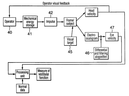
Figure 2 is a schematic block diagram of a method for stimulating the
vestibular
system of the subject,in vertical axis position and measuring the vestibular
and
ocular response;
Figure 3 illustrates hardware including a forehead mounted ring, adjustable
electrode connectors, and a head adaptor capable of measuring eye and head
movements;
Figure 4 shows results obtained from a subject having normal sensitivity;
Figure 5 shows the results obtained from a subject after an acute change in
sensitivity;
Figure 6 shows the results obtained from a subject after a chronic change in
sensitivity; and
Figure 7 is a schematic diagram of an vestibular response evaluation apparatus
with a damping mechanism.
-5-
CA 02382156 2002-02-15
WO 01/97684 PCT/CA01/00894
Detailed Description of the Invention
Figure 1 shows an apparatus for evaluating the vestibular response of a human
subject. The apparatus comprises a rectangular frame 10 having upper and lower
bearings 12 arranged such that the frame is rotatable about symmetrical
vertical
axis 13. In use, a human subject 20 stands in the erect position on platform
14
such that the axis of rotation 13 extends along the longitudinal axis and is
constrained at the hips by brackets 16 and at the head by brackets 18. In use,
an
operator 22 imparts a sudden rotational movement to the frame 10. As a result
the subject rotates about the axis with the head and torso rotating together
in
unison.
In the illustrated embodiment, the frame is rotatable about the vertical axis.
It
will be understood the frame could be mounted so as to allow rotation about
different axes so Iong as they pass through the longitudinal axis of the
subject.
For example, the subject could be rotated in the horizontal position with
suitable
supporting harnesses.
The subject wears a harness 30 with chin strap 32 supporting an electronics
package 34 on the head. The electronics package, which contains an amplifier
and three mutually perpendicular angular velocity sensors, is connected to
ocular
electrodes for recording electrical signals resulting from eye movement of the
subject. The ocular electrodes 37 are conventional stick-on electrodes that
are
mounted to the side and above the eye. The equipment is capable of measuring
eye and head movements with high angular and temporal resolution.
The electrodes 37 are connected by box-shaped connectors 38 to EOG electrode
leads 39 extending into the electronics package 34. The electrode leads are
stabilizedwith a stabilizing ring 31 attached'to the forehead of the subject.
The
stabilizing ring 31, which can conveniently be a redundant electrode that is
not
used for signal collection, reduces signal artifacts that result from the
relative
movement of the connectors 38 and the head of the subject. In this case the
electrode can be stuck on the forehead and the same manner as the active
-6-
CA 02382156 2002-02-15
WO 01/97684 PCT/CA01/00894
electrodes 38. The box-shaped electrode connectors 38 are adjustable so as to
improve the quality of recordings by minimizing motion artifacts caused by
mechanical stress on the electrodes.
As illustrated in the Figure 1, in order to evaluate vestibular response, the
operator at step 40 imparts a sudden angular acceleration to a subject
supported
in a standing position. This can be by done manually or through a controlled
mechanical device that includes a mechanical energy storage device 41, such as
a
flywheel, and an impulse unit 42 for suddenly releasing the stored energy as
rotational energy driving the frame 18. The subject is provided with a visual
target 45 on which to gaze.
The head velocity and eye response to this controlled stimulus are measured
using three mutually perpendicular angular velocity transducers and an electro-
oculogram, which can be included in the electronics package 34 or mounted
externally. In this case, it can communicate with the electronics package by a
wireless Iink. Electro-oculography is a well-known technique to persons
skilled
in the art.
The electrooculographic data (EOG) are processed through various
differentiation and filtering algorithms 46 to derive eye velocity 47.
Software analysis provides decision analysis based on eye versus head
velocity.
Comparison with normal subject data are used to interpret this analysis. The
interactive software can also provide a feedback reference to the operator. A
measure of the vestibular function is then derived.
In the wood prototype apparatus shown in Figure 1, the machine was capable of
reaching angular velocities of 175°/see and displacements of 7°
within 100 msec
while rotating the entire subject as a rigid body. These values are only
slightly
lower than those achieved when only the head is rotated. Furthermore, only
moderate effort is necessary on the part of the operator to apply the impulse
manually and the ride is very benign from the subject's point of view.
CA 02382156 2002-02-15
WO 01/97684 PCT/CA01/00894
The use of digital signal processing techniques on the EOG data both before
and
after differentiation yields exceptionally clean eye velocity information.
This also
yields uniform and automated saccade and blink extraction.
Anah~TSlS:
The description of the data analysis and decision processes of the method is
described in the preliminary results analysis shown in Figures 4, 5, and 6 for
the
vestibular conditions of typical subjects.
In a first experiment shown in Figure 4, a normal subject was exposed to 50
rotations in alternating directions. Eye position was converted to eye
velocity
and the latter was plotted as a function of head velocity on a sample by
sample
basis. This normal subject was able to keep his eye on the target without any
difficulty. Points falling on the diagonal line indicate perfect tracking. The
recording equipment and analytical techniques produced little data scatter,
making it possible to detect changes of the order of 5%.
In a second experiment, an acute change condition is characterized, as
illustrated
in Figure 5. A method known as "torso rotation' is used to induce a small,
temporary and~bilateral reduction in vestibular sensitivity in a subject. The
subject was tested before and immediately after the procedure. In this case,
linear regression (best fit) lines were calculated separately for the points
in the
upper left and lower right quadrants to provide an objective measure of the
gain
(sensitivity) of the system for each direction of rotation.
These results are summarized in Figure 5, including the value of each measured
slope and its standard error. Responses to head rotation to the right were
reduced by 6% and those caused by rotation to the Ieft decreased by 7%. Both
of
these changes 'were statistically significant. A pre-existing directional
asymmetry
was also preserved after "torso rotation'.
Finally, three patients with long-standing, unilateral loss of vestibular
function
were tested to determine if the method could detect and lateralize the lesions
despite compensation developing over a period of one to two years. Figure 6
_g_
CA 02382156 2002-02-15
WO 01/97684 PCT/CA01/00894
indicates that it can. During sudden rotations to.the right (lower right
quadrant),
the first subject performed well if not perfectly. However, rotations to the
left
(upper left quadrant) produced eye movements that were quite slow initially
followed by a sudden attempt to "catch up". This produced a distinctive, hook-
s shaped response that could be seen in all thxee patients when they were
rotated
towards the side of their lesions.
It is also desirable to damp the motion rapidly after the initial sudden
controlled
acceleration since otherwise the rapid motion will make the subject tends
feel. An
effective method for achieving this result is shown in the embodiment of
Figure
7. The platform 14 on the frame 10 is fixed by rods 51 to an array of vertical
cylinders 50 that are suspended in a damping liquid 54 of suitable viscosity,
for
example water, contained in tank 52 below the frame. Alternatively, the frame
could be coupled by suitable mechanical coupling means to an array of such
cylinders located separately from the frame 10 but arranged so as to rotate
with
the frame in the direction A about its longitudinal axis X-X.
This embodiment exploits a property of bodies moving through a fluid. Upon
initial rotation, the liquid flow over the cylinders is laminar and the
resistance to
motion is very small. When the liquid flow becomes turbulent, there is a
sudden
and dramatic increase in resistance. This effect acts as an effective damping
mechanism that permits the initial sudden acceleration sufficient to take the
desired measurements, but ensures that the frame comes to a rapid stop as
quickly as possible but without causing unnecessary discomfort to the subject.
The invention can be employed in the effective clinical diagnosis of dizziness
and
balance disturbances. This involves taking a careful history, performing an
appropriate physical examination, testing vestibular function and hearing and
sometimes using brain imaging. An effective vestibular test can help to
localize
an abnormality. For example, a significant change in the eye movement response
to sudden rotation with all other tests normal would suggest a peripheral
vestibular disturbance. Recording sudden episodes of saccadic eye movements
when the head is placed in certain orientations would be virtually diagnostic
of
-9-
CA 02382156 2002-02-15
WO 01/97684 PCT/CA01/00894
benign positional vertigo. Multiple test abnormalities would suggest a problem
lying deeper within the central nervous system.
The present invention is riot intended to replace the classical, clinical
approach,
but it does permit the improvement in sensitivity and reliability of the basic
vestibular function test battery and thus enhances the ability to diagnose
specifically vestibular abnormalities. This can be accomplished for less money
than prior art techniques, making the method more widely affordable.
It will thus be appreciated that the present invention provides a diagnostics
system for vestibular testing wherein a controlled sudden angular rotation
about
a longitudinal axis produces a significant subject vestibulo-ocular reflex
response
in a subject supported in the standing position. This invention also provides
an
effective means of EOG recording during high-yaw acceleration stimulation.
The application of the stimulus is applied while the subject is in the erect,
and
preferably vertical, position greatly decreases the inertia of the overall
system.
The decreased inertia allows for simple stimulus delivery systems such as a
mechanical, stored energy system, with computerized feedback.
The fastening of the EOG electrode leads via a central stabilizing ring and
adjustable electrode connectors improves the quality of recordings by
minimizing motion artifacts caused by mechanical stress on the electrodes. The
design of the head. adapter provides improved coupling between the stimulus
transducers and the head while allowing for the presence of electrodes.
The use of three mutually perpendicular angular velocity transducers renders
the
head velocity measurement immune to sensor alignment issues.
The method is affordable due to the use of simple mechanical means for
delivering the stimulus and automatiilg the diagnostics. This removes the need
for high-powered turntables and highly trained technicians and support
personnel.
The method of the invention can substantially eliminate any potential inputs
from compensatory mechanisms, such as cervicoocular reflex, by rotating the
-10-
CA 02382156 2002-02-15
WO 01/97684 PCT/CA01/00894
whole subject, from compensatory mechanisms, such as visual tracking or a
contralateral vestibular reference, by looking at only the very early reaction
to a
high-acceleration stimulus thereby restricting the response to only its pure
reflex
component, and. from compensatory mechanisms, such as efference copy, by only
using externally applied passive rotations.
11-