Note: Descriptions are shown in the official language in which they were submitted.
CA 02382276 2006-08-17
METHOD FOR PRODUCING IMPROVED MEDICAL
DEVICES AND DEVICES SO PRODUCED
BACKGROUND OF THE INVENTION
The present invention relates generally to medical devices and products. More
specifically the present invention relates to medical devices and products
that are
coated with a material to provide improved characteristics.
There are literally thousands of products that are used in the medical
industry
for a variety of treatments and therapies. The surface characteristics of some
of these
products may be critical to the ability of the products to function. Such
products run
the gamut from membranes used in blood and cell separation devices, theracyte
devices, dialyzers, arterial filters, catheters, wound drains, vascular
grafts, and heart
valve tissues.
For example, a slippery or low friction surface property is required in
various
medical devices. These devices include wound drains, chest tubes, guide wires,
catheters, and angioplasty products. A lubricious surface is desirable as it
reduces pain
to the patient during insertion and/or removal of the device.
It is also desirable, on a number of medical products, to provide a surface
that
has anti-microbial properties. Likewise, medical devices that have surfaces
that are
non-thrombogenic are valuable in many applications.
In certain applications, it is also desirable to provide a surface that binds
to
certain type of cells or agents. For example, such products may be desirable
for
implantable biological tissue such as bioprosthetic valves.
By way of further and more detailed example, in processing whole blood for
therapeutic administration to patients, it is desirable to separate the
various cellular
1
CA 02382276 2002-02-18
WO 01/15631 PCT/USOO/21285
components. In particular, it is desirable to remove leukocytes because of
their role in
mediating immunologic reactions which can cause adverse clinical events such
as
allosensitization. For a review of adverse clinical sequellae to transfusion,
see Sekiguchi,
et al., Leucocyte-depleted blood products and their clinical usefulness, Ch.
5, pg. 26-33,
from The Role of Leucocyte Depletion in Blood Transfusion Practice (1988).
Furthermore, leukocytes are unessential for therapeutic supplementation of
cell
deficiencies in patients involving platelets and red cells. Thus, filter
systems have been
devised for passaging blood cells in order to remove leukocytes while allowing
platelets
or red blood to pass through for subsequent recovery.
There have been a number of approaches reported for leukocyte depletion. U.S.
Patent No. 4,330,410 discloses a packed fiber mass with leukodepletion
properties
comprising fibers of cellulose acetate, acrylonitrile, polyamide, or
polyester. U.S. Patent
No. 4,925,572 discloses the use of a gelatin coating to inhibit red blood cell
(RBC) and
platelet adhesion. Leukodepletion is accomplished primarily through physical
entrainment of the cells in the fiber body, and adhesion of RBCs and platelets
results
from the gelatin coating. U.S. Patent No. 4,936,998 discloses a strategy for
leukodepletion in which a hydrophilic monomer containing hydroxyl or amido
groups
and functional nitrogen-containing groups such as primary or secondary amino
groups
is coated onto a filter matrix of known fibers such as polyester, polyamide,
etc.
Modification of fiber surfaces has also been used to obtain materials with
improved cell separation properties. For example, U.S. Patent No. 4,130,642
discloses
a packed column in which the packing material comprises an Egyptian cotton
which has
been de-fatted and bleached so that RBC readily pass through the column.
Some separation strategies involve multiple steps. U.S. Patent No. 4,925,572
discloses a multistep method comprising an upstream porous element for removal
of gels,
a second element of finer porosity for removal of aggregated matter, and a
final filtration
step involving common fibers to which surface tension-reducing and improved
wetting
are obtained by radiation grafting of biocompatible moieties. Further
description of
leukodepletion methods is contained in Rikumaru, et al., Advanced methods for
leucocyte
removal by blood filtration, Ch. 6, pgs. 35-40, from The Role of Leucocyte
Depletion in
Blood Transfusion Practice (1988).
2
CA 02382276 2002-02-18
WO 01/15631 PCT/USOO/21285
It is of utmost importance in designing leukodepletion strategies in which one
goal is to obtain good recoveries of platelets and RBCs, to achieve
separations without
activating platelets or complement. It is also important that any coatings
utilized to
enhance the separations not be leached into solution, since the recovered
cells are
intended for intravascular administration to patients. One approach embodies a
filter
composed of a porous polymer material with continuous pore structure having a
coating
combining a nitrogen-containing functional group with a polyethylene oxide
chain
having 2-15 repeating units (See Jap. Kokai Patent Application No. Hei 5
[1993]-
194243). This material is said to entrap leukocytes while giving high yields
of platelets.
The use of polyalkylene oxide polymers is well-known in the construction of
biocompatible materials, because of its low biological activity in activating
cellular and
humoral components of blood, and in stimulating immune responses. However, the
inertness of the polyalkylene oxide polymers may also interfere with the
degree of
separation that can be obtained with cell separation filters, unless combined
with
functional groups that enhance separation parameters. A suitable combination
of coating
components has not heretofore been developed which is efficacious for cell
separations
from whole blood as distinct from semi-purified cell suspension mixtures.
Likewise, for a number of other medical products, a suitable material or
combination for coating products has not been provided.
SUMMARY OF THE INVENTION
The present invention provides improved methods for coating medical products
and devices. Additionally, the present invention provides improved coated
medical
devices and products.
Summarizing briefly, the present invention provides, in an embodiment, medical
devices which are coated, at least in part, with a chemical condensation
product, prepared
by reaction in-situ of a first electrophilically active, high molecular weight
polyalkylene
oxide, and a second high molecular weight polyalkylene oxide derivative. In an
embodiment, the derivative can be either a tetraaminopolyalkylene oxide or a
bifunctional dihydroxy- or diamino- polyoxyalkylene derivative, or combination
thereof.
3
CA 02382276 2002-02-18
WO 01/15631 PCT/US00/21285
In another embodiment, the coating may be an isopolymer of a high molecular
weight
tetraacrylatepolyalkylene oxide, polymerized by exposure to radiation.
The condensation reaction occurs in-situ, e.g. after one polymer is placed
onto a
surface, the second polymer is then contacted with the surface and
specifically the first
polymer, and the condensation reaction occurs spontaneously at a temperature
between
5 degrees and about 200 degrees centigrade. The electrophilically active, high
molecular
weight polyalkylene oxide compound has the general structure Y-PEO-R-PEO-Y
wherein Y is a reactive moiety selected from an oxycarbonylimidazole, tresyl-,
tosyl-, N-
hydroxysuccinimidyl, and p-nitrophenyl-activated esters; acrylates; glycidyl
ethers; and
aldehydes. The oxycarbonylimidazole leaving group is preferred, as will be
apparent
from the detailed specification, R is a spacer molecule (a chemical backbone)
consisting
of either bisphenol A(4,4'-(1-methylethylidene)bisphenol) or bisphenol B(4,4'-
(1-
methylpropylidene)bisphenol), and PEO stands for polyalkylene oxide.
In a method of preparing the material of the present invention, a first
polymer
comprising an electrophilically active, high molecular weight polyalkylene
oxide
compound, having terminal leaving groups as indicated herein above,
oxycarbonylimidazole being preferred, is applied to the surface, then drying
the first
polymer onto the surface, followed by applying a second polymer consisting of
either a
tetraamino-, a diamino-, or a dihydroxy- polyalkylene oxide, or combination
thereof.
The reaction between the polymers occurs spontaneously, and an incubation at a
temperature from about 5 degrees to about 200 degrees Centigrade is continued
for a time
sufficient to obtain substantial completion of crosslinking.
To this end, in an embodiment, the present invention provides a medical device
comprising a body member and a coating on at least a portion of the body
member
comprising an in-situ condensation product of a first electrophilically
active, high
molecular weight polyalkylene oxide and a second high molecular weight
polyoxyalkylene derivative.
In an embodiment, the portion of the body member is constructed at least in
part
from polyvinyl chloride.
In an embodiment, the portion of the body is constructed at least in part from
silicone.
4
CA 02382276 2002-02-18
WO 01/15631 PCT/US00/21285
In an embodiment, the portion of the body is biological tissue.
In an embodiment, the coating provides a lubricious surface.
In an embodiment, the coating includes a third component.
In an embodiment, the coating includes a functional group that modifies the
surface.
In an embodiment, the functional group is chosen from the group consisting of
anti-coagulants, heparin, hirudin, anti-microbial, proteins, peptides, and
biopolymers.
In an embodiment, the coating provides a multilayer structure.
In an embodiment, the coating provides an anti-thrombogenic surface.
In an embodiment, the coating provides a noninflammatory surface.
In an embodiment, the coating provides an anti-bacterial surface.
In another embodiment, the present invention provides a medical device
designed
to be at least partially inserted into a patient comprising a body member that
includes on
a portion thereof a coating of a polyalkylene oxide that is cross-linked with
a
polyalkylene oxide derivative to form a coating that provides a lubricous
surface.
In an embodiment, the coating includes water.
In an embodiment, the coating includes a functional group that provides a
modified surface property to the coating.
In an embodiment, the device is a catheter.
In an embodiment, the device is a wound drain.
In an embodiment, the device is a guide wire.
In an embodiment, the device is a chest tube.
In an embodiment, the portion thereof is constructed from silicone.
In an embodiment, the portion thereof is constructed from polyvinyl chloride.
In another embodiment, the present invention provides an implantable
biological
tissue comprising a biological tissue and a coating thereon including a
multilayer surface
including a high molecular weight polyalkylene oxide derivative and a
biopolymer.
In an embodiment, the tissue is a vascular graft.
In an embodiment, the tissue is a heart valve tissue.
In an embodiment, the tissue is a synthetic membrane.
5
CA 02382276 2006-08-17
In a still further embodiment, the present invention provides a bioprosthetic
device comprising a biological tissue coated at least in part with a coating
including a
polyalkylene oxide derivative that is cross-linked with a polyalkylene oxide
derivative.
Additionally, the present invention provides methods of providing medical
devices. In an embodiment, the method comprising the steps of providing a
medical
device having a body and coating at least a portion of the body with a coating
including a polyalkylene oxide derivative cross-linked with polyalkylene oxide
derivative to modify the surface properties of the portion of the body.
According to an aspect of the present invention, there is provided a medical
device comprising:
a body member, excluding a filter matrix; and
a coating on at least a portion of the body member comprising an in-situ
condensation product of a first electrophilically active, high molecular
weight
polyalkylene oxide and a second high molecular weight polyoxyalkylene
derivative.
According to another aspect of the present invention, there is provided a
medical device designed to be at least partially inserted into a patient
comprising:
a body member that comprises on a portion thereof a coating of a polyalkylene
oxide that is cross-linked with a polyalkylene oxide derivative to form a
coating that
provides a lubricous surface.
According to a further aspect of the present invention, there is provided an
implantable biological tissue comprising:
a biological tissue and a coating thereon comprising a multilayer surface
comprising a layer having a high molecular weight polyalkylene oxide
derivative and
a layer having a biopolymer.
According to another aspect of the present invention, there is provided a
bioprosthetic device comprising:
a biological tissue coated at least in part with a coating comprising a
polyalkylene oxide derivative that is cross-linked with a polyalkylene oxide
derivative.
According to a further aspect of the present invention, there is provided a
method of providing medical devices comprising the steps of:
6
CA 02382276 2006-08-17
providing a medical device having a body, wherein the body excludes a filter
matrix; and
coating at least a portion of the body with a coating comprising a
polyalkylene
oxide derivative cross-linked with polyalkylene oxide derivative to modify the
surface
properties of the portion of the body.
According to another aspect of the present invention, there is provided a
medical device comprising:
a body member, excluding a fiber matrix; and
a coating thereon comprising an irradiated condensation product of a high
molecular weight tetraacrylatepolyalkylene oxide.
According to a further aspect of the present invention, there is provided a
method of coating a surface of a medical device with a crosslinked copolymer
comprising:
applying a first polymer comprising an electrophilically active, high
molecular
weight polyalkylene oxide compound to a surface of a medical device;
drying said first polymer onto said surface;
applying a second high molecular weight polyalkylene derivatives to the
surface; and
incubating at a temperature of from about 5 degrees Centigrade to about 200
degrees Centigrade for a time sufficient to obtain substantial completion of
crosslinking.
According to another aspect of the present invention, there is provided an
implantable biological tissue having improved calcification mitigation
properties,
comprising:
a biological tissue treated with at least one high molecular weight
polyalkylene oxide derivatives.
According to a further aspect of the present invention, there is provided a
method for improving the calcification mitigation properties of an implantable
biological tissue, comprising the step of: treating a biological tissue with
one or more
high molecular weight polyalkylene oxide derivatives.
7
CA 02382276 2006-08-17
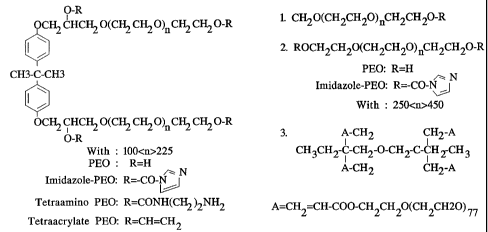
BRIEF DESCRIPTION OF THE DRAWINGS
Figures 1 a-ld illustrate alternative modes of preparing multiple layers of
PEO
and biopolymers onto a surface.
Figure 2 is a schematic of the chemical structure of the polymers of a
preferred
embodiment.
Figure 3 illustrates the relative WBC depletion for PEO-coated and uncoated
Asahi R-2000 filters. Log depletion is illustrated on the right side of the
figure.
Figure 4 illustrates the relative platelet recovery obtained with PEO-coated
and uncoated Asahi R-2000 filters.
Figure 5 illustrates the surface lubricity of a PVC tube having a NH2-PEO
coating using an Instron instrument test pursuant to Example 6.
Figure 6 illustrates the surface lubricity of a tube using an Instron
instrument
test pursuant to Example 6 for a Imz-PEO coating onto a NH2-PEO coated PVC
tubing.
Figure 7 illustrates the surface lubricity of a tube using an Instron
instrument
test pursuant to Example 6 for a Imz-PEO coating and a NH2-PEO coating on a
silicone tubing.
Figure 8 illustrates graphically the effect of heparin coating on fibrinogen
binding to various treated silicone tubings pursuant to Example 6.
Figure 9 illustrates graphically the effect of PEO coating on fibrinogen
absorption into various HVT pursuant to Example 7.
Figure 10 illustrates graphically the binding of HSA-LC-Biotin to Avidin-
coated PVDF with and without pre-treatment with Imz-PEO pursuant to Example 7.
Figure 11 illustrates graphically PEO coating on chitosan-treated glass filter
mats with and without N-acetylation and the effect on WBC recovery from whole
blood pursuant to Example 8.
Figure 12 illustrates graphically PEO coating on chitosan-treated glass filter
mats with and without N-acetylation and the effect on platelet recovery from
whole
blood pursuant to Example 8.
Figure 13 illustrates graphically the effect of PEO coating on fibrinogen
binding to heparinized-HVT (Tissues 4A were sterilized in solution containing
glutaraldehyde, while tissues 3A were in sterile solution without
glutaraldehyde).
8
CA 02382276 2006-08-17
Figure 14 illustrates graphically the effect of PEO coating on fibrinogen
binding from whole blood exposured to Denacol treated bovine pericardial heart
valve
pursuant to Example No. 8.
DETAILED DESCRIPTION OF THE PREFERRED EMBODIMENT
The present invention provides medical products having a coating applied
thereto which changes the surface properties. Additionally, the present
invention
provides methods for producing such products.
Pursuant to the present invention products are provided having a surface
thereof that includes a PEO cross-linked coated surface. Due to the modified
PEO
surface certain advantages are provided.
For example, improved bioprosthetic devices can be provided. In this regard,
the PEO coating technology can be applied to various types of biological
tissues, such
as bovine pericardium or porcine aortic tissues, that have been chemically pre-
treated
with DenacolTM and/or glutaraldehyde for the development of bioprosthetic
heart
valves. The method is based on a preliminary coating of tissue in an aqueous
solution
containing electrophilically active PEO derivatives, preferably a bis-
oxycarbonyl-
diimidazole-active PEO (Imz-PEO), having an average molecular weight of 20,000
daltons. The resulting intermediate Imz-PEO-activated tissue was further cross-
linked
with an amino-PEO derivative (preferably of the same MW) to form a stable PEO-
coated surface.
This intermediate activated Imz-PEO-coated tissue can also be used to couple
proteins, such as avidin into this matrix. This avidin-PEO-coated surface can
be
further employed to bind biotinylated agents, such as peptides like GREDVYTM,
in
order to produce surfaces capable of capturing endothelial cells.
PEO-coated tissues prepared by the above techniques have been shown to
reduce fibrinogen binding, compared to uncoated tissue. Other potential
advantages
of such PEO-modified surfaces include: reduced protein adsorption; limit
complement
activation; eliminate protein aggregation; produce specific ligand or cell
attachment
sites through avidin-biotin chemistry; and intermediate activated PEO-coated-
tissues
can be used for attachment sites to other anti-calcification agents such as 2-
amino-
oleic acid or toluidine blue.
8a
CA 02382276 2006-08-17
By way of further example, the surface of certain devices such as wound
drains, chest tubes, guide wires, catheter, and angioplasty products can be
modified
with a PEO coating to make them lubricious. To this end, such lubricious
surfaces
can be produced using a simple surface modification technology based on a
direct
coating of high molecular weight polyethylene oxide (PEO) derivatives onto
polymeric tubes, using water as a solvent. The polymer materials can be varied
from
polyvinyl chloride (PVC) to silicone or other type of polymers that are
typically used
for medical devices. These materials include polyurethane, polyolefine,
polyethylene,
polypropylene, metal or alloy. The PEO derivatives are functionalized PEO that
could contain an electrophilically active compound such as oxycarbonyl-
imidazoyl-
PEO (Imz-PEO) or nucleophilically active such as amino-PEO (NH2-PEO). This
technology provides a coating that generates low-friction or lubricious
surfaces which
also can limit fibrinogen adsorption. It has been found that the PEO-coated
PVC and
PEO-coated silicone tubes are stable in saline or plasma at 37 C for several
days.
Also, they can be sterilized with ETO without loss of lubricity or of low
protein
adsorption properties.
This technology presents several advantages including a simple coating
technology that uses water as a solvent. It also allows the production of
lubricious
surface on PVC and/or silicone surfaces. The production of products having
surfaces
with low fibrinogen adsorption. It provides the availability of functional
groups that
allow further surface modification (e.g., coupling with anti-coagulant
substances,
heparin or hirudin) or anti-microbial ligand (e.g. chitosan). The technology
also
provides a coating that can be also sterilized with ETO (or gamma) without
loss of
lubricity or low
8b
CA 02382276 2002-02-18
WO 01/15631 PCTIUSOO/21285
protein binding ability. The technology also provides the potential
application to a
variety of other synthetic polymers (polyurethane, polyethylene, polyolefine,
and metal
or alloy).
Still further pursuant to the present invention multilayer coating can be used
to
provide new surface modifications. To this end, the present invention provides
a new
surface modification method that is based on multilayer coatings between high
molecular
weight PEO derivatives and anti-coagulant biopolymers containing terminal
primary
amine groups.
The base material can be a wide variety of materials. For example, the base
material, could be derived from any biological tissue such as vascular grafts
or heart
valve tissues, or synthetic membranes made from various hydrophobic or
hydrophilic
polymers. Biopolymers containing amino-terminal groups can be derived from
carbohydrate structures such as heparin (glycosaminoglycan family) and
chitosan or
proteins such as hirudin.
Figure 1, and specifically Figures la-ld, set forth examples of multilayer
structures that can be produced. In the figures, the base material has thereon
the
multilayer coating. PEO refers of course to the polyethylene oxide coating
discussed
herein. ABP refers to the anticoagulant biopolymers.
These multiple layers of coating may provide numerous advantages. One of the
advantages is to provide a permanent coating technique that assures complete
coverage
of the base material. Additionally, the multiple layers allow the production
of a highly
anti-thrombogenic surface due to the combined presence of PEO and
anticoagulants
(heparin or hirudin). Further, the multiple layers allow the production of a
non-
inflammatory (e.g. non-complement activating) material due to the presence of
PEO and
heparin. Still further the multilayers allow the production of a potential
anti-bacterial
surface because of the presence of chitosan. The multilayer coating has
applicability to
multiple devices, including: membranes; theracyte devices; arterial filter
membranes, and
oxygenators; catheters, wound drains; and vascular grafts or heart valve
tissues.
In another embodiment a blood cell fractionation means is provided comprising
a matrix having a fibrous structure and the matrix further characterized in
having a
coating applied to it which changes its surface properties with respect to
cellular
9
CA 02382276 2002-02-18
WO 01/15631 PCT/US00/21285
adherence of blood cell containing fluid coming into contact therewith. The
matrix can
be a packing material contained within a column, or a fibrous material
compressed into
a filter and held in a filter housing of conventional design and construction,
although
other configurations of a solid matrix contacting a fluid are within the scope
of the
invention. In an embodiment, the coating of polymers and the chemical
reactions which
are carried out to create a generally molecularly continuous polymeric surface
on the
matrix fibers do not require covalent or noncovalent interaction with any
chemical
moiety present on the native surface of the matrix, the coating itself is
independent of the
chemical and physical identity of the matrix. Thus, the coating is intended to
be
universally applicable to any filter available in the cell separation art.
Examples include,
without limitation, filters having a high glass content, as in glass fiber
mats, filters with
less or no glass content such as a filter comprising a mixture of glass and
polyester, and
a polyethylene terephthalate platelet filter coated with hydroxyethylmethyl-
methacrylate.
Filter housings which may be conveniently used are manufactured
conventionally. Examples of such housing are Swinney plastic manifolds
manufactured
by Gelman, pediatric Enterprise Housings, or Intermediate Enterprise Housings.
The
correct size correlations of filters to correspondingly suitable housings will
be apparent
to those skilled in the art. The only limitation applicable to the blood cell
fractionation
means is a surface which is incompatible with the polymer solutions. Even in
the
instance where molecular wetting is not obtainable with the polymer solutions,
techniques utilizing emulsifiers and phase penetrants may be useful in
achieving adequate
coating. To Applicants' knowledge, none of the blood cell fractionation filter
materials
currently available commercially are to be excluded from applicability to the
present
invention.
In the method of separating cells using the product of the invention, a cell
suspension or whole blood is filtered through the filter having the polymer
coating as
disclosed. The leukocytes adhere, and the platelets and RBCs pass through the
in the
filtrate. More generalized methods of contacting the filter with a cell
containing fluid are
contemplated by this invention as well. For example, contracting by passaging
through
a packed column, or mixing cells in bulk with dispersed matrix in solution may
be
employed.
CA 02382276 2002-02-18
WO 01/15631 PCTIUSOO/21285
As noted above, the method of the present invention is applicable to a number
of
products and surfaces. For example, manufacturing ease, chemical condensation
reaction
of the respective polymers can be carried out insitu, i.e. a first free
polymer is laid down
on the matrix and dried, and then the second is contacted in solution with the
matrix. The
ensuing reaction then produces a skin-like sheet or layer of copolymerized
material at the
surface or the matrix. This reaction in a preferred embodiment proceeds
spontaneously
at temperatures generally in the range of 5 to 200 degrees centigrade. It is
evident that
the time for completion of the reaction will be slightly longer at cooler
temperatures than
for higher temperatures in accordance with kinetic thermodynamic principles.
Generally,
these reactions may be carried out at ambient temperatures, as disclosed in
the Examples,
but very little experimentation will be required by those skilled in the art
to adjust the
reaction times to a particular desired temperatures of reaction. The first
polymer to
be contacted with the surface is a high molecular weight electrophilically
active
polyalkylene oxide. Electrophilically active means that a polyalkylene oxide
polymer
contains a oxycarbonyl moiety reactive with a nucleophilic center such as an
amino or
hydroxyl group contained in a second polymer. In a preferred embodiment, a
primary
amine serving as a nucleophile, reacts with the carbonyl group of the
imidazole-
polyalkylene oxide polymer to form, upon reaction, an N-substituted carbamate
bond
where the carbonyl moiety from a cross-linker is incorporated into the new
bond. These
polymer entities must be high molecular weight, in the range of about 13,000
to 24,000
daltons, preferably about 20,000 daltons. Thus preferred molecules shown in
Figure 2
for reaction on surfaces will have n values of about 100-225.
A first electrophilic polyalkylene oxide polymer will have a terminal leaving
group reactive with an amine or hydroxyl containing second polyalkylene oxide.
Suitable leaving groups on the first polymer for achieving acceptable chemical
condensation are imidazoyl-, tresyl-, tosyl-, acryloyl-, and N-
hydroxysuccinimidyl-.
Additionally the structure of the electrophilic polymer can further be defined
by the
general expression: Y-PEO-R-PEO-Y, wherein Y is selected from the following
group
singly or in combination: oxycarbonylimidazole; tresyl-, tosyl-, N-
hydroxysuccinimidyl-,
and p-nitrophenyl- activated esters; acrylates; glycidyl ethers; and
aldehydes, and R is a
spacer defined as a backbone to which the two polyalkylene arms are attached,
consisting
11
CA 02382276 2002-02-18
WO 01/15631 PCT/US00/21285
preferably of bisphenol A or B. Bisphenol A is preferred, as shown in the
structure of
Figure 2.
We have also determined that in certain applications the imidazole derived
polyalkylene oxides provide excellent results, perhaps because the reaction
proceeds
somewhat better, or perhaps because residual unreacted groups improve
leukoadhesion.
In any event, Applicants do not wish to be bound to any particular theory, but
disclose
the result as a guide to those experienced in the art. In general,
polyalkylene means
polyethylene or polypropylene, since these are the most common polyalkylene
oxides
used in biocompatibility applications. However, Applicants consider other
polyalkylene
oxides up to polybutylene oxide to be within the scope of the invention.
In an embodiment, a tetra or diacrylate terminal derivative of polyalkylene
oxide
may be isopolymerized by first contacting with the surface, followed by
irradiation with
UV light or gamma rays to effect free radical polymerization. When used for
blood
filtration, the resulting coated filter matrix is leukodepletive with adequate
recoveries of
platelets and red bloods cells, but is not a efficacious as the other
embodiments of the
invention set forth herein.
In a method of the present invention, insitu chemical condensation can be
carried
out to mold the copolymer skin to the contours of the matrix fiber bed. It is
important
that the electrophilically active polyalkylene oxide be deposited on the
matrix first, dried,
and then further contracted with the second amino- or hydroxy- containing
nucleophilic
polymer. This teaching arises from empirical observation as to which method
steps give
best results in terms of platelet and RBC recovery, and leukodepletion, and
the
mechanistic or molecular basis for the observation is unknown to Applicants.
In the
drying step, drying in ambient air is adequate to "fix" the polymer in
position, but light
to moderate heat at various humidities down to less than 5% humidity or in
vacuo may
be applied to hasten the drying step in a manufacturing context.
The copolymerized material is highly stable to leaching, as shown in some of
the
Examples. In contrast to unreacted single polymer labeled with 1151 which is
readily
leached into filtrate, the fully copolymerized material made according to a
method of the
present invention is highly resistant to leaching, and is stable for
preparation of
therapeutically acceptable cell fractions.
12
CA 02382276 2006-08-17
By way of example, and not limitation, examples of the present invention will
now be given.
EXAMPLE NO. 1
Oxycarbonyl imidazole-polyethylene oxide (Imz-PEO) with an average
molecular weight of 20 K daltons (Sigma Chemical Company), was first coated
onto
existing Asahi R-2000 filters by soaking the filter mats in a 2.5% solution of
Imz-
PEO. The mats were dried under vacuum. The amount of Imz-PEO bound to the mat
was about 70 mg/gram of filter mat. Dried Imz-PEO-coated mats were cross-
linked
with bis[polyoxyethylene bis(amine)] (TAPEOTM, 20 K daltons), obtained from
Sigma Chemical Company. The cross-linking reaction was performed by soaking
the
Imz-PEO-coated mat in a water-methanol (1:1) solution of TAPEOTM at a 2.5 to
5.0
fold molar excess over the bound Imz-PEO. The reaction was allowed to proceed
for
at least 24 hours. The mats were dried again under vacuum. Dried cross-linked
mats
were washed extensively by soaking with water several times to remove any
unbound
PEG. After the final wash, the mats were dried again under a high vacuum.
Cross-
linked mats were stored at room temperature until used for blood filtration.
In this
example, the mats were used with pooled (ABO compatible), one day old, human
whole blood, obtained from Interstate Blood Bank. The pooled whole blood was
suspended about 3 feet above the filter unit, and the blood was allowed to
flow by
gravity through each of the different types of PEG-filter mats. An aliquot of
whole
blood (20 to 30 ml) was taken from the unit before filtration and was saved as
a
control (pre-sample). The filtered blood (post samples) and the pre-samples
were
counted for platelets with a SysmexTM K-1000 cell counter and the WBC
concentrations were determined by staining WBC nuclei (after lysing the
sample)
with propidium iodide and analyzing the stained samples with a FacScanTM flow
cytometer. The results of WBC depletion and platelet recovery are illustrated
in
Figures 3 and 4 respectively. The degree of platelet recovery ranged from 75
to 80%
with Imz-PEO coated mats vs 0.5% for the uncoated mats. The amount of WBC
depletion remained unchanged, in the range of 3 to 4 logs for all of the mats
(Table 1).
13
CA 02382276 2002-02-18
WO 01/15631 PCT/US00/21285
TABLE 1
Filtration of Whole Blood Through PEO-Coated And Uncoated Asahi R-2000 Filter
Mats
WBC Depletion PLATELET
Depletion Recovery
SAMPLE (log) (% Pre)
Imz-PEG
(no crosslinking) 3.25 80
2.5x Crosslinked
(Mat #1) 3.39 74
2.5x Crosslinked
(Mat #1) 3.75 74
Uncoated 3.73 0.5
EXAMPLE NO. 2
In this experiment, variable such as the age of the blood and the storage
temperature were evaluated. The same PEO coated Asahi R-2000 filter mats
described
above were used for these studies. Units of whole blood were obtained fresh in-
house,
and stored at room temperature until used (about 2 hours). One day old blood,
stored at
room temperature or 4 degrees centigrade, were also obtained form Interstate
Blood
Bank. Each unit was allowed to flow through each PEO-coated filter and the
samples
were analyzed as described above. The results, summarized in Table 2, suggest
that
despite the utilization of various units of whole blood stored under different
conditions,
the yield of platelets obtained from PEO-coated Asahi R-2000 filters is
dramatically
improved (68 to 83%) as compared to uncoated mats (2%).
14
CA 02382276 2002-02-18
WO 01/15631 PCT/USOO/21285
TABLE 2
Filtration Of Whole Blood Through PEO-Coated And Uncoated Asahi R-2000
Filters
WBC PLATELET
Depletion Recovery
SAMPLE (log) (% Pre)
PEO-Cross Linked-Mats:
Interstate-RT
(1 day old) #1 -2.63 83
Interstate-RT
(1 day old) #2 -4.01 68
Interstate-4 C
(1 day old) #3 -3.22 80
In-house-RT (-2hrs) #1 -3.25 76
Uncoated Mats:
Interstate-RT
(1 day old) #1 -3.50 02
EXAMPLE NO. 3
In this example, tetraacrylate PEO derivatives were obtained either from
Shearwater Polymer Inc., or synthesized from PEO 20 K daltons obtained from
Sigma
(Figure 2). The acrylate-PEO derivatives were coated onto composite mats by
the same
procedure as described in Example 1. The dried acrylate-PEO-coated mats were
subjected to gamma irradiation at a low dosage (2 megarads) to facilitate
cross-linking
of the PEO coating. The dried, coated mats were cut into circles of about 1.50
inches,
and 3 layers of mats were placed into a small pediatric-sized housing for
whole blood
CA 02382276 2002-02-18
WO 01/15631 PCT/US00/21285
evaluation. One day old pooled whole blood, obtained from Interstate Blood
Bank was
used. The final volume of blood used per housing was about 75 ml. The results
of these
experiments, summarized in Table 3, demonstrate the improvement in platelet
recovery
upon coating mats with the PEO derivatives. However, the improvement in
platelet
recovery seen with the acrylate PEO derivatives is not as good as was observed
with the
Imz-PEO coated mats.
TABLE 3
Filtration Of Whole Blood Through Various Crylate-PEO-Coated and Uncoated
Composite Filters
WBC Depletion PLATELET
Depletion Recovery
SAMPLE (log) (% Pre)
Uncoated -2.20 43
Sigma-Tetra-Acrylate-20K -1.62 69
Shearwater-Tetra-ACR-14K -2.04 56
Sigma-Tetra-Acrylate-20K
Irradiated -1.64 65
Shearwater-Tetra-ACR-14K
Irradiated -1.91 65
EXAMPLE NO. 4
The stability of these PEO coatings was investigated using radioactively
labeled
' Z5I-Imz-PEO and 'ZSI-Tetraamino-PEO. The presence of the bis phenol A units
in the
structure of Imz-PEO or Tetraamino-PEO derivatives permitted conventional
labeling of
these molecules using 125 1 and iodo beads (Pierce Chemical Co.). In the first
set of
16
CA 02382276 2002-02-18
WO 01/15631 PCT/US00/21285
experiments, the'ZSI-Imz-PEO was first coated onto the mats and was cross-
linked with
unlabeled Tetraamino-PEO. In the second set of experiments, unlabeled Imz-PEO
was
coated onto the mats and then cross-linked with 'ZSI-Tetraamino-PEO. Each 125I-
PEO
coated mat was evaluated in a Swinney housing (using a filter about 1 cm in
diameter)
with fresh whole blood. Four fractions of blood filtrate (- 1 ml each) were
collected and
counted for the presence of125I-PEO derivatives with a gamma counter. Each 125
I-PEO-
coated filter mat was also counted for radioactivity, before and after
filtration. The
amount of labeled PEO recovered on the mats after whole blood filtration
varied from
87% to 95%. In contrast, 35% of the labeled Imz-PEO was leached off filter
mats where
no crosslinking reaction was performed.
TABLE 4
Stability Of PEO-Coated Asahi R-2000 Filter Mats Measured With 'ZSI-Imz-PEO
or 125I-Tetraamino-PEO
1251-PEO Coated Mats Recovered
After Filtration
SAMPLE With 125I-Label (% Pre Labeled Mat)
125-Imz-PEO-Tetraamino-PEO 95%
Imz-PEO-125I-Tetraamino-PEO 87%
1251-Imz-PEO (not cross-linked) 65%
EXAMPLE NO. 5
Various pre and post blood samples from the above experiments were further
evaluated for complement activation by measuring C3a and C5a (by RIA) and for
platelet
activation by determining the percentage of platelets positive for the
activation marker
CD62. PLS10A platelet filters (Asahi) were included in this analysis as a
control for
comparison. The results for C3a and C5a is summarized in Table 5.
17
CA 02382276 2002-02-18
WO 01/15631 PCT/USOO/21285
TABLE 5
C3a And C5a Levels In Blood Exposed To PEO-Coated And Uncoated Asahi R-
2000 and PLS-10A Filters
C3a (ng/ml) C5a (ng/ml)
SAMPLE Pre-Samples Post-Samples Pre-Samples Post-Samples
Cross-linked 952 1,276 20 54
Cross-linked 538 614 0 19
Cross-linked 857 1,047 17 13
Cross-linked 1,103 1,149 28 34
Cross-linked 610 619 15 15
Uncoated 319 248 29 19
Uncoated 686 716 15 11
PLS-10A 964 4,057 22 66
PLS-10A 839 2,169 33 34
PLS-10A 328 1,727 9 25
PLS-10A 437 2,572 4 26
High levels of C3a and C5a were found in blood samples obtained from Asahi
platelet filter PLS-10A. Although these PLS-10A filters have not been used
with whole
blood, it appears that the PLS-l0A produces at least a 2 to 4 fold increase in
C3a and C5a
18
CA 02382276 2002-02-18
WO 01/15631 PCT/US00/21285
levels as compared to the corresponding pre-samples. These levels of C3a and
C5a are
higher than the amount of C3a and C5a produced by the PEO-coated Asahi R-2000
filters
are more biocompatible than the PLS-l0A commercial filter used for platelet
concentrate.
The percent of platelets expressing the activation marker, CD62, is a
sensitive
measure of the extent of platelet activation. Samples of whole blood were
analyzed (pre
and post filtration) using a FacScan flow cytometer to determine the
percentage of
platelets positive for CD62. This analysis revealed (Table 6) that no
elevation in the
percentage of CD62 positive platelets occurred during filtration on any of the
mats
investigated.
TABLE 6
Platelet Activation In Whole Blood Samples Exposed To Various Filters
SAMPLE % CD62 in Pre-Samples % CD62 in Post-Samples
Uncoated 5.45 5.88
Cross-linked-PEO 4.45 4.78
Cross-linked-PEO 5.20 5.24
Not Cross-linked-PEO 5.45 3.27
Not Cross-linked-PEO 4.05 2.11
PLS-10A 5.45 2.10
EXAMPLE NO. 6
In this group of examples, polyvinyl chloride and silicone tubes were coated.
A. PEO-Coated Polyvinyl Chloride (PVC) Tubes:
19
CA 02382276 2002-02-18
WO 01/15631 PCT/USOO/21285
PVC tubes (10 or 15 French size) were soaked in a water solution containing
various concentrations of NH2-PEO (1%, 2.5% or 5%). The tubes were incubated
at
55 C overnight, then they were removed. The tubes were allowed to air dry at
room
temperature following by another incubation at 55 C as the curing process.
The NH2-PEO-coated PVC was either used for crosslinking with another PEO
derivative without further washing or was washed extensively with water to
remove free
PEO. Washed tubes were allowed to air dry at room temperature and stored
desiccated
until analysis. Note that the amount of bound PEO was estimated based on the
amount
of radioactive125I-labeled-PEO tracer that was incorporated in the PEO coating
solution.
B. Cross-Linking Of Amino-PEO-Coated PVC:
Dried NH2-PEO-coated PVC (before washing) was soaked in a water solution
containing Imz-PEO at a concentration of 2.5% (or lower). The crosslinking
reaction
was performed at room temperature for 24 hours.
The tubes were removed and allowed to air dry at room temperature. The tubes
were then extensively washed with water to remove free Imz-PEO. The washed
tubes
were dried at room temperature and stored desiccated as described above.
C. PEO-Coated Silicone Tubes:
Silicone tubes (15 French size) were pre-treated with sodium hydroxide before
being treated with a PEO coating. The sodium hydroxide treatment consisted of
soaking
the tubing in 1N sodium hydroxide for 1 hour, following by extensive washing
(until
neutral pH) of the tubes with water.
The method of coating PEO derivatives (Imz-PEO or NH2-PEO) onto silicone
tubes was the same as for PVC above, except that all soaking in PEO solutions
were
performed at room temperature. The step that involved curing at 55 C was
omitted. The
final washed tubes were stored in a desiccated vacuum.
D. Attachment Of Heparin And Imz-PEO Onto Silicone Tubes:
Heparin and Imz-PEO can be incorporated into the silicone matrix by either
reacting heparin with Imz-PEO-coated silicone tubes (a two step process), or
by mixing
Imz-PEO and heparin in the same solution that was used as coating solution (a
one step
process). All heparin attachment was performed at 4 C for 24 hours. The tubes
were
dried and washed as described earlier.
CA 02382276 2002-02-18
WO 01/15631 PCT/US00/21285
E. Fibrinogen Binding Assay:
All PEO or Heparin-PEO-coated tubes were tested for fibrinogen binding against
control uncoated tubes. Each assay was performed with a triplicate sample
using a small
piece of tubing (about 0.4 cm length).
F. Measurement Of Surface Lubricity:
Each tube was cut into about 15 lengths and was placed into a designed flow-
cell
filled with saline (0.9% solution). One end of the tube was connected to an
Instron
instrument that served to pull out the tube from the flow-cell. The maximum
force
required for the Instron to pull the tube out determines the surface lubricity
of the tube.
The force used for pulling the control tube (uncoated PVC or silicone) was set
at
201b. The measurement was performed at two time intervals: 1) at time zero
(t=0) where
the tube was pulled as soon as it was loaded into the flow-cell; and 2) at
rinsed time (t=30
minutes) where the tube was allowed to stay in the flow-cell containing saline
solution
for 30 minutes. Then, the saline solution in the flow-cell was replaced with
new saline,
and finally the tube was pulled out.
G. Stability Study of PEO-Coated PVC or Silicone:
This study was performed in saline and plasma solutions, at 37 C up to 7 days,
using125Imz-PEO or'25I-NHZ-PEO-coated tubes (the radiolabeled PEO was used as
a
tracer). Several sets of small pieces (about 0.4 cm length) of125I-PEO-coated
PVC (or
silicone), and uncoated tubes were soaked in saline or pure plasma solutions.
The
samples were placed on a tube rocker which allows a continuous shaking of the
samples
during the entire incubation period. Each set of tubes (in triplicate) was
removed from
the shaker after day-1 (24 hours), day-3, and day-7. Each sample was counted
for total
radioactivity before removal of saline or plasma solution, then it was washed
twice with
water. The washed piece was counted for the remaining radioactivity. The ratio
between
the remaining radioactivity of PEO-coated tubes after washing and the total
radioactivity
was recorded.
RESULTS
PEO-Coated PVC: The results of Imz-PEO or NH2-PEO-coated PVC tubes are
illustrated graphically in Figures 5 and 6. Figure 5 illustrates graphically
bound NH2-
21
CA 02382276 2002-02-18
WO 01/15631 PCT/US00/21285
PEO (mmoles/cmZ) versus NH2-PEO concentration in the coating solution. Three
solution concentrations are illustrated: 1.0%; 2.5%; and 5.0%. As shown in
Figure 5,
NH2-PEO appeared to bind better to the PVC tubes than the Imz-PEO derivative.
Also,
the amount of bound NH2-PEO onto PVC increased with increasing concentration
of the
NH2-PEO in the coating solutions. However, using high concentration of this
NH2-PEO
(e.g. 10%) in a primary coating solution is not necessary, because it reduced
the amount
of bound Imz-PEO used in the crosslinked reaction; see Figure 6, concentration
of NH2-
PEO in coating solution (before cross-linking).
PEO-Coated Silicone: Figure 7 sets forth two PEO derivatives: Imz-PEO (Imz-PEO
cross-linked with NH2-PEO); and NH2-PEO (NH2-PEO cross-linked with Imz-PEO).
Illustrated in Figure 7, both PEO derivatives were strongly bound to silicone
tubing. The
amount of Imz-PEO bound was about 4 fold higher than the amount of bound NH2-
PEO.
In addition, the results in Figure 6 suggested that the primary coating of PEO
was very
stable since the level of radioactivity was unchanged after the crosslinking
reaction.
Fibrinogen Binding:
The results of fibrinogen binding to PEO-coated PVC tubing are summarized in
Tables 7 and 8 below.
As shown in Table 7, PEO-coated PVC exhibited a great reduction in fibrinogen
binding, compared to control uncoated PVC. Tubings coated with a low
concentration
of NHZ-PEO (1 %) showed the same level of bound fibrinogen, compared to other
tubings
that were coated with higher concentrations of NH2-PEO (2.5% or 5%), and with
or
without crosslinking with Imz-PEO (Table 7).
TABLE 7
Effect Of PEO Coating On Fibrinogen Binding Onto PVC Tubing
[NH2-PEO] in coating Bound Fg (ng/cm2) before Bound Fg (ng/cm2) after
solution crosslinked ( SD) crosslinked ( SD)
Uncoated 670 124
22
CA 02382276 2002-02-18
WO 01/15631 PCTIUSOO/21285
1% 85f9 136f23
2.5% 110 7 110f 19
5.0% 114t27 127 20
Also, the results in Table 8 indicated that there was no change in the level
of
fibrinogen binding to the PVC tubing after ETO sterilization.
TABLE 8
Effect Of ETO Sterilization On Fibrinogen Binding to PEO-Coated PVC Tubing
PVC Tubing Bound Fg (ng/cm2) before Bound Fg (ng/cmZ) after
ETO ( SD) ETO ( SD)
Uncoated 462 43 394 65
NH2-PEO (1%) 80 19 82 f 26
Xlink-PEO (0.5% Imz-PEO) 84 20 27 15
Xlink-PEO (2.5% Imz-PEO) 69 22 46 8
The ability of the PEO coating to reduce fibrinogen binding was also
demonstrated to be obtained with silicone (see Table 9 below). The level of
fibrinogen
bound to the Imz-PEO or NH2-PEO-coated silicone with or without crosslinking
was
about the same. This result suggests that the second crosslinking reaction
with Imz-PEO
derivative may not be necessary in this type of coating.
TABLE 9
Effect of PEO Coating On Fibrinogen Binding Onto PVC Tubing
Silicone Tubing & PEO Bound Fg (ng/cm2) Bound Fg (ng/cm2)
Coating Pre-Crosslinked ( SD) Post-Crosslinked ( SD)
Uncoated 258 173
'ZSI-Imz-PEO 82 27 52 8
125 l-NH2-PEO 62 19 46 2
23
CA 02382276 2002-02-18
WO 01/15631 PCT/US00/21285
However, Imz-PEO coating may be used as crosslinker reagent to the attachment
of heparin, as shown in Figure 8. The results in Figure 8, indicated that
heparin and Imz-
PEO can be incorporated onto silicone tubing by a one step (S1) or two step
(S2)
processes to produce low fibrinogen binding surface. The activity of
immobilized
heparin is under investigation.
Surface Lubricity:
The results of the effect of PEO coating on tubing lubricity are summarized in
Tables 10 and 11 below for PVC and silicone, respectively.
In this analysis, surface lubricity was measured by applying a maximum force
to
pull out the tube from the flow-cell filled with saline solution. The force
for the control
uncoated was set at 201b, on the Instron instrument.
TABLE 10
Effect Of PEO-Coated PVC On Tubing Lubricity
PVC (lOfr.) Pre-ETO Pre-ETO Post-ETO Post-ETO
(t=0) (t=30 min.) (t=0) (t=30 min.)
Experiment #1 (n=3) not not
NH2-PEO (1%) done 7.6 4.3 done 2.6 0.6
Experiment #2 (n=1)
S I(NH2-PEO 1%) 0.75 0.56 0.89 0.94
S2 (Xlink-PEO) 1.71 0.77 1.38 2.27
(Imz-PEO=0.5%)
S3 (Xlink-PEO) 0.78 0.44 0.41 0.59
(Imz-PEO=2.5 %)
TABLE 11
Effect Of PEO-Coated Silicone On Tubing Lubricity
Measured by friction test. Control uncoated = 201b
24
CA 02382276 2002-02-18
WO 01/15631 PCT/US00/21285
Silicone (15fr.) (t=0) (t=30 min.)
(n=2)
Imz-PEO 13.6 9.0 4.4 1.3
NH2-PEO 5.7 3.4 2.4 0.2
ImzPEO-Xlink 5.0 0.6 2.9 0.3
NH2-PEO-Xlink 10.8 0.9 5.3 0.4
The forces required for pulling the PEO-coated PVC or silicone tube were much
lower than the force necessary for the uncoated materials. For both initial
force (t=0) or
rinsed force (t=30 minutes) PVC tubing coated with 1% NH2-PEO solution showed
the
same degree of lubricity, compared to other coatings (Table 10). These results
suggest
that NH2-PEO can be used at low concentration (1%) as a single coating onto
PVC
tubing.
Similar results were also observed with NHZ-PEO-coated silicone tubing (Table
11). This derivative by itself can be used alone for coating silicone tube to
produce
surface with low-friction and low fibrinogen binding.
Stability of PEO Coating:
A. PVC Tubing: The results of the stability study of PEO-coated PVC tubing
in saline and in plasma are summarized in Tables 12 and 13, respectively. As
set forth
in Table 12, all PEO-coated PVC with or without additional cross linking are
very stable
in saline solution, at 37 C up to 7 days.
TABLE 12
Stability of 125 I-PEO-Coated PVC Tubing in Saline Solution at 37 C
PVC Tubing % Of Recovery % Of Recovery % Of Recovery
Day-1 (=L SD) Day-3 ( SD) Day-7 ( SD)
'25I-NH2-PEO-1.0% 95 9 90 9 98 2
125 I-NH2-PEO-2.5% 94 7 94 3 93 5
'25I-NH2-PEO-5.0% 91 6 93 4 96 3
CA 02382276 2002-02-18
WO 01/15631 PCT/US00/21285
Crosslink-1.0% 92 5 102 3 92t5
Crosslink-2.5% 94 8 98 4 95 f 4
Crosslink-5.0% 97 4 97 5 100 7
Also, these tubings (post saline incubation) showed very good reduction in
fibrinogen binding compared to control uncoated tubing (see Table 13 below).
TABLE 13
Stability of'ZSI-PEO-Coated PVC Tubing in Saline Solution at 37 C: Effect On
Fibrinogen Binding
PVC Tubing Bound Fg (ng/cm2) Bound Fg (ng/cm2) Bound Fg (ng/cm2)
Day-1 ( SD) Day-3 ( SD) Day-7 ( SD)
Uncoated 576 45 513 53 561 44
125 1-NH2-PEO-1.0% 75 9 76 16 71 13
125I-NHZ-PEO-2.5% 119 20 100 25 100 18
'25I-NH2-PEO-5.0% 87 9 101 8 93 19
Crosslink-1.0% 73 10 66 11 73 18
Crosslink-2.5% 98 6 85 2 95 9
Crosslink-5.0% 86 16 79 7 92 25
However, in pure human plasma, the percentage of the recovery of bound NH2-
PEO is in the range of 60% to 90% depend on the initial coating concentrations
and the
duration of the incubation (see Table 14 below).
TABLE 14
Stability Of125I-PEO-Coated PVC Tubing in Human Plasma at 37 C
PVC Tubing % Of Recovery % Of Recovery % Of Recovery
Day-1 ( SD) Day-3 ( SD) Day-7 ( SD)
1251-NH2-PEO-1.0% 92 2 77 10 65 5
26
CA 02382276 2002-02-18
WO 01/15631 PCT/USOO/21285
'25I-NHZ-PEO-2.5% 86 f 5 77 f 20 72 f 1
'Z5I-NH2-PEO-5.0% 102 27 79 5 72 f 4
Crosslink-1.0% 95 t 4 91 19 75 f 3
Crosslink-2.5% 91 1 82 6 77 6
Crosslink-5.0% 92 2 82 2 80 7
B. Silicone Tubing: Similar results were obtained with PEO-coated silicone
tubing. In saline the coating is very stable, the percentage of PEO recovery
was all above
95% (see Table 15 below).
TABLE 15
Stability Of125I-PEO-Coated Silicone Tubing in Saline Solution at 37 C
Silicone Tubing % Of Recovery % Of Recovery % Of Recovery
Day-1 ( SD) Day-3 ( SD) Day-7 ( SD)
'25I-NHZ-PEO 101 1 102 3 102 5
1251-Imz-PEO 100 1 100 6 99 5
'ZSI-NH2-PEO-Crosslink 98 4 94 6 92 5
'21I-Imz-PEO-Crosslink 93 4 98 3 88 8
After saline incubation, all PEO-coated silicone tubing still showed good
reduction in fibrinogen binding (see Table 16 below).
TABLE 16
Stability Of125I-PEO-Coated Silicone Tubing in Saline Solution at 37 C: Effect
On
Fibrinogen Binding
Silicone Tubing Bound Fg Bound Fg Bound Fg
(ng/cm2) (ng/cm2) (ng/cm2)
Day-1 ( SD) Day-3 ( SD) Day-7 ( SD)
'25I-NH2-PEO 84 f 21 68 24 89 17
27
CA 02382276 2002-02-18
WO 01/15631 PCT/US00/21285
'ZSI-Imz-PEO 103 43 99 57 102 7
'ZSI-NH2-PEO-Crosslink 70 18 50 17 55 16
'25I-Imz-PEO-Crosslink 99 36 79 4 86 13
In plasma and up to 7 days incubation at 37 C, PEO-coated silicone tubes are
very stable, since the percent recovery of bound PEO varied between 80 and
100% (see
Table 17 below).
TABLE 17
Stability Of125I-PEO-Coated Silicone Tubing in Human Plasma at 37 C
Silicone Tubing % Of Recovery % Of Recovery % Of Recovery
Day-1 ( SD) Day-3 ( SD) Day-7 ( SD)
125I-NH2-PEO 84 2 84 7 80 +7
'25I-Imz-PEO 99 3 94 1 91 4
'25I-NH2-PEO-Crosslink 94 5 94 6 89 10
125I-Imz-PEO-Crosslink 101 4 95 3 97 3
EXAMPLE NO. 7
A. Cross-linked-PEO-Coated Tissues:
Various Denacol pre-treated bovine pericardium heart valve tissues (HVT
obtained from Baxter Edwards) were washed several times with deionized water,
cut into
circles, and pre-coated with an Imz-PEO solution. This was followed by a
reaction with
NHz-PEO, at room temperature.
B. Fibrinogen adsorption:
PEO-coated and uncoated tissues were soaked in a citrate phosphate buffer
solution (pH 7.4) containing 125I-fibrinogen, and incubated at 37 C for one
hour.
Unbound protein was removed by washing extensively with PBS, saline and water.
The
amount of fibrinogen that was bound was calculated from the specific activity
of the
labeled protein and expressed as nanogram of fibrinogen per surface area.
28
CA 02382276 2002-02-18
WO 01/15631 PCT/USOO/21285
C. Avidin cross-linking to Imz-PEO-coated-materials:
PVDF flat sheet membranes (or biological tissues) were cut into circles, pre-
coated with Imz-PEO, and followed by reaction with Avidin, as described with
NH2-PEO
above.
D. Coupling of Avidin-PEO-coated tissue to LC-Biotin-HSA:
Avidin-PEO-coated PVDF, Uncoated PVDF (Millipore) and Avidin-PVDF (with
Avidin non specifically bound) were soaked in PBS (pH 7.4) containing LC-
Biotin-'ZSI-
HSA. The LC-Biotin-125I-HSA was prepared by coupling NHS-LC-biotin to'ZSI-HSA.
Unbound HSA-biotin was washed away with PBS and water. The amount of bound HSA
was expressed as cpm per surface area.
E. Results:
The results are all illustrated graphically in Figure 9. These results
indicate that
all cross-linked-PEO-coated tissues (lA, 2A, 3A, 4A) exhibit about 10 fold
lower
fibrinogen binding than the corresponding tissues without PEO coating.
Figure 10 illustrates graphically the binding of HSA-LC-Biotin to Avidin-
coated
PVDF with and without pre-treatment with Imz-PEO.
The results of the binding of biotin-HSA to Avidin-PEO-coated PVDF
membrane, Figure 10, suggest that Avidin can be covalently attached to the Imz-
PEO-
coated PVDF to improve the binding of Biotin-HSA, compared to Avidin-coated
membranes without PEO treatment.
EXAMPLE NO. 8
A. Cross-Linked Glutaraldehyde-Treated Tissues:
Glutaraldehyde pre-treated bovine pericardium heart valves tissues were washed
several times with de-ionized water, and pre-treated with an Imz-PEO solution.
This was
followed by a reaction with NH2-PEO, at room temperature. The reaction with
NH2-PEO
may be optional. The result is bovine pericardium tissue treated with PEO.
"Treated" in this sense is considered broader than "coated", as the PEO will
tend
to diffuse into the tissue rather than merely collecting on the surface.
Optionally, the
tissue may further be treated with a biologically active recognition sequence,
peptide, or
compound during or after the reaction with NH2-PEO.
29
CA 02382276 2002-02-18
WO 01/15631 PCT/USOO/21285
B. Fibrinogen Adsorption:
PEO-treated and untreated tissues were soaked in a citrate phosphate buffer
solution (pH 7.4) containing 1251-fibrinogen, and incubated at 37 C for one
hour.
Unbound protein was removed by washing extensively with PBS, saline and water.
The
amount of fibrinogen that was bound was calculated from the specific activity
of the
labeled proteins and expressed as nanogram of fibrinogen per surface area.
C. Calcification Assessment:
8 nun disks of PEO-treated and untreated tissues were implanted into the
paravertebral muscle of New Zealand albino (NZA) rabbits. After 30 and 90 days
implantation, the disks were removed and their calcium was quantified by using
known
standards, and the results calculated as ug Ca/mg dry weight tissue.
D. Results
The fibrinogen binding results are provided in Table 18 below. These results
indicate that cross-linked PEO-treated tissues exhibit about six-fold lower
fibrinogen
binding than the corresponding tissues without PEO treatment.
Table 18
Fibrinogen Binding of both PEO-Treated and Untreated Glutaraldehyde-Fixed
Tissue
Bovine Pericardial Tissue Bound Fibrinogen
Untreated 155 43
PEO-Treated 24 3
Table 19 (below) sets forth the results of the calcium content of a number of
explanted PEO-treated tissues at 30 and 90 days. It is apparent that the
average for the
three given samples at 30 days is skewed by the second tissue explant, and it
is believed
that the first and third tissue explants are more representative. This is
borne out by the
more closely grouped results for the three samples at 90 days.
In comparison, the results for a number of control samples is provided in
Table
20 (below). The control samples are glutaraldehyde-treated tissues also
implanted in the
paravertebral muscle of NZA rabbits. The results show that the calcium content
of PEO-
CA 02382276 2002-02-18
WO 01/15631 PCT/US00/21285
treated tissues is reduced significantly compared to glutaraldehyde controls.
Indeed, even
taking into account the seemingly anomalous second PEO-treated tissue sample,
the
average calcium uptake of the PEO-treated tissues was about one-fifth that of
the
untreated tissues at 30 days. The difference at 90 days is even more stark,
with the
average calcium uptake of PEO-treated tissues being about 3% of that of the
untreated
tissues.
Table 19
Calcium Uptake of PEO-Treated Lutaraldehyde-Fixed Tissue from Rabbit
Intramuscular Implant Technique
Rabbit # Sample # Time (days) Total yg/mg Ca Average St. Dev.
703S B2613-05/4 30 2.648
705S B2613-05/2 30 65.445 23.209 t 36.581
707S B2613-05/1 30 1.535
697S B2613-05/4 90 3.41
699S B2613-05/2 90 3.788 6.691 5.359
7015 B2613-05/1 90 12.876
Table 20
Calcium Uptake of Untreated Glutaraldehyde-Fixed Tissue from Rabbit
Intramuscular Implant Technique
Rabbit # Sample # Time (days) Total ,ug/mg Ca Average ~ St. Dev.
703S B2613-07/5 30 109.149
704S B2613-07/6 30 82.031
705S B2613-07/3 30 73.746
706S B2613-07/3 30 152.983 108.182 ~ 32.173
707S B2613-07/2 30 139.964
708S B2613-07/1 30 91.218
31
CA 02382276 2006-08-17
697S B2613-07/5 90 267.791
698S B2613-07/6 90 257.285
699S B2613-07/3 90 267.831
700S B2613-07/3 90 210.112 251.114 32.753
701S B2613-07/2 90 212.551
702S B2613-07/1 90 291.111
It should be noted that the implant methodology wherein the tissues are
implanted in the muscles of rabbits, or of other mammals, is believed to be
more
effective than traditional subcutaneous implant techniques. That is, tissue
implanted
subcutaneously tends to become rapidly encapsulated by the host's natural
immune
response. Because of this encapsulation, and because of the relatively low
presence
of calcium in such interstitial body spaces, the calcium uptake is from
passive
diffusion and is thus relatively slow. Therefore, tissue explanted at 30, 60,
and even
90 days tends to have a calcium content of around 1 micrograms per milligram
dry
weight tissue. Differentiating between different tissues samples is thus
problematic
because of the relatively low resolution of the subcutaneous technique.
Implanting the tissues directly into the animal's muscle, however, vastly
increases the exposure of the tissue to body calcium. It is well known that
calcium
flux within muscles is one of the prime physiological causes of muscle
contraction.
Therefore, tissue implanted into the muscle is regularly exposed to transitory
calcium
flows. Because of the increased calcium exposure, the tissue more rapidly
absorbs the
calcium, and thus exhibits a much higher calcium content at 30, 60 and 90
days. The
sensitivity or resolution of this implant methodology greatly facilitates
differentiation
and analysis of the results for different tissue specimens. A full disclosure
of the
muscle implant methodology is provided in co-pending International Patent
Application No. WO 01/15628, entitled "In vivo Screening Methods for
Predicting
Calcification of Implantable Prosthetic Material" filed on September 1, 2000.
It should also be noted that the PEG treatment as disclosed herein may be
effective in tissues other than bovine pericardium. For example, allograft
tissue,
porcine
32
CA 02382276 2002-02-18
WO 01/15631 PCT/US00/21285
tissue, equine tissue, or other xenograft tissue may be treated with PEO to
obtain the
benefits mentioned herein, in particular calcification mitigation. In
addition, although
PEO treatment has been tested on tissue that has first been pre-treated, or
cross-linked,
with Denacol or glutaralddehyde, the same benefits described herein may also
be
obtained by treating fresh tissue.
EXAMPLE NO. 9
In this example, methods and samples having multiple coatings were prepared
and tested.
A. PEO-coated Chitosan surfaces:
1. Attachment of PEO onto Chitosan-mats:
Glass filter mats (GFM) were cut into circles (about 1 cm in diameter). The
circles were first modified with 1% chitosan solution. The PEO derivatives
with various
lengths (PEO-5K, PEO-18.5K and PEO-20K) were then covalently attached to the
mats
through the amine functional group of the chitosan ligand. At the end of the
coupling
reaction, some mats were treated with an NHS-acetate to acetylate the
unreacted amine
groups of the chitosan polymer.
2. Evaluation with whole blood:
Citrated, fresh whole blood was filtered through various PEO-coated chitosan
mats and uncoated chitosan-mats. Fractions of 1.0 ml (x2) were collected as
post-
samples. The number of white blood cells (WBC) and platelets were determined
on the
pre-samples and post samples using a Sysmex cell counter.
3. Results:
PEO-coated chitosan mats showed an improved recovery of platelets and WBC,
compared to chitosan-coated mats without additional PEO coating. N-acetylation
of the
free amine group of the chitosan molecules appears to improve WBC and platelet
recovery even further compared to non-acetylated materials. Surfaces coated
with HMW
PEO appear to have performed better than surfaces coated with LMW PEO (see
Figures
11 and 12).
B. PEO-Coated Heparin-Surfaces:
33
CA 02382276 2002-02-18
WO 01/15631 PCTIUSOO/21285
1. Attachment of PEO onto heparin-fixed denacol-treated pericardial heart
valve tissues (HVT~
Two types of Heparin fixed Denacol treated tissues (3A and 4A) were obtained
from Baxter CVG and were used for this study. They were washed several times
with
deionized water and were soaked in an oxycarbonyl imidazole-PEO (Imz-PEO)
solution
(pH=8.3) for 24 hours, followed by reaction with an amino-PEO (NH2-PEO) at the
same
pH for at least 24 hours. Incubations were performed at room temperature.
2. Biocompatibility Evaluation:
PEO-coated and non-PEO-coated tissues were tested for their ability to bind
fibrinogen (Fg) from a solution of purified human fibrinogen and from fresh
whole blood
according to the following procedure: PEO-coated and uncoated materials were
soaked
in a citrate phosphate buffer solution (pH=7.4) containing'ZSI-labeled
fibrinogen (Fg),
and were incubated at 37 C for one hour. Unbound fibrinogen was removed from
the
materials by washing extensively with saline, then each sample was counted in
a gamma
counter. The amount of protein adsorbed was calculated from the specific
activity of the
fibrinogen and expressed as ng of protein per mg (or per surface area) of
materials.
3. Results:
The results indicate that PEO-coated heparinized HVT can significantly reduce
fibrinogen binding from both sources, a purified solution of human Fg and Fg
from
whole blood, compared to uncoated tissues (see Figures 13 and 14).
C. Heparin-treated Imz-PEO-coated-HVT:
A Heparin coating procedure, similar to the one described in section 2a above,
was applied in this study. Heparin solution (prepared in bicarbonate buffer
pH=8.3) was
used instead of amino-PEO to react with Imz-PEO-coated HVT.
It will be understood that various modifications to the presently preferred
embodiments described herein will be apparent to those skilled in the art.
Such changes
and modifications can be made without departing from the spirit and scope of
the present
invention and without diminishing its attendant advantages. It is therefore
intended that
such changes and modifications be covered by the appended claims.
34