Note: Descriptions are shown in the official language in which they were submitted.
CA 02382947 2002-02-27
WO 01/27882 PCT/US00/23614
APPARATUS AND METHOD FOR IDENTIFICATION OF INDIVIDUALS
BY NEAR-INFRARED SPECTRUM
Cross Reference to Related Patents and Pending Applications
The present application is related to U.S. Patent Application Serial No.
09/174,812, filed October 19, 1998, entitled "Method for Non-Invasive Analyte
Measurement with Improved Optical Interface", and U.S. Patent Application
Serial
No. 08/871,366, filed June 9, 1997, entitled "Diffuse Reflectance Monitoring
Apparatus", both assigned to the same assignee as the present application.
Technical Field
to The present invention relates generally to methods and systems for
verifying
the identity of an individual utilizing spectral data from a non-invasive near-
infrared
tissue analysis. More specifically, the invention relates to non-invasive
methods and
apparatus for verifying identity of a living individual using near-infrared
absorption of
light energy by tissue with identity verified using multivariate discriminant
analysis
techniques on resulting subcutaneous tissue spectral data as compared to prior
stored
spectral data for that individual.
Background of the Invention
Identity verification is useful in many applications. Examples include
verifying identity prior to activating machinery or gaining entry to a secure
area.
2o Another example would be identity verification of an individual for
matching that
individual to records on file for that individual, such as for matching
hospital patient
records when the individual's identity is unknown. Identity verification is
also useful
to match police records at the time a suspect is apprehended, but true
identity of the
suspect is not known. Passwords, keys, numeric codes and fingerprints are
solutions
currently in use. However, keys and codes can be used by anyone having
possession
-1-
CA 02382947 2002-02-27
WO 01/27882 PCT/US00/23614
of the keys or codes. A requirement that the person physically at a site be
the person
authorized to use the key or password is not easily enforced. Fingerprint
analysis
generally fails to give instant results and security systems relying on
fingerprint
analysis can be circumvented, as disclosed by Osten et al. in U.S. Patent No.
5,719,950.
Living human tissue is recognized as a dynamic system containing a multitude
of components and analyte information that is particularly useful in the
medical
profession for diagnosing, treating and monitoring human physical conditions.
To
this end, effort has been directed toward developing methods for non-invasive
measurement of tissue constituents using spectroscopy. The spectrographic
analysis
of living tissue has been focused on the identification of spectral
information that
defines individual analytes and relates such spectral data to the analyte's
concentration. Concentrations of these analytes vary with time in an
individual
patient. Acquiring tissue spectral data with sufficient accuracy for use in
diagnosis
and treatment has proven difficult. Difficulties in conducting the analysis
have been
found which are related to the fact that the tissue system is a complex matrix
of
materials with differing refractive indices and absorption properties.
Further, because
the constituents of interest are many times present at very low
concentrations, high
concentration constituents, such as water, have had a detrimental impact on
identifying the low level constituent spectral information and giving an
accurate
reading of the desired constituent concentration. Development of these
techniques
has always focused on the changes in spectral output with change in
concentration of
a dynamic analyte of interest, such as glucose. The techniques disclosed are
focused
on identifying concentrations of specific analytes, the concentration of which
is
expected to vary with time.
-2-
CA 02382947 2002-02-27
WO 01/27882 PCT/US00/23614
Improved methods and apparatus for gathering and analyzing a near-infrared
tissue
spectrum for an analyte concentration are disclosed in commonly assigned U.S.
Patent
applications and issued patents. U.S. Patent No. 5,655,530 and U.S. Patent
Application Serial No. 08/844,501, filed April 18, 1997, entitled "Method for
Non-
invasive Blood Analyte Measurement with Improved Optical Interface" relate to
near-
infrared analysis of a tissue analyte concentration which varies with time,
with a
primary focus on glucose concentrations in diabetic individuals. The methods
and
apparatus include placing a refractive index-matching medium between a sensor
and
the skin to improve the accuracy and repeatability of testing. U.S. Patent
Application
Serial No. 09/174,812, filed October 19, 1998, entitled "Method for Non-
Invasive
Blood Analyte Measurement with Improved Optical Interface" discloses
additional
improvements in non-invasive living tissue analyte analysis. The disclosure of
each
of these three applications or patents are hereby incorporated by reference.
U.S. Patent No. 5,636,633 relates, in part, to another aspect of accurate non-
invasive measurement of an analyte concentration. The apparatus includes a
device
having transparent and reflective quadrants for separating diffuse reflected
light from
specular reflected light. Incident light projected into the skin results in
specular and
diffuse reflected light coming back from the skin. Specular reflected light
has little or
no useful information and is preferably removed prior to collection. U.S.
Patent
2o Application Serial No. 08/871,366, filed June 9, 1997, entitled "Improved
Diffuse
Reflectance Monitoring Apparatus", discloses a further improvement for
accurate
analyte concentration analysis which includes a blocking blade device for
separating
diffuse reflected light from specular reflected light. The blade allows light
from the
deeper, inner dermis layer to be captured, rejecting light from the surface,
epidermis
layer, where the epidermis layer has much less analyte information than the
inner
-3-
CA 02382947 2002-02-27
WO 01/27882 PCT/US00/23614
derniis layer, and contributes noise. The blade traps specular reflections as
well as
diffuse reflections from the epidermis. The disclosures of the above patent
and
application, which are assigned to the assignee of the present application,
are also
incorporated herein by reference.
U.S. Patent No. 5,435,309 relates to a system for selecting optimal
wavelengths for multivariate spectral analysis. The use of only one wavelength
gives
insufficient information, especially for solutions having multiple components.
The
use of too many wavelengths can include too much noise and lead to
combinatorial
explosion in calculations. Therefore, the number of wavelengths used should be
limited and the wavelengths well chosen. Genetic algorithms are used in this
reference to select the most fit wavelengths. The disclosure of this patent is
incorporated herein by reference.
Summary of the Invention
In contrast to the above discussed prior art techniques for non-invasive
analysis of a blood or tissue analyte concentration using infrared
spectroscopy, the
present invention is based on applicant's recognition that the resultant
tissue spectrum
of a particular individual includes unique spectral features and combinations
of
spectral features which can be used to identify the individual once the
analytical
device has been trained to identify the individual. Spectral information in
the near
2o infrared range is preferred, however, it is recognized that visible or mid-
infrared light
energy could be used alone or in combination with near infrared. Training of
the
device is accomplished by use of stored spectral data for that individual from
prior
testing. Applicants have been able to achieve essentially zero percent false
positive
error rates with the techniques disclosed herein, even though the tissue being
analyzed
is a dynamic system with analyte concentrations, and thus, tissue spectral
data,
-4-
CA 02382947 2002-02-27
WO 01/27882 PCT/US00/23614
varying considerably over time and between analysis. Success of the method of
the
present invention is believed tied to two components. First, the method
incorporates
an apparatus and technique for accurately and repeatably acquiring a tissue
spectrum
which is stable, while remaining sensitive to slight changes in spectral
output at any
given wave length. The system optimizes optical throughput both into and out
of the
tissue sample. Second, because the spectral features or combinations of
spectral
features that are unique for a particular individual are not readily apparent
or
identified by visual comparison of a spectral result, the present invention
relies on
discriminant analysis techniques to first train the device to identify
spectral features of
1 o significance for the individual and then compare such features to new
spectral data at
the time of attempted verification. The method can incorporate a discriminant
analysis technique based upon Mahalanobis distance technique or other distance
techniques to compare spectral data acquired from an individual with spectral
data
present in a database.
The present invention, thus, includes a method for verifying the identity of
an
individual using non-invasive tissue spectroscopy. A preferred method and
apparatus
illuminates skin with near-infrared radiation and collects the reflected, non-
absorbed
near-infrared radiation. Diffuse, rather than specular, reflected light is
preferably
collected, more preferably light diffusely reflected from the inner dermis
rather than
2o the epidermis. The near-infrared spectral data collected can be stored in a
computer
database. A series of such spectral data are collected from the individual or
individuals for which identity verification is desired. The identity of the
individual is
preferably verified and stored along with the associated spectral data in an
authorization database. Authorized spectra can be collected over a period of
minutes,
or more preferably, a number of spectra can be collected over days and weeks,
which
-5-
CA 02382947 2002-02-27
WO 01/27882 PCT/US00/23614
allows for adjustment of the individual's model for verification to account
for natural
physiological differences at any given time of analysis which will affect a
person's
tissue spectra.
After collection, the authorization spectral database for a particular
individual
can be analyzed, using discriminant analysis tools, relative to new spectral
data from
an individual purporting to be that individual or an unknown individual. When
the
purported identity of a target individual is to be verified or an unknown
individual's
identity is to be checked against a stored database, a target tissue spectrum
can be
taken and processed in a manner similar to the processing of the already
stored
to authorization spectra. In one method, the Mahalanobis distance and spectral
residual
magnitude are used to verify the purported identity or check the unknown
individual's
spectral data against a database. In a preferred method, the Mahalanobis
distance and
spectral residual magnitude are calculated for the target spectrum relative to
the
database spectra for the individual with the purported identity. Identify is
verified
only if the aforementioned distance and magnitude are less than a
predetermined
threshold set for each.
One system for performing identity verification includes: a computer having
an input device and an output device; a database including near-infrared
tissue
spectral data for authorized persons or a collection of spectral data for
individuals
2o against which unknown individual's would be checked; a near-infrared
radiation
source for projecting near-infrared radiation into subcutaneous tissue; a near-
infrared
spectrometer for measuring subcutaneous near-infrared intensity over a
plurality of
wavelengths; and a program running in the computer for discriminating between
a
target individual's spectral data and the authorized spectral data or
collection of
spectra database containing spectra for a group of individuals. The program
can
CA 02382947 2002-02-27
WO 01/27882 PCT/US00/23614
include software for performing discriminant analysis. In one system,
supervised
learning programs can be utilized to assist in associating the various
spectral data for
each identified individual together.
These and various other advantages and features of novelty which characterize
the present invention are pointed out with particularity in the claims annexed
hereto
and forming a part hereof. However, for a better understanding of the
invention, its
advantages, and the object obtained by its use, reference should be made to
the
drawings which form a further part hereof, and to the accompanying descriptive
matter in which there are illustrated and described preferred embodiments of
the
t0 present invention.
Brief Description of the Drawings
In the drawings, in which like reference numerals indicate corresponding parts
or elements of preferred embodiments of the present invention throughout the
several
mews:
t 5 Fig. 1 is a partial cross-sectional view of a sensor element coupled to
the skin
surface via an indexing-matching fluid;
Fig. 2 is a partial cross-sectional view of an alternative embodiment of a
sensor element coupled to opposite sides of a skin surface via an indexing-
matching
fluid; and
2o Fig. 3 is a graphical representation of experimental data showing the
improvement in accuracy and repeatability of a sensor coupled to the skin via
an
index-matching medium.
Detailed Description of the Preferred Embodiments
Detailed embodiments of the present invention are disclosed herein. However,
25 it is to be understood that the disclosed embodiments are merely exemplary
of the
CA 02382947 2002-02-27
WO 01/27882 PCT/US00/23614
present invention which may be embodied in various systems. Therefore,
specific
details disclosed herein are not to be interpreted as limiting, but rather as
a basis for
the claims and as a representative basis for teaching one of skill in the art
to variously
practice the invention.
The present invention is based on Applicant's recognition that an accurate,
precise and repeatable tissue spectra of an individual in the near infrared
range
contains spectral features and combinations of spectral features which are
unique to
that individual. It is further believed that some unique information may be
present in
the visible light region, with the techniques disclosed herein adaptable to
such
analysis. The present invention is further based on a recognition that proper
analysis,
utilizing discriminant analysis techniques, can identify these unique features
or
combinations, which are not readily apparent in visual analysis of a spectral
output, so
that an individual's identity may be verified by comparison of a tissue
spectral data
taken at the time of verification compared to stored tissue spectral data from
prior
t 5 testing. The identification methods can also be used in conjunction, or
simultaneously, with measurement of analyte concentrations in an individual.
The prior spectral data is used to train the device to identify that
particular
person based on features that are recognized unique to that particular
individual.
These unique spectral features have been found to be consistently present even
though
2o the tissue being analyzed at each time of analysis is a dynamic system
which contains
components and analytes whose concentration vary, with resulting tissue
spectral
variations, due to physiological changes in the individual.
As previously stated, there are two components to the success of the method of
the present invention. First, the method incorporates an apparatus and
technique to
25 accurately and repeatably acquire tissue spectral data. The apparatus is
sensitive to
_g_
CA 02382947 2002-02-27
WO 01/27882 PCT/US00/23614
slight changes in spectral output at any given wavelength of input and
optimizes the
overall optical throughput both into and out of the tissue sample. Second, the
method
requires specific techniques for training the instrument to identify spectral
features of
significance for that particular individual, and then to compare such features
to a new
spectral data acquired at the time of attempted verification. Because the
spectral
features or combinations of spectral features that are unique for a particular
individual
are not readily apparent or identified by visual comparison of a spectral
result and the
unique spectral features are present at different wavelengths for different
individuals,
the present invention relies on discriminant analysis techniques to compare
spectral
data. Each component of the apparatus and method of the present invention are
detailed below.
The present invention utilizes an accurate, repeatable and sensitive method
for
non-invasive measurement of a near infrared tissue spectral data. It is
recognized that
the sample is a complex matrix of materials with differing refractive indices
and
I s absorption properties. Further, because many constituents are present at
very low
concentrations, it has been found to be imperative to couple light into and
out from
the tissue in an efficient manner. The method of the present invention
incorporates an
index-matching medium, fluid or deformable solid, to improve the efficiency of
coupling the light both into and out of the tissue sample.
2o The present invention utilizes light energy in the near-infrared region of
the
optical spectrum as an energy source for analysis. Water is by far the largest
contributor to absorption in tissue in the near-infrared region because of its
concentration, as well as its strong absorption coefficient. It has been found
that the
total absorption spectrum of tissue, therefore, closely resembles the water
spectrum.
2s Less than 0.1 percent of the absorption of light is from, for instance, a
constituent
-9-
CA 02382947 2002-02-27
WO 01/27882 PCT/US00/23614
such as glucose. It has been further found that tissue greatly scatters light
because
there are many refractive index discontinuities in a typical tissue sample.
Water is
perfused through the tissue, with a refractive index of 1.33. Cell walls and
other
features of tissue have refractive indices closer to 1.5 to 1.6. These
refractive index
discontinuities give rise to scatter. Although these refractive index
discontinuities are
frequent, they are also typically small in magnitude and the scatter generally
has a
strong directionality towards the forward direction.
This forward scatter has been described in terms of anisotropy, which is
defined as the cosine of the average scatter angle. Thus, for complete
backwards
t o scatter, meaning that all scatter events would cause a photon to divert
its direction of
travel by 180 degrees, the anisotropy factor is -1. Likewise, for complete
forward
scatter, the anisotropy factor is +1. In the near infrared, tissue has been
found to have
an anisotropy factor of around 0.9 to 0.95, which is very forward scattering.
For
instance, an anisotropy factor of .9 means that an average photon of light
only scatters
through an angle of up to 25 degrees as it passes through the sample.
In acquiring tissue spectral data, measurements can be made in at least two
different modes. It is recognized that one can measure light transmitted
through a
section of tissue, or one may measure light reflected or remitted from tissue.
It has
been recognized that transmission is the preferred method of analysis in
spectroscopy
2o because of the forward scattering of light as it passes through the tissue.
However, it
is difficult to find a part of the body which is optically thin enough to pass
near
infrared light through, especially at the longer wave lengths. Thus, the
preferred
method for measurement in the present invention is to focus on the reflectance
of light
from the sample.
-10-
CA 02382947 2002-02-27
WO 01/27882 PCT/US00/23614
Photons reflect and refract at refractive index discontinuities, and so light
impinging on tissue immediately has a small reflectance at the tissue surface.
This is
referred to as specular reflectance. Since this light does not penetrate into
the tissue,
it contains little information about the tissue constituents. This is
especially true in
light of the physiology of skin, which possesses an outward layer which is
essentially
dead and lacks spectral information believed unique to an individual. Thus,
reflected
light energy containing spectral data unique to an individual is believed to
be that
light which is reflected back to the surface through refractive index
discontinuities
deeper within the tissue sample. This reflected light energy is referred to as
diffusely
reflected light.
Applicants have found that a large fraction of incident photons are absorbed
and scattered in the tissue. Those photons which are available for coupling
back out
of the tissue are likely diverted in their angular path. In fact, by
definition, a photon
must change direction in order to exit the tissue in a direction towards the
input optic.
Applicants, however, have found that a large problem with detection is
associated
with the refractive index discontinuity between the average tissue refractive
index and
the refractive index of air outside of the tissue. It has been found that this
discontinuity acting on incident light leads to a refraction and a small
specular
reflectance of less than about 5 percent. However, on the way out, the
discontinuity
2o gives rise to a critical angle phenomenon. Because the photon is traveling
from a high
refractive index medium to a lower one, a critical angle exists above which a
photon
is totally internally reflected and will not escape the tissue sample. This
critical angle
for photons traveling from tissue to air has been found to be about 46
degrees, which
presents a problem. A photon normally incident on the tissue surface must
deviate
2s through a large angle to exit. Because of the forward directionality of
scattering, this
-11-
CA 02382947 2002-02-27
WO 01/27882 PCT/US00/23614
is difficult for a photon to do, and it is very likely to make a grazing or
high angle
incidence with the tissue and air interface. The grazing incidence photons
will not
escape because the critical angle is exceeded.
Applicants have found a solution for the differences in refractive index
associated with coupling light energy exiting tissue to an analytical
instrument. The
solution is the use of an immersion fluid which has very low absorptivity in
the
spectral range of interest, and has a viscosity compatible with good flow and
coverage, while having a refractive index which effectively introduces light
into the
tissues, reduces specular reflection and effectively gets light back out of
the tissue. In
1 o preferred embodiments, the index-matching fluid is preferably minimally or
essentially non-absorbing of light energy in the wavelengths selected as
relevant to
identification of an individual. The fluid is thus non-spectroscopically
active at
desired wavelengths. However, it is believed a minimally absorbing index-
matching
fluid, for example one that absorbs less than about 10% of the light energy of
relevant
wavelengths, could still be utilized. A preferred material is a fluorinated,
chlorinated
hydrocarbon polymer oil manufactured by Occidental Chemical under the
tradename
FLUOROLUBE. FSS is a preferred FLUOROLUBE. These oils have a refractive
index of about 1.38, are non-toxic, and Applicants have found that it has a
spectral
signature in the near infrared region which is minimal.
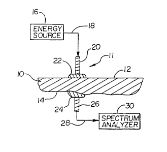
2o Now referring to Figs. 1 and 2, partial cross-sectional views of two
preferred
embodiments of an apparatus for non-invasively acquiring a tissue spectrum are
depicted. The depictions in Figs. 1 and 2 are schematic to depict the concept
of
utilizing an index-matching medium 22 in conjunction with a non-invasive
sensor
element 11 operatively connected to an energy source 16 and a spectrum
analyzer 30.
The relative size, shape and detail of physical components are not depicted.
-12-
CA 02382947 2002-02-27
WO 01/27882 PCT/US00/23614
The apparatus depicted in Fig. 1 and the apparatus depicted in Fig. 2
generally
include three elements, an energy source 16, a sensor element 11, and a
spectrum
analyzer 30. The embodiment of Fig. 1 depicts the sensor element as including
an
input element 20 and an output element 26, which can include a single lens
system for
both input and output light energy. The input element 20 and output element 26
are in
contact with a common skin surface 12 of the selected tissue 10. The
alternative
embodiment of Fig. 2 depicts an alternative sensor element 11 arrangement,
wherein
the input element 20 and output element 26 are arranged on opposing surfaces
12, 14
of tissue 10. Both embodiments function to give a measure of the absorption of
infrared energy by the tissue 10. However, the embodiment of Fig. 1 is
utilized to
measure the quantity of light energy which is reflected from the tissue 10 by
the
components or features therein. In contrast, the embodiment of Fig. 2 measures
the
transmission of light energy through the tissue 10. In either embodiment, the
absorption at various wavelengths can be determined by comparison to the
intensity
of the light energy from the energy source 16.
The energy source 16 is preferably a wide band, infrared black body source.
The optical wavelengths emitted from the energy source 16 are preferably
between
1.0 and 2.5 Vim. The energy source 16 is operatively coupled to a first means
for
transmitting infrared energy 18 from the energy source to the input element
20. In
2o preferred embodiments, this first means 18 is simply the transmission of
light energy
to the input element 20 through air by placing the energy source 16 proximate
the
input element 20.
The input element 20 of the sensor element 11 is preferably an optical lens
which focuses the light energy to a high energy density spot. However, it is
understood that other beam focusing means may be utilized in conjunction with
the
-13-
CA 02382947 2002-02-27
WO 01/27882 PCT/US00/23614
optical lens to alter the area of illumination. For example, a multiple lens
system,
tapered fibers, or other conventional optical beam-shaping devices could be
utilized to
alter the input light energy.
In both embodiments depicted in Figs. 1 and 2, an output sensor 26 is utilized
to receive reflected or transmitted light energy from the tissue 10. In a
preferred
embodiment, a specular control device is incorporated to separate the specular
reflected light from diffusely reflected light. Such devices are disclosed in
co-
pending and commonly assigned application Serial No. 08/871,366, filed June 9,
1997, and entitled "Diffuse Reflectance Monitoring Apparatus", the disclosure
of
which is incorporated herein by reference. As described in conjunction with a
method
of analysis below, the embodiment of Fig. 1 has an output sensor 26 which
receives
reflected light energy, while the embodiment of Fig. 2 includes an output
sensor 26
which receives transmitted light through the tissue 10. As with the input
element 20,
the output element 26 is preferably an optical lens. Other optical collection
means
may be incorporated into an output element 26, such as a multiple lens system,
tapered fiber, or other beam-collection means to assist in directing the light
energy to
the spectrum analyzer 30.
A second means for transmitting infrared energy 28 is operatively connected
to the output element 26. The light transmitted through the second means for
2o transmitting infrared energy 28 is transmitted to the spectrum analyzer 30.
In a
preferred embodiment, the operative connection to the output element includes
transmission of the reflected or transmitted light energy exiting the output
element
through air to the spectrum analyzer 30. A mirror or series of mirrors may be
utilized
to direct this light energy to the spectrum analyzer.
-14-
CA 02382947 2002-02-27
WO 01/27882 PCT/US00/23614
In practicing the method of the present invention, tissue 10 area is selected
as
the point of analysis. This area can include the skin surface 12 on the
forger, earlobe,
forearm, or any other skin surface. Preferably, the area for sampling includes
blood
vessels near the surface, and a relatively smooth, uncalloused surface. A
preferred
sample location is the underside of the forearm. A quantity of an index-
matching
medium 22, whether fluid or deformable solid, is then placed on the skin
surface 12 in
the area to be analyzed to couple the sensor element 11, which includes the
input
element 20 and the output element 26 to the instrument.
In acquiring spectral data of the tissue 10, light energy from the energy
source
l0 16 is transmitted through the first means for transmitting infrared energy
18 into the
input element 20. The light energy is transmitted from the input element 20
through
the index-matching medium 22, to the skin surface 12. The light energy
contacting
the skin surface 12 is differentially absorbed by the various components and
analytes
contained below the skin surface 12. In a preferred embodiment, the non-
absorbed
light energy is reflected back to the output element 26 upon propagating again
through the index-matching medium 22. The non-absorbed light energy is
transmitted
via the second means for transmitting infrared energy 28 to the spectrum
analyzer 30.
In the alternative embodiment of Fig. 2, the light energy propagated through
the input element 20 and first quantity of index-matching medium 22 is
differentially
2o absorbed by the tissue 10, while a quantity of the light energy at various
wavelengths
is transmitted through the tissue 10 to the opposing or second skin surface
14. From
the second skin surface 14, the non-absorbed light energy is propagated
through the
second quantity of index-matching medium 24 to the output element 26 with
subsequent propagation to the spectrum analyzer 30 for producing the tissue
spectrum.
-15-
CA 02382947 2002-02-27
WO 01/27882 PCT/US00/23614
As previously stated, the index-matching medium 22 of the present invention
is a key to the improved accuracy and repeatability of the method described
above.
The index-matching medium can preferably be a fluid composition containing
chlorofluorocarbons. The composition can also be a mixture of
chlorofluorocarbons
and perfluorocarbons. A preferred composition includes
chlorotrifluoroethylene. A
preferred composition contains about 80% to about 99.8% by weight of
chlorofluorocarbons. As previously stated, the present invention utilizes an
index-
matching fluid to optimize the input and output of light energy to and from a
tissue to
be analyzed. In its broadest sense, the index-matching fluid of the present
invention
t o can be any fluid which creates an improved optical interface over that
interface which
results from simply placing the probe of the present invention on a skin
surface.
Absent the index-matching fluid of the present invention, this interface can
include
gaps which are air filled and cause detrimental refraction of light both going
into the
tissue and exiting the tissue. Thus, any index-matching fluid having a
refractive index
t5 closer to that of the tissue at about 1.38 versus the refractive index of
air of about 1.0
would provide an improved interface.
An optimum system includes an index-matching fluid which effectively
introduces light into the tissue, reduces specular reflection, and effectively
gets light
back out of the tissue. The selection of the refractive index for the fluid
must be
20 optimized by a taking into account the refractive index of the tissue and
the lens
system. The process of maximizing the throughput of the system from an index-
matched perspective is governed by the equation:
N 2 = N ~ ~5 3 Equation 1
Where Ni is the refractive index of the tissue, N3 is the refractive index of
the optical
system and NZ is the refractive index of the optical coupling medium. Although
a
-16-
CA 02382947 2002-02-27
WO 01/27882 PCT/US00/23614
wide range of matching fluid indexes can be used with little percentage change
in
overall transmission into the tissue, a key is the amount of back reflected
light which
contributes to unwanted specular light. With the above controlling equation,
in a
system with a tissue index of 1.38 and a lens index of 1.42, the ideal
matching fluid
index is 1.39986. Using this as a reference, the amount of light reflected
from the
interface will double if the fluid value of 1.38 or 1.42 is utilized.
It has been found that minimization of specular light via appropriate index
matching is critical due to the fact that specular artifacts are difficult to
model with
conventional spectrographic modeling tools. Specular light is additive in
intensity
t 0 units, but non-linear in absorbance units. As partial least squares
analysis is
conducted in absorbance space, such non-linearities are detrimental to the
analysis
due to the fact that a partial least squares analysis is a linear model.
Applicants have also recognized that the usefulness of the apparatus of the
present invention requires that the coupling of the sensor be repeatable and
that the
results be an accurate reflection of the tissue constituents of the patient.
To this end,
Applicants have found that it is preferable for the index-matching fluids of
the present
invention to contain diagnostic additives and/or physiological additives. The
diagnostic additives provide an assessment of the quality of the lens to
tissue interface
and/or an assessment of the instrument's present performance, while the
physiological
additives alter the physiology of the tissue to correct for differences in
tissue analyte
concentration versus blood analyte concentration. A discussion of these
additives
follows.
The non-invasive measurement of tissue spectral data by the present invention
is improved by placing an additive into the index-matching fluid that allows
evaluation of the thickness of the fluid when the tissue is placed in contact
with the
-17-
CA 02382947 2002-02-27
WO 01/27882 PCT/US00/23614
instrument. In preferred embodiments, the additive also provides a calibration
of the
instrument by including a compound of known high absorption at a specified
wavelength of light. Such additives also further assure that the correct index-
matching fluid is being utilized for the instrument.
Since an index-matching fluid inherently causes a change of height in the
tissue above the sample probe, the measurement of this height can aid in the
overall
glucose or other analyte measurement, while allowing a path length correction
to be
applied to the spectral measurement as a function of the tissue height above
the
sampler. This can insure a reproducible, consistent height is achieved before
commencing the spectral measurement of the tissue, and further allows for the
adjustment of the height before commencing the spectral measurement of the
tissue.
In this way, the user can be certain that spurious results are not achieved
due to excess
matching fluid height, insufficient index-matching fluid being utilized, or
some other
misplacement of the tissue surface relative to the analyzer.
Laboratory spectrometers utilize a Fourier Transform system which
incorporates a laser reference signal to establish the wavelengths and
guarantees that
the instrument is calibrated. However, it is likely, instruments that are
affordable for
an end user will not use a laser, but rather will be dispersion type
instruments such as
gratings, CCD arrays and others. With such instruments, it is important to
make
2o certain that calibration is proper prior to each analysis of tissue
spectral data. To this
end, Applicants have found that the addition of an additive which includes a
well-
defined spectral feature at a known wavelength of light can be utilized to
assure
calibration.
The use of a known spectrally active additive to the index-matching fluid also
insures that the end user is using a correct index-matching fluid for which
the
-18-
CA 02382947 2002-02-27
WO 01/27882 PCT/US00/23614
instrument has been calibrated and programmed. The use of a different index-
matching fluid could result in an error in the non-invasive tissue spectrum by
absorbing light energy in the areas of interest for identifying an individual.
To accomplish the above repeatability, accuracy and quality assurance, a
s spectroscopically active agent is preferably added to the index-matching
fluid. The
agent preferably has sharp bands of absorption outside the region of interest
to be
measured. For example, in a preferred method for identification of
individuals, the
agent would be active outside the range of 4200 to 7200 wave numbers. The
agent
could also be active within this range so long as there is no significant
overlap with
t o wavelengths actually used to verify an individual's identity. The additive
can be
manufactured by placing an appropriate functional group on perfluorinated
hydrocarbons. The perfluorinated hydrocarbons are spectrally inactive in the
region
of interest, however, the functional group placed upon the perfluorinated
hydrocarbons may be spectrally active. Further, these functional groups do not
1 s interfere with the analysis of the blood analyte of interest. Exemplary
compounds
include perfluoro-2-butyltetrahydrofuran and perfluorosuccinyl chloride.
In an alternative embodiment, the index-matching fluid and diagnostic
additive can comprise the same fluid which provides both functions. For
example,
perfluoro-2-butyltetrahydrofuran can be utilized as an index-matching medium
which
2o improves the optical interface, and at the same time includes a functional
group which
makes the compound spectrographically active in a desired range for diagnostic
purposes.
Applicants believe that vasodilating agents which are topically applied can be
used in conjunction with the present analysis. These agents can be
incorporated into
2s the index-matching medium. These agents work by diffusing into the skin and
-19-
CA 02382947 2002-02-27
WO 01/27882 PCT/US00/23614
blocking the adrenergic receptors on the small arterioles that feed the
capillary
vessels. This results in dilation of the arterial sphincters, a reduction of
resistance to
flow, and an increase in pressure and size of the capillaries. A number of
preferred
vasodilating agents include: methyl nicotinamide, minoxidil, nitroglycerin,
histamine,
menthol, and capsaicin.
In practicing the present invention, the tissue spectral data is determined by
measuring the light intensity received by the output sensor at the various
wavelengths
which give indications of the absorption at such wavelengths of the infrared
energy as
a function of the composition of the tissue sample. As is well known in the
art, a
t o spectrum analyzer 30 of the present invention is able to convert the
intensity of the
infrared energy incident on the detector into a proportional amplitude of
voltage. In
this way, an output spectrum is defined for the tissue under analysis.
Experimental
results documenting the improvements associated with the above-identified
method
for obtaining a tissue spectral data are documented in Fig. 3. The top trace,
labeled
50, shows the result obtained when sampling in the previously described mode
in the
absence of an index-matching medium. In the bottom trace, labeled 52, 100
microliters of chlorotrifuoroethylene polymer was applied to the surface of
the input
and output device prior to placing the arm. First, each of the lines drawn, 50
and 52,
are each comprised of multiple spectra. With the index-matching fluid, all of
the
spectra overlay each other quite closely. This is a good indication that the
interface is
quite stable. Without the index-matching medium, the interface is extremely
unstable
and it is clear that the data at a particular wavelength would not be
particularly
accurate when dealing with small changes in concentration of specific
constituents
that would be indicative of an individual's identity.
-20-
CA 02382947 2002-02-27
WO 01/27882 PCT/I1S00/23614
Once accurate and repeatable spectral data for tissue analysis is acquired,
the
second key element of the present invention is to define a methodology for
training
the device or instrument to identify spectral features or combinations of
features that
are unique for that particular individual and then to compare the database's
spectral
data and its unique features to new spectral data from supposedly the same
individual
to determine whether or not the spectral data in fact came from the same
individual.
In a preferred method, the verification task is implemented when a person
seeks to perform an operation for which there are a limited number of people
authorized (e.g., perform a spectroscopic measurement, gain entry into a room,
achieve control over an interlocked vehicle or piece of machinery, etc.). The
person's
NIR spectral data is used for verification of the person's identity. In this
preferred
method, the person uses a spectroscopic measurement device to collect one or
more
tissue spectra. Before, during, or after the measurement, the person also
states who
they are (e.g. "person X") by some means (personal ID number, name, badge,
etc.).
1 s The verification task is then the confirmation that the person is who they
stated by
comparison of the near-infrared spectrum with one or more previously recorded
and
verified spectra from person X. Equivalently, if the verification task is
associated
with an operation for which only a single person is authorized, then the task
simplifies
to an assurance that the sole authorized individual is attempting the
operation.
2o All preferred implementations of the proposed verification methodology
generate a difference spectrum, D(v), using the spectrum just collected from
the
person wishing authorization, V(v), and the prestored authorized spectrum,
A(v), or
spectra corresponding to the person whose identification was stated:
D(v) = V(v) - A(v), Equation 2
-21-
CA 02382947 2002-02-27
WO 01/27882 PCT/US00/23614
where v is a variable designating the spectral frequency or wavelength, and D,
V, A
are spectral values in absorbance units or some related quantities.
Alternatively, D,
V, and A could be spectral intensity values, and the "difference" operation
becomes
an element-by-element ratio:
D(v) = V(v) / A(v) Equation 3
Other mathematical operations of a similar nature would also be possible to
use for
this application.
The other key element of a preferred verification method is a spectral
difference database that was developed using the same mathematical operation
as
t 0 used for generating D(v). The spectral differences (or ratio, etc.) in the
authorization
database are preferably formed from one or more people measured multiple times
each. For robustness, the sampling of an individual person should span
expected
changes in the person's physiology, expected changes in or across the
spectroscopic
measurement devices, and changes in the measurement environment. In one
preferred
~ 5 embodiment, spectral differences can be generated in a multitude of
combinations of
spectra from a given person, but should never be formed using spectra from
different
people. By filling the database with intra-patient difference spectra, typical
inter-
patient spectral differences are removed, and the resulting database contains
only
intra-patient spectral features as well as instrumental and environmental
effects.
20 The verification task is accomplished through determining if the spectral
difference, D(v), is consistent with the spectral difference database for the
individual.
If the identification that the person stated is accurate, the resulting
difference
spectrum, D(v), will contain only intra-patient spectral features, and thus,
be
consistent with the database. Conversely, if the identification is not
accurate, D(v)
-22-
CA 02382947 2002-02-27
WO 01/27882 PCT/US00/23614
will contain inter-patient spectral features and be incompatible with the
infra-patient
spectral difference database for the individual. In this case, the
verification will fail.
Consistency with the database can be ascertained in a variety of ways. In
preferred methods discriminant analysis techniques incorporated in computer
programs are used. These methods rely upon establishing the underlying
spectral
shapes (factors, loading vectors, eigenvectors, latent variables, etc.) in the
spectral
database, and then using standard outlier methodologies (spectral F ratios,
Mahalanobis distances, Eucliden distances, etc.) to determine the consistency
of D(v)
with the database. The underlying spectral shapes can be generated by multiple
t o means as disclosed herein. First, the underlying spectral shapes can be
generated
based upon simple spectral decompositions (eigen analysis, Fourier analysis,
etc.)
The second method of generating underlying spectral shapes relates to the
development of a generic model as described in co-pending U.S. Patent
Application
Serial No. 09/415,432, filed on October 8, 1999, entitled "Methods and
Apparatus for
Tailoring Spectroscopic Calibration Models," the disclosure of which is
incorporated
by reference. In this application, the underlying spectral shapes are
generated through
a calibration procedure performed on infra-patient spectral features. The
calibration is
based upon measured analyte concentration features.
In the third method the underlying spectral shapes can be generated by the
2o development of a calibration based upon simulated constituent variation.
The
simulated constituent variation can model the variation introduced by real
analyte
variation or can be simply be an artificial spectroscopic variation. In either
situation
the variation must be added in a manner that allows calibration development.
For
example the spectroscopic variation introduced by changing alcohol
concentration can
artificially be added onto the data in a manner consistent with Beer's law.
Results
-23-
CA 02382947 2002-02-27
WO 01/27882 PCT/US00/23614
from several different calibrations and corresponding verification results may
be able
to be combined to enhance the accuracy of the verification.
It is recognized that other means of classifying whether the spectral
difference
D(v), is or is not consistent with the database would be applicable to the
verification
method of the present invention. These methods could be used either in
conjunction
with, or in lieu of the aforementioned techniques.
Many variations in the methodology are possible within the scope of the
present invention. In one embodiment, the entire spectra is stored at
substantially even
wavelength intervals. In another embodiment, only preselected wavelengths of
likely
t o interest are recorded. In yet another embodiment, the spectral data are
analyzed and
stored as parameters capable of substantially regenerating the various
spectra. In this
later embodiment, measurements at specific wavelengths outside of the
parameters
are not stored. The verified spectra can be stored in a database. In one
embodiment, a
number of spectra are obtained at one sitting and used to populate the
verified spectra
database. In another embodiment, spectra are obtained over several sittings
for an
individual.
As previously stated, spectral differences or distances can be obtained by
performing calculations on different measurements taken at the same wavelength
for
the same individual. Variations in defining the spectral difference are
possible. For
2o the purpose of illustrating the invention consider first the case of
measurement
samples taken at a single wavelength. The spectral difference can take the
form of a
statistical analysis of the sample population, such as a mean measurement
value and
the standard deviation about the mean relative to a new spectral value at that
wavelength. Various wavelengths can be evaluated in an attempt to maximize or
minimize the standard deviation for the sample population. It may be desirable
to
-24-
CA 02382947 2002-02-27
WO 01/27882 PCT/US00/23614
select a wavelength to minimize the variation for that wavelength for samples
taken
for a single individual. At the same time, it is desirable to select a
wavelength that
varies inter-person, to allow for distinguishing or discriminating bettveen
the verified
person and an impostor. For example, a wavelength that did not vary intra-
person
would not be useful for discrimination between persons. At the same time, it
is
desirable to select a wavelength that does not also vary a great deal between
measurements for the same individual, as the intra-person differences can
swamp the
inter-person differences.
In the simple, single wavelength case discussed above, a wavelength could be
1 o selected that maximized inter-person spectral differences while minimizing
intra
person spectral differences. In this one-dimensional example, a wavelength
could be
selected that tended to cluster the measurements for each individual tightly
about a
single point along an axis, while spreading these tight clusters along the
axis for the
numerous individuals. When a target sample is introduced, the measurement
taken
t 5 can be compared with the cluster of values for the individual for that
purported
identity. A threshold can be established for each cluster of values for a
verified
individual. For example, a threshold could be set at two standard deviations
for the
sample population, with any measurements falling outside of this range being
rejected, and the target individual verification refused.
2o From the above simplified single wavelength example, the theory of
analyzing
the spectral data can be expanded. In a two-wavelength example, two
wavelengths
could be selected and the two wavelength measurements plotted against each
other as
X-Y coordinates in a two-dimensional plot in a plane. The two-dimensional plot
would preferably show a series of clusters widely separated from each other. A
25 threshold can be established for each cluster, for example, using a
probability
-25-
CA 02382947 2002-02-27
WO 01/27882 PCT/US00/23614
distribution function. The measurement values from a target individual could
be
analyzed for membership within the sample population, to a certain
probability. In
one example, membership is confirmed if the target measurement falls within a
99%
probability threshold. In another example, the geometric center of the cluster
is
calculated and stored. The two wavelength measurements taken for an individual
could then be plotted, and the spectral distance in two-dimensional space from
the
cluster center determined. The verification in this example is based on
whether the
data point for the target individual is judged to be within or without the
cluster.
Similarly, a three-wavelength example of the application of this analysis can
t o be envisioned, represented by clusters of data points being plotted in
three-
dimensional space, and the geometric distance of a target point from a cluster
being
determined. By extension, ten wavelengths could be selected and the distance
of a
target point from a cluster calculated in ten-dimensional space. While not as
easily
envisioned, multiple wavelengths are used in preferred embodiments. In a
preferred
t 5 embodiment, factors, or combinations of measurements taken at a number of
wavelengths, are used to simplify analysis, and, at lower dimensions, human
visualization.
In an alternative method, functions are used to preprocess spectral
measurement values and the resulting function value used rather than the
direct
20 measurement. For example, measurement values taken at two wavelengths may
be
observed to vary up and down, opposite from one another, for an individual,
but the
average or total of these two values may be seen to be remain constant for
that
individual. In this example, a plot of the two measurements against each other
from
several sittings could show a cluster about a line segment having negative
slope. A
25 one-dimensional plot of the total or average would show a tight cluster
about a single
-26-
CA 02382947 2002-02-27
WO 01/27882 PCT/US00/23614
point. In this way, multiple wavelengths may be preprocessed with functions to
result
in a single value, and the single value used in place of the direct
measurements.
In another alternative method, measurements are used to determine an analyte
concentration for an individual, and the analyte concentrations used in place
of some
direct measurements. In this method, multiple tissue spectra and calibration
blood
samples are taken for a person having a known identity. The calibration
samples are
used to generate a function that can receive a tissue spectra as input, and
output an
analyte concentration. A single value can thus be used in place of the
multiple
wavelength measurements. In use, the purported identity of a target individual
is used
to preprocess the tissue spectra and arrive at an analyte concentration value.
Selection of which wavelengths to use is important. One method of selecting
wavelengths is discussed in U.S. Patent No. 5,435,309. In one method,
wavelengths
of interest are selected a priori and used for all samples. In another method,
the
measurements are periodically used to recalculate inter-person and intra-
person
~ 5 differences. The addition of new otherwise closely clustered or even
overlapping,
individuals into an authorization database can be remedied by choosing
different
wavelengths, or different functions operating upon these wavelengths.
In use, tissue spectral data can be taken from forearm undersides of
individuals, as previously described. The tissue spectral data can then be
stored in a
2o computer database. In general, either before or after storage, the
underlying spectral
shapes and properties such as factors, loading vectors, eigenvectors, and
latent
variables can be established. Standard outlier methodologies such as spectral
F ratios,
Mahalanobis distances, and Euclidean distances can be used to determine the
consistency of the target spectrum with the spectral database for the person
with the
25 purported identity.
-27-
CA 02382947 2002-02-27
WO 01/27882 PCT/US00/23614
In one method, after a sufficient number of spectra have been collected, the
database is operated upon by software and discriminant analysis performed on
the
data, generating the appropriate factors. Discriminant analysis is performed
in this
method to generate factors useful in clustering the intra-person data points
together,
while separating the intra-person clusters at a large inter-person distance
apart.
Examples of discriminant analysis methods useful in conjunction with the
present
invention include linear discriminant analysis and non-linear discriminant
analysis.
In one method, when identity verification is desired, a tissue spectrum and
purported identity are obtained from the target individual. The tissue
spectrum is
operated on to generate the same factors used to cluster the datapoints in the
spectral
database. The spectral difference between the target spectnim and the database
spectra are calculated. One calculation measures the Mahalanobis distance
between
the target spectrum and the database spectra for the purported identity. If
the distance
is less than a threshold distance, then the purported identity can be
positively verified.
Another spectral difference includes computing a spectral residual, or
difference
spectrum between the target spectrum and a cumulative spectrum, for the
purported
individual from the database. If the spectral residual is less than a preset
threshold,
then the identity can be positively identified. In one method, both the
spectral
residual and a difference, such as the Mahalanobis distance, must be below
their
2o respective thresholds before identity is positively established. In one
method,
threshold values were set for both spectral distance and spectral residual
magnitude to
include 99% of the database spectra. In another method, threshold values were
set for
both spectral distance and spectral residual magnitude to include 95% of the
database
spectra.
-28-
CA 02382947 2002-02-27
WO 01/27882 PCT/US00/23614
Experimental Results
An experiment was conducted to determine the viability of utilizing the
methodology disclosed herein to verify the identification of an individual.
The
instrumentation utilized was a near infrared Fourier transfer
spectrophotometer
manufactured by Perkin Elmer. The specific model used as a Perkin Elmer 2000.
The sampling of the human tissue was done on the volar side of the forearm.
The
optical sampling device was a fiber optic sampling device which had separate
fibers
for launching light into the tissue and fibers for collecting the light
exiting the tissue.
An index matching fluid was placed between the arm and the fiber optic
sampling
head. The resulting intensity spectra were converted into absorbance spectra
and
scaled by a vector wavelength. Spectra were recorded and subsequently
processed in
the wavelength range of 4,200 to 7,200 cm-~. The data consisted of sitting
average
spectra (5 samples per sitting) measured for 288 different people. Each were
measured for a single sitting sometime within a 5 week time span. As well,
there
t 5 were three patients measured for multiple sittings over the same 5 week
span
(nominally 10 times).
The framework for the investigation assumed a calibration model, a spectral
database consisting of spectra from a large number of individuals against whom
matching was performed, and a spectrum from an unknown individual (target
2o spectrum). The verification task was to properly identify the target
spectrum as either
the specified patient or to determine that the patient did not properly
identify himself.
The discrimination method applied in this case relied on Mahalanobis distance
and the spectral residual magnitude that were generated when a difference
spectrum
was presented to the calibration model. The spectral difference was formed
between
25 the target spectrum and a test spectrum in the database. If the value of
the
-29-
CA 02382947 2002-02-27
WO 01/27882 PCT/US00/23614
Mahalanobis distance and the spectral residual for a given spectral difference
pair
were both below a prescribed level, the two spectra were determined to have
come
from the same individual. If one or both metrics were greater than their
respective
thresholds, the determination was made that the two spectra came from
different
individuals.
Thresholds for the two metrics were set by examining the respective
cumulative distribution functions for the full-model calibration data. Two
threshold
values were used for this investigation: one pair that each encompassed 99% of
the
calibration data ("lenient") and one pair such that each encompassed only 95%
of the
1 o calibration data ("stringent")
The false positive error rate was examined by using the 288 individual patient
spectra in a round-robin fashion. Each was pulled out of the database and an
evaluation made of how many of the remaining people in the database matched
this
spectrum at each of the two similarity thresholds. The false negative error
rate was
examined by looking at the degree of matching observed between sittings of the
same
patient (performed for each of the three repeat patients).
When the threshold values were set to the more lenient threshold (99%), the
round-robin results showed the number of "matches" that occurred when each of
the
288 patients is pulled out from the spectral library and evaluated relative to
the
2o remaining 287 patient spectra. On average, each patient matches 0.5 of
another
patient within this database, yielding a false positive rate of 0.17%. This is
the error
rate that occurs when a patient not in the database incorrectly specifies that
he is one
of the library patients and the measurement confirms this.
In a subsequent test, one of the patients, who was measured repeatedly over
the 5 week data collection period, was compared to all other observation using
the
-30-
CA 02382947 2002-02-27
WO 01/27882 PCT/US00/23614
same verification methodology described above. Using the lenient threshold,
every
sitting matches with every other sitting, resulting in a false negative error
rate of
0.0%. Results from the other two repeat patients were similar.
When the verification threshold was set to the slightly more stringent
standard
(95%), the cross-person and same person results showed there were no matches
observed across people, resulting in a false positive error rate of 0.0%. The
same
person, cross-sitting results show a diminished ability to match any one
sitting with
any other one sitting, leading to a single-sample false negative error rate of
greater
than 30%. However, if the spectral library consists of multiple samplings of
the
patient in different physiologic states, the verification results can be
greatly improved.
In this case, if the spectral library consists of all nine of the remaining
samples, then
100% of the time one or more (actually 3 or more) of the spectral library
entries
match the target spectrum, resulting in a false negative error rate of 0.0%.
Results
from the other two repeat patients were similar.
The present invention has been disclosed with focus on in-vivo analysis. It
is,
however, recognized that the present methods and techniques can be used for in-
vitro
analysis of blood, tissue or fluid samples.
New characteristics and advantages of the invention covered by this document
have been set forth in the foregoing description. It will be understood,
however, that
2o this disclosure is, in many respects, only illustrative. Changes may be
made in details,
particularly in matters of shape, size, and arrangement of parts, without
exceeding the
scope of the invention. The scope of the invention is, of course, defined in
the
language in which the appended claims are expressed.
-31-