Note: Descriptions are shown in the official language in which they were submitted.
CA 02383423 2002-03-13
WO 01/20015 PCTNS00/25457
REVERSE TRANSFECTION METHOD
BACKGROUND OF THE INVENTION
Genome and expressed sequence tag (EST) projects are rapidly cataloging
and cloning the genes of higher organisms, including humans. The emerging
challenge is to uncover the functional roles of the genes and to quickly
identify gene
products with desired properties. The growing collection of gene sequences and
cloned cDNAs demands the development of systematic and high-throughput
approaches to characterizing the gene products. The uses of DNA microarrays
for
transcriptional profiling and of yeast two-hybrid arrays for determining
protein-
protein interactions are recent examples of genomic approaches to the
characterization of gene products (Schena, M., et al., Nature, 10.623 (2000)).
Comparable strategies do not exist to analyze the function, within mammalian
cells,
of large sets of genes. Currently, in vivo gene analysis can be done --on a
gene-by-
gene scale-- by transfecting cells with a DNA construct that directs the
overexpression of the gene product or inhibits its expression or function. The
effects
on cellular physiology of altering the level of a gene product is then
detected using a
variety of functional assays.
A variety of DNA transfection methods, such as calcium phosphate
coprecipitation, electroporation and cationic liposome-mediated transfection
(e.g.,
CA 02383423 2002-03-13
WO 01/20015 PCT/US00/25457
-2-
lipofection) can be used to introduce DNA into cells and are useful in
studying gene
regulation and function. Additional methods, particularly high throughput
assays
that can be used to screen large sets of DNAs to identify those encoding
products
with properties of interest, would be useful to have available.
SUMMARY OF THE INVENTION
Described herein is a strategy for the high throughput analysis of gene
function in mammalian cells. A method to create transfected cell microarrays
that
are suitable for rapidly screening large sets of cDNAs or DNA constructs for
those
encoding desired products or for causing cellular phenotypes of interest is
described.
Using a slide printed with sets of cDNAs in expression vectors, a living
microarray
of cell clusters expressing the gene products has been generated. The cell
clusters
can be screened for any property detectable on a surface and the identity of
the
responsible cDNA(s) determined form the coordinates of the cell cluster with a
phenotype of interest.
Accordingly, the present invention relates to a method, referred to as a
reverse transfection method, in which a defined nucleic acid (a nucleic acid
of
known sequence or source), also referred to as a nucleic acid of interest or a
nucleic
acid to be introduced into cells, is introduced into cells in defined areas of
a lawn of
eukaryotic cells, in which it will be expressed or will itself have an effect
on or
interact with a cellular component or function. Any suitable nucleic acid such
as an
oligonucleotide, DNA and RNA can be used in the methods of the present
invention.
The particular embodiments of the invention are described in terms of DNA.
However, it is to be understood that any suitable nucleic acid is encompassed
by the
present invention.
In one embodiment, the present invention relates to a method in which
defined DNA (DNA of known sequence or source), also referred to as DNA of
interest or DNA to be introduced into cells, is introduced into cells in
defined areas
of a lawn of eukaryotic cells, in which it will be expressed or will itself
have an
effect on or interact with a cellular component or function. In the method, a
mixture,
defined below, comprising DNA of interest (such as cDNA or genomic DNA
incorporated in an expression vector) and a carrier protein is deposited
(e.g., spotted
CA 02383423 2002-03-13
WO 01/20015 PCT/US00/25457
-3-
or placed in small defined areas) onto a surface (e.g., a slide or other flat
surface,
such as the bottoms of wells in a multi-welled plate) in defined, discrete
(distinct)
locations and allowed to dry, with the result that the DNA-containing mixture
is
affixed to the surface in defined discrete locations.
Such locations are referred to herein, for convenience, as defined locations.
The DNA-containing mixture can be deposited in as many discrete locations as
desired. The resulting product is a surface bearing the DNA-containing mixture
in
defined discrete locations; the identity of the DNA present in each of the
discrete
locations (spots) is known/defmed. Eukaryotic cells, such as mammalian cells
(e.g.,
human, monkey, canine, feline, bovine, or marine cells), bacterial, insect or
plant
cells, are plated (placed) onto the surface bearing the DNA-containing mixture
in
sufficient density and under appropriate conditions for introduction/entry of
the
DNA into the eukaryotic cells and expression of the DNA or its interaction
with
cellular components. Preferably, the eukaryotic cells (in an appropriate
medium)
are plated on top of the dried DNA-containing spots at high density (e.g., 1 x
105/cmz), in order to increase the likelihood that reverse transfection will
occur. The
DNA present in the DNA-containing mixture affixed to the surface enters
eukaryotic
cells (reverse transfection occurs) and is expressed in the resulting reverse
transfected eukaryotic cells.
In one embodiment of the method, referred to as a "gelatin-DNA"
embodiment, the DNA-containing mixture, referred to herein as a gelatin-DNA
mixture, comprises DNA (e.g., DNA in an expression vector) and gelatin, which
is
present in an appropriate solvent, such as water or double deionized water.
The
mixture is spotted onto a surface, such as a slide, thus producing a surface
bearing
(having affixed thereto) the gelatin -DNA mixture in defined locations. The
resulting product is allowed to dry sufficiently that the spotted gelatin -DNA
mixture
is affixed to the slide and the spots remain in the locations to which they
have
become affixed, under the conditions used for subsequent steps in the method.
For
example, a mixture of DNA in an expression vector and gelatin is spotted onto
a
slide, such as a glass slide coated with E poly-L-lysine (e.g., Sigma, Inc.),
for
example, by hand or using a microarrayer. The DNA spots can be affixed to the
slide
by, for example, subjecting the resulting product to drying at room
temperature, at
CA 02383423 2002-03-13
WO 01/20015 PCT/US00/25457
-4-
elevated temperatures or in a vacuum-dessicator. The length of time necessary
for
sufficient drying to occur depends on several factors, such as the quantity of
mixture
placed on the surface and the temperature and humidity conditions used.
The concentration of DNA present in the mixture will be determined
empirically for each use, but will generally be in the range of from about
0.01 ~,g/~,1
to about 0.2 ~,g/~,l and, in specific embodiments, is from about 0.02 ~,g/~,l
to about
0.10 ~,g/~,1. Alternatively, the concentration of DNA present in the mixture
can be
from about 0.01 ~,g/~,1 to about 0.5 ~,g/~,1, from about 0.01 ~,g/~,1 to about
0.4
~,g/~,l and from about 0.01 ~,g/~,l to about 0.3 ~g/~,1. Similarly, the
concentration
of gelatin, or another earner macromolecule, can be determined empirically for
each
use, but will generally be in the range of 0.01% to 0.5% and, in specific
embodiments, is from about 0.05% to about 0.5%, from about 0.05% to about 0.2%
or from about 0.1 % to about 0.2%. The final concentration of DNA in the
mixture
(e.g., DNA in gelatin) will generally be from about 0.02 ~,g/~,1 to about 0.1
~,g/~,1
and in a specific embodiment described herein, DNA is diluted in 0.2% gelatin
(gelatin in water) to produce a final concentration of DNA equal to
approximately
0.05 ~,g/~,1.
If the DNA used is present in a vector, the vector can be of any type, such as
a plasmid or viral-based vector, into which DNA of interest (DNA to be
expressed in
reverse transfected cells) can be introduced and expressed (after reverse
transfection)
in recipient cells. For example, a CMV-driven expression vector can be used.
Commercially available plasmid-based vectors, such as pEGFP (Clontech) or
pcDNA3 (Invitrogen), or viral-based vectors can be used. In this embodiment,
after
drying of the spots containing the gelatin-DNA mixture, the surface bearing
the
spots is covered with an appropriate amount of a lipid-based transfection
reagent and
the resulting product is maintained (incubated) under conditions appropriate
for
complex formation between the DNA in the spots (in the gelatin-DNA mixture)
and
the lipid-based transfection reagent. In one embodiment, the resulting product
is
incubated for approximately 20 minutes at 25°C. Subsequently,
transfection reagent
is removed, producing a surface bearing DNA (DNA in complex with transfection
reagent), and cells in an appropriate medium are plated onto the surface. The
CA 02383423 2002-03-13
WO 01/20015 PCT/US00/25457
-5-
resulting product (a surface bearing DNA and plated cells) is maintained under
conditions that result in entry of the DNA into plated cells.
A second embodiment of the method is referred to as a "lipid -DNA"
embodiment. In this embodiment, a DNA-containing mixture (referred to herein
as a
S lipid-DNA mixture) which comprises DNA (e.g., DNA in an expression vector);
a
Garner protein (e.g., gelatin); a sugar, such as sucrose; a buffer that
facilitates DNA
condensation and an appropriate lipid-based transfection reagent is spotted
onto a
surface, such as a slide, thus producing a surface bearing the lipid-DNA
mixture in
defined locations. The resulting product is allowed to dry sufficiently that
the
spotted lipid-DNA mixture is affixed to the slide and the spots remain in the
locations to which they have become affixed, under the conditions used for
subsequent steps in the method. For example, a lipid-DNA mixture is spotted
onto a
slide, such as a glass slide coated with E poly-L-lysine (e.g., Sigma, Inc.),
for
example, by hand or using a microarrayer. The DNA spots can be affixed to the
slide as described above for the gelatin-DNA method.
The concentration of DNA present in the mixture will be determined
empirically for each use, but will generally be in the range of 0.5 ~,g/~,1 to
1.0 ~.g/~,1.
A range of sucrose concentrations can be present in the mixture, such as from
about
O.1M to about 0.4M. Similarly, a range of gelatin concentrations can be
present in
the mixture, such as from about 0.01% to about 0.05%. In this embodiment, the
final concentration of DNA in the mixture will vary and can be determined
empirically. In specific embodiments, final DNA concentrations range from
about
0.1 ~,g/~,l to about 2.0 ~g/~,1. If a vector is used in this embodiment, it
can be any
vector, such as a plasmid, or viral-based vector, into which DNA of interest
(DNA to
be expressed in reverse transfected cells) can be introduced and expressed
(after
reverse transfection), such as those described for use in the gelatin-DNA
embodiment.
After drying is complete (has occurred to a sufficient extent that the DNA
remains affixed to the surface under the conditions used in the subsequent
steps of
the method), eukaryotic cells into which the DNA is to be reverse transfected
axe
placed on top of the surfaces onto which the DNA-containing mixture has been
affixed. Actively growing cells are generally used and are plated, preferably
at high
CA 02383423 2002-03-13
WO 01/20015 PCT/US00/25457
-6-
density (such as 1 x 1 OS /cmz ), on top of the surface containing the affixed
DNA-
containing mixture in an appropriate medium, such as Dulbecco's Modified
Eagles
Medium (DMEM) containing 10% heat-inactivated fetal serum (IFS) with L-
glutamine and penicillin/streptomycin (pen/strep). Other media can be used and
their components can be determined based on the type of cells to be
transfected. The
resulting slides, which contain the dried lipid-DNA mixture and cells into
which the
DNA is to be reverse transfected, are maintained under conditions appropriate
for
growth of the cells and entry of DNA, such as an entry of an expression vector
containing the DNA, into cells. In the present method, approximately one to
two
cell cycles are sufficient for reverse transfection to occur, but this will
vary with the
cell type and conditions used and the appropriate length of time for a
specific
combination can be determined empirically. After sufficient time has elapsed,
slides
are assessed for reverse transfection (entry of DNA into cells) and expression
of the
encoded product or effect of the introduced DNA on reverse-transfected cells,
using
known methods. This can be done, for example, by detecting immunofluorescence
or enzyme immunocytochemistry, autoradiography, in situ hybridization or other
means of detecting expression of the DNA or an effect of the encoded product
or of
the DNA itself on the cells into which it is introduced. If immunofluorescence
is
used to detect expression of an encoded protein, an antibody that binds the
protein
and is fluorescently labeled is used (e.g., added to the slide under
conditions suitable
for binding of the antibody to the protein) and the location (spot or area of
the
surface) containing the protein is identified by detecting fluorescence. The
presence
of fluorescence indicates that reverse transfection has occurred and the
encoded
protein has been expressed in the defined locations) which show fluorescence.
The
presence of a signal, detected by the method used, on the slides indicates
that reverse
transfection of the DNA into cells and expression of the encoded product or an
effect
of the DNA in recipient cells has occurred in the defined locations) at which
the
signal is detected. As described above, the identity of the DNA present at
each of
the defined locations is known; thus, when expression occurs, the identity of
the
expressed protein is also known.
Thus, the present invention relates, in one embodiment, to a method of
expressing defined DNA, such as cDNA or genomic DNA, in defined locations or
CA 02383423 2002-03-13
WO 01/20015 PCT/US00/25457
_'7_
areas of a surface onto which different DNAs, such as DNA in a vector, such as
an
expression vector, has been affixed, as described herein. Because each area of
the
surface has been covered/spotted with DNA of known composition, it is a simple
matter to identify the expressed protein. In addition, the present method is
useful to
identify DNAs whose expression alters (enhances or inhibits) a pathway, such
as a
signaling pathway in a cell or another property of a cell, such as its
morphology or
pattern of gene expression. The method is particularly useful, for example, as
a
high-throughput screening method, such as in a microarray format. It can be
used in
this format for identifying DNAs whose expression changes the phosphorylation
state or subcellular location of a protein of interest or the capacity of the
cell to bind
a reagent, such as a drug or hormone ligand. In a second embodiment, which is
also
useful as a high-throughput screening method, DNA reverse transfected into
cells
has an effect on cells or interacts with a cellular components) without being
expressed, such as through hybridization to cellular nucleic acids or through
antisense activity.
Also the subject of this invention are arrays, including microarrays, of
defined DNAs spotted onto (affixed to) a surface and array: including
microarrays
of reverse transfected cells spotted to (affixed to) a surface by the method
described
herein. Such arrays can be produced by the gelatin-DNA embodiment or the lipid-
DNA embodiment of the present method. Arrays of this invention are surfaces,
such
as slides (e.g., glass or E poly-L-lysine coated slides) or wells, having
affixed
thereto (bearing) in discrete, defined locations DNAs, such as cDNAs or
genomic
DNA, or cells containing DNA of interest introduced into the cells by the
reverse
transfection method described herein.
A method of making arrays of the present invention is also the subject of this
invention. The method comprises affixing DNAs or reverse transfected cells
onto a
surface by the steps described herein for the gelatin-DNA embodiment or the
lipid-
DNA embodiment.
A DNA array of the present invention comprises a surface having affixed
thereto, in discrete, defined locations, DNA of known sequence or source by a
method described herein. In one embodiment, DNA is affixed to a surface, such
as a
slide, to produce an array (e.g., a macro-array or a micro-array) by spotting
a gelatin-
CA 02383423 2002-03-13
WO 01/20015 PCT/US00/25457
_g_
DNA mixture, as described herein, onto the surface in distinct, defined
locations
(e.g., by hand or by using an arrayer, such as a micro-arrayer) and allowing
the
resulting surface bearing the gelatin-DNA mixture to dry sufficiently that the
spots
remain affixed to the surface under conditions in which the arrays are used.
In an
alternative embodiment, DNA is affixed to a surface, such as a slide, to
produce an
array by spotting a lipid-DNA mixture, as described herein, onto the surface
in
distinct defined locations (e.g., by hand or by using an arrayer, such as a
micro-
arrayer) and allowing the resulting surface bearing the lipid-DNA mixture to
dry
sufficiently that the spots remain affixed to the surface under the conditions
in which
the arrays are used. This result in production of a surface bearing (having
affixed
thereto) DNA-containing spots.
An array of reverse transfected cells can also be produced by either
embodiment described herein. In the gelatin-DNA embodiment, the steps
described
above for producing DNA arrays are carried out and subsequently, the surface
bearing the DNA-containing spots is covered with an appropriate amount of a
lipid-
based transfection reagent and the resulting product is maintained (incubated)
under
conditions appropriate for complex formation between DNA in the spots and the
reagent. After sufficient time (e.g., about 20 minutes at 25°C) for
complex
formation to occur, transfection reagent is removed, producing a surface
bearing
DNA and cells in an appropriate medium are added. The resulting product (a
surface bearing DNA and plated cells) is maintained under conditions that
result in
entry of DNA into plated cells, thus producing an array (a surface bearing an
array)
of reverse transfected cells that contain defined DNA and are in discrete,
defined
locations on the array. Such cell arrays are the subject of this invention.
In the lipid-DNA embodiment, the steps described above for producing DNA
arrays are carried out and subsequently (after drying is sufficient to affix
the DNA-
containing spots to the surface, such as a slide or well bottom), cells are
plated on
top of the surface bearing the DNA-containing spots and the resulting slides,
which
contain the dried lipid-DNA mixture and cells to be reverse transfected, are
maintained under conditions appropriate for growth of the cells and entry of
DNA
into the cells, thus producing an array (a surface bearing an array) of
reverse
CA 02383423 2002-03-13
WO 01/20015 PCT/US00/25457
_9_
transfected cells that contain defined DNA and are in discrete, defined
locations on
the array. Such arrays are the subject of this invention.
BRIEF DESCRIPTION OF THE DRAWINGS
The file of this patent contains at least one drawing executed in color.
Copies of this patent with color drawings) will be provided by the Patent and
Trademark Office upon request and payment of the necessary fee.
Figure 1 is a schematic representation of one embodiment of the present
method of reverse transfection, in which cDNA (HA-GST, HA-FKBP12 or myc-
FRB) in an expression vector (prk5) was introduced into cells by the following
procedures: combining cDNA in an expression vector, a lipid-based transfection
reagent and a carrier protein, to produce a mixture; spotting the mixture onto
a glass
slide; allowing the spotted mixture to dry on the slide surface; plating human
embryonic kidney (HEK 293T) cells into which cDNA is to be introduced onto the
slide; maintaining the resulting slide under conditions appropriate for
reverse
transfection to occur; and detecting immunofluorescence using a fluorescently
labeled antibody that binds HA but not myc, demonstrating the presence and
location of expressed cDNA.
Figure 2 shows the results of reverse transfection of HEK293T cells with HA-
GST, as demonstrated using anti-HA imunofluorescence.
Figure 3 shows the results of reverse transfection of HEK293T cells with
pBABE EGFP, as demonstrated by detecting endogenous fluorescence of EGFP.
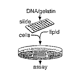
Figure 4A is a schematic for making transfected cell microarrays using a
well-less transfection of plasmid DNAs in defined areas of a lawn of mammalian
cells. Plasmid DNA dissolved in an aqueous gelatin solution is printed on a
glass
slide using a robotic arrayer. The slide is dried and the printed array
covered with a
lipid transfection reagent. After removal of the lipid, the slide is placed in
a culture
dish and covered with cells in media. The transfected cell microarray forms in
1-2
days and is then ready for downstream assays. An alternative method in which
the
lipid is added to the DNA/gelatin solution prior to printing is also
described.
Figure 4B is a GFP-expressing microarray made from a slide printed in a 12
x 8 pattern with a GFP expression construct.
CA 02383423 2002-03-13
WO 01/20015 PCT/iJS00/25457
-10-
Figure 4C is a higher magnification image obtained with fluorescence
microscopy of the cell cluster boxed in Figure 4B. Scale bar equals 100 Vim.
Figure 4D is a graph of GFP cDNA (picograms) versus mean signal intensity
+/- S.D. showing expression levels of clusters in a transfected cell
microarray are
proportional, over a four-fold range, to the amount of plasmid DNA printed on
the
slide. Arrays were printed with elements containing the indicated amounts of
the
GFP construct. Amount of DNA assumes a one nanoliter printing volume. After
transfection, the mean +/- S.D. of the fluorescence intensities of the cell
clusters
were determined. Arrays were prepared as described in Example 3 except that
the
concentration of the GFP expression plasmid was varied from 0.010-0.050 pg/~.1
while the total DNA concentration was kept constant at 0.050 ~g/p,l with empty
vector (prk5). Cell clusters were photographed and the signal intensity
quantitated
with Image Quant (Fuji). The fluorescent image is from a representative
experiment.
Figure 4E is a scan image showing that by printing mixtures of two
plasmids, cotransfection is possible with transfected cell microarrays. Arrays
with
elements containing expression constructs for HA-GST, GFP or both were
transfected and processed for anti-myc immunofluorescence. For
immunofluorescence staining the cells were fixed as described in Example 3,
permeabilized in 0.1% Triton X-100 in PBS for 15 minutes at room temperature
and
probed with primary and secondary antibodies as described. Primary antibodies
were used for 1 hour at room temperature at the following concentrations:
1:500
anti-HA ascites (BaBCo), 2 ~g/ml anti-myc 9E-10 (Calbiochem), 2 ~.g/ml anti-V5
(Invitrogen), or 10 ug/ml 4610 anti-phosphotyrosine (Upstate Biotechnologies).
The secondary antibody used was Cy3 pg/ml labeled anti-mouse antibody (Jackson
Immunoresearch) at 3.1 qg/ml for 40 minutes at room temperature. Panels
labeled
Cy3 and GFP show location of clusters expressing HA-GST and GFP, respectively.
Merged panel shows superimposition of Cy3 and GFP signals and yellow color
indicates co-expression. Scale bar equals 100 Vim.
Figure 4F is an enlarged view of boxed area of scan image from Figure 4E.
Figure 5A is a laser scan showing detection of the receptor for FK506.
Arrays with elements containing expression constructs for GFP, myc-FKBP 12 or
CA 02383423 2002-03-13
WO 01/20015 PCT/US00/25457
-11-
both were printed and transfected with HEK293 cells. SnM dihydro-FK506
[propyyl-3H] (NEN) was added to the culture media 1 hour prior to fixation and
processing for immunofluorescence and autoradiography. Slides were process for
anti-myc immunofluorescence, scanned at 5 ~m resolution and photographed using
a fluorescent microscope, and then exposed to tritium sensitive film
(Hyperfilm,
Amersham) for 4 days. Autoradiographic emulsion was performed as described by
the manufacturer (Amersham). Laser scans show expression pattern of GFP and
FKBP12 and superimposition of both (merged). Film autoradiography detects
binding of tritiated FK506 to the same array (autorad film).
Figure SB is a higher magnification image obtained by fluorescent
microscopy of an FKBP12-expressing cluster (FKBP12). Emulsion
autoradiography detects, with cellular resolution, binding of tritiated FK506
to the
same cluster (autorad emulsion).
Figure SC is a scan showing detected components of tyrosine kinase
signaling cascades. 192 VS-epitope-tagged cDNAs in expression vectors were
printed in two 8 x 12 subgrids named array 1 and 2. For ease of determining
the
coordinates of cell clusters within the arrays a border around each array was
printed
with the GFP expression construct. After transfection, separate slides were
processed for anti-VS or anti-phosphotyrosine immunofluorescence and Cy3 and
GFP fluorescence detected. Merged images of array 1 show location of clusters
expressing VS-tagged proteins (left panel) and having increased levels of
phosphotyrosine (right panel). No DNA was printed in coordinates F10-12.
Figure SD show two examples of the morphological phenotypes detectable in
the transfected cell microarrays described in Figure SC. Clusters shown are E8
and
F7 from array 2.
DETAILED DESCRIPTION OF THE INVENTION
A microarray-based system was developed to analyze the function in
mammalian cells of many genes in parallel. Mammalian cells are cultured on a
glass
slide printed in defined locations with solutions containing different DNAs.
Cells
growing on the printed areas take up the DNA, creating spots of localized
transfection within a lawn of non-transfected cells. By printing sets of
CA 02383423 2002-03-13
WO 01/20015 PCT/US00/25457
-12-
complementary DNAs (cDNAs) cloned in expression vectors, micorarrays which
comprise groups of live cells that express a defined cDNA at each location can
be
made. Transfected cell microarrays can be of broad utility for the high-
throughput
expression cloning of genes, particularly in areas such as signal transduction
and
drug discovery. For example, as shown herein, transfected cell microarrays can
be
used for the unambiguous identification of the receptor for the
immunosuppressant
FK506 and components of tyrosine kinase pathways.
The present invention relates to a method of introducing defined DNAs into
cells at specific discrete, defined locations on a surface by means of a
reverse
transfection method. That is, the present method makes use of DNAs, of known
sequence and/or source, affixed to a surface (DNA spots), such as a slide or
well
bottom, and growing cells that are plated onto the DNA spots and maintained
under
conditions appropriate for entry of the DNAs into the cells. The size of the
DNA
spots and the quantity (density) of the DNA spots affixed to the surface can
be
1 S adjusted depending on the conditions used in the methods. For example, the
DNA
spots can be from about 100 ~.m to about 200 ~,m in diameter and can be
affixed
from about 200 ~,m to about 500 ~,m apart on the surface. The present method
further includes identification or detection of cells into which DNA has been
reverse
transfected. In one embodiment, DNA introduced into cells is expressed in the
cells,
either by an expression vector containing the DNA or as a result of
integration of
reverse transfected DNA into host cell DNA, from which it is expressed. In an
alternative embodiment of the present method, DNA introduced into cells is not
expressed, but affects cell components and/or function itself. For example,
antisense
DNA can be introduced into cells by this method and affect cell function. For
example, a DNA fragment which is anti-sense to an mRNA encoding a receptor for
a drug can be introduced into cells via reverse transfection. The anti-sense
DNA
will decrease the expression of the drug receptor protein, causing a decrease
in drug
binding to cells containing the anti-sense DNA. In the method, a mixture
comprising
DNA of interest (such as cDNA or genomic DNA incorporated in an expression
vector) and a carrier protein is deposited (e.g., spotted or placed in small
defined
areas) onto a surface (e.g., a slide or other flat surface, such as the
bottoms of wells
in a multi-welled plate) in defined, discrete (distinct) locations and allowed
to dry,
CA 02383423 2002-03-13
WO 01/20015 PCT/US00/25457
-13-
with the result that the DNA-containing mixture is affixed to the surface in
defined
discrete locations.
Detection of effects on recipient cells (cells containing DNA introduced by
reverse transfection) can be carned out by a variety of known techniques, such
as
immunofluorescence, in which a fluorescently labeled antibody that binds a
protein
of interest (e.g., a protein thought to be encoded by a reverse transfected
DNA or a
protein whose expression or function is altered through the action of the
reverse
transfected DNA) is used to determine if the protein is present in cells grown
on the
DNA spots.
The nucleic acid used in the methods of the present invention can be
oligonucleotides, DNA and/or RNA. The nucleic acid of interest introduced by
the
present method can be nucleic acid from any source, such as nucleic acid
obtained
from cells in which it occurs in nature, recombinantly produced nucleic acid
or
chemically synthesized nucleic acid. For example, the nucleic acid can be cDNA
or
genomic DNA or DNA synthesized to have the nucleotide sequence corresponding
to that of naturally-occurring DNA. The nucleic acid can also be a mutated or
altered form of nucleic acid (e.g., DNA that differs from a naturally
occurring DNA
by an alteration, deletion, substitution or addition of at least one nucleic
acid
residue) or nucleic acid that does not occur in nature. Nucleic acid
introduced by the
subject method can be present in a vector, such as an expression vector (e.g.,
a
plasmid or viral-based vector), but it need not be. Nucleic acid of interest
can be
introduced into cells in such a manner that it becomes integrated into genomic
DNA
and is expressed or remains extrachromosomal (is expressed episomally). The
nucleic acid for use in the methods of the present invention can be linear or
circular
and can be of any size. For example, the nucleic acid can be from about 3 kb
to
about lOkb, from about 5 kb to about 8 kb and from about 6 kb to 7 kb. Nucleic
acid introduced into cells by the method described herein can further comprise
nucleic acid (e.g., DNA) that facilitates entry of the nucleic acid into cells
or passage
into the cell nucleus (nuclear localization elements).
The carrier for use in the methods of the present invention can be, for
example, gelatin or an equivalent thereof.
CA 02383423 2002-03-13
WO 01/20015 PCT/US00/25457
-14-
Eukaryotic cells, such as mammalian cells (e.g., human, monkey, canine,
feline, bovine, or marine cells), bacterial, insect or plant cells, are plated
(placed)
onto the surface bearing the DNA-containing mixture in sufficient density and
under
appropriate conditions for introduction/entry of the DNA into the eukaryotic
cells
and expression of the DNA or its interaction with cellular components.
Preferably,
the eukaryotic cells (in an appropriate medium) are plated on top of the dried
DNA-
containing spots at high density (e.g., 0.5-1 x 10⁵/cm2), in order to increase
the
likelihood that reverse transfection will occur. For example, the density of
cells can
be from about 0.3 x 10⁵/cm2 to about 3 x 10⁵/cmz, and in specific embodiments,
is
from about 0.5 x 10⁵/cm2 to about 2 x 10⁵/cm2 and from about 0.5 x 10⁵/cm2 to
about
1 x 10⁵/cmz. The appropriate conditions for introduction/entry of DNA into
cells
will vary depending on the quantity of cells used.
Two embodiments of the present method are described in detail herein: a
DNA-gelatin method, in which a mixture comprising DNA (e.g., DNA in an
expression vector, such as, a plasmid-based or viral-based vector) and a
carrier
protein (e.g., gelatin) is used and a lipid vector-DNA method, in which a
mixture
comprising DNA, such as DNA in an expression vector (e.g., a plasmid); a
carrier
protein (e.g., gelatin); a sugar (e.g., sucrose); DNA condensation buffer; and
an
appropriate lipid-containing transfection reagent is used. Any suitable
gelatin which
is non-toxic, hydrated, which can immobilize the nucleic acid mixture onto a
surface
and which also allows the nucleic acid immobilized on the surface to be
introduced
over time into cells plated on the surface can be used. For example, the
gelatin can
be a crude protein gelatin or a more pure protein based gelatin such as
fibronectin.
In addition, a hydrogel, a sugar based gelatin (polyethylene glycol) or a
synthetic or
chemical based gelatin such as acrylamide can be used.
In the first embodiment, a mixture comprising two components (DNA such
as DNA in an expression vector and a Garner protein) is spotted onto a surface
(e.g.,
a slide) in discrete, defined locations or areas and allowed to dry. One
example of
this embodiment is described in Example 1. After the carrier (e.g., gelatin)-
DNA
mixture has dried sufficiently that it is affixed to the surface, transfection
reagents (a
lipofection mixture) and cells to be reverse transfected are added, preferably
sequentially. The transfection mixture can be one made from available
components
CA 02383423 2002-03-13
WO 01/20015 PCT/US00/25457
-15-
or can be a commercially available mixture, such as EffecteneTM (Qiagen),
FugeneTM
6 (Boehringer Mannheim) or LipofectamineTM (Gibco/BRL-Life Technologies). It
is added in an appropriate quantity, which can be determined empirically,
taking into
consideration the amount of DNA in each defined location. A wax barrier can be
drawn around the locations on the surface which contain the vector-DNA
mixture,
prior to addition of the transfection mixture, in order to retain the mixture
or the
solution can be kept in place using a cover well. Generally, in this
embodiment, the
transfection reagent is removed, such as by vacuum suctioning, prior to
addition of
cells into which DNA is to be reverse transfected. Actively growing cells are
plated
on top of the locations, producing a surface that bears the DNA-containing
mixture
in defined locations. The resulting product is maintained under conditions
(e.g.,
temperature and time) which result in entry of DNA in the DNA spots into the
growing cells. These conditions will vary according to the types of cells and
reagents used and can be determined empirically. Temperature can be, for
example,
room temperature or 37°C, 25°C or any temperature determined to
be appropriate
for the cells and reagents.
A variety of methods can be used to detect protein expression in the DNA-
containing spots. For example, immunofluorescence can be used to detect a
protein.
Alternatively, expression of proteins that alter the phosphorylation state or
subcellular localization of another protein, proteins that bind with other
proteins or
with nucleic acids or proteins with enzymatic activity can be detected.
In the second embodiment, one example of which is described in Example 2,
a mixture comprising DNA in an expression vector; a carrier protein (e.g.,
gelatin); a
sugar (e.g., sucrose); DNA condensation buffer; and a lipid-based transfection
reagent is spotted onto a surface, such as a slide, in discrete, defined
locations and
allowed to dry. Actively growing cells are plated on top of the DNA-containing
locations and the resulting surface is maintained under conditions (e.g.,
temperature
and time) which result in entry of DNA in the DNA spots into the growing cells
(reverse transfection). Expression of DNA in cells is detected using known
methods, as described above.
CA 02383423 2002-03-13
WO 01/20015 PCT/US00/25457
-16-
Any suitable surface which can be used to affix the DNA containing mixture
to its surface can be used. For example, the surface can be glass, polystyrene
or
plastic. In addition, the surface can be coated with, for example, polylysine.
The present invention also encompasses methods of making arrays which
comprise DNA affixed to a surface such that when cells are plated onto the
surface
bearing the DNA, the DNA can be introduced (is introducible) into the cells
(i.e., the
DNA can move from the surface into the cells). The present invention also
encompasses a DNA array comprising a surface having affixed thereto, in
discrete,
defined locations, DNA of known sequence or source by a method described
herein.
The methods of this invention are useful to identify DNAs of interest (DNAs
that are expressed in recipient cells or act upon or interact with recipient
cell
constituents or function, such as DNAs that encode a protein whose function is
desired because of characteristics its expression gives cells in which it is
expressed).
They can be used in a variety of formats, including macro-arrays and micro-
arrays.
They permit a DNA array to be converted into a protein or cell array, such as
a
protein or cell microarray.
The present invention is illustrated by the following examples, which are not
intended to be limiting in any way.
Example 1 Reverse Transfection: "Gelatin-DNA" Method
Materials
[DNA]: lpg/~L (eg., HA-GST pRKS, pBABE CMV GFP)
Gelatin (ICN, cat.# 901771): 0.2% stock in ddH20, all dilutions made in PBS-
0.20% gelatin = O.Sg gelatin + 250mL ddH20
Effectene Transfection Kit (Qiagen, cat.# 301425)
Plasmid-DNA: grown in 100mL L-amp overnight from glycerol stock, purified by
standard Qiaprep Miniprep or Qiagen Plasmid Purification Maxi protocols
Cell Type: HEK 293T cultured in DMEM/10%IFS with L-glut and pen/strep
Diluting and Spotting DNA
~ Dilute DNA in 0.2% gelatin* to give final [DNA]=O.OS~g/~L**
~ Spot DNA/gelatin mix on ~ poly-L-lysine slides using arrayer
CA 02383423 2002-03-13
WO 01/20015 PCT/US00/25457
-17-
~ Allow slides to dry in vacuum-dessicator overnight***
* range of gelatin concentration that worked under the conditions used =
0.05% to 0.5%
** range of DNA concentrations that worked under the conditions used = 0.01
S ~g/~l to O.l O~g/~.1
*** range of drying time = 2 hours to 1 week
Adding Tx. Reagents to Gelatin -DNA Spots
~ In eppendorf tube, mix 300~L DNA-condensation buffer (EC Buffer)+ 16~L
Enhancer
~ Mix by vortexing. Incubate for 5 minutes
~ Add SO~L Effectene and mix by pipetting
~ Draw a wax circular barrier on slide around spots to apply the transfection
reagent
~ Add 366~L mix to wax-enclosed region of spots
~ Incubate at room temperature forl0 to 20 minutes
~ Meanwhile, split cells to reverse-transfect
~ Vacuum-suction off reagent in hood
Place slides in dish and add cells for reverse transfection
Splitting Cells
~ Split actively growing cells to [cell] = 10' cells in 25mL
~ Plate cells on top of slides) in square 100x100x15mm petri dish
~ Allow reverse transfection to proceed for 40 hours = approx. 2 cell cycles
~ Process slides for immunofluorescence
Example 2 Reverse Transfection: "Lipid - DNA" Method
Materials
[DNA]:lqg/~L (eg., HA-GST pRKS, pBABE CMV GFP)
Gelatin (ICN, cat.# 901771): 0.2% stock in ddH20, all dilutions made in PBS-
0.05% gelatin = 250~L 0.2% + 750~L PBS-
CA 02383423 2002-03-13
WO 01/20015 PCT/US00/25457
-18-
Effectene Transfection Kit (Qiagen, cat.# 301425):
EC Buffer in 0.4M sucrose = 273.6~L 50% sucrose + 726.4~,L EC Buffer
Plasmid-DNA: grown in 100mL L-amp overnight from glycerol stock, purified by
standard Qiaprep Miniprep or Qiagen Plasmid Purification Maxi protocols
Cell Type: HEK 293T cultured in DMEM/10%IFS with L-glut and pen/strep
Reverse Transfection Protocol with Reduced Volume
~ Aliquot l.6pg DNA in separate eppendorf tubes
~ Add 15 ~.L of pre-made DNA-condensation buffer (EC Buffer) with 0.4M
sucrose * to tubes
~ Add l.6uL of Enhancer solution and mix by pipetting several times. Incubate
at
room temperature for 5 minutes
~ Add 5 ~L of Effectene Transfection Reagent to the DNA-Enhancer mix and mix
by pipetting. Incubate at room temperature for 10 minutes
~ Add 23.2uL of 0.05% gelatin** to total transfection reagent mix (i.e. 1:1
dilution)
~ Spot lipid-DNA on E poly-L-lysine slides mix using arrayer
~ Allow slides to dry in vacuum-dessicator overnight***
EffecteneTM kit (Qiagen) used includes Enhancer solution, which was used
according to Qiagen's instructions.
* range of sucrose that worked under the conditions used = O.1M to 0.4M
** range of gelatin concentration that worked under the conditions used =
0.01% to 0.05%
*** range of drying time = 2 hours to 1 week
Splitting Cells
~ Split actively growing cells to [cell] = 10' cells in 25mL
~ Plate cells on top of slides) in square 100x100x15mm petri dish
~ Allow reverse transfection to proceed for 40 hours = approx. 2 cell cycles
~ Process slides for immunofluorescence
CA 02383423 2002-03-13
WO 01/20015 PCT/US00/25457
-19-
Example 3 Transfected Cells Micorarrays: a genomics approach for the analysis
of gene products in mammalian cells
Lipid-DNA Method
I. Gelatin Preparation and DNA Purification
Materials:
Gamma-Amino Propyl Silane (GAPS) slides (Corning catalog #2550),
Purified cDNA,
Gelatin, Type B: 225 Bloom (Sigma, catalog #G-9391),
Methods:
0.2% Gelatin was made by incubation in a 60°C water bath for 15
minutes. The
gelatin was cooled slowly to 37°C at which point it was filtered
through 0.45~,m
cellular acetate membrane (CA).
Bacterial clones with DNA plasmids were grown in a 96 Deep-Well Dish for 18 to
24 hours in 1.3mL of ternfic broth (TB) shaking at 250rpm at 37°C. The
plasmids
were miniprepped and optical density (OD) was taken. DNA purity, as indicated
by
final 280nm/260nm absorbance ratio, was greater than 1.7.
Storage:
For storage purposes, gelatin was kept at 4°C and miniprepped DNA kept
at -20°C.
II. Sample Preparation and Array Printing
Materials:
Effectene Transfection Reagent (Qiagen catalog #301425),
Sucrose (Life Technologies),
INTEGRID 100mm x l5mm Tissue Culture Square Petri Dishes (Becton Dickinson:
Falcon catalog #35-1012),
Costar 384-well plates (VWR catalog #7402),
Stealth Micro Spotting Pins, (Telechem International, Inc. catalog #SMP4),
PixSys 5500 Robotic Arrayer (Cartesian Technologies, Model AD20A5),
CA 02383423 2002-03-13
WO 01/20015 PCT/US00/25457
-20-
Vacuum Dessicator with Stopcock 250mm, NALGENE (VWR catalog #24987-
004),
DRIERITE Anhydrous Calcium Sulfate (VWR catalog #22890-229)
Forceps to hold slides,
Human Embryonic Kidney (HEK) 293T cells,
Tissue Culture hood,
Cover Slips (SOmm x 25mm),
Methods:
For each DNA-containing spot, 15,1 of pre-made DNA-condensation buffer (Buffer
EC) with 0.2M to 0.4M sucrose was added to 0.80~g to 1.60~.g DNA in a separate
eppendorf tube. Subsequently, 1.5,1 of the Enhancer solution was added to the
tube
and mixed by pipetting. This was let to incubate at room temperature for 5
minutes.
5~,1 Effectene transfection reagent was added, mixed and let to incubate at
room
temperature for 10 minutes with the DNA-Enhancer mixture. 1X volume of 0.05%
gelatin was added, mixed and the appropriate amount was aliquoted into a 384-
well
plate for arraying purposes.
The PixSys 5500 Robotic Arrayer was used with Telechem's ArrayIt Stealth Pins
(SMP4) with each spot spaced 400~,m apart with a SOms to SOOms delay time of
the
pin on the slide for each spot. A 55% relative humidity environment was
maintained during the arraying. A thorough wash step was implemented betv~.-
een
each dip into a DNA sample in the 384-well plate to avoid clogging of the pins
that
would result in missing spots in the array.
In a tissue culture hood, 10x106 Human Embryonic Kidney (HEK) 293T cells were
prepared in 25m1 DME media with 10% IFS, pen/strep and glutamine for every 3
slides that were to be processed. After arraying, the slides were simply
placed array-
side facing up on a sterile 100x100x10mm square dish (3 slides per plate) and
the
cells were poured gently on the slides while avoiding direct pouring on the
arrays
themselves. If the number of slides were not a multiple of 3, dummy slides
were
placed to cover the square dish.
CA 02383423 2002-03-13
WO 01/20015 PCT/US00/25457
-21-
The cells were let to grow on the arrays for approximately 2 cell cycles
(~40hours
for 293T). Subsequently, the slides were very gently rinsed with PBS in a
Coplin
jar, and then fixed in 3.7% paraformaldehyde/4.0% sucrose for 20 minutes in a
Coplin jar, and then transferred back to a jar with PBS-.
Storage:
After arraying, slides were stored at room temperature in a vacuum dessicator
with
anhydrous calcium sulfate pellets. After fixation, slides were kept in PBS- at
4°C
until analyses were completed (maximum of 5 days).
III. Methods of Detection
Immunofluorescence
Fluorescence Microscopy
Laser Scanning
Radiolabelling and detection with sensitive film or emulsion
If the expressed proteins to be visualized are fluorescent proteins, they can
be
viewed and photographed by fluorescent microscopy. For large expression array,
slides may be scanned with a laser scanner for data storage. If a fluorescent
antibody can detect the expressed proteins, the protocol for
immunofluorescence can
be followed. If the detection is based on radioactivity, the slides can be
fixed as
indicated above and radioactivity detected by autoradiography with film or
emulsion.
Immunofluorescence:
After fixation, the array area was permeabilized in 0.1% Triton X-100 in PBS
for 15
minutes. After two rinses in PBS-, the slides were blocked for 60 minutes,
probed
with a primary antibody at 1:200 to 1:500 dilution for 60 minutes, blocked for
20
minutes, probed with a fluorescent secondary antibody at 1:200 dilution for 40
minutes. The slides can be transferred to a Coplin jar in PBS- and visualized
under
an upright fluorescent microscope. After analyses, the slides can be mounted
and
stored in the dark at 4°C.
CA 02383423 2002-03-13
WO 01/20015 PCT/LJS00/25457
-22-
To create these microarrays, distinct and defined areas of a lawn of cells
were simultaneously transfected with different plasmid DNAs (Figure 4A). This
is
accomplished without the use of individual wells to sequester the DNAs.
Nanoliter
volumes of plasmid DNA in an aqueous gelatin solution are printed on a glass
slide.
A robotic arrayer (PixSys 5500, Cartesian Technologies) equipped with stealth
pins
(SMP4, Telechem) was used to print a plasmid DNA/gelatin solution contained in
a
384-well plate onto CMT GAPS glass slides (Corning). The pins deposited ~ 1 n1
volumes 400 ~m apart using a 25 ms pin down slide time in a 55% relative
humidity
environment. Printed slides were stored at room temperature in a vacuum
desiccator
until use. Preparation of aqueous gelatin solution is important and is as
follows.
0.02% gelatin (w/v) (Sigma G-9391) was dissolved in MilliQ water by heating
and
gentle swirling in a 60°C water bath for 15 minutes. The solution was
cooled slowly
to room temperature and filtered through a 0.45~um cellular acetate membrane
and
stored at 4°C. Plasmid DNA was purified with the Plasmid Maxi or
QIAprep 96
Turbo Miniprep kits (Qiagen), and always had an A260/A280>1.7. Concentrated
solutions of DNA were diluted in the gelatin solution so to keep the gelatin
concentration >0.017% and, unless otherwise specified, final plasmid DNA
concentrations were 0.033 ~,g/~,1. To express GFP the EGFP construct in
pBABEpuro was used.
After drying, the DNA spots are briefly exposed to a lipid transfection
reagent, the slide is placed in a culture dish and covered with adherent
mammalian
cells in media. The Effectene transfection kit (301425, Qiagen) was used as
follows.
In a 1.5 ml microcentrifuge tube, 16 ~1 enhancer was added to 150 ~.1 EC
buffer,
mixed, and incubated for S minutes at room temperature. 25 ~l effectene lipid
was
added, mixed and the entire volume pipetted onto a 40 x 20 mm cover well
(PC200,
Grace Bio-Labs). A slide with the printed side down was placed on the cover
well
such that the solution covers the entire arrayed area while also creating an
airtight
seal. After a 10 minute incubation, the cover well was pried off the slide
with a
forceps and the transfection reagent removed carefully by vacuum aspiration.
The
slide was placed printed side up in a 100 x 100 x 10 mm square tissue culture
dish
and a 1 x 10' actively growing HEK293T cells in 25 ml media (DMEM with 10%
FBS, 50 units/ml penicillin and 50 ~g/ml streptomycin) were poured into the
dish.
CA 02383423 2002-03-13
WO 01/20015 PCT/US00/25457
-23-
Three slides can be transfected side-by-side in this fashion. The cells grew
on the
slide for 40 hours prior to fixing for 20 minutes at room temperature in 3.7%
paraformaldehyde/4.0% sucrose in PBS. Other commonly used mammalian cells
lines, such as HeLa and A549 cells, were also tested and similar results were
obtained but with transfection efficiencies of 30-50% of those obtained with
HEK293 cells. The DNA in the gelatin gel is insoluble in cell culture media
but
readily enters cells growing on it to create the transfected cell microarray.
To illustrate the method, an array with elements containing an expression
construct for the green fluorescent protein (GFP) was printed. HEK293 cells
were
plated on the slide for transfection and the fluorescence of the cells
detected with a
laser fluorescence scanner. Microarrays were imaged at a resolution of S~.m
with a
laser fluorescence scanner (ScanArray 5000, GSI Lumonics). GFP and cy3
emission was measured separately after sequential excitation of the two
fluorophores. To obtain images at cellular resolution, cells were photographed
with
a conventional fluorescent microscope. All images were pseudocolored and
superimposed using Photoshop 5.5 (Adobe Systems).
A low magnification scan showed a regular pattern of fluorescent spots that
matches the pattern in which the GFP expression construct was printed (Figure
4B).
A higher magnification image obtained via fluorescence microscopy showed that
each spot is about 150 ~.m in diameter and consists of a cluster of 30-80
fluorescent
cells (Figure 4C). As in a conventional transfection, the total expression
level in the
clusters is proportional over a defined range to the amount of plasmid DNA
used
(Figure 4D). Since it may be useful to express two different plasmids in the
same
cells, whether the technique is compatible with cotransfection was examined.
Arrays with elements containing expression constructs for GFP, an epitope-
tagged
protein or both were prepared and transfected. The cells growing on elements
printed with both cDNAs express both encoded proteins, indicating that
cotransfection had occurred (Figure 4E).
Whether transfected cell microarrays could be used to clone gene products
based on their intrinsic properties was also determined. As a test case, an
array to
identify the receptor for FK506, a clinically important immunosuppressant
whose
pharmacologically relevant target, FKBP 12, is an intra-cellular protein, was
used
CA 02383423 2002-03-13
WO 01/20015 PCT/US00/25457
-24-
(Kino, T., et al., J. Antiobiot., 40:1256 (1987); Handing, M.W., et al.,
Nature,
26:755 (1989)). Elements containing expression constructs for FKBP12, GFP, or
both were printed on a slide, in an easily recognizable pattern. After the
transfected
cell microarray formed, radiolabeled FK506 was added to the tissue culture
media
for one hour prior to processing the slide for autoradiography and
immunofluorescence. The radiolabeled FK506 bound to the array in a pattern of
spots that exactly matches the pattern of cell clusters expressing FKBP12
(Figure
SA). Detection of the bound FK506 with autoradiographic emulsion confirmed, at
the cellular level, colocalization between FKBP12 expression and FK506 binding
(Figure SB). The binding is specific because the GFP-expressing clusters and
the
non-transfected cells surrounding the clusters showed only background levels
of
signal (Figure SA). Furthermore, the prior addition of excess rapamycin, a
competitive antagonist of FK506, completely eliminated the signal. 1 ~M
rapamycin was added to the cell culture media 30 minutes before the addition
of
radiolabeled FK506.
The utility of transfected cell microarrays for identifying gene products that
induce phenotypes of interest in mammalian cells or have a distinct sub-
cellular
localization was also explored. Arrays with a collection, enriched for
signaling
molecules, of 192 distinct epitope-tagged cDNAs in expression vectors were
printed.
192 Genestrom expression constructs (Invitrogen) in bacteria were cultured in
two
96-well plates and plasmid DNA was purified using the Turbo Miniprep Kit
(Qiagen). Plasmid DNA was diluted with 0.02% gelatin to a final concentration
of
0.040 p,g/~1 and printed. Cellular phosphotyrosine levels were determined by
immunofluorescence staining and scanning. Cell morphology and subcellular
localization of expressed proteins was assessed by visual inspection via
fluorescence
microscopy of the cells in the clusters after their detection with anti-VS
immunofluorescence.
After transfection, their effects on cellular phosphotyrosine levels and
morphology as well as their subcellular localization were determined. Five
cell
clusters on grid 1 (A2, C7, C9, Cl l, and F6) had phosphotyrosine levels above
background (Figure SC). The coordinates of the clusters match those of the
wells of
a microtiter plate containing the source cDNAs and were used to look up the
identity
CA 02383423 2002-03-13
WO 01/20015 PCTNS00/25457
-25-
of the transfected cDNAs. This revealed that four of these clusters were
transfected
with known tyrosine kinases (trkC, syk, syn, and blk) while the fifth (C11)
encodes
a protein of unknown function. Simple visual examination of the morphology of
the
cells in the transfected clusters revealed a diversity of cellular phenotypes
even in
this small set of clones. In array 2, cluster E8 had fragmented cells
characteristic of
apoptosis while in two clusters (D10 and F7) the cells were closely attached
to each
other (Figure SD). The presence of apoptotic cells was confirmed by TLTNEL
(Terminal deoxynucleotidyl transferase mediated dUTP-biotin nick end labeling
method) staining. TIJNEL staining was performed as described (Y. Gavrieli, Y.
Sherman, S.A. Ben-Sasson. J. Cell Biol. 119, 493 (1992)).
The observed phenotypes are consistent with the presumed functions of the
cDNAs expressed in these clusters (the Table). Subcellular localization of the
expressed proteins were examined through visual inspection the and those with
distinct patterns were noted (the Table). This revealed that several proteins
that are
known transcription factors were mainly located in the cell nucleus. This was
also
true for other proteins, such as phosphatase 1-beta, whose subcellular
distribution
has not been previously ascertained.
CA 02383423 2002-03-13
WO 01/20015 PCT/US00/25457
-26-
TABLE
Description of selected cDNAs expressed in the transfected cell microarray.
Shown are the
coordinates, the phenotype or property detected, the Genbank accession number
and the name of the
cDNA. nuc/cyto means nuclear and cytoplasmic staining was visible.
Grid: Phenotype/propertAccession Function
Coordinatey number
2:E8 apoptosis AF016266 TRAIL receptor 2
2:D10 cell adhesionX97229 NK receptor
2:F7 cell adhesionM98399 CD36
1:A9 nuclear U11791 Cyclin H
1:BS nuclear M60527 deoxycytidine kinase
1:B 12 nuclear M60724 p70 S6 kinase kinase
cx 1
1:C12 nuclear M90813 D-type cyclin
1:E4 mitochondria)U54645 methylmalonyl-coA mutase
1:E10 mitochondria)J05401 creatine kinase
1:G9 nuc/cyto U40989 tat interactive protein
1:610 nuc/cyto U09578 MAPKAP (3pk) kinase
2:A9 nuclear X83928 TFIID subunit TAFII28
2:A12 nuc/cyto M62831 ETR101
2:B6 nuc/cyto X06948 IgE receptor a-subunit
2:B 12 nuclear X63469 TFIIE (3 subunit
2:C5 nuclear M76766 General transcription
factor IIB
2:C7 nuc/cyto M15059 CD23A
2:C12 nuclear X80910 PP1, ~3 catalytic subunit
2:D4 nuclear AF017307 Ets-related transcription
factor
2:E7 nuclear X63468 TFIIE a
2:E12 nuclear U22662 Orphan receptor LXR-a
2:F8 nuclear L08895 MEF2C
2:F12 nuclear AF028008 SP1-like transcription
factor
2:G2 nuc/cyto U37352 PP2A, regulatory B'
a 1 subunit
2:G3 nuc/cyto L14778 PP2B, catalytic (x
subunit
The microarrays can be printed with the same robotic arrayers as traditional
DNA arrays, so it is feasible to achieve densities of up 10,000-15,000 cell
clusters
CA 02383423 2002-03-13
WO 01/20015 PCT/US00/25457
-27-
per standard slide. At these densities the entire set of human genes can be
expressed
on a small number of slides, allowing rapid pan-genomic screens. Thus,
comprehensive collections of full-length cDNAs for all mammalian genes can be
generated (Strausberg, R.L., et al., Science, 15:455 (1999);
www.hip.harvard.edu/research.html. www.guthrie.or~/cDNA.) and will be valuable
tools for making such arrays.
Transfected cell microarrays have distinct advantages over conventional
expression cloning strategies using FACs or sib selection (Simonsen, H., et
al.,
Ttrends Pharmacol. Sci., 15:437 (1994)). First, cDNAs do not need to be
isolated
from the cells exhibiting the phenotype of interest. This allows for screens
using a
variety of detection methods, such as autoradiography or irz situ
hybridization, and
significantly accelerates the pace of expression cloning. The experiments
described
herein took days to perform instead of the weeks to months necessary with
other
expression cloning strategies. Second, transfected cell microarrays can also
be used
to screen living cells, allowing the detection of transient phenotypes, such
as
changes in intracellular calcium concentrations. Third, being compact and easy
to
handle, transfected cell microarrays have economies of scale. The arrays are
stable
for months and can be printed in large numbers, allowing many phenotypes to be
screened in parallel, with a variety of methods, in a small number of tissue
culture
plates.
Described herein are arrays in which the transfected plasmids direct gene
overexpression. However, as antisense technology improves or other methods
emerge for decreasing gene function in mammalian cells, it is likely that
transfected
cell microarrays can be used to screen for phenotypes caused by loss of gene
function. Lastly, the immobilization of the plasmid DNA in a degradable gel is
the
key to spatially restricting transfection without wells.
While this invention has been particularly shown and described with
references to preferred embodiments thereof, it will be understood by those
skilled
in the art that various changes in form and details may be made therein
without
departing from the scope of the invention encompassed by the appended claims.