Note: Descriptions are shown in the official language in which they were submitted.
CA 02383499 2005-09-12
E
DRUG DELIVERY DEVICE
Field of the Invention
The present invention generally pertains to biocompatible implants for
localized
delivery of pharmaceutically active agents to body tissue. More particularly,
but not by
way of limitation, the present invention pertains to biocompatible implants
for localized
delivery of pharmaceutically active agents to the posterior segment of the
eye.
Description of the Related Art
Several diseases and conditions of the posterior segment of the eye threaten
vision.
Age related macular degeneration (ARMD), choroidal neovascularization (CNV),
retinopathies (i.e. diabetic retinopathy, vitreoretinopathy), retinitis (i.e.
cytomegalovirus
(CMV) retinitis), uveitis, macular edema, and glaucoma are several examples.
Age related macular degeneration (ARMD) is the leading cause of blindness in
the
elderly. ARMD attacks the center of vision and blurs it, making reading,
driving, and
other detailed tasks difficult or impossible. About 200,000 new cases of ARMD
occur
each year in the United States alone. Current estimates reveal that
approximately forty
percent of the population over age 75, and approximately twenty percent of the
population
over age 60, suffer from some degree of macular degeneration. "Wet" ARMD is
the type
of ARMD that most often causes blindness. In wet ARMD, newly formed choroidal
blood vessels (choroidal neovascularization (CNV)) leak fluid and cause
progressive
damage to the retina.
In the particular case of CNV in ARMD, two main methods of treatment are
currently being developed, (a) photocoagulation and (b) the use of
angiogenesis inhibitors.
I
CA 02383499 2002-02-22
WO 01/28472 PCT/US00/24983
However, photocoagulation can be harmful to the retina and is impractical when
the CNV
is near the fovea. Furthermore, photocoagulation often results in recurrent
CNV over
time. Oral or parenteral (non-ocular) administration of anti-angiogenic
compounds is also
being tested as a systemic treatment for ARMD. However, due to drug-specific
metabolic
restrictions, systemic administration usually provides sub-therapeutic drug
levels to the
eye. Therefore, to achieve effective intraocular drug concentrations, either
an
unacceptably high dose or repetitive conventional doses are required.
Periocular
injections of these compounds often result in the drug being quickly washed
out and
depleted from the eye, via periocular vasculature and soft tissue, into the
general
circulation. Repetitive intraocular injections may result in severe, often
blinding,
complications such as retinal detachment and endophthalmitis.
In order to prevent complications related to the above-described treatments
and to
provide better ocular treatment, researchers have suggested various implants
aimed at
localized delivery of anti-angiogenic compounds to the eye. U.S. Patent No.
5,824,072 to
Wong discloses a non-biodegradable polymeric implant with a pharmaceutically
active
agent disposed therein. The pharmaceutically active agent diffuses through the
polymer
body of the implant into the target tissue. The pharmaceutically active agent
may include
drugs for the treatment of macular degeneration and diabetic retinopathy. The
implant is
placed substantially within the tear fluid upon the outer surface of the eye
over an
avascular region, and may be anchored in the conjunctiva or sclera;
episclerally or
intrasclerally over an avascular region; substantially within the
suprachoroidial space over
an avascular region such as the pars plana or a surgically induced avascular
region; or in
direct communication with the vitreous.
U.S. Patent No. 5,476,511 to Gwon et al. discloses a polymer implant for
placement under the conjunctiva of the eye. The implant may be used to deliver
2
WO 01/28472 CA 02383499 2002-02-22 PCT/USOO/24983
neovascular inhibitors for the treatment of ARMD and drugs for the treatment
of
retinopathies, retinitis, and CMV retinitis. The pharmaceutically active agent
diffuses
through the polymer body of the implant.
U.S. Patent No. 5,773,019 to Ashton et al. discloses a non-bioerodable polymer
implant for delivery of certain drugs including angiostatic steroids and drugs
such as
cyclosporine for the treatment of uveitis. Once again, the pharmaceutically
active agent
diffuses through the polymer body of the implant.
All of the above-described implants require careful design and manufacture to
permit controlled diffusion of the pharmaceutically active agent through a
polymer body
(matrix devices) or polymer membrane (reservoir devices) to the desired site
of therapy.
Drug release from these devices depends on the porosity and diffusion
characteristics of
the matrix or membrane, respectively. These parameters must be tailored for
each drug
moiety to be used with these devices. Consequently, these requirements
generally increase
the complexity and cost of such implants.
U.S. Patent No. 5,824,073 to Peyman discloses an indentor for positioning in
the
eye. The indentor has a raised portion that is used to indent or apply
pressure to the sclera
over the macular area of the eye. This patent discloses that such pressure
decreases
choroidal congestion and blood flow through the subretinal neovascular
membrane,
which, in turn, decreases bleeding and subretinal fluid accumulation.
Therefore, a need exists in the biocompatible implant field for a surgically
implantable drug delivery device capable of safe, effective, rate-controlled,
localized
delivery of a wide variety of pharmaceutically active agents to any body
tissue. The
surgical procedure for implanting such a device should be safe, simple, quick,
and capable
of being performed in an outpatient setting. Ideally, such a device should be
easy and
economical to manufacture. Furthermore, because of its versatility and
capability to
3
CA 02383499 2005-09-12
deliver a wide variety of pharmaceutically active agents, such an implant
should be
capable of use in clinical studies to deliver various agents that create a
specific physical
condition in a patient or animal subject. In the particular field of
ophthalmic drug
delivery, such an implantable drug delivery device is especially needed for
localized
delivery of pharmaceutically active agents to the posterior segment of the eye
to combat
ARMD, CNV, retinopathies, retinitis, uveitis, macular edema, and glaucoma.
Summaa of the Invention
Certain exemplary embodiments can provide an ophthalmic drug delivery device,
comprising: a body having: a scleral surface having a radius of curvature that
facilitates
contact with a sclera of a human eye; a well having an opening to said scleral
surface;
and a geometry that facilitates disposing said device on an outer surface of
said sclera,
below a Tenon's capsule of said eye, and in a posterior segment of said eye;
and an inner
core disposed in said well and comprising a pharmaceutically active agent.
Certain exemplary embodiments can provide a method of delivering a
pharmaceutically active agent to an eye, said eye having a sclera, comprising
the steps
of: providing a drug delivery device comprising: a body having a scleral
surface and a
well having an opening to said scleral surface; and an inner core disposed in
said well
comprising a pharmaceutically active agent; and disposing said device within
said eye so
that said pharrnaceutically active agent is in communication with said sclera
through said
opening.
Certain exemplary embodiments can provide a method of delivering a
pharmaceutically active agent to the eye, said eye having a sclera, a Tenon's
capsule, and
a macula, comprising the steps of: providing a drug delivery device comprising
a body
having a pharmaceutically active agent disposed therein; and disposing said
device on an
outer surface of said sclera and below said Tenon's capsule.
Certain exemplary embodiments can provide an ophthalmic drug delivery device,
comprising: a body having a scleral surface for placement proximate a sclera
in a
posterior segment of an eye and a well having an opening to said scleral
surface; and an
inner core disposed in said well comprising a pharmaceutically active agent,
wherein
4
CA 02383499 2005-09-12
said pharmaceutically active agent is selected from the group consisting of
anti-
infectives, steroidal anti-inflammatory agents, non-steroidal anti-
inflammatory agents,
combinations of anti-infectives and anti-inflammatory agents, agents for the
treatment of
macular degeneration, agents for the treatment of choroidial
neovascularization, agents
for the treatment of retinopathies, agents for the treatment of retinitis,
agents for the
treatment of uveitis, agents for the treatment of macular edema, agents for
the treatment
of glaucoma, angiogenesis inhibitors, antimetabolites, and neuroprotective
drugs.
In various embodiments, the present invention includes a method of delivering
a
pharmaceutically active agent to an eye having a sclera, a Tenon's capsule,
and a macula.
A drug delivery device comprising a body having a pharmaceutically active
agent
disposed therein is provided. The device is disposed on an outer surface of
the sclera,
below the Tenon's capsule, and proximate the macula.
Brief Description of the Drawin .gs
For a more complete understanding of the present invention, and for further
objects and advantages thereof, reference is made to the following description
taken in
conjunction with the accompanying drawings in which:
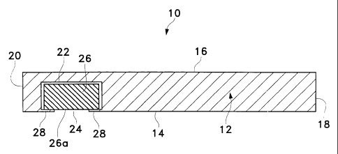
FIG. I is a side sectional view of a drug delivery device according to a
preferred
embodiment of the present invention;
FIG. 2 is a side sectional view of a second drug delivery device according to
a
preferred embodiment of the present invention;
FIG. 3 is a side sectional view schematically illustrating the human eye;
FIG. 4 is detailed cross-sectional view of the eye of FIG. 3 along line 4-4;
FIG. 5 is a perspective view of an ophthalmic drug delivery device according
to a
preferred embodiment of the present invention;
FIG. 6A is a side sectional view of the ophthalmic drug delivery device of
FIG. 5;
FIG. 6B is an enlarged cross-sectional view of the ophthalmic drug delivery
device of FIG. 6A taken along line 6B-6B; and
5
WO 01/28472 CA 02383499 2002-02-22 PCT/USOO/24983
FIG. 7 is a graphical illustration of the results of a pharmacokinetic study
with
New Zealand White rabbits implanted with the ophthalmic drug delivery device
of FIGS.
through 6B showing the mean concentration of a pharmaceutically active agent
at a
target site in the retina and choroid of the rabbits as a function of time.
5 Detailed Description of the Preferred Embodiments
The preferred embodiments of the present invention and their advantages are
best
understood by referring to FIGS. 1 through 7 of the drawings, like numerals
being used
for like and corresponding parts of the various drawings.
FIG. 1 schematically illustrates a drug delivery device 10 according to a
preferred
embodiment of the present invention. Device 10 may be used in any case where
localized
delivery of a pharmaceutically active agent to body tissue is required. By way
of
example, device 10 may be used to treat a medical disorder of the eye, ear,
nose, throat,
skin, subcutaneous tissue, or bone. Device 10 may be used in humans or
animals.
Device 10 generally includes a body 12 having an internal surface 14 and an
external surface 16. As shown in FIG. 1, body 12 preferably has a generally
rectangular
three-dimensional geometry with a proximal end 18 and a distal end 20. Body 12
may
have any other geometry that has an internal surface 14 for placement
proximate a target
tissue in the body of a patient. By way of example, body 12 may have a
cylindrical, an
oval, a square, or other polygonal three-dimensional geometry.
Body 12 includes a well or cavity 22 having an opening 24 to internal surface
14.
An inner core 26 is preferably disposed in well 22. Inner core 26 is
preferably a tablet
comprising one or more pharmaceutically active agents. Alternatively, inner
core 26 may
comprise a conventional hydrogel having one or more pharmaceutically active
agents
disposed therein. A retaining member 28 is preferably disposed proximate
opening 24.
Retaining member 28 prevents inner core 26 from falling out of well 22. When
inner core
6
CA 02383499 2002-02-22
WO 01/28472 PCT/US00/24983
26 is a cylindrical tablet, retaining member 28 is preferably a continuous rim
or lip
disposed circumferentially around opening 24 having a diameter slightly less
than the
diameter of tablet 26. Alternatively, retaining member 26 may comprise one or
more
members that extend from body 12 into opening 24. Although not shown in FIG.
1, inner
core 26 may alternatively comprise a suspension, solution, powder, or
combination
thereof containing one or more pharmaceutically active agents. In this
embodiment,
internal surface 14 is formed without opening 24, and the suspension,
solution, powder, or
combination thereof diffuses through the relatively thin portion of internal
surface 14
below inner core 26. Still further in the alternative, device 10 may be formed
without
wel122 or inner core 26, and the pharmaceutically active agent(s) in the form
of a
suspension, solution, powder, or combination thereof may be dispersed
throughout body
12 of device 10. In this embodiment, the pharmaceutically active agent
diffuses through
body 12 into the target tissue.
The geometry of device 10 maximizes communication between the
pharmaceutically active agent of inner core 26 and the tissue underlying
internal surface
14. Internal surface 14 preferably physically contacts the target tissue. By
way of
example, if the target tissue has a generally flat surface, device 10 would be
appropriate
for the delivery of a pharmaceutically active agent. As another example, if
the target
tissue has a generally convex surface, a device l0a shown in FIG. 2 having a
generally
concave internal surface 14a designed to mate with such a target surface may
be utilized.
Corners 30 of proximal end 18a, and corners 32 of distal end 20a, may be
slanted and/or
rounded off to facilitate surgical placement of device l0a and to maximize
comfort to the
patient. Retaining member 28 is preferably designed with a minimum thickness
necessary
to retain inner core 26 so as to dispose a surface 26a of inner core 26 in
close proximity to
7
CA 02383499 2005-09-12
the target tissue. Although not shown in FIGS. 1 or 2, inner core 26 may be
formed so
that surface 26a physically contacts the target tissue.
Alternatively, device 10 or 10a may be disposed in the body of a patient so
that
internal surface 14 or 14a is disposed proximate the target tissue. In this
case, internal
surface 14 or 14a physically contacts intermediate tissue disposed between it
and the
target tissue. The pharmaceutically active agent of inner core 26 communicates
with the
target tissue through opening 24 and this intermediate tissue.
Referring again to FIG. 1, body 12 preferably comprises a biocompatible, non-
bioerodable material. Body 12 more preferably comprises a biocompatible, non-
bioerodable polymeric composition. Said polymeric composition may be a
homopolymer,
a copolymer, straight, branched, cross-linked, or a blend. Examples of
polymers suitable
for use in said polymeric composition include silicone, polyvinyl alcohol,
ethylene vinyl
acetate, polylactic acid, nylon, polypropylene, polycarbonate, cellulose,
cellulose acetate,
polyglycolic acid, polylactic-glycolic acid, cellulose esters,
polyethersulfone, acrylics,
their derivatives, and combinations thereof. Examples of suitable soft
acrylics are more
fully disclosed in U.S. Patent No. 5,403,901. Said polymeric composition
most preferably comprises silicone. Of course, said polymeric composition
may also comprise other conventional materials that affect its
physical properties, including, but not limited to, porosity, tortuosity,
permeability,
rigidity, hardness, and smoothness. Exemplary materials affecting certain ones
of these
physical properties include conventional plasticizers, fillers, and
lubricants. Said
polymeric composition may comprise other conventional materials that affect
its chemical
properties, including, but not limited to, toxicity, hydrophobicity, and body
12 - inner
core 26 interaction. Body 12 is preferably impenneable to the pharmaceutically
active
agent of inner core 26. When body 12 is made from a generally elastic
polymeric
8
CA 02383499 2005-09-12
composition, the diameter of well 22 may be slightly less than the diameter of
inner core
26. This frictional fit secures inner core 26 within well 22. In this
embodiment, body 12
may be formed without retaining member 28, if desired.
Inner core 26 may comprise any pharmaceutically active agents suitable for
localized delivery to a target tissue. Examples of pharmaceutically active
agents suitable
for inner core 26 are anti-infectives, including, without limitation,
antibiotics, antivirals,
and antifungals; antiallergenic agents and mast cell stabilizers; steroidal
and non-steroidal
anti-inflammatory agents; combinations of anti-infective and anti-inflammatory
agents;
decongestants; anti-glaucoma agents, including, without limitation,
adrenergics, ~-
adrenergic blocking agents, a-adrenergic agonists, parasypathomimetic agents,
cholinesterase inhibitors, carbonic anhydrase inhibitors, and prostaglandins;
combinations
of anti-glaucoma agents; antioxidants; nutritional supplements; drugs for the
treatment of
cystoid macular edema including, without limitation, non-steroidal anti-
inflammatory
agents; drugs for the treatment of ARMD, including, without limitation,
angiogenesis
inhibitors and nutritional supplements; drugs for the treatment of herpetic
infections and
CMV ocular infections; drugs for the treatment of proliferative
vitreoretinopathy
including, without limitation, antimetabolites and fibrinolytics; wound
modulating agents.
including, without limitation, growth factors; antimetabolites;
neuroprotective drugs,
including, without limitation, eliprodil; and angiostatic steroids for the
treatment of
diseases or conditions of the posterior segment of the eye, including, without
limitation,
ARMD, CNV, retinopathies, retinitis, uveitis, macular edema, and
glaucoma. Such angiostatic steroids are more fully disclosed in U.S.
Patent Nos. 5,679,666 and 5,770,592. Preferred ones of such
angiostatic steroids include 4,9(1 I)-Pregnadien-17a,21-diol-3,20-dione and
4,9(11)-
Pregnadien-17a,21-diol-3,20-dione-21-acetate. Inner core 26 may also comprise
9
CA 02383499 2002-02-22
WO 01/28472 PCT/US00/24983
conventional non-active excipients to enhance the stability, solubility,
penetrability, or
other properties of the active agent or the drug core.
If inner core 26 is a tablet, it may further comprise conventional excipients
necessary for tableting, such as fillers and lubricants. Such tablets may be
produced using
conventional tableting methods. The pharmaceutically active agent is
preferably
distributed evenly throughout the tablet. In addition to conventional tablets,
inner core 26
may comprise a special tablet that bioerodes at a controlled rate, releasing
the
pharmaceutically active agent. By way of example, such bioerosion may occur
through
hydrolosis or enzymatic cleavage. If inner core 26 is a hydrogel, the hydrogel
may
bioerode at a controlled rate, releasing the pharmaceutically active agent.
Alternatively,
the hydrogel may be non-bioerodable but allow diffusion of the
pharmaceutically active
agent.
Device 10 may be made by conventional polymer processing methods, including,
but not limited to, injection molding, extrusion molding, transfer molding,
and
compression molding. Preferably, device 10 is formed using conventional
injection
molding techniques. Inner core 26 is preferably disposed in well 22 after the
formation of
body 12 of device 10. Retaining member 28 is preferably resilient enough to
allow inner
core 26 to be inserted through opening 24 and then to return to its position
as shown in
FIG. 1.
Device 10 is preferably surgically placed proximate a target tissue. The
surgeon
first makes an incision proximate the target tissue. Next, the surgeon
performs a blunt
dissection to a level at or near the target tissue. Once the target tissue is
located, the
surgeon uses forceps to hold device 10 with internal surface 14 facing the
target tissue and
distal end 20 away from the surgeon. The surgeon then introduces device 10
into the
dissection tunnel, and positions device 10 with internal surface 14 facing the
target tissue.
CA 02383499 2002-02-22
WO 01/28472 PCT/US00/24983
Once in place, the surgeon may or may not use sutures to fix device 10 to the
underlying
tissue, depending on the specific tissue. After placement, the surgeon sutures
the opening
and places a strip of antibiotic ointment on the surgical wound.
The physical shape of body 12, including the geometry of internal surface 14,
well
22, opening 24, and retaining member 28, facilitate the unidirectional
delivery of a
pharmaceutically effective amount of the pharmaceutically active agent from
inner core
26 to the target tissue. In particular, the absence of a polymer layer or
membrane between
inner core 26 and the underlying tissue greatly enhances and simplifies the
delivery of an
active agent to the target tissue.
Device 10 can be used to deliver a pharmaceutically effective amount of a
pharmaceutically active agent to target tissue for many years, depending on
the particular
physicochemical properties of the pharmaceutically active agent employed.
Important
physicochemical properties include hydrophobicity, solubility, dissolution
rate, diffusion
coefficient, and tissue affinity. After inner core 26 no longer contains
active agent, a
surgeon may easily remove device 10. In addition, the "pre-formed" tunnel
facilitates the
replacement of an old device 10 with a new device 10.
FIGS. 3 through 6B schematically illustrate an ophthalmic drug delivery device
50
according to a preferred embodiment of the present invention. Device 50 may be
used in
any case where localized delivery of a pharmaceutically active agent to the
eye is
required. Device 50 is particularly useful for localized delivery of active
agents to the
posterior segment of the eye. A preferred use for device 50 is the delivery of
pharmaceutically active agents to the retina proximate the macula for treating
ARMD,
choroidial neovascularization (CNV), retinopathies, retinitis, uveitis,
macular edema, and
glaucoma. Of course, device 50 may also be utilized for localized delivery of
pharmaceutically active agents to body tissue other than the eye, if desired.
11
CA 02383499 2002-02-22
WO 01/28472 PCT/US00/24983
Referring first to FIG. 3, a human eye 52 is schematically illustrated. Eye 52
has a
cornea 54, a lens 56, a sclera 58, a choroid 60, a retina 62, and an optic
nerve 64. An
anterior segment 66 of eye 52 generally includes the portions of eye 52
anterior of a line
67. A posterior segment 68 of eye 52 generally includes the portions of eye 52
posterior
of line 67. Retina 62 is physically attached to choroid 60 in a
circumferential manner
proximate pars plana 70. Retina 62 has a macula 72 located slightly lateral to
its optic
disk. As is well known in the ophthalmic art, macula 72 is comprised primarily
of retinal
cones and is the region of maximum visual acuity in retina 62. A Tenon's
capsule or
Tenon's membrane 74 is disposed on sciera 58. A conjunctiva 76 covers a short
area of
the globe of eye 52 posterior to limbus 77 (the bulbar conjunctiva) and folds
up (the upper
cul-de-sac) or down (the lower cul-de-sac) to cover the inner areas of upper
eyelid 78 and
lower eyelid 79, respectively. Conjunctiva 76 is disposed on top of Tenon's
capsule 74.
As is shown in FIGS. 3 and 4, and as is described in greater detail
hereinbelow, device 50
is preferably disposed directly on the outer surface of sclera 58, below
Tenon's capsule 74
for treatment of most posterior segment diseases or conditions. In addition,
for treatment
of ARMD in humans, device 50 is preferably disposed directly on the outer
surface of
sclera 58, below Tenon's capsule 74, with an inner core of device 50 proximate
macula
72.
FIGS. 5, 6A, and 6B schematically illustrate drug delivery device 50 in
greater
detail. Device 50 generally includes a body 80 having a scleral surface 82 and
an orbital
surface 84. Scleral surface 82 is preferably designed with a radius of
curvature that
facilitates direct contact with sclera 58. Orbital surface 84 is preferably
designed with a
radius of curvature that facilitates implantation under Tenon's capsule 74.
Body 80
preferably has a curved, generally rectangular three-dimensional geometry with
rounded
sides 86 and 88, proximal end 90, and distal end 92. As shown best in the side
sectional
12
WO 01/28472 CA 02383499 2002-02-22 pCT/US00/24983
view of FIG. 6A, orbital surface 84 preferably has tapered surfaces 94 and 96
proximate
proximal end 90 and distal end 92, respectively, that facilitate sub-Tenon
implantation of
device 50 and enhance the comfort of the patient. Body 80 may alternatively
have a
geometry similar to that of device 10a shown in FIG. 2. In addition, body 80
may have
any other geometry that has a curved scleral surface 82 for contact with
sclera 58. By way
of example, body 80 may have a generally cylindrical, oval, square, or other
polygonal
three-dimensional geometry.
Body 80 includes a well or cavity 102 having an opening 104 to scleral surface
82.
An inner core 106 is preferably disposed in well 102. Inner core 106 is
preferably a tablet
comprising one or more pharmaceutically active agents. Alternatively, inner
core 106
may comprise a conventional hydrogel having one or more pharmaceutically
active agents
disposed therein. A retaining member 108 is preferably disposed proximate
opening 104.
Retaining member 108 prevents inner core 106 from falling out of well 102.
When inner
core 106 is a cylindrical tablet, retaining member 108 is preferably a
continuous rim or lip
disposed circumferentially around opening 104 having a diameter slightly less
than the
diameter of tablet 106. Alternatively, retaining member 108 may comprise one
or more
members that extend from body 80 into opening 104. Although not shown in FIG.
6A,
inner core 106 may alternatively comprise a suspension, solution, powder, or
combination
thereof containing one or more pharmaceutically active agents. In this
embodiment,
scleral surface 82 is formed without opening 104, and the suspension,
solution, powder,
or combination thereof diffuses through the relatively thin portion of scleral
surface 82
below inner core 26. Still further in the alternative, device 50 may be formed
without
well 102 or inner core 106, and the pharmaceutically active agent(s) in the
form of a
suspension, solution, powder, or combination thereof may be dispersed
throughout body
13
WO 01/28472 CA 02383499 2002-02-22 PCT/USOO/24983
80 of device 50. In this embodiment, the pharmaceutically active agent
diffuses through
body 80 into the target tissue.
The geometry and dimensions of device 50 maximize communication between the
pharmaceutically active agent of inner core 106 and the tissue underlying
scleral surface
82. Scleral surface 82 preferably physically contacts the outer surface of
sciera 58.
Although not shown in FIGS. 6A or 6B, inner core 106 may be formed so that
surface
106a physically contacts the outer surface of sciera 58. Alternatively,
scleral surface 82
may be disposed proximate the outer surface of sclera 58. By way of example,
device 50
may be disposed in the periocular tissues just above the outer surface of
sclera 58 or intra-
lamellarly within sclera 58.
Body 80 preferably comprises a biocompatible, non-bioerodable material. Body
80 more preferably comprises a biocompatible, non-bioerodable polymeric
composition.
The polymeric composition comprising body 80, and the polymers suitable for
use in the
polymeric compositions of body 80, may be any of the compositions and polymers
described hereinabove for body 12 of device 10. Body 80 most preferably is
made from a
polymeric composition comprising silicone. Body 80 is preferably impermeable
to the
pharmaceutically active agent of inner core 106. When body 80 is made from a
generally
elastic polymeric composition, the diameter of well 102 may be slightly less
than the
diameter of inner core 106. This frictional fit secures inner core 106 within
well 102. In
this embodiment, body 80 may be formed without retaining member 108, if
desired.
Inner core 106 may comprise any ophthalmically acceptable pharmaceutically
active agents suitable for localized delivery. Exemplary pharmaceutically
active agents
include the pharmaceutically active agents listed hereinabove for inner core
26 of device
10. Inner core 106 may also comprise conventional non-active excipients to
enhance the
stability, solubility, penetrability, or other properties of the active agent.
14
CA 02383499 2002-02-22
WO 01/28472 PCTIUSOO/24983
If inner core 106 is a tablet, it may further comprise conventional excipients
necessary for tableting, such as fillers and lubricants. Such tablets may be
produced using
conventional tableting methods. The pharmaceutically active agent is
preferably
distributed evenly throughout the tablet. In addition to conventional tablets,
inner core
106 may comprise a special tablet that bioerodes at a controlled rate,
releasing the
pharmaceutically active agent. By way of example, such bioerosion may occur
through
hydrolosis or enzymatic cleavage. If inner core 106 is a hydrogel, the
hydrogel may
bioerode at a controlled rate, releasing the pharmaceutically active agent.
Alternatively,
the hydrogel may be non-bioerodable but allow diffusion of the
pharmaceutically active
agent.
Device 50 may be made by conventional polymer processing methods, including,
but not limited to, injection molding, extrusion molding, transfer molding,
and
compression molding. Preferably, device 50 is formed using conventional
injection
molding techniques as described hereinabove for device 10.
Device 50 is preferably surgically placed directly on the outer surface of
sclera 58
below Tenon's capsule 74 using a simple surgical technique that is capable of
being
performed in an outpatient setting. The surgeon first performs a peritomy in
one of the
quadrants of eye 52. Preferably, the surgeon performs the peritomy in the
infra-temporal
quadrant, about 3 mm posterior to limbus 77 of eye 52. Once this incision is
made, the
surgeon performs a blunt dissection to separate Tenon's capsule 74 from sclera
58,
forming an antero-posterior tunnel. Once the tunnel is formed, the surgeon
uses forceps
to hold device 50 with scleral surface 82 facing sclera 58 and distal end 92
away from the
surgeon. The surgeon then introduces device 50 into the tunnel in a generally
circular
motion to position inner core 106 of device 50 generally above the desired
portion of
retina 62. The surgeon then closes the peritomy by suturing Tenon's capsule 74
and
CA 02383499 2002-02-22
WO 01/28472 PCT/US00/24983
conjunctiva 76 to sclera 58. After closing, the surgeon places a strip of
antibiotic
ointment on the surgical wound. Alternatively, the surgeon may suture proximal
end 90
of device 50 to sclera 58 to hold device 50 in the desired location before
closure of the
tunnel.
In the case of ARMD in the human eye, the surgeon utilizes the above-described
technique to position inner core 106 of device 50 in one of two preferred
locations in the
infra-temporal quadrant of eye 52. One preferred location is directly on the
outer surface
of sclera 58, below Tenon's capsule 74, with inner core 106 positioned
proximate to, but
not directly above, macula 72. A surgeon may position inner core 106 of device
50 at this
location by moving distal end 92 of device 50 below the inferior oblique
muscle in a
direction generally parallel to the lateral rectus muscle. A second preferred
location is
directly on the outer surface of sclera 58, below Tenon's capsule 74, with
inner core 106
positioned directly above macula 72. A surgeon may position inner core 106 of
device 50
at this location by moving distal end 92 of device 50 toward macula 72 along a
path
generally between the lateral and inferior rectus muscles and below the
inferior oblique
muscle. For ARMD, the pharmaceutically active agent of inner core 106 is
preferably one
of the angiostatic steroids disclosed in U.S. Patent Nos. 5,679,666 and
5,770,592.
The physical shape of body 80 of device 50, including the geometry of scleral
surface 82, well 102, opening 104, and retaining member 108, facilitate the
unidirectional
delivery of a pharmaceutically effective amount of the pharmaceutically active
agent from
inner core 106 through sclera 58, choroid 60, and into retina 62. In
particular, the absence
of a polymer layer or membrane between inner core 106 and sclera 58 greatly
enhances
and simplifies the delivery of an active agent to retina 62.
It is believed that device 50 can be used to deliver a pharmaceutically
effective
amount of a pharmaceutically active agent to retina 62 for many years,
depending on the
16
WO 01/28472 CA 02383499 2002-02-22 PCTIUSOO/24983
particular physicochemical properties of the pharmaceutically active agent
employed.
Important physicochemical properties include hydrophobicity, solubility,
dissolution rate,
diffusion coefficient, and tissue affinity. After inner core 106 no longer
contains active
agent, a surgeon may easily remove device 50. In addition, the "pre-formed"
tunnel
facilitates the replacement of an old device 50 with a new device 50.
The following example illustrates effective drug delivery to a rabbit retina
using a
preferred embodiment and surgical technique of the present invention, but are
in no way
limiting.
EXAMPLE
A device 50 was surgically implanted on the outer surface of the sciera, below
the
Tenon's capsule, generally along the inferior border of the lateral rectus
muscle of the
right eye of twenty (20) New Zealand White rabbits using a procedure similar
to that
described hereinabove for implantation of device 50 on sclera 58 of eye 52.
Device 50
was constructed as shown in FIGS. 5 through 6B, with the following dimensions.
Body
80 had a length 110 of about 15 mm, a width 112 of about 7.0 mm, and a maximum
thickness 114 of about 1.8 mm. Retaining member 108 had a thickness 116 of
about 0.15
mm. Scleral surface 82 had a radius of curvature of about 8.5 mm and an arc
length of
about 18 mm. Inner core 106 was a cylindrical tablet with a diameter of about
5.0 mm
and a thickness of about 1.5 mm. Opening 104 had a diameter of about 3.8 mm.
Well
102 had a diameter of about 4.4 mm. The pharmaceutically active agent used in
tablet
106 was 4,9(I 1)-Pregnadien-17a,21-diol-3,20-dione, an angiostatic steroid
sold by
Steraloids, Inc. of Wilton, New Hampshire, and which is more fully disclosed
in U.S.
Patent Nos. 5,770,592 and 5,679,666. The formulation of tablet 106 consisted
of 99.75
weight percent 4,9(11)-Pregnadien-17a,21-diol-3,20-dione, and 0.25 weight
percent
magnesium stearate.
17
WO 01/28472 CA 02383499 2002-02-22 PCT/USOO/24983
At one week after implantation, 4 rabbits were euthanized and their right eyes
were enucleated. The device 50 was removed from the eyes, and the location of
tablet
106 was marked on their sclerae. Following the removal of the anterior segment
and the
vitreous of each eye and inversion of the thus formed eye-cup, a 10 mm
diameter circular
zone of retinal tissue, concentric with and below the location of tablet 106
on the sclera,
was harvested (the "target site"). A 10 mm diameter circular zone of retinal
tissue was
also harvested from a second site located remote from the target site and on
the other side
of the optic nerve. In addition, a 10 mm diameter circular zone of retinal
tissue was
harvested from a third site located between the second site and the target
site. Similar 10
mm diameter circular zones of choroidal tissue were also harvested at the
target site,
second site, and third site. All these tissues were separately homogenized,
and the
concentration of angiostatic steroid in each of these tissues was determined
via an ocular
pharmacokinetic study using high performance liquid chromatography and mass
spectrometry analysis (LC-MS/MS). This procedure was repeated at 3, 6, 9, and
12
weeks after implantation.
FIG. 7 shows the mean concentration of 4,9(11)-Pregnadien-17a,21-diol-3,20-
dione in the retina and the choroid at the target site as a function of time.
The "error bars"
surrounding each data point represent standard deviation. As shown in FIG. 7,
device 50
delivered a pharmaceutically effective and generally constant amount of
4,9(11)-
Pregnadien-17a,21-diol-3,20-dione to the retina and the choroid at the target
site for a
time period of up to twelve weeks. In contrast, the levels of 4,9(11)-
Pregnadien-17a,21-
diol-3,20-dione in the retina and the choroid at the second and third sites
were at or near
zero. Therefore, device 50 also delivered a localized dose of angiostatic
steroid to the
retina and the choroid at the target site.
18
WO 01/28472 CA 02383499 2002-02-22 PCTIUSOO/24983
From the above, it may be appreciated that the present invention provides
improved devices and methods for safe, effective, rate-controlled, localized
delivery of a
variety of pharmaceutically active agents to any body tissue. The surgical
procedure for
implanting such devices is safe, simple, quick, and capable of being performed
in an
outpatient setting. Such devices are easy and economical to manufacture.
Furthermore,
because of their capability to deliver a wide variety of pharmaceutically
active agents,
such devices are useful in clinical studies to deliver various agents that
create a specific
physical condition in a patient or animal subject. In the particular field of
ophthalmic
drug delivery, such devices are especially useful for localized delivery of
pharmaceutically active agents to the posterior segment of the eye to combat
ARMD,
CNV, retinopathies, retinitis, uveitis, macular edema, and glaucoma.
It is believed that the operation and construction of the present invention
will be
apparent from the foregoing description. While the apparatus and methods shown
or
described above have been characterized as being preferred, various changes
and
modifications may be made therein without departing from the spirit and scope
of the
invention as defined in the following claims.
19