Note: Descriptions are shown in the official language in which they were submitted.
CA 02383939 2002-02-27
1
DESCRIPTION
NOVEL NUCLEIC ACID PROBES, METHOD FOR DETERMINING
NUCLEIC ACIDS BY USING THE PROBES, AND METHOD
FOR ANALYZING DATA OBTAINED BY THE METHOD
Technical Field
This invention relates to novel nucleic acid probes each
of which is labeled with a fluorescent dye and/or a quencher
substance. Specifically, a single-stranded oligonucleotide
is labeled with the fluorescent dye and/or the quencher
substance such that the intensity of fluorescence in a
hybridization reaction system increases or decreases when the
nucleic acid probe is hybridized with a target nucleic acid.
This invention also relates to a method for determining a
nucleic acid by using the nucleic acid probe. The present
invention is also concerned with determination kits,
determination devices and various measurement systems
associated with such kits or devices, all of which are useful
in the method. The present invention also pertains to a method
for analyzing the kinds and amounts of various nucleic acids,
a method for analyzing data obtained by such methods, and
computer-readable recording media with procedures, which are
required to have steps of the analysis method performed by a
computer, recorded as a program.
CA 02383939 2002-02-27
2
Background Art
A variety of methods are conventionally known to
determine a concentration of a nucleic acid by using a nucleic
acid probe labeled with a fluorescent dye. These methods
include:
(1) Dot blotting assay
After a target nucleic acid and a nucleic acid probe
labeled with a fluorescent dye are hybridized on a membrane,
unreacted nucleic probe is washed off. The intensity of
fluorescence only from fluorescent dye molecules, by which the
nucleic acid probe hybridized with the target nucleic acid is
labeled, is measured.
(2) Method making use of an Yntercalator: Glazer et al., Nature,
359, 959, 1992
A certain specific fluorescent dye called "intercalator"
emits strong fluorescence upon its insertion into a double
strand of a nucleic acid. This method measures an increase in
fluorescence from the fluorescent dye. Examples of the
fluorescent dye can include ethidium bromide [Jikken Igaku
(Laboratory Medicine), 15(7), 46-51, Yodosha (1997) ) and SYBR
R Green I(LightCyclerTm System, April 5, 1999; pamphlet
distributed by Roche Diagnostics).
(3) Method making use of FRET (fluorescence energy transfer):
Mergny et al., Nucleic Acid Res., 22, 920-928, 1994
This method comprises hybridizing two nucleic acid probes
CA 02383939 2002-02-27
3
to a target nucleic acid. These two nucleic acid probes are
labeled by different fluorescent dyes, respectively. The
fluorescent dye of one of the two probes can transfer energy
to the fluorescent dye of the other probe such that the latter
fluorescent dye is caused to emit fluorescence. These two
probes are designed such that they hybridize with their
fluorescent dyes being located opposite each other and apart
from each other by 1 to 9 bases. When these two nucleic acid
probes hybridize to the target nucleic acid, emission of
fluorescence from the latter fluorescent dye takes place. The
intensity of this fluorescence emission is proportional to the
number of replications of the target nucleic acid.
(4) Molecular beacon method: Tyagi et al., Nature Biotech., 14,
303-308, 1996
A nucleic acid probe for use in this method is labeled
at an end thereof with a reporter dye and at an opposite end
thereof with a quencher dye. As both end portions of the probe
are complementary with each other in their base sequences, the
overall base sequence of the probe is designed to form a hairpin
stem. Owing to this structure, emission from the reporter dye
is suppressed by the quencher dye under Forster resonant energy
in a state suspended in a liquid. When the probe hybridizes
to a target nucleic acid, the hairpin stem structure is broken.
This leads to an increase in the distance between the reporter
pigment and the quencher pigment, so that the transfer of
CA 02383939 2002-02-27
4
Forster resonant energy no longer takes place. This allows
the reporter dye to make emission.
(5) Davis's method: Davis et al., Nucleic Acids Res., 24,
702-706, 1996
Davis prepared a probe with a fluorescent dye attached
to the 3' end of an oligonucleotide via a spacer having 18 carbon
atoms. The probe was applied to flow cytometry. Its
hybridization resulted in a 10-fold increase in fluorescence
intensity compared to a probe with the fluorescent dye directly
attached to the 3'end.
Applied to various determination methods for nucleic
acids, Fish methods (fluorescent in situ hybridization assays),
PCR methods, LCR methods (ligase chain reactions), SD methods
(strand displacement assays), competitive hybridization and
the like, significant developments have been made on these
methods.
(6) Substantial technical improvements have been made on
methods for amplifying a target gene by PCR [Tanpakushitsu,
Kakusan, Koso (Proteins, Nucleic Acids, Enzymes), 35(17),
KYORITSU SHUPPAN CO., LTD. (1990)] and conducting a
polymorphous analysis on the target gene so amplified, and these
polymorphous analysis methods have now found wide-spread
utility in various fields such as medical field [Jikken Igaku
(Laboratory Medicine), 15(7), Yodosha (1997)]. Various
diseases, especially immune-related diseases have hence been
CA 02383939 2002-02-27
elucidated from genes, thereby obtaining certain successful
outcomes.
Although these methods are now widely used, they include
a disadvantageous step that, subsequent to hybridization
5 reaction between a nucleic acid probe labeled with a fluorescent
dye and a target nucleic acid, an unhybridized portion of the
nucleic acid probe has to be washed out of the reaction system.
Obviation of this step can apparently bring about shorter
determination time, simplified determination, and accurate
determination. There is, accordingly, a long-standing desire
for the development of a nucleic acid determination method which
does not include such a step.
Further, conventional polymorphous analyses are each
conducted using PCR which does not have quantitativeness with
respect to amplification of a gene. Therefore, it has
heretofore been unable to make an analysis to such an extent
as the amount or polymorphous composition of the initial gene
before the amplification of the target gene.
With the foregoing in view, the present invention has as
an object thereof the provision of a method for determining a
concentration of a target nucleic acid by using a nucleic acid
probe labeled with a fluorescent dye, which makes it possible
to determine the concentration of the target nucleic acid in
a shorter time, more easily and more accurately, and also the
provision of nucleic acid probes useful for the practice of the
CA 02383939 2002-02-27
6
method and various devices making use of the probes.
The present invention also has as a second object thereof
the provision of a novel polymorphous analysis method for easily
and quickly performing determination of a polymorphous
composition of a target gene and reagent kits useful in the
method, a computer-readable recording medium with programmed
procedures, which are required to make a computer perform a
method for analyzing data obtained by the quantitative
polymorphous analysis method, and an analysis system for the
quantitative polymorphous analysis.
Disclosure of the Invention
To achieve the above-described objects, the present
inventors have proceeded with a variety of investigations and
have obtained findings as will be described below.
A detailed study was conducted on a variety of nucleic
acid probes, and in a trial and error manner, many probes were
prepared. As a result, it has been found that, even in the case
of a nucleic acid probe composed of an oligonucleotide which
does not form a stem-loop structure between nucleotide chains
at positions where the oligonucleotide is labeled with a
fluorescent dye and a quencher substance, respectively,
labeling by the dye and substance at specific positions may
allow the quencher substance to act on the emission of
fluorescence from the fluorescent dye and may give quenching
CA 02383939 2002-02-27
7
effect on the emission of fluorescence.
The present inventors have proceeded with an
investigation on methods for determining a concentration of a
nucleic acid by using a nucleic acid probe. As a result, it
was found that emission of fluorescence from a fluorescent dye
decreases (quenching phenomenon of fluorescence) when a nucleic
acid probe labeled with the fluorescent dye hybridizes to a
target nucleic acid. It was also found that this decrease is
significant with certain specific dyes. It was also found that
the extent of this decrease varies depending on bases in a probe
portion, to which the fluorescent dye is conjugated, or on the
sequence of the bases.
30 Performance of a polymorphous analysis on a target gene
after amplifying the target gent by a quantitative gene
amplification method makes it possible to easily and quickly
determine the pre-amplification amount and polymorphous
composition of the target gene with good quantitativeness.
The present invention has been completed based on the
above-described findings.
Therefore, the present invention provides the following
(novel) nucleic acid probes, methods, kits and devices:
1) A novel nucleic acid probe for determining a
concentration of a target nucleic acid, comprising: a
single-stranded oligonucleotide capable of hybridizing to the
target nucleic acid, and a fluorescent dye and a quencher
CA 02383939 2002-02-27
8
substance, both of which are labeled on the oligonucleotide,
wherein the oligonucleotide is labeled with the fluorescent dye
and the quencher substance such that an intensity of
fluorescence in a hybridization reaction system increases when
the nucleic acid probe is hybridized with the target nucleic
acid; and the oligonucleotide forms no stem-loop structure
between bases at positions where the oligonucleotide is labeled
with the fluorescent dye and the quencher substance,
respectively.
2) A nucleic acid probe for determining a concentration of
a target nucleic acid, the probe being labeled with a
fluorescent dye, wherein the probe is labeled at an end portion
thereof with the fluorescent dye, and the probe has a base
sequence designed such that, when the probe hybridizes at the
end portion thereof to the target nucleic acid, at least one
G (guanine) base exists in a base sequence of the target nucleic
acid at a position 1 to 3 bases apart from an end base of the
target nucleic acid hybridized with the probe, whereby the
fluorescent dye is reduced in fluorescence emission when the
probe labeled with the fluorescent dye hybridizes to the target
nucleic acid.
3) A nucleic acid probe for determining a concentration of
a target nucleic acid, the probe being labeled with a
fluorescent dye, wherein the probe is labeled at an end portion
thereof with the fluorescent dye, and the probe has a base
CA 02383939 2002-02-27
9
sequence designed such that, when the probe hybridizes to the
target nucleic acid, plural base pairs in a probe-nucleic acid
hybrid complex form at least one G (guanine) and C (cytosine)
pair at the end portion, whereby the fluorescent dye is reduced
in fluorescence emission when the probe labeled with the
fluorescent dye hybridizes to the target nucleic acid.
4) A nucleic acid probe for determining a concentration of
a target nucleic acid, the probe being labeled with a
fluorescent dye, wherein the probe is labeled at a modification
portion other than a 5' end phosphate group or a 3' end OH group
thereof with the fluorescent dye, and the probe has a base
sequence designed such that, when the probe hybridizes to the
target nucleic acid, plural base pairs in a probe-nucleic acid
hybrid complex form at least one G (guanine) and C (cytosine)
pair at the modification portion, whereby the fluorescent dye
is reduced in fluorescence emission when the probe labeled with
the fluorescent dye hybridizes to the target nucleic acid.
5) A nucleic acid probe as described above under any one of
1) to 4) for determining a concentration of a target nucleic
acid, wherein the oligonucleotide of the nucleic acid probe for
the measurement of the nucleic acid is a chemically-modified
nucleic acid.
6) A nucleic acid probe as described above under any one of
1) to 5) for determining a concentration of a target nucleic
acid, said nucleic acid probe being labeled with a fluorescent
CA 02383939 2002-02-27
dye, wherein the oligonucleotide of the nucleic acid probe
for the determination of the nucleic acid is a chimeric
oligonucleotide which comprises a ribonucleotide and a
deoxyribonucleotide.
5 7) A method for determining a concentration of a target
nucleic acid, which comprises hybridizing a nucleic acid probe
as described above under any one of 1) to 6) to the target nucleic
acid, and measuring an intensi.ty of fluorescence in a measuring
system.
10 8) A method for determining a concentration of a target
nucleic acid, which comprises hybridizing a nucleic acid probe
as described above under any one of 1) to 6) to the target nucleic
acid, and measuring a change in fluorescence emission from the
fluorescent dye after the hybridization relative to
fluorescence emission from the fluorescent dye before the
hybridization.
9) A method for determining a concentration of a target
nucleic acid by using a nucleic acid probe as described above
under any one of 1) to 6), wherein the nucleic acid probe and
the target nucleic acid are hybridized to each other after
subjecting the target nucleic acid to heat treatment under
conditions suited for sufficient degradation of a high-order
structure of the target nucleic acid.
10) A method as described above under 9) for measuring a
concentration of a target nucleic acid, wherein a helper probe
CA 02383939 2002-02-27
11
for the practice of a hybridization reaction is added to a
hybridization reaction system before the hybridization
reaction.
11) A methodfor analyzing or determining polymorphism and/or
mutation of a target nucleic acid, which comprises hybridizing
a nucleic acid probe as described above under any one of 1) to
6) to the target nucleic acid, and measuring a change in an
intensity of fluorescence.
12) A novel quantitative, polymorphous analysis method
comprising amplifying a target gene by a quantitative gene
amplification method, and performing a polymorphous analysis
with respect to the target gene to determine an amount of the
target gene and a polymorphous composition or amounts of
individual components of the target gene.
13) A quantitative, polymorphous analysis method as
described above under 12), wherein the polymorphous analysis
is T-RELP (terminal restriction fragment length polymorphism),
RFLP (restriction fragment length polymorphism), SSCP (single
strand conformation) or CFLP (cleavase fragment length
polymorphism).
14) A quantitative, polymorphous analysis method as
described above under 12) or 13), wherein the quantitative gene
amplification method is quantitative PCR or real-time
monitoring quantitative PCR.
15) A kit for determining a concentration of a target nucleic
CA 02383939 2002-02-27
12
acid, wherein the kit includes or is accompanied by a nucleic
acid probe as described above under any one of 1) to 6) or a
nucleic acid probe as described above under any one of 1) to
6) and a helper probe.
16) A kit for analyzing or determining polymorphism and/or
mutation of a target nucleic acid, comprising a nucleic acid
probe as described above under any one of 1) to 6) or a nucleic
acid probe as described above under any one of 1) to 6) and a
helper probe.
17) A reagent kit for use in quantitative PCR, wherein the
kit includes or is accompanied by a nucleic acid probe as
described above under any one of 1) to 6) or a nucleic acid probe
as described above under any one of 1) to 6) and a helper probe.
18) A device for determining a concentration of at least one
target nucleic acid out of plural nucleic acids, comprising:
a solid support, and a like plural number of nucleic acid probes
as described above under any one of 1) to 6) bound on a surface
of the solid support such that the concentration of the target
nucleic acid can be determined by hybridizing the target nucleic
acid to the corresponding one of the probes and determining a
change in an intensity of fluorescence.
19) A method for determining a concentration of a target
nucleic acid, which comprises determining the concentration of
the target nucleic acid or analyzing or determining
polymorphism and/or mutation of the target nucleic acid by using
CA 02383939 2002-02-27
13
a nucleic acid determination device as described above under
18), or a quantitative, polymorphous analysis method of a target
nucleic acid, which comprises performing a quantitative,
polymorphous analysis of the target nucleic acid by using a
nucleic acid determination device as described above under 18).
20) A nucleic acid determination method, a method for
analyzing or determining polymorphism and/or mutation of a
target nucleic acid, or a quantitative, polymorphous analysis
method as described above under any one of 7) to 14), wherein
the target nucleic acid is a nucleic acid contained in cells
derivedfrom a microorganism or animal obtained by single colony
isolation or a nucleic acid contained in a homogenate of the
cells.
21) A method for determining a concentration of a target
nucleic acid by using PCR, which comprises conducting reactions
in PCR by using a nucleic acid probe as described above under
any one of 1) to 6), and determining an initial concentration
of the amplified target nucleic acid from percentage of a change
in an intensity of fluorescence occurred as a result of
hybridization between the probe and the amplified target
nucleic acid.
22) A method for determining a concentration of a target
nucleic acid by using PCR, which comprises conducting reactions
in PCR by using as a primer a nucleic acid probe as described
above under any one of 1) to 6), and determining an initial
CA 02383939 2002-02-27
14
concentration of the amplified target nucleic acid from
percentage of a change in an intensity of fluorescence occurred
as a result of hybridization between the primer or an amplified
nucleic acid amplified from the primer and the amplified target
nucleic acid.
23) A method for determining an initial concentration of a
target nucleic acid amplified in PCR, which comprises
conducting reactions in PCR by using a nucleic acid probe as
described above under any one of 1) to 6) , measuring an intensity
of fluorescence in a reaction system in which in a course of
a nucleic acid extending reaction, the probe has been degraded
out by polymerase or in which a nucleic acid denaturing reaction
is proceeding or has been completed and also an intensity of
fluorescence in the reaction system in which the target nucleic
acid or amplified target nucleic acid is hybridized with the
nucleic acid probe, and then calculating percentage of a change
in the latter intensity of fluorescence from the former
intensity of fluorescence.
24) A method for determining an initial concentration of a
nucleic acid amplified in PCR, which comprises conducting
reactions in PCR by using, as a primer, a nucleic acid probe
as described above under any one of 1) to 6), measuring an
intensity of fluorescence in a reaction system in which t,he
probe and the target nucleic acid or amplified nucleic acid have
not hybridized with each other and also an intensity of
CA 02383939 2002-02-27
fluorescence in the reaction system in which the probe and
the target nucleic acid or amplified nucleic acid are hybridized
with each other, and then calculating percentage of a decrease
of the former intensity of fluorescence from the latter
5 intensity of fluorescence.
25) A method as described above under 23) or 24) for
determining a concentration of a nucleic acid amplified in PCR,
wherein the PCR is real-time quantitative PCR.
26) A method for analyzing data obtained by a nucleic acid
10 determination method as described above under any one of 23)
to 25), further comprising correcting an intensity value of
fluorescence in a reaction system, said intensity value being
available after the target nucleic acid has hybridized to the
nucleic acid probe labeled with the fluorescent dye, in
15 accordance with an intensity value of fluorescence in the
reaction system available after a probe-nucleic acid hybrid
complex so formed has been denatured.
27) A method for analyzing data obtained by a real-time
quantitative PCR method as described above under any one of 23)
to 25), further comprising, as a processing step (hereinafter
called "the correction processing step"), correcting an
intensity value of fluorescence in a reaction system, said
intensity being available in each cycle after the amplified
nucleic acid has conjugated to the fluorescent dye or after the
amplified nucleic acid has hybridized to the nucleic acid probe
CA 02383939 2002-02-27
16
labeled with the fluorescent dye, in accordance with an
intensity value of fluorescence in the reaction system
available after a nucleic acid-fluorescent dye conjugate or
probe-nucleic acid hybrid complex so formed has been denatured
in the cycle.
28) A method for analyzing a melting curve of a target nucleic
acid, which comprises performing PCR on the target nucleic acid
by using a nucleic acid probe as described above under to any
one of 1) to 6), and analyzing the melting curve of the target
nucleic acid to determine a Tm value of each amplified nucleic
acid.
Brief Description of the Drawings
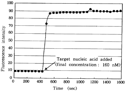
FIG. 1 diagrammatically shows changes in the intensity
of fluorescence in a solution system with a nucleic acid probe
according to the present invention contained therein when a
target nucleic acid was added, in which time (sec) is plotted
along the abscissa and intensities of fluorescence are plotted
along the ordinate;
FIG. 2 shows a working curve for a target nucleic acid
by a nucleic acid probe according to the present invention, in
which concentrations of the target nucleic acid are plotted
along the abscissa and intensities of fluorescence are plotted
along the ordinate;
FIG. 3 illustrates probe designs and target nucleic acid
CA 02383939 2002-02-27
17
designs for studying effects of the distance (the number of
bases) between a fluorescent dye (Texas Red) and a quencher
substance (Dabcyl) on the emission rate of fluorescence from
a fluorescence emitting probe making use of interaction between
the fluorescent dye and the quencher substance;
FIG. 4 is a diagram illustrating effects of the distance
(the number of bases) between the fluorescent dye (Texas Red)
and the quencher substance (Dabcyl) on the emission rate of
fluorescence from the fluorescence emitting probe making use
of interaction between the fluorescent dye and the quencher
substance;
FIG. 5 illustrates probe designs, in each of which bases
in a deoxyribooligonucletide chain were modified with both
fluorescent dye (Texas Red) and quencher substance (Dabcyl),
respectively, and target nucleic acid designs;
FIG. 6 is a diagram illustrating effects of the distance
(the number of bases) between the fluorescent dye (Texas Red)
and the quencher substance (Dabcyl) on the emission rate of
fluorescence as observed using a probe in which bases in a
deoxyribooligonucletide chain were modified with both of the
fluorescent dye and the quencher substance, respectively;
FIG. 7 is a diagram showing measurement data of
fluorescence intensity when the sequence of bases in 16S rRNA
of Escherichia coli, said bases ranging from the 335`" base to
the 35 8`h base as counted from the 5'end, was determined using
CA 02383939 2002-02-27
18
a nucleic acid probe obtained in Example 7;
FIG. 8 diagrammatically illustrates effects of heat
treatment of a target nucleic acid on hybridization of a
35-nucleotides-chained2-0-Me probe to the target nucleic acid;
FIG. 9 diagrammatically shows effects of the number of
bases in a nucleotide chain of a probe, a helper probe and
methylation of an OH group on the 2' carbon of ribose at the 5' end
of the probe on the hybridization between the probe and a target
nucleic acid, 16S rRNA;
FIG. 10 shows a working curve for rRNA assay by an
invention method;
FIG. 11 diagrammatically shows analysis results of the
time-dependent rRNA amount of strains, KYM-7 and KYM-8, in
co-cultivation by a FISH method according to the present
invention;
FIG. 12 is a schematic illustration of a DNA chip
according to the present invention;
FIG. 13 is a schematic illustration of equipment for an
SNAPs detection or determination making use of the DNA chip
according to the present invention, and illustrates an erect
microscope 1, a transparent warming plate 2 for the microscope,
a control unit 3, a cooled CCD camera 4, and an image analyzer
5;
FIG. 14 is a diagram showing experimental results of the
SNAPs detection or determination making use of the DNA chip
CA 02383939 2002-02-27
19
according to the present invention;
FIG. 15 diagrammatically illustrates a relationship
between cycles and a decrease in fluorescence emission from a
fluorescent dye in a quantitative PCR method making use of
primers 1 and 2 labeled with "BODIPY FL/C6";
FIG. 16 diagrammatically shows a relationship between
cycles and the logarithm of a decrease in fluorescence emission
from a fluorescent dye in the quantitative PCR making use of
primers 1 and 2 labeled with "BODIPY FL/C6", in which signs 1~
to have the same meanings as in FIG. 15;
FIG. 17 is a diagram showing a working line for 16S rDNA
of Escherichia coli, which was prepared using the quantitative
PCR according to the present invention;
FIG. 18 (upper diagram) depicts decreases (%) in
fluorescence intensity in real-time quantitative PCR in which
a single probe of the present invention was used as opposed to
two probes labeled with a fluorescent dye and required for a
real-time quantitative PCR method using FRET, and FIG. 18 (lower
diagram) shows a working line prepared by calculating numbers
of cycles (threshold numbers: Ct values) at which decreases in
fluorescence intensity were begun to be significantly observed;
FIG. 19 depicts fluorescence decrease curves obtained by
real-time quantitative PCR, which used an invention primer
labeled with "BODIPY FL/C6", without performing correction
processing according to the present invention;
CA 02383939 2002-02-27
FIG. 20 shows fluorescence decrease curves obtained by
real-time quantitative PCR conducted in a similar manner as the
curves in FIG. 19 except that on each of the curves, each decrease
(%) in fluorescence emission was corrected assuming that the
5 corresponding value in the 10th cycle was 1;
FIG. 21 shows curves obtained by calculating, with
respect to the individual plotted values on the respective
curves in FIG. 20, the rates of decreases (the rates of changes)
in fluorescence intensity in accordance with the formula (9)
10 and then plotting the thus-calculated values;
FIG. 22 shows a working line for human genome DNA as
obtained from the data in FIG. 21;
FIG. 23 depicts curves obtained by subjecting the
measurement values in the individual cycles in FIG. 19 to
15 correction processing in accordance with the formula (1) and
then plotting the corrected values relative to their
corresponding cycles;
FIG. 24 illustrates curves obtained by plotting values,
which had been obtained by processing the processed values of
20 the individual cycles in FIG. 23 in accordance with the formula
(3), against their corresponding cycles;
FIG. 25 shows curves obtained by subjecting the corrected
values in the individual cycles in FIG. 24 to correction
processing in accordance with the formula (6) and then plotting
the corrected values relative to their corresponding cycles;
CA 02383939 2002-02-27
21
FIG. 26 shows working lines drawn corresponding to 0.1,
0.3, 0.5, 0.7, 0.9 and 1.2 chosen at will as candidates for Ct
values from the respective values of log (changes in
fluorescence, %) in FIG. 24, in which the individual working
lines have the following correlation coefficients;
FIG. 27 depicts fluorescence decrease curves when
real-time quantitative PCR was conducted on human genome DNA
of 1 copy and 10 copies by using an invention primer labeled
with"BODIPY FL/C6" and the correction processing of the formula
(1) was applied;
FIG. 28 illustrates melting curves of nucleic acids when
a melting curve analysis was conducted with respect to the PCR
amplification products shown in FIG. 27;
FIG. 29 illustrates curves obtained by differentiating
the curves of FIG. 28 and showing Tm values as valleys;
FIG. 30 shows amplification curves of 16S rRNA genes
(cDNAs) obtained using quantitative PCT according to the
present invention;
FIG. 31 illustrates a working line for cDNA, which was
prepared by a data analysis method according to the present
invention;
FIG. 32 illustrates an analysis pattern by polymorphous
T-RELP according to the present invention;
FIG. 33 diagrammatically illustrates results of
quantitative PCR making use of a fluorescence emitting probe
CA 02383939 2002-02-27
22
as a primer (fluorescence emitting primer) (exponential
graph);
FIG. 34 shows a working line for 16S rRNA gene
(fluorescence emitting primer: 0 pM), in which:
A: Number of copies in an artificial co-cultivation
system of microorganisms (about 296,000 copies);
FIG. 36 illustrates results of real-time monitoring on
PCR amplification products obtained by real-time quantitative
PCR making use of a fluorescence emitting primer, and a working
line obtained by the real-time monitoring;
FIG. 36 diagrammatically shows results of an SNPs
detection by a fluorescence emitting probe;
FIG. 37 diagrammatically illustrates results of an SNPs
detection by a DNA chip with fluorescence emitting probes fixed
thereon;
FIG. 38 diagrammatically shows results of real-time
monitaring of PCR reaction using DNA array havig fixed
fluorescence emitting probes and fluorescence quenching
probes; and
FIG. 39 depicts melting curves of PCR products using DNA
array havig fixed fluorescence emitting probes and fluorescence
quenching probes.
Best Modes for Carrying Out the Invention
The present invention will next be described in further
CA 02383939 2002-02-27
23
detail based on certain preferred embodiments.
The present invention has three aspects.
The present invention, in the first aspect thereof,
relates to a novel nucleic acid probe for determining a target
nucleic acid, comprising: a single-stranded oligonucleotide
capable of hybridizing to the target nucleic acid, and a
fluorescent dye and a quencher substance, both of which are
labeled on the oligonucleotide, wherein the oligonucleotide is
labeled with the fluorescent dye and the quencher substance such
that an intensity of fluorescence in a hybridization reaction
system increases when the nucleic acid probe is hybridized with
the target nucleic acid, and the oligonucleotide forms no
stem-loop structure between bases at positions where the
oligonucleotide is labeled with the fluorescent dye and the
quencher substance, respectively. For thesake of brevity, the
nucleic acid probe according to the present invention may
therefore be called a "fluorescence emitting probe" or a
"nucleic acid probe according to the first aspect of the present
invention" in t.he subsequent description.
The present invention, in the second aspect thereof,
relates to a nucleic acid probe labeled with a fluorescent dye,
which is characterized in that, when the nucleic acid probe
hybridizes to a target nucleic acid, emission of fluorescence
from the fluorescent dye decreases after the hybridization. It
is to be noted that the nucleic acid probe according to the
CA 02383939 2002-02-27
24
present invention may also be called a"fluorescence quenching
probe" or a "nucleic acid probe according to the second aspect
of the present invention" for the sake of brevity.
The present invention, in the third aspect thereof,
relates to a variety of use of the fluorescence emitting probe
and fluorescence quenching probe.
A description will now be made about technical terms
employed in the present invention.
The term "probe-nucleic acid hybrid complex" as used
herein means one (complex) in which a nucleic acid probe
according to the present invention, which is labeled with a
fluorescent dye, and a target nucleic acid are hybridized with
each other. For the same of brevity, it will hereinafter be
called a"nucleic acid hybrid complex" in a shortened form.
Further, the term "fluorescent dye-nucleic acid
conjugate" as used herein means a conjugate in which a
fluorescent dye is bound with a target nucleic acid.
Illustrative is a conjugate in which an intercalator is bound
in a double-stranded nucleic acid.
The terms as used herein - such as nucleic acid probes,
to hybridize, hybridization, stem-loop structures, quenching,
quenching effects, DNAs, RNAs, cDNAs, mRNAs, rRNAs, XTPs, dXTPs,
NTPs, dNTPs, nucleic acid probes, helper nucleic acid probes
(or nucleic acid helper probes, or simply helper probes), to
hybridize, hybridization, intercalators, primers, annealing,
CA 02383939 2002-02-27
extending reactions, thermal denaturing reactions, nucleic
acid melting curves, PCR, RT-PCR, RNA-primed PCR, stretch PCR,
reverse PCR, PCR using Alu sequence(s), multiple PCR, PCR using
mixed primers, PCR using PNA,-hybridization assays, FISH
5 methods (fluorescent in situ hybridization assays), PCR methods
(polymerasechain assays), LCR methods (ligase chain reactions),
SD methods (strand displacement assays), competitive
hybridization, DNA chips, nucleic acid detecting (gene-
detecting) devices, SNP (single nucleotide polymorphism), and
10 co-cultivation systems of plural microorganisms - have the same
meanings as the corresponding terms generally employed these
days in molecular biology, genetic engineering, bioengineering
and the like.
The term "target gene" or "target nucleic acid" as used
15 herein means a nucleic acid or a gene the quantitation or
qualitative detection or mere detection of which is intended,
irrespective whether it is in a purified form or not and further
irrespective of its concentration. Various other nucleic
acids may also exist together with the target nucleic acid. For
20 example, the target nucleic acid may be a specific nucleic acid
in a co-cultivation system microorganisms (a mixed system of
RNAs or gene DNAs of plural microorganisms) or a symbiotic
cultivation system of microorganisms (a mixed system of RNAs
or gene DNAs of plural animals, plants and/or microorganisms),
25 the quantitation or qualitative detection or mere detection of
CA 02383939 2002-02-27
26
which is intended. Purification of the specific nucleic acid,
if needed, can be conducted by a method known per se in the art.
For example, purification can be effected using a purification
kit or the like available on the market. Specific examples of
the above-described nucleic acid can include DNAs, RNAs, PNAs,
oligodeoxyribonucleotides, and oligoriboxynucleotides.
Other examples can include chimera nucleic acids of the
above-exemplified nucleic acids.
The expression "to determine a concentration of a target
nucleic acid" as used herein means to quantitatively determine
concentration(s), to perform qualitative detection, to simply
detect, or to perform an analysis for polymorphism and/or
mutation, all with respect to one or more nucleic acids in a
measurement system. In the case of plural nucleic acids,
quantitative detection of the plural nucleic acids at the same
time, simple detection of the plural nucleic acids at the same
time and an analysis for the polymorphism, mutation and/or the
like of the plural nucleic acids at the same time obviously fall
within the technical scope of the present invention.
The term "device for the measurement of a concentration
of a target nucleic acid" as used herein mean various DNA chips.
Specific examples of the device can obviously include a variety
of DNA chips. The present invention include all DNA chips
irrespective of their types insofar as the nucleic acid probe
according to the present invention can be applied to them.
CA 02383939 2002-02-27
27
The expression "method for determining a concentration
of a target nucleic acid by using a nucleic acid probe labeled
with a fluorescent dye (hereinafter simply called a "nucleic
acid probe according to the present invention" or a "probe
according to the present invention") means to determine the
concentration of the target nucleic acid by a hybridization
assay, FISH method (fluorescent in situ hybridization assay),
PCR method (polymerase chain assay), LCR method (ligase chain
reaction), SD method (strand displacement assay), competitive
hybridization or the like.
A description will first be made of the fluorescence
emitting probes.
This probe is characterized in that, when the probe is
not hybridized with a target nucleic acid, emission of
fluorescence from the fluorescent dye is inhibited by the
quencher dye but, when the probe is hybridized with the target
nucleic acid, the inhibition is rendered ineffective to result
in an increase in the intensity of fluorescence.
The term "fluorescent dye" as used herein means
fluorescent dyes or the like, which are generally used for the
determination or detection of nucleic acids by labeling nucleic
acid probes. Illustrative of such fluorescent dyes are
fluorescein and derivatives thereof [for example, fluorescein
isothiocyanate (FITC) and its derivatives]; Alexa 488, Alexa
532, cy3, cy5, 6-joe, EDANS; rhodamine 6G (R6G) and its
CA 02383939 2002-02-27
28
derivatives [for example, tetramethylrhodamine (TMR),
tetramethylrhodamine isothiocyanate (TMRITC), x-rhodamine,
Texas red, "BODIPY FL" (trade name, product of Molecular Probes,
Inc., U.S.A.), "BODIPY FL/C3" (trade name, product of Molecular
Probes, Inc., U.S.A.), "BODIPY FL/C6" (trade name, product of
Molecular Probes, Inc., U.S.A.), "BODIPY 5-FAM" (trade name,
product of Molecular Probes, Inc., U.S.A.), "BODIPY TMR" (trade
name, product of Molecular Probes, Inc., U.S.A.), and
derivatives thereof (for example, "BODIPY TR" (trade name,
product of Molecular Probes, Inc., U.S.A.), "BODIPY R6G" (trade
name, product of Molecular Probes, Inc., U.S.A.), "BODIPY 564"
(trade name, product of Molecular Probes, Inc.,' U.S.A.), and
"BODIPY 581" (trade name, product of Molecular Probes, Inc.,
U.S.A.) J. Among these, FITC, EDANS, Texas red, 6-joe, TMR,
Alexa 488, Alexa 532, "BODIPY FL/C3" and "BODIPY FL/C6" are
preferred, with EDANS, Texas red, FITC, TMR, 6-joe, "BODIPY
FL/C3" and "BODIPY FL/C6" being more preferred.
The term "quencher substance" means a substance which
acts on the above-described fluorescent dye and inhibits or
quenches emission of fluorescence from the fluorescent dye.
Illustrative are Dabcyl, "QSY7" (Molecular Probes), "QSY33"
(Molecular Probes), Ferrocene and its derivatives, methyl
viologen, and N,N'-dimethyl-2,9-diazopyrenium, with Dabcyl
and the like being preferred.
By labeling an oligonucleotide at specific positions
CA 02383939 2002-02-27
29
thereof with such fluorescent dye and quencher substance as
described above, the emission of fluorescence from the
fluorescent dye is subjected to quenching effect by the quencher
substance.
The expression "single-stranded oligonucleotide, which
forms a nucleic acid probe according to the present invention
and forms no stem-loop structure between bases at positions
where the oligonucleotide is labeled with the fluorescent dye
and the quencher substance, respectively" means an
oligonucleotide which - owing to the complementation of base
sequences at at least two positions between the bases at
positions where the oligonucleotide is labeled with the
fluorescent dye and the quencher substance, respectively -
forms double strands in its own chain and forms no stem-loop
structure.
To label an oligonucleotide useful in the practice of the
present invention with a fluorescent dye and a quencher
substance such that the intensity of fluorescence in a
hybridization reaction system increases when the resulting
nucleic acid probe according to the present invention is
hybridized with a target nucleic acid, the labeling can be
conducted as will be described hereinafter.
The distance between the bases at the positions where the
single-stranded oligonucleotide is labeled with the
fluorescent dye and the quencher substance, respectively, is
CA 02383939 2002-02-27
zero (0) in terms of the number of bases, that is, the
single-stranded oligonucleotide is labeled at the same position
of the same nucleotide thereof with the fluorescent dye and the
quencher substance. As an alternative, the distance is 1 to
5 20 or {(a desired integer of from 3 to 8) + lOn) (n: an integer
> 0) in terms of the number of bases. More preferably, the
single-stranded oligonucleotide can be labeled at the same
position of the same nucleotide thereof or can be labeled with
a distance of from 4 to 8 or a number obtained by adding 10 to
10 a desired number in this range. Preferably, the single-
stranded oligonucleotide can be labeled at the same position
of the same nucleotide thereof or can be labeled with a distance
of from 4 to 8. It is desired to label an oligonucleotide with
a fluorescent dye and a quencher substance, respectively, as
15 described above. However, the distance between the bases
depends strongly upon the base sequence of the probe, the
fluorescent dye and quencher substance to be used for
modification, the lengths of linkers adapted to bind them to
the oligonucleotide, and the like. It is, therefore, difficult
20 to fully specify the base-to-base distance. It is to be noted
that the above-described base-to-base distances are merely
general examples and the distance between the bases includes
many exceptions.
Concerning the labeling positions, it is preferred that,
25 when a single-stranded oligonucleotide is labeled at the
CA 02383939 2002-02-27
31
position of the same nucleotide thereof, one of a fluorescent
dye and a quencher substance is labeled to a base and the other
is labeled to a portion other than bases, specifically to a
phosphate portion or to a ribose portion or deoxyribose portion.
In this case, labeling to the 3'end portion or 5'end portion
is preferred.
When it is desired to set the distance between the bases
labeled with the fluorescent dye and quencher substance,
respectively, at 1 to 20 or {(a desired integer of from 3 to
8) + lOn} (n: an integer > 0), preferably at 4 to 8 or a number
obtained by adding 10 to a desired number in this range, more
preferably at 4 to 8 in terms of the number of bases, the
oligonucleotide may be labeled in its chain with both of the
fluorescent dye and quencher substance or may be labeled at the
5'end or 3'end thereof with one of the fluorescent dye and
quencher substance and in the chain thereof with the other one.
It is preferred to label the oligonucleotide at the 5'end or
3'endthereof with the fluorescent dye or the quencher substance
and at a base, which is apart by the above-described number of
bases from the end, with the quencher substance or the
fluorescent dye. When it is desired to label the 3' e.nd or 5' end
in this case, the labeling can be effected to a base, a phosphate
portion, a ribose portion or a deoxyribose portion, preferably
to the phosphate portion, the ribose portion or the deoxyribose
portion, more preferably to the phosphate portion. When it is
CA 02383939 2002-02-27
32
desired to conduct the labeling into the chain, the labeling
can be effected preferably to bases in the chain.
When the bases are modified in each of the above-described
cases, the modification can be effected to any bases insofar
as the modification is feasible. It is, however, preferred to
effect the modification to the OH group, amino group, 2-N, 7-N
and/or 8-C of a purine base or to the OH group, amino group,
methyl group and/or 2-N of a pyrimidine base [ANALYTICAL
BIOCHEMISTRY, 225, 32-38 (1998)].
The nucleic acid probe according to the present invention,
which is to be hybridized to the target nucleic acid, may be
formed of either an oligodeoxyribonucleotide or an
oligoribonucleotide. The nucleic acid probe may be a chimeric
oligonucleotide which contains both of them. These
oligonucleotides may be in chemically-modified forms. Such
chemically-modified oligonucleotides may be inserted in
chimeric oligonucleotides.
Examples of the modified positions of the chemically-
modified oligonucleotide can include an end hydroxyl group or
end phosphate group of an end portion of an oligonucleotide,
the position of a phosphate portion of an internucleoside, the
5-carbon of a pyrimidine ring, and the position of a saccharide
(ribose or deoxyribose) in a nucleoside. Preferred examples
are the positions of ribose or deoxyribose. Specific examples
can include 2'-O-alkyloligoribonucleotides ("2'-O-" will
CA 02383939 2002-02-27
33
hereinafter be abbreviated as "2-0-"), 2-0-alkyleneoligo-
ribonucleotides, and 2-O-benzyloligoribonucleotides. The
oligonucleotide is modified at the OH group(s) on the
2' carbon (s) of one or more ribose molecules at desired positions
thereof with alkyl group(s), alkylene group(s) or benzyl
group ( s)( via ether bond ( s)). Preferred examples use ful in the
present invention can include, among 2-0-alkyloligo-
ribonucleotides, 2-0-methyloligoribonucleotide, 2-0-ethyl-
oligoribonucleotide and 2-0-butyloligoribonucleotide; among
2-O-alkyleneoligoribonucleotides, 2-0-ethyleneoligo-
ribonucleotide; and 2-O-benzyloligoribonucleotide.
Particularly preferably, 2-0-methyloligoribonucleotide
(hereinafter simply abbreviated as "2-0-Me-oligo-
ribonucleotide") can be used. Application of such chemical
modification to an oligonucleotide enhances its affinity with
a target nucleic acid so that the efficiency of hybridization
with a nucleic acid probe according to the present invention
is improved. The improved efficiency of hybridization leads
to a further improvement in the rate of a decrease in the
intensity of fluorescence from the fluorescent dye of the
nucleic acid probe according to the present invention. As a
consequence, the accuracy of determination of the concentration
of the target nucleic acid is improved further.
Incidentally, it is to be noted that the term
"oligonucleotide" as used herein means an oligodeoxy-
CA 02383939 2002-02-27
34
ribonucleotide or an oligoribonucleotide or both of them and
hence, is a generic term for them.
2-0-alkyloligoribonucleotides, 2-0-alkyleneoligo-
ribonucleotides and 2-0-benzyloligoribonucleotide can be
synthesized by a known process [Nucleic Acids Research, 26,
2224-2229 (1998)]. As custom DNA synthesis services are
available from GENSET SA, France, they can be readily obtained.
The present inventors have completed the present invention by
conducting experiments with the compounds furnished by this
company pursuant to our order.
Incidentally, use of a nucleic acid probe according to
the present invention with modified DNA, such as 2-0-
methyloligoribonucleotide (hereinafter simply called "2-0-
Me-oligoribonucleotide), inserted in an oligodeoxy-
ribonucleotide primarily for the determination of RNA,
especially for the determination of rRNA can provide preferred
results.
Upon determination of RNA by the nucleic acid probe
according to the present invention, it is preferred to subject
an RNA solution as a sample to heat treatment at 80 to 1009C,
preferably 90 to 1001C, most preferably 93 to 97~C for 1 to 15
minutes, preferably 2 to 10 minutes, most preferably 3 to 7
minutes before hybridization with the probe such that the
higher-order structure of RNA can be degraded, as this heat
treatment makes it possible to improve the efficiency of
CA 02383939 2002-02-27
hybridization.
It is also preferred to add a helper probe to a
hybridization reaction mixture for raising the efficiency of
hybridization of the nucleic acid probe of this invention to
5 the hybridization sequence region. In this case, the
oligonucleotide of the helper probe can be an oligodeoxy-
ribonucleotide, an oligoribonucleotide or an oligonucleotide
subjected to similar chemical modification as described above.
Examples of the above-described oligonucleotides can include
10 those having the base sequence of (5')TCCTTTGAGT TCCCGGCCGG
A(3') as the forward type and those having the base sequence
of (5')CCCTGGTCGT AAGGGCCATG ATGACTTGAC GT(3') as the backward
type or the reverse type. Preferred examples of the
chemically-modified oligonucleotide can include 2-0-alkyl-
15 oligoribonucleotides, notably 2-0-Me-oligoribonucleotide.
Where the base strand of the nucleic acid probe according
to the present invention is formed of 35 or fewer bases, use
of a helper probe is particularly preferred. When a nucleic
acid probe according to the present invention longer than a
20 35-base strand is used, however, it may only be necessary to
thermally denature target RNA in some instances.
When the nucleic acid probe according to the present
invention is hybridized to RNA as described above, the
efficiency of hybridization is enhanced. The fluorescence
25 intensity, therefore, decreases corresponding to the
CA 02383939 2002-02-27
36
concentration of RNA in the reaction mixture so that RNA can
be determined up to a final RNA concentration of about 150 pM.
Accordingly, the present invention also relates to a kit
for determining a concentration of a target nucleic acid, which
includes or is accompanied by the above-described helper probe
in addition to a kit which is adapted to determine the
concentration of the target nucleic acid and which includes or
is accompanied by the nucleic acid probe of this invention.
In determination of RNA by a conventional hybridization
assay making use of a nucleic acid probe, an oligodeoxy-
ribonucleotide or oligoribonucleotide has beefi used as the
nucleic acid probe. Because RNA itself has a higher-order solid
structure, the efficiency of hybridization between the probe
and the target RNA was poor, resulting in quantitation of low
accuracy. The conventional methods, therefore, are
accompanied by irksomeness that a hybridization reaction is
conducted after denaturing RNA and immobilizing denatured RNA
on a membrane. The method according to the present invention,
on the other hand, uses a nucleic acid probe a ribose portion
of which has been modified to have high affinity to a particular
structural part of RNA, so that a hybridization reaction can
be conducted at a higher temperature compared with the
conventional methods. The above-mentioned adverse effects of
the high-order structure of RNA can be overcome by simply
conducting thermal denaturation as pretreatment and using a
CA 02383939 2002-02-27
37
helper probe in combination. As a consequence, the efficiency
of hybridization in the method according to the present
invention is practically as high as 100%, leading to
improvements in the accuracy of quantitation. Further, the
method according to the present invention is far simpler and
easier than the conventional methods.
The probe according to the present invention is formed
of 5 to 50 bases, preferably 10 to 25 bases, most preferably
to 20 bases. A base number greater than 50 leads to lower
10 permeability through a cell membrane when employed in the FISH
method, thereby narrowing an applicable range of the present
invention. A base number smaller than 5, on the other hand,
tends to induce non-specific hybridization and, therefore,
results in a large determination error.
15 The oligonucleotide in the nucleic acid probe in the
present invention can be produced by a conventional production
process for general oligonucleotides. It can be produced, for
example, by a chemical synthesis process or by a microbial
process which makes use of a plasmid vector, a phage vector or
the like (Tetrahedron Letters, 22, 1859-1862, 1981; Nucleic
Acids Research, 14, 6227-6245, 1986) Further, it is suitable
to use a nucleic acid synthesizer currently available on the
market (for example, "ABI 394", manufactured by Perkin-Elmer
Corp., U.S.A.). Further, there are some enterprises which
offer custom DNA synthesis services on commercial basis. It
CA 02383939 2002-02-27
38
is, therefore, most convenient to conduct only the designing
of base sequences and to order their synthesis to such
enterprises. Illustrative of such enterprises are Takara
Shuzo Co., Ltd., Japan and Espec Oligo Service Corp..
To label the oligonucleotide with the fluorescent dye and
the quencher substance, desired one of conventionally-known
labeling methods can be used (Nature Biotechnology, 14, 303-308,
1996; Applied and Environmental Microbiology, 63, 1143-1147,
1997; Nucleic Acids Research, 24, 4532-4535, 1996). To
conjugate a fluorescent dye and a quencher substance to the
5' end, a linker or spacer, for example, - (CHz)õ-SH or -(CH2) n-NH2
is first introduced into a phosphate group at the 5'end by a
method known per se in the art. As such a linker- or
spacer-introduced derivative is available on the market, a
commercial product may be purchased (Midland Certified Reagent
Company). In the above-mentioned example, n ranges from 3 to
8 with 6 or 7 being preferred. The oligonucleotide can be
labeled by reacting an SH- or NH2- reactive fluorescent dye or
quencher substance to the linker or spacer. Reversed phase
chromatography or the like to provide a nucleic acid probe for
use in the present invention can purify the thus-synthesized
oligonucleotide, which is labeled with the fluorescent dye.
Further, to conjugate the fluorescent dye or quencher
substance to the 3'end of the oligonucleotide, a linker, for
example, -(CH2) n-NHZ o20r -(CH2) n-SH is introduced onto an OH
CA 02383939 2002-02-27
39
group on the C atom at the 2' -position or 3' -position of ribose
or onto an OH group on the C atom at the 3'-position of
deoxyribose. As such a linker-introduced derivative is also
available on the market like the above-described ones, a
commercial product may be purchased (Midland Certified Reagent
Company) . As an alternative, a phosphate group may be
introduced onto the OH group, followed by the introduction of
a linker, for example, -(CH2) n-SH or -(CH2) n-NHZ onto the OH group
of the phosphate group. In these cases, n ranges from 3 to 9,
with 4 to 8 being preferred. The oligonucleotide can be labeled
by reacting an NH2- or SH-reactive fluorescent dye or quencher
substance to the linker.
For the introduction of the amino group, it is convenient
to use a kit reagent [for example, "Uni-link Aminomodifier"
(product of Clontech Laboratories, Inc., U.S.A.), or
"FluoReporter Kit F-6082, F-6083, F-6084 or F-10220" (product
of Molecular Probes, Inc. , U. S.A. )]. In a manner known per se
in the art, molecules of the fluorescent dye can then be
conjugated to the oligo-ribonucleotide. It is also possible
to introduce molecules of the fluorescent dye into strands of
the probe nucleic acid (ANALYTICAL BIOCHEMISTRY, 225, 32-38,
1998).
When it is desired to introduce an amino group onto a
ribose portion, deoxyribose portion, phosphate portion or base
portion of an oligonucleotide, a linker, a fluorescent dye or
CA 02383939 2002-02-27
a quencher substance to enhance its reactivity, use of a kit
reagent (for example, "Uni-link Aminomodifier" (product of
Clontech Laboratories, Inc., U.S.A.),"FluoReporterKit F-6082,
F-6083, F-6084 or F-10220" (product of Molecular Probes, Inc.,
5 U.S.A.)) is convenient. The fluorescent dye and the quencher
substance can then be bound to the oligoribonucleotide by a
method known per se in the art.
In the above-described synthesis, the introduction of a
protecting group to each function group and the elimination of
10 the protecting group can be conducted by conventional, known
methods.
The oligonucleotide labeled with the fluorescent dye and
the quencher substance can be synthesized as described above.
It is desired to purify intermediate synthesis products and the
15 completed synthesis product by gel filtration, liquid
chromatography such as reversed phase liquid chromatography or
the like. The nucleic acid probe according to the present
invention can be obtained as described above.
As is appreciated from the foregoing, the nucleic acid
20 probe according to the present invention can be designed by
simply labeling an oligonucleotide, which has a base sequence
hybridizable to a target nucleic acid, with a fluorescent dye
and a quencher substance. Its designing is therefore simple.
The nucleic acid probe according to the present invention
25 can also be readily obtained by ordering its synthesis like the
CA 02383939 2002-02-27
41
synthesis of the oligonucleotide, provided that only the
designing of the probe can be completed.
A description will next be made about the fluorescence
quenching probe according to the second aspect of the present
invention.
This probe is characterized in that it is an
oligonucleotide labeled with a single fluorescent dye and, when
hybridized with a target nucleic acid, the intensity of its
fluorescence decreases. It, therefore, has a property
opposite to the fluorescence emitting probe.
The oligonucleotide of the fluorescence quenching probe
of this invention, which is hybridized to a nucleic acid, is
similar to that in the above-described fluorescence emitting
probe. Specifically, it can be a chimeric oligonucleotide or
a chemically-modified oligonucleotide. As a still further
alternative, an oligonucleotide with such a chimeric
oligonucleotide or chemically-modified oligonucleotide
inserted in its chain can also be used.
The position of the oligonucleotide, where the oligo-
nucleotide is modified by a fluorescent dye, is the same as that
of the above-described fluorescence emitting probe.
Similarly to the above-described invention, it is also
possible to add a helper probe to a hybridization reaction
mixture to further improve the efficiency of hybridization of
the nucleic acid probe of this invention to the hybridization
= CA 02383939 2002-02-27
42
sequence region. Further, the base sequence and the number
of base chains of the helper probe, the usability of a
chemically-modified oligonucleotide, and the like are also as
described above in connection with the above-described
invention. When hybridized to RNA, the efficiency of
hybridization is increased. The intensity of fluorescence is
thus decreased corresponding to the amount of RNA in the
reaction mixture, thereby making it possible to determine RNA
up to a final concentration of about 150 pM.
The thermal modification of RNA and the addition of the
helper probe in the determination method of RNA by using the
fluorescence quenching probe of this invention are also similar
to those described above in connection with the fluorescence
emitting probe.
As a consequence, the efficiency of hybridization also
reaches substantially 100% in this invention method, leading
to an improvement in quantitativeness. In addition, the method
is far simpler than the conventional methods.
The number of bases in the probe according to the present
invention is similar to that in the above-described invention.
No particular limitation is imposed on the base sequence of the
probe insofar as it specifically hybridizes to the target
nucleic acid. Preferred examples of the base sequence of the
probe can include:
(1) a base sequence designed such that at least one G
, = CA 02383939 2002-02-27
43
(guanine) base exists in the base sequence of the target
nucleic acid at a position 1 to 3 bases apart from the end base
portion of the target nucleic acid hybridized to the probe,
(2) a base sequence designed such that plural base pairs
of a nucleic acid hybrid complex forms at least one G (guanine)
and C (cytosine) pair at an end portion of the probe, and
(3) a base sequence designed such that in the probe
modified with the fluorescent label at a portion other than the
5'end phosphate group or the 3'end OH group, base pairs in the
fluorescence-labeled portion forms at least one G (guanine) and
C (cytosine) pair,
when the nucleic acid probe labeled with the fluorescent
dye is hybridized with the target nucleic acid.
The preparation process of the oligonucleotide of the
nucleic acid probe according to the present invention and the
labeling method of the oligonucleotide with the fluorescent dye
are similar to those described above in connection with the
above-described invention.
Further, fluorescent dye molecules can also be introduced
into the chain of the nucleic acid probe [ANALYTICAL
BIOCHEMISTRY, 225, 32-38 (1998)].
The nucleic acid probe according to the present invention
can be prepared as described above. A preferred probe form is
one labeled with a fluorescent dye at the 3' or 5'end and
containing G or C as the base at the labeled end. If the 5' end
CA 02383939 2002-02-27
44
is labeled and the 3'end is not labeled, the OH group on the
C atom at the 3'-position of the 3'end ribose or deoxyribose
or the OH group on the C atom at the 2'-position of the 3'end
ribose may be modified with a phosphate group or the like
although no limitation is imposed in this respect.
In addition, a nucleic acid probe according to the present
invention can also be prepared by modifying C or G in the chain
of a probe.
The present invention, in the third aspect thereof, is
an invention making use of such fluorescence emitting probes
and fluorescence quenching probes as described above.
1) Determination kits and determination devices
A target nucleic acid can be easily and accurately
determined in a short time when a fluorescence emitting probe
or a fluorescence quenching probe (hereinafter collectively
called a "nucleic acid probe of the present invention" for the
sake of brevity unless otherwise specifically indicated) is
hybridized with the target nucleic acid and a change in the
intensity of fluorescence after the hybridization (an increase
in the intensity of fluorescence in the case of the fluorescence
emitting probes; a decrease in the intensity of fluorescence
in the case of the fluorescence quenching probes) is measured.
Use of the nucleic acid probe of the present invention also makes
it possible to determine RNA although its determination has
heretofore been difficult.
CA 02383939 2002-02-27
Accordingly, the present invention also relates to a
kit for measuring a concentration of a target nucleic acid,
which includes or is accompanied by the nucleic acid probe
according to the present invention.
5 Use of the nucleic probe according to the present
invention is not limited to the determination of a nucleic acid,
but it can also be suitably applied to methods for analyzing
or determining polymorphism or mutation of a target nucleic acid.
In particular, its application to a device for the determination
10 of a concentration of a target nucleic acid (a DNA chip
[Tanpakushitsu, Kakusan, Koso (Proteins, Nucleic Acids,
Enzymes), 43, 2004-2011 (1998)]} provides a more convenient
device for the determination of the concentration of the target
nucleic acid. The method for analyzing or determining
15 polymorphism and/or mutation of the target nucleic acid by using
the device is an extremely convenient method. Described
specifically, when the nucleic acid probe of this invention is
a fluorescence quenching probe, the intensity of fluorescence
upon its hybridization with the target nucleic acid varies
20 depending on whether or not a GC pair is formed. It is,
therefore, possible to analyze or determine polymorphism and/or
mutation of a target nucleic acid by hybridizing the nucleic
acid probe according to the present invention to the target
nucleic acid and then measuring the intensity of fluorescence.
25 Specific methods will be described in Examples. In this case,
CA 02383939 2002-02-27
46
the target nucleic acid can be an amplified or extracted
product obtained by desired one of nucleic acid amplification
methods. Further, no particular limitation is imposed on the
kind of the target nucleic acid. They are however required to
contain a guanine base or cytosine base in strands thereof or
at ends thereof, because the intensity of fluorescence would
otherwise not decrease. The method of the present invention
can, therefore, analyze or determine a mutation or substitution
such as G->A, G-A, C-T, C-T, G-C or G-C, specifically,
polymorphism such as single nucleotide polymorphism (SNP).
Incidentally, it is the current practice to perform an analysis
of polymorphism by determining the base sequence of a target
nucleic acid in accordance with the Maxam-Gilbert method or the
dideoxy method.
Inclusion of the nucleic acid probe according to the
present invention in a kit for analyzing or determining
polymorphism and mutation of a target nucleic acid, therefore,
makes it possible to suitably use the kit as a kit for the
analysis or determination of the polymorphism and/or mutation
of the target nucleic acid.
When analyzing data obtained by the method of the present
invention for the analysis or determination of polymorphism
and/or mutation of a target nucleic acid, a processing step may
be added to correct the intensity of fluorescence, which is
emitted from the reaction system when the target nucleic acid
CA 02383939 2002-02-27
47
is hybridized with the nucleic acid probe of the present
invention by the intensity of fluorescence emitted from the
reaction system when the target nucleic acid and the nucleic
acid probe are not hybridized with each other. The data so
processed are provided with high reliability.
Accordingly, the present invention also provides a data
analysis method for the method which analyzes or measures
polymorphism and/or mutation of a target nucleic acid.
The present invention also features a system for
analyzing or determining polymorphism and/or mutation of a
target nucleic acid, which has processing means for correcting
a fluorescence intensity of a reaction system, in which the
target nucleic acid is hybridized with the nucleic acid probe
according to the present invention, in accordance with a
fluorescence intensity of the reaction system in which the
target nucleic acid is not hybridized with the nucleic acid
probe according to the present invention.
The present invention further features a computer-
readable recording medium with procedures recorded as a program
therein for making a computer perform a processing step in which,
when analyzing data obtained by the method for analyzing or
determining polymorphism and/or mutation of a target nucleic
acid, a fluorescence intensity of a reaction system, in which
the target nucleic acid is hybridized with the nucleic acid
probe according to the present invention, is corrected in
CA 02383939 2002-02-27
48
accordance with a fluorescence intensity of the reaction
system in which the target nucleic acid or gene is not hybridized
with the nucleic acid probe according to the present invention.
The nucleic acid probe according to the present invention
may be immobilized on a surface of a solid (support layer) , for
example, on a surface of a slide glass. In this case, the probe
may preferably be immobilized on the end not labeled with the
fluorescent dye. The probe of this form is now called a "DNA
chip". These DNA chips can be used for monitoring gene
expressions, determining base sequences, analyzing mutations
or analyzing polymorphisms such as single nucleotide
polymorphism (SNP) . Needless to say, they can also be used as
devices (chips) for determining nucleic acids.
To bind the nucleic acid probe of the present invention,
for example, to a surface of a slide glass, a slide glass coated
with polycations such as polylysine, polyethyleneimine or
polyalkylamine, a slide glass with aldehyde groups introduced
thereon, or a slide glass with amino groups introduced thereon
is first provided. Binding can then be achieved, for example,
by i ) reacting phosphate groups of the probe to the slide glass
coated with the polycations, ii ) reacting a probe, in which amino
groups have been introduced, to the slide glass on which
aldehyde groups have been introduced or iii) reacting a probe,
in which PDC (pyridinium dichromate) residual groups, amino
groups or aldehyde groups have been introduced, to the slide
CA 02383939 2002-02-27
49
glass on which amino groups have been introduced (Fodor, P.A.,
et al., Science, 251, 767-773, 1991; Schena, W., et al., Proc.
Natl. Acad. Sci., U. S.A. , 93, 10614-10619, 1996; McGal, G., et
al., Proc. Natl. Acad. Sci., U.S.A., 93, 13555-13560, 1996;
Blanchad, A.P., et al., Biosens. Bioelectron., 11, 687-690,
1996).
A device having nucleic acid probes of the invention
arranged and bound in an arrayed form on a surface of a solid
support permits more convenient determination of a nucleic
acid.
In this case, the formation of a device by individually
binding many probes of this invention, the base sequences of
which are different from each other, on a surface of the same
solid support makes it possible to simultaneously detect and
quantitate a variety of target nucleic acids.
Preferably, this device may be designed such that each
probe is provided on a side of the solid support, said side being
opposite to the side to which the nucleic acid probe according
to the present invention is bound, with at least one temperature
sensor and at least one heater at an area of the solid support,
where the probe is bound, can be controlled to meet optimal
temperature conditions.
For this device, probes other than nucleic acid probes
of the present invention, for example, nucleic acid probes of
a construction designed such that two different fluorescent
= CA 02383939 2002-02-27
dyes are contained per molecule and each of the probes either
quenches or emits fluorescence owing to interaction between the
two fluorescent dyes when the probe is not hybridized with its
corresponding target nucleic acid but either emits fluorescence
5 or quenches when the probe hybridizes to the target nucleic acid,
specifically, a device with molecular beacons described above
(Tyagi et al., Nature Biotech., 14, 303-308, 1996) or the like
bound thereon can also be used suitably. These devices,
therefore, are embraced within the technical scope of the
10 present invention.
Fundamental operations in the determination method
making use of the device according to the present invention are
simply to place a solution, which contains a target nucleic acid
such as mRNA, cDNA or rRNA, on the solid support on which the
15 nucleic probes are bound and then to induce hybridization. As
a result, a change in the intensity of fluorescence takes place
corresponding to the concentration of the target nucleic acid,
and the target nucleic acid can then be detected and quantitated
from the change in the intensity of fluorescence. Further,
20 binding of many nucleic acid probes according to the present
invention of different base sequences on a surface of a single
solid support makes it possible to determine concentrations of
many target nucleic acids at the same time. As this device can
be used for exactly the same application as a DNA chip, that
25 is, for the determination of the concentrations of the target
= CA 02383939 2002-02-27
51
nucleic acids, it is a novel DNA chip. Under reaction
conditions optimal for the target nucleic acid, the intensities
of fluorescence emitted from the nucleic acids other than the
target nucleic acid remain unchanged. No operation is,
therefore, needed for washing off the unreacted nucleic acids.
Further, independent temperature control of the individual
nucleic acid probes according to the present invention by their
corresponding microheaters makes it possible to control the
probes under their optimal reaction conditions, respectively.
Accurate determination of concentrations is therefore feasible.
In addition, a denaturation curve between each nucleic acid
probe of this invention and its corresponding target nucleic
acid can be analyzed by continuously changing the temperature
with the microheater and measuring the intensity of
fluorescence during the changing of the temperature. From
differences in such denaturation curves, it is possible to
determine properties of the hybridized nucleic acid and also
to detect SNP.
Further, the device also makes it possible to conduct
amplification of a gene by PCR or the like and detection of the
gene at the same time.
According to each conventional device for determining a
concentration of a target nucleic acid, a nucleic acid probe
not modified with a fluorescent dye is bound or fixed on a surface
of a solid support and, subsequent to hybridization with the
+ CA 02383939 2002-02-27
52
target nucleic acid labeled with the fluorescent dye, an
unhybridized portion of the target nucleic acid is washed off,
followed by the measurement of the intensity of fluorescence
from the remaining fluorescent dye.
To label the target nucleic acid with the fluorescent dye,
the following steps can be followed, for example, when specific
mRNA is chosen as a target: (1) mRNA extracted from cells is
extracted in its entirety, and (2) using a reverse transcriptase,
cDNA is synthesized while inserting a nucleoside modified by
the fluorescent dye. These operations are not needed in the.
present invention.
A number of various probes are applied in spots on the
device. Optimal hybridization conditions, for example,
temperatures or the like for nucleic acids to be hybridized to
the individual probes are different from each other.
Theoretically speaking, it is therefore necessary to conduct
a hybridization reaction and a washing operation under optimal
conditions for each probe (at each spot). This is however
physically impossible. For all the probes, hybridization is
conducted at the same temperature and further, washing is also
carried out at the same temperature with the same washing
solution. The device is, therefore, accompanied by a drawback
that a nucleic acid does not hybridize although its
hybridization is desired or that, even if its hybridization
takes place, the nucleic acid is readily washed off as the
CA 02383939 2002-02-27
53
hybridization is not strong. For these reasons, the accuracy
of quantitation of the nucleic acid is low. The present
invention does not have such a drawback because the above-
mentioned washing operation is not needed. Further, a
hybridization reaction can be conducted at an optimal
temperature for each probe of the present invention by
independently arranging a microheater at the bottom of each spot
and controlling the hybridization temperature. Accordingly,
the accuracy of quantitation has been significantly improved
in the present invention.
2) Determination method of a target nucleic acid
In the present invention, use of the above-described
nucleic acid probe, determination kit or device according to
the present invention makes it possible to specifically
determine a concentration of a target nucleic acid with ease
in a short time. A description will hereinafter be made of the
determination method.
In the determination method according to the present
invention, the nucleic acid probe of the present invention is
firstly added to a measurement system and is caused to hybridize
to a target nucleic acid. This hybridization can be effected
by a conventionally-known method (Analytical Biochemistry, 183,
231-244, 1989; Nature Biotechnology, 14, 303-308, 1996; Applied
and Environmental Microbiology, 63, 1143-1147, 1997). As
conditions for hybridization, the salt concentration may range
= CA 02383939 2002-02-27
54
from 0 to 2 molar concentration, preferably from 0.1 to 1.0
molar concentration, and the pH may range from 6 to 8, preferably
from 6.5 to 7.5.
The reaction temperature may preferably be in a range of
the Tm value of the nucleic acid hybrid complex, which is to
be formed by hybridization of the nucleic acid probe of the
present invention to the specific site of the target nucleic
acid, 109C. This temperature range can prevent non-specific
hybridization. A reaction temperature lower than Tm-109C
allows non-specific hybridization, while a reaction
temperature higher than Tm+10 +C allows no hybridization.
Incidentally, a Tm value can be determined in a similar manner
as in an experiment which is needed to design the nucleic acid
probe for use in the present invention. Described specifically,
an oligonucleotide which is to be hybridized with the nucleic
acid probe of this invention (and has a complementary base
sequence to the nucleic acid probe) is chemically synthesized
by the above-described nucleic acid synthesizer or the like,
and the Tm value of a nucleic acid hybrid complex between the
oligonucleotide and the nucleic acid probe is then measured by
a conventional method.
The reaction time may range from 1 second to 180 minutes,
preferably from 5 seconds to 90 minutes. If the reaction time
is shorter than 1 second, a substantial portion of the nucleic
acid probe according to the present invention remains unreacted
CA 02383939 2002-02-27
in the hybridization. On the other hand, no particular
advantage can be brought about even if the reaction time is set
excessively long. The reaction time varies considerably
depending on the kind of the nucleic acid, namely, the length
5 or base sequence of the nucleic acid.
In the present invention, the nucleic acid probe is
hybridized to the target nucleic acid as described above. The
intensity of fluorescence emitted from the fluorescent dye is
measured by a fluorimeter both before and after the
10 hybridization, and a change in fluorescence intensity is then
calculated. As the decrease is proportional to the
concentration of the target nucleic acid, the concentration of
the target nucleic acid can be determined.
The concentration of the target nucleic acid in the
15 reaction mixture may preferably range from 0. 1 to 10. 0 nM, while
the concentration of the nucleic acid probe in the reaction
mixture may preferably range from 1.0 to 25.0 nM. Upon
preparation of a working curve, the nucleic acid probe of the
present invention may desirably be used at a ratio of from 1.0
20 to 2.5 relative to the target nucleic acid.
Upon actually determining the concentration of a target
nucleic acid, the concentration of which is unknown, in a sample,
a working curve is first prepared under the below-described
conditions. A corresponding probe according to the present
25 invention is added at plural concentrations to aliquots of the
= CA 02383939 2002-02-27
56
sample, respectively, followed by the measurement of changes
in the intensity of fluorescence from the respective aliquots.
The probe concentration, which corresponds to the greatest one
of the changes in fluorescence intensity so measured, is chosen
as a preferred probe concentration. Based on the change in
fluorescence intensity measured at the preferred probe
concentration, a quantitated value of the target nucleic acid
can be determined from the working curve.
A description has been made about the principle of the
method of the present invention for the determination of a
concentration of a nucleic acid. The present invention can be
applied to various nucleic acid determination methods, for
example, FISH methods, PCR methods, LCR methods, SD methods,
competitive hybridizations, and TAS methods.
Examples of these applications will hereinafter be
described.
(D Application to FISH methods
The method of the present invention can be applied to
nucleic acids contained in cells of microorganisms, plants or
animals or those contained in homogenates of the respective
cells. The method of the present invention can also be suitably
applied to nucleic acids in cells of a cultivation system of
microorganisms (e.g., a co-cultivation system of
microorganisms or a symbiotic cultivation system of
microorganisms) , in which various kinds of microorganisms are
= CA 02383939 2002-02-27
57
contained together or a microorganism and other animal- or
plant-derived cells are contained together and cannot be
isolated from each other, or in a homogenate or the like of the
cells of the cultivation system. The term "microorganisms" as
used herein means microorganisms in general sense, and no
particular limitation is imposed thereon. Examples of such
microorganisms can include eukaryotic microorganisms and
prokaryotic microorganisms, and also mycoplasmas, virus and
rickettsias. The term "a target nucleic acid" as used in
connection with such a microorganism system means a nucleic acid
with a base sequence specific to cells of a cell strain which
is desired to be investigated, for example, as to how it is acting
in the microorganism strain. Illustrative examples can
include 5S rRNAs, 16S rRNAs and 23S rRNAs of certain specific
cell strains and particular sequences of their gene DNAs.
According to the present invention, a nucleic acid probe
is added to a co-cultivation system of microorganisms or a
symbiotic cultivation system of microorganisms and the amount
of 5S rRNA, 16S rRNA or 23S rRNA of a particular cell strain
or its gene DNA, thereby making it possible to determine the
viable count of the particular strain in the system.
Incidentally, a viable count of a particular cell strain in a
co-cultivation system of microorganisms or a symbiotic
cultivation system of microorganisms can be determined by
adding the nucleic acid probe to a homogenate of the system and
= CA 02383939 2002-02-27
58
then measuring the intensity of fluorescence emission from
the fluorescent dye before and after hybridization. It is to
be noted that this method also falls within the technical scope
of the present invention.
The above-described determination method can be carried
out as will be described hereinafter. Before the addition of
the nucleic acid probe of the present invention, the temperature,
salt concentration and pH of the co-cultivation system of
microorganisms or the symbiotic cultivation system of
microorganisms are adjusted to meet the conditions described
above. It is also preferable to adjust the concentration of
the specific cell strain, which is contained in the co-
cultivation system of microorganisms or the symbiotic
cultivation system of microorganisms, to 10' to 1012 cells/mL,
preferably 109 to 1010 cells/mL in terms of viable count. These
adjustments can be achieved by dilution, centrifugal or like
concentration, or the like. A viable count smaller than 10'
cells/mL results in low fluorescence intensity and greater
determination error. A viable count greater than 1012 cells/mL,
on the other hand, leads to excessively high fluorescence
intensity from the co-cultivation system of a microorganism or
the symbiotic cultivation system of microorganisms, so that the
viable count of the particular microorganism cannot be
determined quantitatively. However, this range depends upon
the performance of a fluorimeter to be used.
CA 02383939 2002-02-27
59
The concentration of the nucleic acid probe of the
present invention to be added depends upon the viable count of
the particular cell strain in the co-cultivation system of
microorganisms or the symbiotic cultivation system of
microorganisms and, at a viable count of 108 cells/mL, may be
in a range of from 0.1 to 10.0 nM, preferably in a range of from
0. 5 to 5 nM, more preferably 1. 0 nM. A probe concentration lower
than 0.1 nM cannot provide any data which accurately reflects
the viable count of the particular microorganism. The optimal
concentration of the nucleic acid probe according to the present
invention, however, cannot be specified in any wholesale manner
because it depends upon the concentration of a target nucleic
acid in cells.
Upon hybridizing the nucleic acid probe to the 5S rRNA,
16S rRNA or 23S rRNA of the particular cell strain or its gene
DNA in the present invention, the reaction temperature may be
set as described above. Further, the hybridization time may
also be set as described above.
The nucleic acid probe according to the present invention
is hybridized to the 5S rRNA, 16S rRNA or 23S rRNA of the
particular cell strain or its gene DNA under such conditions
as described above. Intensities of fluorescence from the
fluorescent dye in the co-cultivation system of microorganisms
or the symbiotic cultivation system of microorganisms before
and after the hybridization are then measured.
= CA 02383939 2002-02-27
In the present invention, no particular limitation is
imposed on components other than the microorganisms in the
co-cultivation system of microorganisms or the symbiotic
cultivation system of microorganisms, insofar as the components
5 do not interfere with the hybridization between the nucleic acid
probe according to the present invention and the 5S rRNA, 16S
rRNA or 23S rRNA of the particular cell strain or its gene DNA
and further, do not inhibit the emission of fluorescence from
the fluorescent dye or the action of the quencher substance
10 labeled on the oligonucleotide. For example, phosphates such
as KH2PO4, K2HPO4, NaH2PO9 and Na2HPO9, inorganic nitrogens such
as ammonium sulfate, ammonium nitrate and urea, various salts
of ions such as magnesium, sodium, potassium and calcium,
various salts such as the sulfates, hydrochlorides, carbonates
15 and the like of trace metal ions such as manganese, zinc, iron
and cobalt, and vitamins may be contained to adequate extent.
If the above-described interference or inhibition is observed,
it may be necessary to separate cells of the plural
microorganisms from the cultivation system by an operation such
20 as centrifugal separation and then to resuspend them in a buffer
or the like.
Usable examples of the buffer can include various buffers
such as phosphate buffer, carbonate buffer, Tris-HC1 buffer,
Tris-glycine buffer, citrate buffer, and Good's buffer. The
25 buffer should be adjusted to a concentration not inhibiting the
= CA 02383939 2002-02-27
61
hybridization or the emission of fluorescence from the
fluorescent dye. This concentration depends upon the kind of
the buffer. The pH of the buffer may range from 4 to 12, with
to 9 being preferred.
5 (2)Application to PCR methods
The present invention can be applied to any method insofar
as it is a PCR method. A description will hereinafter be made
of an application of the present invention to a real-time
quantitative PCR method.
In the real-time quantitative PCR method, PCR is
conducted using a specific nucleic acid probe according to the
present invention, and a change in fluorescence emission from
the florescent dye after a reaction relative to fluorescence
emission from the fluorescent dye before the reaction is
determined in real time.
The term "PCR" as used herein means a variety of PCR
methods. Examples can include RT-PCR, RNA-primed PCR, stretch
PCR, reverse PCR, PCR making use of an Alu sequence, multiple
PCR, PCR making use of a mixed primer, and PCR making use of
PNA. Further, the term "quantitative" means, in addition to
quantitation in general sense, quantitation of such an extent
as detection as described above.
As described above, the term "target nucleic acid" as used
herein means a nucleic acid the existing amount of which is
intended to be determined, irrespective whether it is in a
= CA 02383939 2002-02-27
62
purified form or not and further irrespective of its
concentration. Various other nucleic acids may also exist.
For example, the target nucleic acid may be a specific nucleic
acid in a co-cultivation system microorganisms (a mixed system
of RNAs or gene DNAs of plural microorganisms) or a symbiotic
cultivation system of microorganisms (a mixed system of RNAs
or gene DNAs of plural animals, plants and/or microorganisms),
the amplification of which is intended. Purification of the
target nucleic acid, if needed, can be conducted by a method
known per se in the art. For example, purification can be
effected using a purification kit or the like available on the
market.
The conventionally-known quantitative PCR methods
individually amplify, in the presence of Mg ions, a target
nucleic acid (DNA or RNA) by using dATP, dGTP, dCTP, dTTP or
dUTP, the target nucleic acid, Taq polymerase, a primer, and
a nucleic acid labeled with a fluorescent dye or an intercalator
while repeatedly changing the temperature between low and high
levels, and monitor increases in fluorescence emission from the
fluorescent dye in real time in the course of the amplification
[Jikken Igaku (Laboratory Medicine), 15(7), 46-51, Yodosha
(1997)).
On the other hand, the quantitative PCR method according
to the present invention is characterized in that the target
nucleic acid is amplified by using the nucleic probe of the
= CA 02383939 2002-02-27
63
present invention and a change in fluorescence emission from
the fluorescent dye, specifically, an increase in the intensity
of fluorescence emission in the case of the fluorescence
emission or a change in the intensity of fluorescence in the
case of the fluorescence quenching probe is determined. The
number of bases in a preferred probe of the present invention
for use in the quantitative PCR according to the present
invention may be from 5 to 50, preferably from 10 to 25, notably
from 15 to 20. No particular limitation is imposed on the probe
insofar as it hybridizes to amplification products of the target
nucleic acid in PCR cycles. The probe may be designed in either
a forward type or a reverse type.
For example, the following designs can be mentioned when
the nucleic acid probe is a fluorescence emitting probe. The
above-described fluorescence emitting probes are all usable.
Most suitable ones are those not labeled at the 3'ends for the
reasons to be mentioned next. As the probe is used as a primer,
the fluorescent dye of the present invention, namely, the target
nucleic acid labeled with the quencher substance and
fluorescent dye increases with the cycle of the reaction, so
that the intensity of florescence in the reaction system at the
time of the hybridization increases with the cycle of the
reaction. Needless to say, those labeled at the 3' end can also
be used sufficiently. In this case, they can be used as simple
nucleic acid probes.
CA 02383939 2002-02-27
64
The followings can be mentioned as illustrative
examples of the fluorescence quenching probe:
(1) A probe labeled, at an portion, preferably, an end
thereof, with a fluorescent dye useful in the practice of the
present invention. The base sequence of the probe is designed
such that, when the probe hybridizes to a target nucleic acid,
at least one G (guanine) base exists in the base sequence of
the target nucleic acid at a position 1 to 3 bases apart from
the end base of the target nucleic acid hybridized on the end
portion or end of the probe where the probe is labeled with the
fluorescent dye.
(2) A probe similar to the probe (1) except that the probe
is labeled at the 3'end thereof with the fluorescent dye.
(3) A probe similar to the probe (1) except that the probe
is labeled at the 5'end thereof with the fluorescent dye.
(4) A nucleic acid probe the base sequence of which is
designed such that, when the probe hybridizes to a target
nucleic acid, plural base pairs in a nucleic acid hybrid complex
form at least one G (guanine) and C (cytosine) pair at the end
portion of the probe.
(5) A nucleic acid probe similar to the probe (4) , wherein
the probe has G or C as a 3' end base of the probe and is labeled
at the 3'end thereof with a fluorescent dye.
(6) A nucleic acid probe similar to the probe (4) , wherein
the probe has G or C as a 5'end base thereof and is labeled at
CA 02383939 2002-02-27
the 5'end thereof with a fluorescent dye.
(7) A nucleic acid probe similar to any one of the probes
(1) -( 6) except that the OH group on the C atom at the 3' -position
of ribose or deoxyribose at the 3'end or the OH group on the
5 C atom at the 3' - or 2' -position of ribose at the 3' end has been
phosphorylated.
(8) a nucleic acid probe labeled with the fluorescent dye
at a portion other than the phosphate group on the 5'end or the
OH group on the 3' end and having a base sequence designed such
10 that, when the probe hybridizes to a target nucleic acid, plural
base pairs in a probe-nucleic acid hybrid form at least one G
(guanine) and C (cytosine) pair at the modification portion.
(9) a nucleic acid probe similar to any one of the probes
(1) -( 6) except that the oligonucleotide of the probe has been
15 chemically modified.
(10) A nucleic acid probe the base sequence of which is
designed such that, when the probe hybridizes to a target
nucleic acid, plural base pairs in a nucleic acid hybrid complex
form at least one G (guanine) and C (cytosine) pair and the G
20 or C forming the base pair is modified by a fluorescent dye at
a position other than the phosphate group on the 5'end or the
OH group on the 3'end.
In the case of the probe (6) , the 3' or 5' end may not be
designed to G or C due to the base sequence of a target nucleic
25 acid. If this should be the case, 5' -guanylic acid or guanosine
= ' CA 02383939 2002-02-27
66
or 5'-cytidylic acid or cytidine may be added to the 5'end
of an oligonucleotide designed as a primer from the base
sequence of the target nucleic acid. The probe so obtained can
still achieve the objects of the present invention adequately.
The objects of the present invention can also be adequately
achieved by adding 5' -guanylic acid or 5' -cytidylic acid to the
3'end. The expression "nucleic acid probe designed such that
the 3' end or 5' end base thereof becomes G or C" as used herein
is, therefore, defined to embrace not only probes designed based
on the base sequence of the target nucleic acid but also probes
added at the 5' end thereof with 5' -guanylic acid or 5' -cytidylic
acid or guanosine or 5' -cytidylic acid or cytidine and probes
added at the 5' end thereof with 5' -guanylic acid or 5' -cytidylic
acid.
In particular, the above-described probe (7) of the
present invention is designed such that it is not used as a primer.
PCR is conducted by using a single probe of the present invention
as opposed to two (fluorescent-dye-labeled) probes needed in
a real-time quantitative PCR method making use of the FRET
phenomenon. The probe is added to a PCR reaction system, and
PCR is then conducted. During a nucleic acid extending reaction,
the probe which has been in a form hybridized with the target
nucleic acid or amplified target nucleic acid is degraded by
polymerase and is dissociated off from the nucleic acid hybrid
complex. The intensity of fluorescence of the reaction system
= CA 02383939 2002-02-27
67
at this time or the reaction system in which a nucleic acid
denaturing reaction has completed is measured. Further, the
intensity of fluorescence of the reaction system in which the
target nucleic acid or amplified target nucleic acid has
hybridized with the probe ( i. e., the reaction system at the time
of an annealing reaction or at the time of the nucleic acid
extending reaction until the probe is eliminated from the
nucleic acid hybrid complex by polymerase) . By calculating the
rate of a decrease of the latter fluorescence intensity from
the former fluorescence intensity, the amplified nucleic acid
is determined. The intensity of fluorescence is high when the
probe has completely dissociated from the target nucleic acid
or amplified target nucleic acid by the nucleic acid denaturing
reaction or when the probe has been degraded out from the hybrid
complex of the probe and the target nucleic acid or amplified
nucleic acid at the time of extension of the nucleic acid.
However, the intensity of fluorescence of the reaction system
in which an annealing reaction has been completed and the probe
has fully hybridized to the target nucleic acid or amplified
target nucleic acid or of the reaction system until the probe
is degraded out of the hybrid complex of the probe and the target
nucleic acid or amplified target nucleic acid by polymerase at
the time of a nucleic acid extending reaction is lower than the
former. The decrease in the intensity of fluorescence is
proportional to the concentration of the amplified nucleic
CA 02383939 2002-02-27
68
acid.
In this case, the base sequence of the probe (7) may
desirably be designed such that the Tm of a nucleic acid hybrid
complex, which is available upon hybridization of the probe with
the target nucleic acid, falls within a range of the Tm value
of the nucleic acid hybrid complex as a primer 15 C, preferably
5`C. If the Tm of the probe is lower than (the Tm value of
the primer - 59C) , especially (the Tm value of the primer - 159C) ,
the probe does not hybridize so that no decrease takes place
in the fluorescence emission from the fluorescent dye. If the
Tm of the probe is higher than (the Tm value of the primer +
59C) , especially (the Tm value of the primer + 15 C ), on the other
hand, the probe also hybridizes to nucleic acid or acids other
than the target nucleic acid so that the specificity of the probe
is lost.
The probes other than the probe (7), especially the probe
(6) is added as a primer to PCR reaction systems. Except for
the PCR method according to the present invention, no PCR method
is known to make use of a primer labeled with a fluorescent dye.
As the PCR reaction proceeds, the amplified nucleic acid is
progressively labeled with the fluorescent dye useful in the
practice of the present invention. Accordingly, the intensity
of fluorescence of the reaction system in which the nucleic acid
denaturing reaction has completed is high but, in the reaction
system in which the annealing reaction has completed or the
CA 02383939 2002-02-27
69
nucleic acid extending reaction is proceeding, the intensity
of fluorescence of the reaction system is lower than the former
intensity of fluorescence.
The PCR reaction can be conducted under similar
conditions as in conventional PCR methods. It is, therefore,
possible to conduct amplification of a target nucleic acid in
a reaction system the concentration of Mg ions in which is low
(1 to 2 mM) . Needless to say, the present invention can also
be conducted even in a reaction system in which Mg ions are
contained at such a high concentration (2 to 4 mM) as that
employed in the conventionally-known quantitative PCR methods.
In the PCR method according to the present invention, Tm
value can be determined by conducting the PCR of the present
invention and then analyzing the melting curve of the nucleic
acid with respect to the amplification products. This method
is a novel analysis method of a melting curve of a nucleic acid.
In this method, the nucleic acid probe employed as a nucleic
acid probe or primer in the PCR method of the present invention
can be used suitably.
In this case, designing of the base sequence of the
nucleic acid probe according to the present invention into a
sequence complementary with a region containing SNP (single
nucleotide polymorphism) makes it possible to detect SNP from
a difference, if any, in a dissociation curve of the nucleic
acid from the nucleic acid probe of the present invention by
CA 02383939 2002-02-27
analyzing the dissociation curve after completion of PCR. If
a base sequence complementary with an SNP-containing sequence
is used as a sequence for the nucleic acid probe of the present
invention, a Tm value available from a dissociation curve
5 between the sequence of the probe and the SNP-containing
sequence becomes higher than a Tm value available from a
dissociation curve between the sequence of the probe and the
SNP-free sequence.
30 Data analysis method
10 The present invention, in the third aspect thereof,
relates to the method for analyzing data obtained by the
above-described real-time quantitative PCR method.
A real-time quantitative PCR method is now practiced in
real time by a system which is composed of a reactor for
15 conducting PCR, equipment for detecting fluorescence emission
from a fluorescent dye, a user interface, namely, a
computer-readable recording medium with various procedures of
a data analysis method recorded as a program (also called
"sequence detection software system"), and a computer for
20 controlling them and analyzing data. Determination by the
present invention is also conducted by such a system.
A description will first be made of an analyzer for
real-time quantitative PCR. Any system can be used in the
present invention insofar as it can monitor PCR in real time.
25 Particularly suitable examples can include "ABI PRISMT'" 7700
CA 02383939 2002-02-27
71
Sequence Detection System SDS 7700" (manufactured by
Perkin-Elmer Applied Biosystems, Inc., U.S.A.) and
"LightCyclerTM System" (manufactured by Roche Diagnostics,
Germany).
The above-described PCR reactor is an apparatus for
repeatedly conducting a thermal denaturing reaction of a target
nucleic acid, an annealing reaction and an extending reaction
of the nucleic acid (these reactions can be repeatedly conducted,
for example, by successively changing the temperature to 959C,
609C and 72 C) . The detection system comprises a fluorescence
emitting argon laser, a spectrograph and a CCD camera. Further,
the computer-readable recording medium with the various
procedures of the data analysis method recorded as the program
is used by installing it in the computer, and contains a program
recorded therein for controlling the above-described system via
the computer and also for processing and analyzing data
outputted from the detection system.
The data analysis program recorded in the computer-
readable recording medium comprises the following steps:
measuring the intensity of fluorescence cycle by cycle,
displaying each measured fluorescence intensity as a function
of cycles, namely, as a PCR amplification plot on a display of
the computer, calculating a threshold cycle number (Ct) at which
the intensity of fluorescence is begun to be detected, forming
a working line useful in determining from Ct values the number
CA 02383939 2002-02-27
72
of copies of the nucleic acid in the sample, and printing data
and plot values in the respective steps. When PCR is
exponentially proceeding, a linear relationship is established
between the logarithm of the number of copies of the target
nucleic acid at the time of initiation of PCR and Ct. It is
therefore possible to calculate the number of copies of the
target nucleic acid at the time of initiation of PCR by forming
a working line based on known copy numbers of the target nucleic
acid and detecting the Ct of a sample which contains the target
nucleic acid the number of copies of which is unknown.
The PCR-related invention such as the above-described
data analysis method is an invention for analyzing data obtained
by such a real-time quantitative PCR method as described above.
Its respective features will be described hereinafter.
("Sequence Detection System SDS 7700") (manufactured by Perkin
Elmer - Applied Biosystem, Inc., CA, U.S.A.) and "LightCyclerTM
System" (manufactured by Roche Diagnostic GmbH, Mannheim,
Germany) can be mentioned as particularly preferred examples.
The above-described reactor is an apparatus for
repeatedly conducting a thermal denaturing reaction of a target
nucleic acid, an annealing reaction and an extending reaction
of the nucleic acid (these reactions can be repeatedly conducted,
for example, by successively changing the temperature to 95 C,
60 C and 729C) . The detection system comprises a fluorescence
emitting argon laser, a spectrograph and a CCD camera. Further,
= CA 02383939 2002-02-27
73
the computer-readable recording medium with the various
procedures of the data analysis method recorded as the program
is used by installing it in the computer, and contains a program
recorded therein for controlling the above-described system via
the computer and also for processing and analyzing data
outputted from the detection system.
The data analysis program recorded in the computer-
readable recording medium comprises the following steps:
measuring the intensity of fluorescence cycle by cycle,
displaying each measured fluorescence intensity as a function
of cycles, namely, as a PCR amplification plot on a display of
the computer, calculating a threshold cycle number (Ct) at which
the intensity of fluorescence is begun to be detected, forming
a working line useful in determining from Ct values the number
of copies of the nucleic acid in the sample, and printing data
and plot values in the respective steps. When PCR is
exponentially proceeding, a linear relationship is established
between the logarithm of the number of copies of the target
nucleic acid at the time of initiation of PCR and Ct. It is
therefore possible to calculate the number of copies of the
target nucleic acid at the time of initiation of PCR by forming
a working line based on known copy numbers of the target nucleic
acid and detecting the Ct of a sample which contains the target
nucleic acid the number of copies of which is unknown.
The PCR-related invention such as the above-described
CA 02383939 2002-02-27
74
data analysis method is a method for analyzing data obtained
by such a real-time quantitative PCR method as described above.
Its respective features will be described hereinafter. A first
feature resides in a processing step for correcting a
fluorescence intensity of a reaction system, which is measured
when the nucleic acid amplified in each cycle is conjugated with
the fluorescent dye or when the amplified nucleic acid
hybridizes to a nucleic acid probe according to the present
invention in the method for analyzing data obtained by the
real-time quantitative PCR method, by a fluorescence intensity
of the reaction system as obtained when the above-described
conjugate of the fluorescent dye and the nucleic acid or the
fluorescent dye-nucleic acid conjugate or the above-described
hybrid complex of the nucleic acid probe of the present
invention and the target nucleic acid or the nucleic acid hybrid
complex has dissociated in each cycle, namely, the first feature
resides in a correction-processing step.
As a specific example of "the reaction system ...... when the
amplified target nucleic acid is conjugated with the
fluorescent dye or when the amplified target nucleic acid
hybridizes to a nucleic acid probe according to the present
invention", a reaction system upon conducting a nucleic acid
extending reaction or annealing at 40 to 859C, preferably 50
to 809C in each cycle of PCR can be mentioned. It also means
a reaction system in which such a reaction has been completed.
CA 02383939 2002-02-27
The actual temperature depends upon the length of the amplified
nucleic acid.
Further, "the reaction system ...... when the above-described
fluorescent dye-nucleic acid conjugate or the above-described
5 nucleic acid hybrid complex has dissociated" can be a reaction
system upon conducting thermal denaturation of the nucleic acid
in each cycle of PCR, specifically at a reaction temperature
of from 90 to 1009C, preferably 94 to 96r. Illustrative is a
system in which the reaction has been completed.
10 Any correction processing can be used as the correction
processing in the correction processing step insofar as it
conforms with the objects of the present invention.
Specifically, correction processing including a processing
step by the following formula (1) or formula (2) can be
15 exemplified.
fn = fhyb nlfden.n (1)
fn = fden,nlfhyb,n (2)
where
f correction-processed value in an n"' cycle as
20 calculated in accordance with the formula (1) or
formula (2),
fhyb,n= intensity value of fluorescence of the reaction system
available after the amplified nucleic acid has
conjugated to the fluorescent dye or the amplified
25 nucleic acid has hybridized to the nucleic acid probe
CA 02383939 2002-02-27
76
labeled with the fluorescent dye in the n`" cycle, and
fden,n: intensity value of fluorescence of the reaction system
available after the fluorescent dye-nucleic acid
conjugate or the nucleic acid hybrid complex has
dissociated in the n`h cycle.
This step includes a sub-step in which correction-
processed values obtained by the above-described processing are
displayed on a computer display and/or the correction-processed
values are likewise displayed and/or printed in the form of a
graph as a function of cycles.
A second feature resides in a data analysis method, which
comprises introducing correction-processed values, which have
been calculated in accordance with the formula (1) or formula
(2) in individual cycles, into the following formula (3) or
formula (4) to calculate rates or percentages of changes in
fluorescence between samples in the individual cycles:
Fn = fn/fa (3)
Fn = fa/fn (4)
where
Fn: rate or percentage of a change in fluorescence in an ntn
cycle as calculated in accordance with the formula (3)
or formula (4),
f,,: correction-processed value calculated in the nt'' cycle
as calculated in accordance with the formula (1) or
formula (2), and
CA 02383939 2002-02-27
77
fa: correction-processed value calculated in a given cycle
before a change in fn is observed as calculated in
accordance with the formula (1) or formula (2), and in
general, a correction-processed value, for example, in
one of 10`h to 40`" cycles, preferably one of 15`h to 30`h
cycles, more preferably one of 20t'' to 30`h cycles is
adopted; and
comparing the rates or percentages of changes in fluorescence.
This step includes a sub-step in which calculated values
obtained by the above-described processing are displayed on a
computer display and/or are printed or comparative values or
the calculated values are likewise displayed and/or printed in
the form of a graph as a function of cycles. This sub-step may
be applied or may not be applied to the correction-processed
values obtained by the formula (1) or formula (2).
A third feature resides in a data analysis method, which
comprises the following processing steps: 3.1) performing
processing in accordance with the following formula (5), (6)
or (7) by using data of rates or percentages of changes in
fluorescence as calculated in accordance with said formula (3)
or (4):
logb(Fõ), ln(Fn) (5)
logb{ (1-Fn) x A}, ln{ (1-Fn) x A} (6)
logb{ (Fn-1) x A}, ln{ (Fn-1) x A} (7)
where
CA 02383939 2002-02-27
78
A,b: desired numerical values, preferably integers, more
preferably natural numbers and, when A=100, b=10, {(Fn-1)
x A} is expressed in terms of percentage (%), and
F,,: rate or percentage of a change in fluorescence in an
n`h cycle as calculated in accordance with the formula
(3) or formula (4),
3.2) determining a cycle in which said processed value
of said processing step 3.1) has reached a constant value,
3.3) calculating a relational expression between cycle
of a nucleic acid sample of a known concentration and the number
of copies of said target nucleic acid at the time of initiation
of a reaction, and
3.4) determining the number of copies of said target
nucleic acid in an unknown sample upon initiation of PCR.
Preferably, these steps are performed in the order of 3.1)
3.2)-. 3.3)-. 3.4).
Each of these steps 3.1) to 3.3) may include a sub-step
in which processed values obtained by the corresponding
processing are displayed on a computer display and/or the
processed values are likewise displayed and/or printed in the
form of a graph as a function of cycles. The step 3.4) should
include at least a printing sub-step as the processed values
obtained in the step 3.4) have to be printed, although the
processed values obtained in the step 3.4) may also displayed
on a computer display.
CA 02383939 2002-02-27
79
Incidentally, thecorrection-processed values obtained
by the formula (1) or (2) and the calculated values obtained
by the formula (3) or (4) may be or may not be displayed on a
computer display and/or printed in the form of graphs as a
function of cycles, respectively. These displaying and/or
printing sub-steps may, therefore, be added as needed.
The above-described data analysis method is particularly
effective when decreases in fluorescence emission from the
fluorescent dye are measured in the real-time quantitative PCR
method, that is, when fluorescence quenching probes are used.
As a specific example, the real-time quantitative PCR method
according to the present invention, which makes use of a
fluorescence quenching probe, can be mentioned.
A fourth feature resides in an analysis system for
real-time quantitative PCR, which comprises processing and
storing means for performing a data analysis method for the
above-described real-time quantitative PCR method of the
present invention.
A fifth feature resides in a computer-readable recording
medium with individual procedures of a data analysis method,
which is adapted to analyze PCR by using the analysis system
for the real-time quantitative PCR, stored as a program therein,
wherein the program is designed to make a computer perform the
individual procedures of the data analysis method of the present
invention.
CA 02383939 2002-02-27
A sixth feature resides in a novel method for determining
a nucleic acid, which comprises using the data analysis method,
determination and/or analysis system and/or recording medium
of the present invention in the nucleic acid determination
5 method.
A seventh feature resides in a method for analyzing data
obtained by the above-described method of the present invention
for the analysis of a melting curve of a nucleic acid, namely,
data obtained by the method of the present invention in which
10 the Tm value of the nucleic acid is determined by conducting
PCR.
Specifically, the seventh feature resides in an analysis
method, which comprises the following steps: gradually heating
a nucleic acid, which has been amplified by the PCR method of
15 the present invention, from a low temperature until complete
denaturation of the nucleic acid (for example, from 50 C to
95 C); measuring an intensity of fluorescence at short time
intervals (for example, at intervals equivalent to a
temperature rise of from 0.2C to 0.5 C) during the heating step;
20 displaying results of the measurement as a function of time on
a display, namely, a melting curve of the nucleic acid;
differentiating the melting curve to obtain differentiated
values (-dF/dT, F: intensity of fluorescence, T: time);
displaying the differentiated values as derivatives on the
25 display; and determining a point of inflection from the
CA 02383939 2002-02-27
81
derivatives. In the present invention, the intensity of
fluorescence increases as the temperature rises. Preferable
results can be obtained in the present invention by adding to
the above-described step a further processing step in which in
each cycle, the intensity of fluorescence at the time of the
nucleic acid extending reaction, preferably at the time of
completion of the PCR reaction is divided by the value of
fluorescence intensity at the time of the thermal denaturing
reaction.
A measurement and/or analysis system for the real-time
quantitative PCR of the present invention, said real-time
quantitative PCR including the method of the present invention
for the analysis of the melting curve of a nucleic acid added
to the above-described novel method of the present invention
for the analysis of data obtained by a PCR method, also falls
within the technical scope of the present invention.
A still further feature of the present invention resides
in a computer-readable recording medium with the individual
procedures of the method of the present invention for the
analysis of the melting curve of a nucleic acid recorded therein
as a program such that the procedures can be performed by a
computer or a computer-readable recording medium with the
individual procedures of the method of the present invention
for the analysis of data obtained by a PCR method recorded
therein as a program such that the procedures can be performed
CA 02383939 2002-02-27
82
by a computer, wherein a program designed to make the computer
perform the individual procedures of the method of the present
invention for the analysis of the melting curve of the nucleic
acid is additionally recorded.
The above-described data analysis methods, systems and
recording media of the present invention can be used in a variety
of fields such as medicine, forensic medicine, anthropology,
paleontology, biology, genetic engineering, molecular biology,
agricultural science and phytobreeding. They can be suitably
applied to microorganism systems called "co-cultivation
systems of microorganisms" or "symbiotic cultivation systems
of microorganisms", in each of which various kinds of
microorganisms are contained together or a microorganism and
other animal- or plant-derived cells are contained together and
cannot be isolated from each other. The term "microorganisms"
as used herein means microorganisms in general sense, and no
particular limitation shall be imposed thereon. Illustrative
are eukaryotic microorganisms, prokaryotic microorganisms,
mycoplasmas, viruses and rickettsias.
The vial count of a particular cell strain in a co-
cultivation system of microorganisms or a symbiotic cultivation
systems of microorganisms can be determined by determining the
number of copies of the 5S rRNA, 16S rRNA or 23S rRNA of the
particular cell strain or its gene DNA in the system by using
one or more of the above-described data analysis methods,
CA 02383939 2002-02-27
83
systems and recording media of the present invention, because
the number of copies of the gene DNA of 5S rRNA, 16S rRNA or
23S rRNA is specific to each cell strain. In the present
invention, the vial count of a particular cell strain can also
be determined by applying the real-time quantitative PCR of the
present invention to a homogenate of a co-cultivation system
of microorganisms or a symbiotic cultivation systems of
microorganisms. It shall also be noted that this method also
falls with the technical scope of the present invention.
Polymorphous analysis method
The feature of the polymorphous analysis method according
to the present invention resides in the use of the nucleic acid
probe of this invention in a polymorphous analysis method to
determine a nucleic acid. The term "polymorphous" or
"polymorphism" as used herein means biological polymorphous or
polymorphism. In the present invention, it means especially
the polymorphism of a gene (RNA, DNA, gene) on which the
polymorphism is brought about. It has the same meaning as
commonly employed these days in molecular biology.
The term "polymorphous analysis" means to analyze and/or
determine what polymorphism a gene has.
Currently-available examples of the polymorphous method
include SSOP (sequence specific oligonucleotide probe) method,
RELP (restriction fragment length polymorphism) method, T-RFLP
(terminal restriction fragment length polymorphism) method,
CA 02383939 2002-02-27
84
SSCP (single strand conformation) method, MPH method, CFLP
(cleavase fragment length polymorphism) method, SSP (ssequence
specific primer) method, PHFA (preferential homoduplex
formation formation assay) method, SBT (sequence base typing)
method [PCR Ho, Riyo no Tebiki(PCR Methods, Manual for Their
Use), Chugai Medical Publishing Co., Ltd. (1998) ; Tanpakushitsu,
Kakusan, Koso (Proteins, Nucleic Acids, Enzymes), 35(17),
KYORITSU SHUPPAN CO., LTD. (1990); Jikken Igaku (Laboratory
Medicine), 15 (7) (special number), Yodosha (1997) ) . T-RELP
method or CFLP method can be especially suitably applied,
although the methods currently used in polymorphous analyses
are all usable in the present invention
Features of the polymorphous analysis method will
hereinafter be described specifically in order.
The first feature resides in a quantitative gene
amplification method making use of the nucleic acid probe of
this invention. Any quantitative gene amplification method
can be adopted insofar as it has quantitativeness. For example,
PCR methods can be adopted suitably. Among these, quantitative
PCR methods and real-time monitoring, quantitative PCR
methods are more preferred.
Examples of conventionally-known, quantitative PCR
methods can include RT-PCR, RNA-primed PCR, Stretch PCR,
reversed PCR, PCT making use of an Alu sequence, multiple PCR,
PCR making use of a mixed primer, and PCR making use of PNA.
CA 02383939 2002-02-27
According to these conventionally-known, quantitative
PCR methods, a target gene is amplified by cycling the
temperature between a low temperature and a high temperature
in the presence of Mg ions while using dATP, dGTP, dCTP and dTTP
5 or dUTP, a target gene (DNA or RNA), Taq polymerase, a primer
and a nucleic acid probe labeled with a fluorescent dye or an
intercalator, and an increase in the emission of fluorescence
from the fluorescent dye in the course of the amplification is
monitored in a real-time manner [Jikken Igaku (Laboratory
10 Medicine), 15(7), 46-51, Yodosha (1997)].
The quantitative PCR method according to the present
invention, which makes use of the invention nucleic acid probe,
is a method in which the probe labeled with the fluorescent dye
is used. It is a quantitative PCR method that makes use of a
15 probe designed such that the intensity of fluorescence from the
fluorescent dye changes (specifically, increases in the case
of a fluorescence emitting probe or decreases in the case of
a fluorescence quenching probe) when the probe hybridizes to
a target nucleic acid.
20 For example, as has been described in detail in connection
with the second aspect of the invention, a fluorescence
quenching probe is labeled at an end thereof with a fluorescent
dye, and its base sequence is designed such that, when the probe
hybridizes at the end portion thereof to a target gene, at least
25 one G (guanine) exists in a base sequence of the target gene
CA 02383939 2002-02-27
86
at a position 1 to 3 bases apart from the portion of an end
base pair of the target gene hybridized with the probe, whereby
the fluorescent dye is reduced in fluorescence emission when
the probe hybridizes to the target gene. Among these,
fluorescence quenching probes each of which is labeled at a
3'end or 5'end thereof with a fluorescent dye are more
preferred.
Preferably, the fluorescence quenching probe is labeled
at the end thereof with the fluorescent dye, and its base
sequence is designed such that, when the probe hybridizes to
the target gene, base pairs of the hybrid complex of the probe
and the target gene forms at least one G (guanine) and C
(cytosine) pair (GC base pair) at the end thereof, whereby the
fluorescent dye is reduced in the intensity of fluorescence when
the probe hybridizes to the target gene.
If the 5' or 3' end cannot be designed to G or C due to
the base sequence of a target gene, the objects of the present
invention can also be adequately achieved by adding 5'-guanylic
acid or 5'-cytidylic acid to the 5'end of an oligonucleotide
designed as a primer from the base sequence of the target nucleic
acid. The expression "fluorescence quenching probe" as used
herein is, therefore, defined to embrace not only probes
designed based on the base sequence of the target nucleic acid
but also probes added at the 3' or 5'ends thereof, preferably
the 5' ends thereof with 5'-guanylic acid or 5'-cytidylic acid.
CA 02383939 2002-02-27
87
If it is inconvenient to form the end into C or G, similar
fluorescence quenching effect can also be obtained by
fluorescence labeling C or G in the chain of a probe or primer.
The nucleic acid probe of this invention to be used
contains 5 to 50 bases, preferably 10 to 25 bases, especially
preferably 15 to 20 bases. No particular limitation is imposed
on its base sequence insofar as the probe hybridizes
specifically to the target gene.
According to the quantitative PCR method making use of
the fluorescence quenching probe, the target gene can be easily
and specifically amplified in short time. When a fluorescence
quenching probe labeled at the 5'end thereof with a fluorescent
dye is used, a target gene labeled at the 5'end thereof with
the fluorescent dye is amplified [Jikken Igaku (Laboratory
Medicine), 15(7), Yodosha (1997)].
As a thermal cycler for use in the quantitative PCR method,
any one of various equipment currently available on the market
can be conveniently used no matter whether or not it permits
real-time monitoring. Particularly preferred examples of
equipment, which permit real-time monitoring, can include "ABI
PRISMT"'7700Sequence Detection System" (SDS 7700) (Perkin-Elmer
Applied Biosystem, Inc., U.S.A.) and "LightCycler"T" System"
(Roche Diagnostic GmbH, Germany).
Amplification of a gene can be attained under amplifying
reaction conditions known to date. It is generally desired
CA 02383939 2002-02-27
88
to proceed with amplification to an amplification degree which
is commonly used. In the course of the amplification of the
target gene, the intensity of fluorescence is measured by a
fluorimeter. Changes in the intensity of fluorescence are
proportional with amplified amounts of the gene. Plotting of
the changes in the intensity of fluorescence as a function of
time (cycles in the case of PCR) on an ordinary graph paper gives
an S-shaped (sigmoid) curve, whereas their plotting on a semilog
graph paper gives a line, which linearly increases in the
beginning like an exponential function but then forms a curve
which reaches a gentle plateau.
As the degree of amplification of the target gene, in
other words, the time to stop the amplifying reaction of the
gene to improve the quantitativeness of the initial amount of
the gene before starting PCR depends upon the purpose of the
polymorphous analysis, no particular limitation is imposed
thereon. Described specifically, when a polymorphous system
is analyzed for only priority polymorphism, it is suited to
amplify the target gene for a desired time from the initial
observation of a change in the intensity of fluorescence until
before the above-described plateau is reached. It is most
preferable to stop the reaction in an exponential growth phase
[ i. e., before reaching a midpoint of the sigmoid curve (a point
where a derivative of the curve becomes 0) ]. When it is desired
to analyze all polymorphous species contained in the
CA 02383939 2002-02-27
89
polymorphous system, it is desired to conduct several
experiments in a trial and error manner to determine a degree
of amplification considered to be the best and then to amplify
the gene to such extent that genes, which show polymorphism in
the reaction system, can all be observed. A method - in which
amplification is conducted by dividing it in plural stages, in
other words, an experiment is conducted at plural degrees of
amplification and the results are analyzed as a whole - can also
be adopted appropriately, because minor polymorphous species
tend to draw a sigmoid curve having large time lags.
When the quantitative PCR method, especially the
real-time monitoring quantitative PCR method is performed using
the fluorescence quenching probe of this invention as a primer,
the fluorescence quenching probe as the primer is used
repeatedly for the amplification of the target gene so that the
target gene labeled at the 5'end thereof with the fluorescent
dye is amplified. The amplified target gene then hybridizes
to the corresponding target gene. When this hybridization
takes place, the intensity of fluorescence decreases. It is
therefore only required to conduct the amplifying reaction to
the best degree of amplification in a similar manner as
described above while tracing decreases in the intensity of
fluorescence. This quantitative PCR method can also be
conducted under similar reaction conditions as the conventional
PCR methods. Accordingly, amplification of a target gene can
CA 02383939 2002-02-27
be conducted in a reaction system the Mg ion concentration
of which is low (1 to 2 mM) or, as was known conventionally,
is high (2 to 4 mM).
It is preferred to prepare a working line for the target
5 gene by using a target gene before the amplifying reaction of
the target gene. A description will now be made about an
illustrative case in which the above-described fluorescence
quenching probe was used as a primer and the real-time
monitoring quantitative PCR method was conducted.
10 Plotting of decreases in the intensity of fluorescence
as a function of cycles on an ordinary graph paper gives an
S-shaped (sigmoid) curve. An exponential relation exists
between the number of cycles at a time point where the rate of
decrease was the greatest and the initial number of copies of
15 the target gene (the number of copies before the initiation of
PCR) , that is, the target gene in the initial stage. Advanced
preparation of a target straight line, which represents the
correlation between the number of cycles and the number of
copies at that time point, makes it possible to determine the
20 initial number of copies of the target gene in an unknown sample,
namely, the initial amount of the target gene.
Incidentally, the above-described quantitative PCR
method making use of the fluorescence quenching probe is a novel
method developed by the present inventors.
25 As the second feature of the quantitative polymorphous
CA 02383939 2002-02-27
91
analysis method, it is an analysis method for analyzing data
obtained by the quantitative PCR method.
As a matter of fact, it is nothing but a method for
analyzing data obtained by the above-described quantitative PCR
method. This analysis method is currently most suited for
determining the initial amount of the target gene as accurately
as possible.
This invention also relates to a reagent kit for use in
the above-described quantitative gene amplification method and
also to a computer-readable recording medium characterized in
that a program for making a computer perform the above-
described data analysis method is recorded.
Moreover, the present invention also relates to a data
analysis system characterized in that the system is provided
with means for conducting the above-described data analysis
method.
The third feature of the present invention relates to a
method for analyzing polymorphism with respect to genes
amplified by the quantitative PCR method according to the
present invention.
Now, this polymorphous analysis method will be described
specifically. Among various polymorphous analysis methods,
T-RFLP can be suitably used in the.present invention. As an
example of the present invention, a gene is amplified by a
quantitative PCR method making use of a fluorescence quenching
CA 02383939 2002-02-27
92
probe as a primer, especially by a real-time monitoring
quantitative PCR method, and the initial amount of the gene
before PCR is determined. Further, a detailed description
will be made about a method for analyzing polymorphism of the
amplification products by T-RFLP. Incidentally, the gene
amplified by using the fluorescence quenching probe as a primer
is labeled at the 5'end thereof with the fluorescent dye useful
in the practice of the present invention.
(1) Firstly, the amplification products are digested by
a restriction endonuclease. As this restriction endonuclease,
the currently known restriction endonucleases are all usable.
Illustrative are Bso FI, Hha I, Hph I, Mnl I, Rca I, Alu I and
Msp I. Among these, preferred are Hha I, Alu I and MSp I, with
Hha I being most preferred. As digesting reaction conditions,
conditions generally employed for the currently known genes can
be used. If Hha I is chosen as a restriction endonuclease, for
example, it is reacted at 37 +C for 6 hours at a restriction
endonuclease concentration of 10 units.
(2) Gene fragments digested as described above can
preferably be thermally modified into single-stranded forms.
This modification treatment can also be conducted under usual
conditions known to the public. For example, they are treated
at 97~C for 5 minutes and then chilled in ice.
(3) Analysis and determination of gene fragments
In the polymorphous analysis method of the present
CA 02383939 2002-02-27
93
invention, only the gene fragments labeled with the
fluorescent dye are analyzed and determined by electrophoresis,
HPLC, sequencer or the like.
Described specifically, individual bands and band peaks
are detected based on fluorescence intensities. This
detection can be conducted using an ordinary analyzer currently
available on the market. Examples of the analyzer can include
"ABI 373A" (a sequencer manufactured by Applera Corp-Applied
Biosystems Group), "ABI 377" (a sequencer manufactured by
Applera Corp-Applied Biosystems Group), and "Biofocus 3000"
(manufactured by Bio-Rad Laboratories, Inc.).
In the present invention, appearance of plural bands or
plural peaks in the above-described analysis means existence
of polymorphism. A single band or a single peak means non-
existence of polymorphism. A fluorescence intensity ratio of
individual bands or peaks obviously means a polymorphous ratio.
As the amount of a target gene before PCR is determined in the
quantitative PCR method of the present invention,
multiplication of the determined value by the above-described
polymorphous ratio makes it possible to determine the initial
amounts of the individual species of the polymorphous gene.
A method for obtaining data with respect to polymorphism
as described above has been successfully provided for the first
time owing to the use of the quantitative PCR method making use
of the fluorescence quenching probe of the present invention.
CA 02383939 2002-02-27
94
Further, a convenient reagent kit for quantitative
polymorphous analysis can also be provided by either including
or attaching a reagent kit for the quantitative PCR method.
In addition, additional recording of a program, which is
adapted to make a computer perform an analysis of data of the
above-described real-time monitoring quantitative PCR, in a
computer-readable recording medium - in which a program for
making the computer perform the analysis method of data obtained
by the above-described polymorphous analysis method has already
been recorded - can provide a more convenient, computer-
readable recording medium for the analysis of data obtained by
the quantitative polymorphous analysis method.
Moreover, combined arrangement of a data analyzer for PCR
with a polymorphous analyzer equipped with means for performing
the quantitative polymorphous analysis method can provide a
more convenient polymorphous analyzer.
Examples
The present invention will next be described more
specifically based on the following Examples. Examples 1-7
relate to fluorescence emitting probes according to the present
invention.
Example 1
Synthesis of nucleic acid probe
Assuming that the base sequence of a target nucleic acid
CA 02383939 2002-02-27
was (5')GGGGGGAAAAAAAAA(3') formed of an oligodeoxy-
ribonucleotide, synthesis of a nucleic acid probe according to
the present invention was conducted in the following order.
Designing of nucleic acid probe
5 As the base sequence of the target nucleic acid was
(5')GGGGGGAAAAAAAAA(3'), it was possible to readily design the
base sequence of the nucleic acid probe as
(5' ) TTTTTTTTTCCCCCC (3' ) formed of an oligodeoxyribonucleotide.
The nucleic acid probe according to the present invention was
10 designed further as will be described hereinafter. It was
decided to label a fluorescent dye, Texas Red, to a phosphate
group on the 5'end and a quencher substance, Dabcyl, to an OH
group on the 6-C of a base ring of the 6th thymine from the 5'end
(Design of Texas Red-(5')TTTTTT(Dabcyl-)TTTCCCCCC(3')).
15 Using "5'Amino-Modifier C6 Kit" (Glen Research
Corporation, U.S.A.), the phosphate group of thymidylic acid
was modified with an amino linker (protecting group: MMT).
Using "'Amino-Modifier C2dT Kit" (Glen Research Corporation,
U. S.A. ), the OH group on the 6-C of the base ring of thymidine
20 was modified with an amino linker (protecting group: TFA).
Using those modified thymidylic acid and thymidine, an
oligonucleotide having the following base sequence was
synthesized by a DNA synthesizer ("ABI 394") (PerkinElmer Japan
Co., Ltd.). Specifically, it was a deoxyribooligonucleotide
25 having the base sequence of (5')TTTTTTTTTCCCCCC(3'), the
CA 02383939 2002-02-27
96
phosphate group on the 5' end was modified with the amino linker
(protecting group MMT) , and the OH group on the 6-C of the base
ring of the 6`" thymine from the 5'end was modified with the
amino linker (protecting group TFA). Incidentally, the
synthesis of DNA was conducted by the R-
cyanoethylphosphoramidate method. After the synthesis,
elimination of the protecting groups was conducted with 28%
aqueous ammonia at 559C for 5 hours.
Purification of synthesized product
The synthetic oligonucleotide obtained as described
above was dried into a dry product. The dry product was
dissolved in 0.5 M NaHCOj/Na2CO3 buffer (pH 9.0) . The solution
was subjected to gel filtration through "NAP-25 Column"
(product of Pharmacia AB), whereby unreacted substances were
removed.
Labeling with quencher substance
The filtrate was dried into solid, and dissolved in
sterilized water (150 pL) (oligonucleotide A solution).
"Dabcyl-NHS" (Molecular Probes, Inc., U.S.A.) (1 mg) was
dissolved in DMF (dimethylformamide) (100 pL), and the
oligonucleotide A solution and 1 M NaHC03/NazCO3 buffer (150 L)
were added. The resulting mixture was stirred, followed by a
reaction overnight at room temperature.
Purification of synthesized product
The reaction product was subjected to gel filtration
CA 02383939 2002-02-27
97
through "NAP-25" (product of Pharmacia AB) to remove unreacted
substances. Then, the protecting group (MMT) on the 5'end was
eliminated with 2% TFA. Reversed phase HPLC was conducted using
"SEP-PAC C18 column" to fractionate the target product in which
the quencher substance, "Dabcyl" was bound to the linker -
(CHZ),-NH2 of the oligonucleotide. The fractionated product was
subjected to gel filtration through "NAP-10" (product of
Pharmacia AB).
Labeling with fluorescent dye
The gel filtrate was dried into solid, and dissolved in
sterilized water (150 pL) (oligonucleotide B solution).
"Sulforhodamine 101 Acid Chloride" (Dojindo Laboratories,
Japan) (1 mg) was dissolved in DMF (100 pL), and the
oligonucleotide B solution and 1 M NaHCO3/NaZCO3 buffer (150 uL)
were added. The resulting mixture was stirred, followed by a
reaction overnight at room temperature to have the fluorescent
dye, Texas Red, bound to the amino linker on the 5'end.
Purification of synthesized product
The reaction product was subjected to gel filtration
through "NAP-25" (product of Pharmacia AB) to remove unreacted
substances. Reversed phase HPLC was conducted in a similar
manner as described above, and a nucleic acid probe according
to the present invention, which was an oligonucleotide with the
quencher substance bound to the 7 th thymine base from the 5' end
and also with the fluorescent dye, Texas Red, added to the 5'end,
CA 02383939 2002-02-27
98
namely, an nucleic acid probe labeled with the fluorescent
dye and the quencher substance was obtained. Incidentally, the
invention nucleic acid probe was eluted with a lag from the
oligonucleotide with the quencher substance bound thereon.
Quantitation of the invention nucleic acid probe was
conducted by measuring a value at 260 nm with a
spectrophotometer. With respect to the probe, scanning of an
absorbance over 650 nm to 220 nm was also conducted using the
spectrophotometer. As a result, absorptions ascribed to
Dabcyl, Texas Red and DNA, respectively, were confirmed.
Further, the purity of the purified product was tested by
similar reversed phase HPLC as in the above. As a result, it
was confirmed that the purified product gave a single peak.
The invention nuclear acid probe synthesized as described
above is free of any base sequence having complementation at
at least two positions between the base chains at positions
where the probe was labeled with Texas Red as a fluorescent dye
and Dabcyl as a quencher substance, respectively. The
invention nuclear acid probe, therefore, does not form any
double-stranded chain in its own chain. In other words, the
invention nuclear acid probe does not form any stem-loop
structure.
The above-described reversed phase chromatography was
conducted under the following conditions:
Eluting solvent A: 0.05 N TEAA 5% CH3CN
CA 02383939 2002-02-27
99
Eluting solvent B (for gradient elution) 0.05 N TEAA
40% CH3CN
Column: "SEP-PAK C18", 6 x 250 mm
Elution rate: 1.0 mL/min
Temperature: 40 C
Detection: 254 nm
Example 2
Synthesis of target nucleic acid
An oligonucleotide the base sequence of which was
(5')GGGGGGAAAAAAAAA(3') was synthesized in a similar manner as
in the synthesis of the above-described oligonucleotide, and
provided as a target nucleic acid to which the present invention
is applicable.
Example 3
Measurement of the intensity of fluorescence from a reaction
system in which a probe according to the present invention had
been hybridized with the target nucleic acid
Buffer (2 M NaCl, 200 mM Tris-HC1; pH 7.2)(500 pL) was
added to a quartz cell (10 mm x 10 mm) (capacity: 4 mL) , followed
by the addition of sterilized distilled water (1460 }iL) . The
resulting mixture was then stirred. While maintaining the
mixture at 359jC, the intensity of fluorescence was measured in
the course of time [exciting wavelength: 581 nm (8 nm wide);
wavelength of measuring fluorescence: 603 nm (8 nm wide)]. A
target nucleic acid solution the concentration of which was 160
CA 02383939 2002-02-27
100
nM (32.0 uL) was then added, followed by stirring. The
intensity of fluorescence was measured in the course of time
under the same conditions as described above. The results are
diagrammatically shown in FIG. 1. It is understood from the
diagram that the addition of a target nucleic acid leads to an
increase in the intensity of fluorescence and this increment
is leveled off in an extremely short time, specifically in 100
seconds (1 minute and 40 seconds) [incidentally, about 15
minutes are required in the case of a molecule beacon: Nature
Biotechnology, 14, 303-308 (1998)]. This indicates that the
method of the present invention for the measurement of a nucleic
acid can be performed in a short time.
Example 4
Measurement of the target nucleic acid
Under similar conditions as described above except that
the concentration of the target nucleic acid were varied in
various ways, the intensity of fluorescence was measured at the
varied concentrations. The results are diagrammatically
illustrated in FIG. 2. It has been found from the diagram that
the intensity of fluorescence increases with the concentration
of a target nucleic acid and their relationship is proportional.
From the foregoing results, it has been confirmed that
the use of a nucleic acid probe according to the present
invention permits measurement of a nucleic acid with good
accuracy.
CA 02383939 2002-02-27
101
Example 5
(Effect of the distance between a fluorescent dye and a
quencher substance)
A deoxyribooligonucleotide the base sequence of which is
shown in FIG. 3 was synthesized as in Example 1. In a similar
manner as in Example 1, a probe was prepared by labeling a
phosphate group at the 5'end of the deoxyribooligonucleotide
with a fluorescent dye, Texas Red, and also by labeling an OH
group on the C atom at the 6-position of the thymine base with
a quencher substance, Dabcyl. The labeled thymine base was then
shifted one base by one base toward the 3' end. In this manner,
twenty (20) probes according to the present invention were
synthesized. To those probes, complementary target
deoxyribooligonucleotides were hybridized, respectively.
Changes in the intensity of fluorescence through the
hybridization were measured.
Tris buffer (2 M NaCl, 200 mM Tris-HC1; pH 7.2) (500 uL)
was added to a quartz cell (the same cell as that used in Example
3), followed by the addition of sterilized distilled water (1460
pL) The resulting mixture was then stirred. A 10 pM solution
of the probe according to the present invention (8.0 pL) was
added to the mixture, followed by stirring (final concentration
of the probe: 40 nM) While maintaining the mixture at 35r,
fluorescence was measured [exciting wavelength: 581 nm;
wavelength of measuring fluorescence: 603 nm; slit width: 8 nm
= 4
CA 02383939 2002-02-27
102
(both) ]. A 10 pM solution of the target deoxyribooligo-
nucleotide (32.0 uL) was then added, followed by stirring (final
concentration of the target deoxyribooligonucleotide: 160 nM).
Measurement of fluorescence was thereafter conducted in the
course of time under the same conditions as described above.
The results are diagrammatically shown in FIG. 4. As is
appreciated readily from the diagram, it was observed that in
most of the fluorescence emitting probes of the present
invention dually modified by Dabcyl and Texas Red,
hybridization with the target deoxyribooligonucleotide leads
to an increase in the emission of fluorescence compared with
the emission of fluorescence before the hybridization.
Further, maximum emission of fluorescence was observed when the
inter-base distance from the base having the phosphate group
labeled with Texas Red to the Dabcyl-labeled base (when counted
by assuming that the base number of the base labeled with Text
Red was the 0"' base) was 6 bases long. The emission of
fluorescence at that time was about 11 times. When the
inter-base distance was 16 bases long, large emission of
fluorescence was also observed. The emission of fluorescence
at that time was about 11 times as in the case of the 6 bases.
As a DNA helix makes a turn with 10 bases, the 6 th and 16`h bases
as observed from the 5'end base are located substantially on
the opposite side of the helix. It is therefore considered that,
when the 6 `h and 16th bases were labeled with the quencher
CA 02383939 2002-02-27
103
substance, quenching of fluorescence took place based on
transfer of electrons between Dabcyl and Texas Red when the
deoxyribooligonucleotide is in the single-stranded form, but
as a result of physical separation of Dabcyl and Texas Red from
each other by the hybridization, the quenching of fluorescence
based on the transfer of electrons was canceled and Texas Red
emitted fluorescence.
Example 6
(Relationship between fluorescent dye and intensity of
emitted fluorescence)
An investigation was conducted about the kinds of
fluorescent dyes in fluorescent emitting probes according to
the present invention. An experiment was carried out in a
similar manner as in Example 5 except that the inter-base
distance between each fluorescent dye and Dabcyl was set at 6
bases long and the width of a slit for fluorescence measurement
was set at 5 nm in both excitation and measurement. The results
are presented in Table 1.
An absorption of Dabcyl as a quencher appears at 400 to
500 nm. Many of probes with large emission of fluorescence,
however, emitted fluorescence at wavelengths substantially
shifted from the absorption of Dabcyl, that is, at wavelengths
longer than 550 nm. In the case of a fluorescent dye which emits
fluorescence at wavelengths longer than 550 nm, the mechanism
of fluorescence quenching by DABCYL is considered to be
CA 02383939 2002-02-27
104
attributable primarily to transfer of photoexited electrons
rather than FRET. Since Dabcyl and the fluorescent dye are
physically separated from each other as a result of a change
in the stereostructure of the probe, the fluorescence quenching
phenomenon by the transfer of photoexcited electrons is
cancelled. In the case of a fluorescent dye such as FITC which
emits fluorescence at wavelengths close to the absorption of
Dabcyl, it is considered that, even when Dabcyl and a
fluorescent dye are physically separated as a result of a change
in the stereostructure and the fluorescence quenching
phenomenon based on transfer of photoexcited electrons is hence
cancelled, no substantial emission of fluorescence takes place
from quenching of fluorescence by FRET because the quenching
of fluorescence by FRET prevails. Accordingly, a dye capable
of satisfying the following three conditions is desired as a
fluorescent dye for use in a fluorescence emitting probe
according to the present invention: (1) the fluorescence
quenching phenomenon based on transfer of photoexcited
electrons occurs between the fluorescent dye and Dabcyl; (2)
Fluorescence of wavelengths substantially shifted from the
absorption of Dabcyl is emitted; and (3) a strong interaction
exists between the fluorescent dye and Dabcyl to reduce the
intensity of fluorescence before hybridization (in other words,
to further facilitate the occurrence of the fluorescence
quenching phenomenon by transfer of photoexcited electrons).
CA 02383939 2002-02-27
C4 l0 m 61 r- O l0 01 Ol O O
a) ~ . . . . . . . . . ~
U FC .-~ .-I ~i M v M M l0 Vs~
N
U
U)
a) S4 4-)
~4 a) (D
4--) 0)
0 4-I " s4
o(aro
+J
w >, a) =.4
4-) 044 0 LO o O m lO
a) -.A I~ O ~ . . . . -4
4-J m N oo ao r ~ lo ` O ~ rn N
U- U N r-i
N a) O -r1
,1 a) =,-I a)
f:: s4 4-J -I
r+ 0 -rl 0
44
0
4-I ra
>1
~
1 v 4-)
U) ~4
r O a)
a) y,,l 4 I y,a
4-) O a) fo
Q
>t
t~ ~ U ~~ ~ dl O l0 ("1 OD N lq` r-1
rC) U)~
F_: ('') CV --I 1~ d' N N N
s~ U ~ M
~-+ N O
U) .i1 -4 4)
N a)
~ ~ >1
r-1 ~4 =.-1 U
~ o ~ ro
w
a~
U O
~ C U
~
-P
0 0) a)
r- 0 O O O C) N m O m iI) M
~ a) 44 U) .-i (") v' l0 00 l'M Ql O r=i C)
4+ ~ 0 ~ Ln Ln Ln Ln un LO ~ ko w ~o
~ > 0
(a
0 3
-14 4-I
~4
(a
~ M 4-)
~ S~ 101
a) =11 c: O O O O Ln O O Lrn LO LO
3 ~) a) OO O (N LO O l0 [- 00 aO
7-1 U~ v un LO LO un a) LO ul LO LO
a)
x >
--I
~
4J
-~ N >1. <,) rn Ea-~ N
C~ f~ LO
~ ?, H W W >, ~ LY. k C]. v)
N ~-1 "0 w E-~ a) I r I rt!
0 Ooo w a) x
~ ~ 0 ~
w
CA 02383939 2002-02-27
106
Example 7
(Probe with intra-chain bases modified by fluorescent dye
and quencher)
A probe with intra-chain bases modified by a fluorescent
dye and a quencher and a target deoxyribooligonucleotide, such
as those shown in FIG. 5, were synthesized in a similar manner
as in Example 1 with the following exceptions: (1) using
"Amino-Modifier C6 dT" (product of Glen Research Corporation,
U.S.A) in place of "5'Amino-Modider Cy Kit" (product of Glen
Research Corporation, U.S.A), the probe was modified with Texas
Red; (2) Dabcyl was introduced directly into the base chain by
using "Dabcyl dT" (product of Glen Research Corporation, U.S.A)
instead of modifying the probe with Dabcyl by means of
"5'Amino-Modifier C6 Kit" (product of Glen Research Corporation,
U.S.A); and (3) the Dabcyl-modification step and the subsequent
purification step were omitted accordingly.
An investigation was then conducted in a similar manner
as in Example 5 to determine whether or not the probe so obtained
would be actually usable. A further investigation was also made
for possible effects of the distance between the bases labeled
with the quencher (Dabcyl) and the fluorescent dye (Texas Red),
respectively. The results are diagrammatically illustrated in
FIG. 6. As is evident from the results, it has been found that
even a probe with intra-chain bases modified with a fluorescent
dye and a quencher, respectively, is actually usable. Like
CA 02383939 2002-02-27
107
the probe with the 5'end phosphate group modified with Texas
Red, maximum emission of fluorescence was observed when the
inter-base distance between Texas Red and Dabcyl was 6 bases
long or 16 bases long. The intensity of fluorescence emitted
at that time was about 10 times higher compared with the
fluorescence intensity before the hybridization.
Examples 8-31 and Comparative Experiment 1 relate to
fluorescence quenching probes according to the present
invention.
Example 8
Preparation of a nucleic acid probe to be hybridized to
the nucleic acid base sequence 16S rRNA of Escherichia coli,
namely, having the base sequence of (5')CTGCCTCCCG
TAGGAGT(3')was conducted as will be described hereinafter.
Preparation of nucleic acid probe
An oligonucleotide, which was composed of an
oligodeoxyribonucleotide having the base sequence of
(5')CTGCCTCCCG TAGGAGT(3') and -(CH2),-NHZ bonded to the OH
group at the 5' position of the oligodeoxyribonucleotide, was
purchased from Midland Certified Reagent Company, U.S.A. From
Molecular Probes, Inc.,`'F1uoReporter Kit F-6082" (trade name)
was also purchased, which contained not only "BODIPY FL"
propionic acid succinimidyl ester but also a reagent for
conjugating the compound to the amine derivative of the
oligonucleotide. The kit was caused to act on the above-
CA 02383939 2002-02-27
108
purchased oligonucleotide, whereby a nucleic acid probe labeled
with "BODIPY FL" was synthesized for use in this Example.
Purification of synthesized product
The synthesized product was dried into a dry product. The
dry product was dissolved in 0.5 M NaHCO3/NaZCO3 buffer (pH 9.0) .
The solution was subjected to gel filtration through "NAP-25
Column" (product of Pharmacia AB), whereby unreacted substances
were removed. Further, reversed phase HPLC (B gradient: 15 to
65%, 25 minutes) was conducted under the below-described
conditions. An eluted main fraction was collected. The
collected fraction was lyophilized, whereby a nucleic acid
probe was obtained with a yield of 23% as calculated relative
to 2 mM of the starting oligonucleotide.
The above-described reversed phase chromatography was
conducted under the following conditions:
Eluting solvent A: 0.05 N TEAA 5% CH3CN
Eluting solvent B (for gradient elution): 0.05 N TEAA
40% CH3CN
Column: "CAPCEL PAK C18" , 6 x 250 mm
Elution rate: 1.0 mL/min
Temperature: 401C
Detection: 254 nm
Example 9
Using a 200-mL Erlenmeyer flask which had been sterilized
and which contained sterilized nutrient broth (NB) (50 mL;
CA 02383939 2007-12-07
109
product of Difco; composition: NB, 0.08 g/100 mL), Escherichia
coli JM109 was cultured overnight at 37C under shaking. To the
culture, an equivalent amount of 99.7% ethanol was then added.
A 2-mL aliquot of the ethanol-added culture was centrifuged in
a 2.0-rnL Eppendorfl" centrifuge tube, whereby cells were obtained.
The cells were washed once with 30 mM phosphate buffer (sodium
salt )(100 ;.c L; pH 7.2) . The cells were suspended in the
phosphate buffer (100 L) which contained 130 mM NaCl. The
suspension was ultrasonicated for 40 minutes under ice cooling
(output: 33 W, oscillating frequency: 20 kHz, oscillation
method: 0. 5-second oscillation, followed by a0.5-second pause),
whereby a homogenate was prepared.
After the homogenate was centrifuged, the supernatant was
collected and was then transferred into a cell of a fluorimeter.
The cell with the supernatant placed therein was controlled at
36 C. A solution of the above-described nucleic acid probe,
said solution having had been controlled to 36 C beforehand,
was added to the supernatant to give a final concentration of
5 nM. While controlling at 36 C, E. coli 16S rRNA and the nucleic
acid probe were hybridized for 90 minutes. Intensity of
fluorescence emission from the fluorescent dye was then
measured by the fluorimeter.
As the intensity of fluorescence emission from the
fluorescent dye before the hybridization, a value measured by
using 30 mM phosphate buffer (sodium salt), which contained 130
CA 02383939 2002-02-27
110
mM NaCl, (pH: 7.2) instead of the above-described supernatant
was adopted. Intensity of fluorescence emission was measured
by changing the ratio of the amount of the nucleic probe to the
amount of the supernatant (exciting light: 503 nm; measured
fluorescence color: 512 nm) . The results are shown in FIG. 7.
As is appreciated from FIG. 7, the intensity of fluorescence
emission from the fluorescent dye decreased as the ratio of the
amount of the supernatant increased. Namely, it is understood
that in the present invention, the magnitude of a decrease in
fluorescence emission from a fluorescent dye becomes greater
in proportion to the amount of a target nucleic acid to which
a nucleic acid probe hybridizes.
Example 10
Preparation of nucleic acid probe
An oligonucleotide, which was to be hybridized to 23S rRNA
of Escherichia coli JM109, had a base sequence of
(5')CCCACATCGTTTTGTCTGGG(3') and contained -(CHZ),-NH2 bonded
to the OH group on the carbon atom at the 3' position of the
5'end nucleotide of the oligonucleotide, was purchased from
Midland Certified Reagent Company, U.S.A. as in Example 8. From
Molecular Probes, Inc., "FluoroReporter Kit F-6082" was also
purchased as in Example 8, which contained not only "BODIPY FL"
propionic acid succinimidyl ester but also a reagent for
conjugating the compound to the amine derivative of the
oligonucleotide. The kit was caused to act on the
CA 02383939 2002-02-27
111
oligonucleotide, whereby a nucleic acid probe labeled with
"BODIPY FL" was synthesized. The synthesized product so
obtained was purified as in Example 8, whereby the nucleic acid
probe labeled with "BODIPY FL" was obtained with a yield of 25%
as calculated relative to 2 mM of the starting oligonucleotide.
Example 11
With Escherichia coli JM109 cells obtained in Example 9,
cells of Pseudomonas paucimobilis (now called "Sphingomonas
paucimobilis) 421Y (FERM P-5122), said cells having have been
obtained using the same culture medium and cultivation
conditions as in Example 9, were mixed at the same concentration
as Escherichia coli JM109 in terms of OD660 value, whereby a
co-cultivation system of the microorganisms was prepared.
From the resulting mixed system in which the cell concentration
of Escherichia coli JM109 was the same as that in Example 9,
a homogenate was prepared in the same manner as in Example 9.
An experiment was conducted in a similar manner as in Example
9 except that the nucleic acid probe prepared in Example 10 was
used, results similar to those obtained in Example 9 were
obtained.
Example 12
The base selectivity of a target nucleic acid in the
fluorescence quenching phenomenon, that is, the base
selectivity according to the present invention was investigated.
Ten kinds of synthetic target deoxyribooligonucleotides (30
CA 02383939 2002-02-27
112
mer; poly a to poly j), which will be described subsequently
herein, were prepared by a DNA synthesizer, "ABI 394"
(manufactured by Perkin-Elmer Corp., U.S.A.)
Also prepared were the below-described probes according
to the present invention, which were labeled with "BODIPY FL"
at the 5'ends of deoxyribooligonucleotides corresponding to the
above-described synthetic deoxyribooligonucleotides (target
genes or target nucleic acids), respectively.
Primer deoxyribooligonucleotides, which corresponded to
the above-described synthetic deoxyribooligonucleotides and
contained -(CHZ)6-NH2 bonded to the phosphate groups at the
5'ends of the primer deoxyribooligonucleotides, were purchased
from Midland Certified Reagent Company, U.S.A. From Molecular
Probes, Inc., "FluoroReporter Kit F-6082" was also purchased,
which contained not only "BODIPY FL" propionic acid
succinimidyl ester but also a reagent for conjugating the
compound to the amine derivative of the deoxyribooligo-
nucleotide. The kit was caused to act on the above-purchased
primer deoxyribooligonucleotides, whereby invention nucleic
acid probes labeled with "BODIPY FL" (probes a to d, f to h)
were synthesized. An investigation was made under the
below-described conditions to determine how much the
fluorescence emission from the fluorescent dye would decrease
(in other words, the degree of quenching) when the probes were
caused to hybridize to their corresponding synthetic
CA 02383939 2002-02-27
113
deoxyribooligonucleotides, and the specificity of the
invention probes was studied. Fundamentally, purification was
conducted in a similar manner as in Example 8.
Name Target deoxyribooligonucleotide
poly a 5'ATATATATTTTTTTTGTTTTTTTTTTTTTT3'
poly b 5'ATATATATTTTTTTTTGTTTTTTTTTTTTT3'
poly c 5'ATATATATTTTTTTTTTGTTTTTTTTTTTT3'
poly d 5'ATATATATTTTTTTTTTTGTTTTTTTTTTT3'
poly e 5'ATATATATTTTTTTTTTTTGTTTTTTTTTT3'
Name Target deoxyribooligonucleotide
poly f 5'ATATATATTTTTTTTCTTTTTTTTTTTTTT3'
poly g 5'ATATATATTTTTTTTTCTTTTTTTTTTTTT3'
poly h 5'ATATATATTTTTTTTTTCTTTTTTTTTTTT3'
poly i 5'ATATATATTTTTTTTTTTCTTTTTTTTTTT3'
poly j 5'ATATATATTTTTTTTTTTTCTTTTTTTTTT3'
Name Invention probe
Probe a 3'TATATATAAAAAAAACAA5'-BODIPY FL/C6
Probe b 3'TATATATAAAAAAAAACA5'-BODIPY FL/C6
Probe c 3'TATATAT C5'-BODIPY FL/C6
Probe d 3'TATATAT 5'-BODIPY FL/C6
CA 02383939 2002-02-27
114
Name Invention probe
Probe f 3'TATATATAAAAAAAAGAA5'-BODIPY FL/C6
Probe g 3'TATATATAAAAAAAAAGA5'-BODIPY FL/C6
Probe h 3'TATATATAAAAAAAAAAG5'-BODIPY FL/C6
(1) Components of hybridization mixture
Synthetic DNA 320 nM (final concentration)
Nucleic acid probe 80 nM (final concentration)
NaCl 50 mM (final concentration)
MgC12 1 mM (final concentration)
Tris-HC1 buffer (pH 7.2) 100 mM (final concentration)
"MiliQ" purified water 1.6992 mL
Final whole volume 2.0000 mL
= CA 02383939 2002-02-27
115
(2) Hybridization temperature: 519C
Table 2
Decrease in
Nucleic acid probe Target nucleic acid Fluorescence
intensity (%)
a a -10
b b 2
c c 75
d d 48
d e 18
f f -8
g g -2
h h 70
d i -6
d j -5
The results are shown in Table 2. As is appreciated from
Table 2, it is preferred to design the base sequence of a nucleic
acid probe labeled with a fluorescent dye such that, when the
nucleic acid probe hybridizes to a target DNA
(deoxyribooligonucleotide), at least one G (guanine) exists in
the base sequence of the target DNA at a position 1 to 3 bases
apart from an end base portion where the probe and the target
DNA are hybridized with each other. From Table 2, it is also
understood to be desired to design the base sequence of a nucleic
CA 02383939 2002-02-27
116
acid probe labeled with a fluorescent dye such that, when the
nucleic acid probe is hybridized with a target DNA, base pairs
in the nucleic acid hybrid complex form at least one G (guanine)
and C (cytosine) pair at the end portion.
Example 13
Target nucleic acids and invention nucleic acid probes
of the below-described base sequences were prepared. In a
similar manner as in the preceding Example, an investigation
was made about effects of the number of G(s) in each target
nucleic acid and the number of G(s) in its corresponding
invention nucleic acid probe.
Name Target deoxyribooligonucleotide
poly k 5'TATATATATATTTTTGGGGG3'
poly 1 5'TATATATATATTTTTTGGGG3'
poly m 5'TATATATATTTTTTTTTGGG3'
poly n 5'TATATATATTTTTTTTTTGG3'
poly o 5'TATATATATTTTTTTTTTTG3'
Name Target deoxyribooligonucleotide
poly p 5'TATATATATATTTTTCCCCC3'
poly q 5'TATATATATATTTTTTCCCC3'
poly r 5'TATATATATTTTTTTTTCCC3'
poly s 5'TATATATATTTTTTTTTTCC3'
poly t 5'TATATATATTTTTTTTTTTC3'
poly u 5'TATATATATTTTTTTTTTTT3'
= CA 02383939 2002-02-27
117
Name Invention probe
probe k 3'ATATATATATAAAAACCCCC5'-BODIPY FL/C6
probe 1 3'ATATATATATAAAAAACCCC5'-BODIPY FL/C6
probe m 3'ATATATATATAAAAAAACCC5'-BODIPY FL/C6
probe n 3'ATATATATATAAAAAAAACC5'-BODIPY FL/C6
probe o 3'ATATATATATAAAAAAAAAC5'-BODIPY FL/C6
Name Invention probe
probe p 3'ATATATATATAAAAAGGGGG5'-BODIPY FL/C6
probe q 3'ATATATATATAAAAAAGGGG5'-BODIPY FL/C6
probe r 3'ATATATATATAAAAAAAGGG5'-BODIPY FL/C6
probe s 3'ATATATATATAAAAAAAAGG5'-BODIPY FL/C6
probe t 3'ATATATATATAAAAAAAAAG5'-BODIPY FL/C6
probe u 3'ATATATATATAAAAAAAAAA5'-BODIPY FL/C6
CA 02383939 2002-02-27
118
Table 3
Decrease in
Nucleic acid probe Target nucleic acid Fluorescence
intensity (%)
k k 93
1 1 92
m m 94
n n 92
0 0 87
p p 61
q q 68
r r 69
s s 75
t t 73
u u 2
As is appreciated from Table 3, neither the number of G(s)
in a target nucleic acid nor the number of G( s) in an invention
probe substantially affects a decrease in fluorescence
intensity.
Example 14
Target nucleic acids and invention nucleic acid probes
of the below-described base sequences were prepared in a similar
manner as described above. The invention nucleic acid probes
in this Example were each labeled"at the 5'end portion of
~ = CA 02383939 2002-02-27
119
oligonucleotide with "BODIPY FL/C6". In a similar manner as
in the preceding Example, an investigation was made about
effects of the kind of bases in each target nucleic acid and
the kind of bases in its corresponding invention nucleic acid
probe.
Name Target deoxyribooligonucleotide
poly W 5'CCCCCCTTTTTTTTTTTT3'
poly X 5'GGGGGGAAAAAAAAAAAA3'
poly Y 5'TTTTTTCCCCCCCCCCCC3'
poly Z 5'AAAAAAGGGGGGGGGGGG3'
Name Invention probe
probe w BODIPY FL/C6-5'AAAAAAAAAGGGGGG3'
probe x BODIPY FL/C6-5'TTTTTTTTTCCCCCC3'
probe y BODIPY FL/C6-5'GGGGGGGGGAAAAAA3'
probe z BODIPY FL/C6-5'CCCCCCCCCTTTTTT3'
Y = CA 02383939 2002-02-27
~
O U
-~ U
dP
~ O
(a U) :~, Lf) (N
0) a) w N al
~-1 ~4 -ri
U O tn
N ~3
4-4 Q)
4-)
=~
4-) O
-r-I ti)
U) ~4
m
w
~ O
.,-1 - -I
a) p ro o 0 0 0 0
oD C) Lf) l") p
U M
a) r1 -r{
U N x
(n r-i
~4 4
O a) r: ~
P~l
134
O N V
C9 ~ N O ~ II
U 14
(a r,: 4-4
F' a) a) U
U>' O CD C) O O
OJ -r-I = I M ~T c. ~ oW
~4 U) M d' M
::3 N a) >1
H 4J A 4-+
O =r-I
Q ~
N
4-J
U
v a)
~ U
U
a)
U
4-J (0 ~
a)
0) ~4
ia O (a :5
E- r-4
4-4
r-
=H r1
U ~
~ a) U)
O '-' ~C }+ N N
p ~4
Q4
U q
2 .~
CA 02383939 2002-02-27
121
As is appreciated from Table 4 and the preceding Example,
a substantial decrease takes place in fluorescence intensity
( i ) when an end of an invention probe labeled with a fluorescent
dye is composed of C and hybridization of a target nucleic acid
forms a G-C pair, or ( ii ) when an end of an invention probe labeled
with a fluorescent dye is composed of a base other than C and
at least one G exists on a side closer to the 3'end of a target
nucleic acid than a base pair formed of a base at a location
where the invention probe is labeled with the fluorescent dye
and a base of the target nucleic acid.
Example 15
Concerning the kinds of dyes usable for labeling nucleic
acid probes of the present invention, an investigation was made
in a similar, manner as in the preceding Examples. As an
invention probe, the probe z of Example 14 was used. As a target
nucleic acid, on the other hand, the oligonucleotide z of
Example 14 was employed.
The results are shown in Table 5. As is readily envisaged
from this table, illustrative fluorescent dyes suitable for use
in the present invention can include FITC, "BODIPY FL", "BODIPY
FL/C3", ~"BODIPY FC/C6", 6-joe, and TMR.
CA 02383939 2002-02-27
122
Table 5
Fluorescent dye Decrease in fluorescence intensity ( s)
FITC 90
"BODIPY FL" 95
"BODIPY FL/C3" 98
"BODIPY FL/C6" 97
6-joe 75
TMR 93
Incidentally, the decreases (%) in fluorescence
intensity were calculated in a similar manner as in Example 14.
Example 16 [Experiment on effects of heat treatment of target
nucleic acid (16S rRNA)]
Preparation of invention nucleic acid probe
An oligonucleotide was purchased from Midland Certified
Reagent Company, U.S.A. as in Example 8. The oligonucleotide
had a base sequence of (5')CATCCCCACC TTCCT CCGAG TTGACCCCGG
CAGTC(3')(35 base pairs) hybridizable specifically to the 16S
rRNA base sequence of KYM-7 strain, said base sequence being
equivalent to the base sequence ranging from the 1156`" base
to the 1190`h base of the 16S rRNA of Escherichia coli JM109,
contained deoxyribonucleotides at the ls` to 16 th bases and the
25"' to 35`h bases, respectively, and a methyl-modified
ribooligonucleotide at the 17th to 24`h bases, said methyl-
modified ribooligonucleotide being modified with a methyl group
CA 02383939 2002-02-27
123
(modified with an ether bond) at the OH group on the carbon atom
at the 2'position, and was modified with -(CHZ) 7-NH2 at the OH
group of the phosphate group at the 5' end. On the other hand,
2-0-Me-oligonucleotide for use in the 2-0-Me probe (a probe
formed of a 2-0-Me-oligonucleotide will be simply called
"2-0-Me probe") was obtained from GENSET SA, Paris, France by
relying upon their custom DNA synthesis services.
From Molecular Probes, Inc., "FluoroReporter Kit F-6082"
was also purchased, which contained not only "BODIPY FL/C6"
propionic acid succinimidyl ester but also a reagent for
conjugating the compound to the amine derivative of the
oligonucleotide. The kit was caused to act on the above
oligonucleotide, whereby a nucleic acid probe labeled with
"BODIPY FL/C6" was synthesized. The synthesized product so
obtained was purified as in Example 8, whereby the nucleic acid
probe according to the present invention labeled with "BODIPY
FL/C6" was obtained with a yield of 23% as calculated relative
to 2 mM of the starting oligonucleotide. This probe was named
"',35-nucleotides-chained 2-0-Me probe".
Using a DNA synthesizer, an oligoribonucleotide having
a base sequence of (5')TCCTTTGAGT TCCCGGCCGG A(3') was
synthesized as in the above to provide it as a forward-type
hepter probe. On the other hand, an oligoriboynucleotide
having a base sequence of (5' ) CCCTGGTCGT AAGGGCCATG ATGACTTGAC
GT (3' ) was synthesized by using a DNA synthesizer, in a similar
_ .. ~
CA 02383939 2002-02-27
124
manner as described above to provide it as a backward-type, in
other words, reverse-type helper probe.
The 35-nucleotides-chained 2-0-Me probe, the forward-
type helper probe and the reverse-type helper probe were
dissolved in buffer of the below-described composition such
that their concentrations reached 25 nM, respectively, and the
solution so obtained was heated at 751C (probe solution).
The above-described 16S rRNA was subjected to heat
treatment at 95 C for 5 minutes, and was then added to the probe
solution which had been maintained under the below-described
reaction conditions. By a fluorescence measuring instrument
"Perkin-Elmer LS-50B", the intensity of fluorescence was
measured. The results are shown in FIG. 8. Incidentally, data
obtained by using 16S rRNA which was not subjected to the
above-described heat treatment are plotted as a control. It
is understood from FIG. 8 that substantial decreases in
fluorescence intensity took place in the experimental group
subjected to heat treatment. These results indicate that heat
treatment of 16S rRNA at 950C induces stronger hybridization
with the probe according to the present invention.
Reaction conditions:
16S rRNA: 10.0 nm
Probe: 25 nM, each
Buffer: 100 mM succinic acid, 125 mM
lithium hydroxide, 8.5% lithium
CA 02383939 2002-02-27
125
dodecylsulfite, pH 5.1
Temperature: 75 C
Example 17 (Experiment on contribution of 2'-O-Me-
oligonucleotide and helper probe to the efficiency
of hybridization)
Various invention probes and helper probes, which were
to be hybridized to the above-described 16S rRNA, were prepared
in a similar manner as in Example 16. The 2-0-Me-
oligonucleotides for use in 2-0-Me probes were all obtained by
ordering their synthesis to GENSET SA, Paris, France. Under
conditions to be described subsequently herein, an
investigation was made about effects of the 2'-O-Me probes of
the present invention, effects of the lengths of nucleotide
chains in the probes and effects of helper probes in the
experiment groups of diagrams A, B, C and D in FIG. 9 in a similar
manner as in Example 16. The results are presented in FIG. 9.
It is appreciated from these diagrams that the 2-0-Me
probes according to the present invention contribute to the
efficiency of hybridization. It is also understood that these
helper probes are effective in increasing the efficiency of
hybridization when the base strands of the 2-0-Me probes are
short.
1) 35-Nucleotides-chained 2-0-Me probe: Same probe as in
Example 16.
2) 35-Nucleotides-chained DNA probe: A probe having the
a r 4
CA 02383939 2002-02-27
126
same base sequence as the 35-nucleotides-chained 2-0-Me probe
described above under 1) except that the oligonucleotide is
formed of a deoxyribose.
3) 17-Nucleotides-chained 2-0-Me probe: A probe having
the same base sequence as the 35-nucleotides chained 2-0-Me
probe described above under 1) except that the nucleotides
ranging over 8 bases from the 5' end and 10 bases from the 3' end
were removed.
4) 17-Nucleotides-chained DNA probe: A probe having the
same base sequence as the 33-nucleotides-chained DNA probe
described above under 2) except that a nucleotide ranging over
16 bases from the 3'end was removed.
5) Forward-type 2-0-Me-helper probe: A helper probe
obtained by modifying (via an ether bond) the OH group on the
carbon atom at the 2' -position of ribose over the central 8 bases
(the 9"' base to the 16"' base counted from the 5'end) of the
forward-type helper probe in Example 16 with a methyl group.
6) Reverse-type 2-0-Me-helper probe: A helper probe
obtained by modifying (via an ether bond) the OH group on the
carbon atom at the 2' -position of ribose over the central 8 bases
(the 9"' base to the 16`h base counted from the 5' end) of the
reverse-type helper probe in Example 16 with a methyl group.
7) Forward-type DNA helper probe: A helper probe having
the same base sequence as the forward-type helper probe in
Example 16 except that the oligonucleotide is formed of a
~ = ~ CA 02383939 2002-02-27
127
deoxyribonucleotide.
8) Reverse-type DNA helper probe: A helper probe having
the same base sequence as the reverse-type helper probe in
Example 16 except that the oligonucleotide is formed of a
deoxyribonucleotide.
9) 35-Base oligoribonucleotide: An oligoribonucleotide
having a base sequence of (5' ) CATCCCCACC TTCCTCCGAG TTGACCCCGG
CAGTC ( 3' ) .
10) 17-Base oligoribonucleotide: An oligoribonucleotide
having a base sequence of (5')CCTTCCTCCG AGTTGAC(3').
Reaction conditions:
Concentration of 16S rRNA: 10 nM
Concentration of probe: 25 nM
Helper probe concentration: 1 M
Buffer composition: 100 mM succinic acid,
125 mM lithium hydroxide,
8.5% lithium dodecyl-
sulfite, pH 5.1
Reaction temperature:
759C (for 35-nucleotides-chained 2-0-Me probe)
709C (for 17-nucleotides-chained 2-0-Me probe)
759C (for 33-nucleotides-chained DNA probe)
609C (for 17-nucleotides-chained oligoribo-
nucleotide probe)
CA 02383939 2002-02-27
128
Experiment system, FIG. 9A:
HP(M)+: 16S rRNA, 35-nucleotides-chained DNA probe,
forward-type 2-0-Me helper probe, reverse-type
2-0-Me helper probe,
HP(D)+: 16S rRNA, 35-nucleotides-chained DNA probe,
forward-type DNA helper probe, reverse-type DNA
probe,
HP-: 16S rRNA, 35-nucleotides-chained DNA probe, and
Ref (Control): 35-nucleotides-chained DNA oligoribo-
nucleotide, 35-nucleotides-chain.
Experiment system, FIG. 9B:
HP(M)': 16S rRNA, 35-nucleotides-chained 2-0-Me probe,
forward-type 2-0-Me helper probe, reverse-type
2-0-Me helper probe,
HP(D)+: 16S rRNA, 35-nucleotides-chained 2-0-Me probe,
forward-type DNA helper probe, reverse-type DNA
probe,
HP-: 16S rRNA, 35-nucleotides-chained 2-0-Me probe,
and
Ref (Control): 35-nucleotides-chained DNA oligoribo-
nucleotide, 35-nucleotides-chained 2-0-Me
probe.
CA 02383939 2002-02-27
129
Experiment system, FIG. 9C:
HP+(M): 16S rRNA, 17-nucleotides-chained DNA probe,
forward-type 2-0-Me helper probe, reverse-type
2-0-Me helper probe,
HP+(D): 16S rRNA, 17-nucleotides-chained DNA probe,
forward-type DNA helper probe, reverse-type DNA
probe,
HP-: 16S rRNA, 17-nucleotides-chained DNA probe, and
Ref (Control): 17-nucleotides-chained DNA oligoribo-
nucleotide, 17-nucleotides-chain.
Experiment system, FIG. 9D:
HP+(M): 16S rRNA, 17-nucleotides-chained 2-0-Me probe,
forward-type 2-0-Me helper probe, reverse-type
2-0-Me helper probe,
HP+(D): 16S rRNA, 17-nucleotides-chained 2-0-Me probe,
forward-type DNA helper probe, reverse-type DNA
probe,
HP-: 16S rRNA, 17-nucleotides-chained 2-0-Me probe,
and
Ref (Control): 17-nucleotides-chained DNA oligoribo-
nucleotide, 17-nucleotides-chained 2-0-Me
probe.
Example 18
(Preparation of working curve for rRNA determination)
At diverse concentrations within a range of from 0.1 to
CA 02383939 2002-02-27
130
nM, the above-described rRNA was heated at 95 C for 5 minutes.
The resulting nucleic acid solutions were added to aliquots of
a reaction mixture, respectively. The reaction mixture had
been prepared and maintained under the below-described reaction
5 conditions. Upon elapsed time of 1,000 seconds, decreases in
fluorescence intensity were measured using "Perkin-Elmer
LS-50B". The results are plotted in FIG. 10. It is appreciated
from the diagram that the working curve shows linearity in the
range of from 0.1 to 10 nM. Incidentally, the following
10 35-nucleotides-chained 2-0-Me probe was the same probe as that
prepared in Example 16.
Reaction conditions:
35-nucleotides-chained
2-0-Me probe: 1.0 to 25 nM
Buffer: 100 mM succinic acid,
125 mM lithium hydroxide,
8.5% lithium dodecyl-
sulfite, pH 5.1
Reaction temperature: 75 C
Example 19
(FISH method)
In a similar manner as described above, the below-
described 35- and 36-nucleotides-chained
oligodeoxyribonucleotide 2-0-Me probes according to the
present invention were prepared for hybridization to the
CA 02383939 2002-02-27
131
respective rRNAs of Cellulomonas sp. KYM-7 (FERM P-11339) and
Agrobacteriurn sp. KYM-8 (FERM P-16806), respectively. Those
probes had the following base sequences:
35-nucleotides-chained oligodeoxyribonucleotide 2-0-Me
probe for assaying the rRNA of Cellulomonas sp. KYM-7:
(5')CATCCCCACC TTCCTCCGAG TTGACCCCGG CAGTC(3')
(the underlined portion is modified with a methyl group)
36-nucleotides-chained oligodeoxyribonucleotide 2-0-Me
probe for assaying the rRNA of Agrobacterium sp. KYM-8:
(5')CATCCCCACC TTCCTCTCGG CTTATCACCG GCAGTC(3')
(the underlined portion is modified with a methyl group)
Cellulomonas sp. KYM-7 and Agrobacterium sp. KYM-8 were
co-cultured on a culture medium of the below-described
composition under the below-described cultivation conditions.
Co-cultures were sampled at various phases of the co-
cultivation. From each of the co-cultures, rRNAs were prepared
using "RNeasy Maxikit" (QIAGEN GmbH) . Those rRNAs were heated
at 95~C for 5 minutes, and then added to the reaction mixture
which had been maintained under the reaction conditions. After
they were reacted at 709C for 1,000 seconds, the intensity of
fluorescence was measured using "Perkin-Elmer LS-50B". The
results are plotted in FIG. 11. Incidentally, the total rRNA
was measured using ""RiboGreen Total RNA Quantification Kit"
(Company name: Molecular Probe, Inc., location: Eugene, Oregon,
U.S.A.).
CA 02383939 2002-02-27
132
As is appreciated from the diagram, the mobilizations of
the rRNAs of the respective cell strains were consistent with
that of the total rRNA. This indicates that the method of the
present invention can be effectively used in the FISH method.
Composition of culture medium (g/L):
Starch, 10.0; aspartic acid, 0.1 ; K2HPO4, 5.0; KH2PO91
2.0; MgSO9 7H201 0.2; NaCl, 0.1; (NH4) ZSO9, 0. 1.
Aliquots (100 mL, each) of the culture medium were poured
in 500-mL Erlenmeyer flasks, and were sterilized in an
autoclave at 120'C for 10 minutes.
Cultivation conditions:
The above-described cell strains were cultivated
beforehand on a slant medium. One roopful of cells was
collected from the slant medium, and was then inoculated to the
above-described sterilized nutrient broth (NB) in the
Erlenmeyer flask. The strains were cultured at 309C and 150 rpm
under shaking.
Reaction conditions:
35-nucleotides-chained
oligodeoxyribonucleotide
2-0-Me probe: 1.0 to 10 nM
Buffer: 100 mM succinic acid,
125 mM lithium hydroxide,
8.5% lithium dodecyl-
sulfite, pH 5.1
CA 02383939 2002-02-27
133
Reaction temperature: 750C
Example 20
(Example directed to intra-chain modified fluorescence
quenching probes)
Target nucleic acids and invention nucleic acid probes,
which had the below-described base sequences, were prepared.
To provide probes a),b), amino linkers were introduced
into the corresponding base sequences by using "Amino-Modifier
C6 dC" (product of Glen Research Corporation), and the amino
linkers were labeled with BODIPY FL. Except for these, the
probes a) b) were synthesized in a similar manner as in Example
8. Therefore, the probe a) was modified with the fluorescent
dye on the C base at the 5' end rather than the phosphate group
at the 5'end. Modification with BODIPY FL, purification and
the like were conducted in a similar manner as described above.
Probe a): 5'C(-BODIPY FL)TTTTTTTTTCCCCCCCCC3'
Probe b): 5'TTTC(-BODIPY FL)TTTTTTCCCCCCCCC3'
Target nucleic acid c) for Probe a):
5'GGGGGGGGAAAAAAAAAG3'
Target nucleic acid d) for Probe b):
5'GGGGGGGGAAAAAAGAAA3'
<Experimenting method>
An experiment was conducted in a similar manner as in
Example 9.
CA 02383939 2002-02-27
134
<Results of the experiment>
As is readily envisaged from the table described below,
it has been found that the probe a) and the probe b) are both
reduced in the intensity of fluorescence when they hybridize
to the corresponding target nucleic acids. It has also been
found from the results on the probe b) that modification of a
cytosine base in a DNA chain at a position other than the 5' end
or 3'end with a fluorescent dye also permits functioning as a
fluorescence quenching probe. It has also been found from the
results on the probe a) that, even in the case of an end cytosine,
modification at a position other than the phosphate group at
the 5' end or the OH group at the 3'end with a fluorescent dye
makes it possible to obtain a fluorescence quenching probe.
Table 6
Results of Example 20
Intensity of Intensity of Quenching rate
fluorescence fluorescence of
before after fluorescence
hybridization hybridization ($)
Probe a)
+ 410 75 81.7
Target nucleic
acid c)
Probe b)
+ 380 82 78.4
Target nucleic
acid d)
A method for analyzing or determining polymorphism and
mutation of target nucleic acids will hereinafter be described
CA 02383939 2002-02-27
135
in Example 21.
Example 21
Four oligonucleotides with the below-described base
sequences were synthesized using the same DNA synthesizer as
that employed in Example 12. Further, an invention nucleic acid
probe having the below-described base sequence was also
synthesized in a similar manner as in Example 12. The
oligonucleotides were separately hybridized with the probe in
solutions. An investigation was then made as to whether or not
a single base substitution can be determined from a change in
fluorescence intensity. The base sequence of the nucleic acid
probe according to the present invention is designed such that,
if G exists at the 3' end of any one of the target oligonucleotides,
it matches 100% with the base sequence of the particular
oligonucleotide. The hybridization temperature was set at409C
at which all base pairs between the probe and the target
oligonucleotide can hybridize 100%. The concentrations of the
probe and target oligonucleotides, the concentration of a
buffer solution, a fluorimeter, fluorescence measuring
conditions, experimental procedures, and the like were set or
chosen as in Example 12.
CA 02383939 2002-02-27
136
Invention probe: 3'TTTTTTTTGGGGGGGGC5'BODIPY FL/C6
Target nucleotide No. 1: 5'AAAAAAAACCCCCCCCA3'
Target nucleotide No. 2: 5'AAAAAAAACCCCCCCCC3'
Target nucleotide No. 3: 5'AAAAAAAACCCCCCCCI3'
(I: hypoxanthine)
Target nucleotide No. 4: 5'AAAAAAAACCCCCCCCG3'
The results are shown in Table 7. As is appreciated from
the table, no change in fluorescence intensity was observed in
the case of the target oligonucleotides Nos. 1 to 3, but in the
case of the target oligonucleotide No. 4, a decrease as much
as 84% was observed.
Table 7
Target oligo- Initial Fluorescence
nucleotide fluorescence intensity after (A-B)/B
intensity (A) hybridization (B)
No. 1 340 350 -0.03
No. 2 332 328 0.01
No. 3 343 336 0.02
No. 4 345 52 0.84
In the method of the present invention for analyzing data
(for example, the data in columns A and B in Table 7) obtained
by the method for analyzing or determining polymorphism and/or
mutation of a target nucleic acid (for example, the target
CA 02383939 2002-02-27
137
oligonucleotide No. 1, 2, 3 or 4), the processing to correct
a fluorescence intensity of a reaction system, said
fluorescence intensity being obtained when a target nucleic
acid is hybridized with a nucleic acid probe according to the
present invention (for example, the above-described nucleic
acid probe), by a fluorescence intensity of the same reaction
system when the target nucleic acid is not hybridized with the
nucleic acid probe means the calculation of (A-B)/B in Table
4.
From the above results, it has been found that, when a
target nucleic acid is a double-stranded nucleic acid,
substitutions of G-*A, G<--A, C-*T, C-T, G--->C and G-C can be
detected.
Example 22
One example of a DNA chip model according to the present
invention is illustrated in FIG. 12. Firstly, a modified probe
and a surface-treated slide glass were provided. The modified
probe had been prepared by introducing an amino group onto the
OH group on the carbon atom at the 3' position of ribose at the
3'end of the invention probe, 3'TTTTTTTTGGGGGGGGC5'BODIPY
FL/C6, prepared in Example 21. On the other hand, the
surface-treated slide glass had been prepared by treating a
slide glass on a surface thereof with a silane coupling agent
which contained epoxy groups as reactive groups. A solution
with the modified probe contained therein was applied in spots
CA 02383939 2002-02-27
138
onto the surface-treated slide glass by a DNA chip production
apparatus, "'GMSTM 417 ARRAYER" (TAKARA) . As a result, the
modified probes were bound at the 3'end onto the surface of the
slide glass. The slide glass was then placed for 4 hours or
so in a closed vessel to bring the reaction to completion. The
slide glass was alternately dipped in 0.2% SDS solution and
water, twice in each of the solution and water, for about 1 minute
each time. Further, the slide glass was immersed for about 5
minutes in a boron solution, which had been prepared by
dissolving NaBH4 (1.0 g) in water (300 mL). Shortly after the
slide glass was placed for 2 minutes in water of 950C, the slide
glass was alternately dipped in 0.2% SDS solution and water,
twice in each of the solution and water, for about 1 minute each
time, so that reagents were washed off. The slide glass was
then dried. As a result, a DNA chip according to the present
invention was prepared.
Further, arrangement of a minute temperature sensor and
a microheater on the lower side of the slide glass at a position
corresponding to each spot of the modified probe makes it
possible to provide the DNA chip of the present invention with
high performance.
A description will next be made of determination of a
target nucleic acid by the DNA chip. No change takes place in
fluorescence intensity where the target nucleic acid is not
hybridized with the probe, where no G-C pair is formed at the
CA 02383939 2002-02-27
139
fluorescent-dye-labeled end, or where at least one G (guanine)
or C (cytosine ) base does not exist in the base sequence of the
target nucleic acid at a position 1 to 3 bases from an end base
portion where the probe and the target nucleic acid are
hybridized with each other. However, the intensity of
fluorescence decreases conversely where the target nucleic acid
is hybridized with the probe, where a G-C pair is formed at the
fluorescent-dye-labeled end even if they are hybridized
together, or where at least one G (guanine) or C (cytosine) base
exists in the base sequence of the target nucleic acid at a
position 1 to 3 bases from an end base portion where the probe
and the target nucleic acid are hybridized with each other.
This fluorescence intensity can be measured by using a DNA chip
analyzer, "GMSTM 418 Array Scanner" (Takara)
Example 23
[Experimental detection of single nucleotide
polymorphism (SNPs)]
I) Preparation of target nucleic acid
An oligodeoxyribonucleotide having the base sequence of
(5')AAACGATGTG GGAAGGCCCA GACAGCCAGG ATGTTGGCTT
AGAAGCAGCC(3') was synthesized using a DNA synthesizer "ABI
394" (manufactured by Perkin-Elmer Inc., U.S.A.), and was
provided as a target nucleic acid.
II) Preparation of nucleic acid probes
The following six oligodeoxyribonucleotides, which had
CA 02383939 2002-02-27
140
base sequences hybridizable to a sequence of 15 bases
(underlined portion) from the 5'end of the target nucleic acid,
were synthesized using the DNA synthesizer "ABI 394"
(manufactured by Perkin-Elmer Inc., U.S.A.). Using "3'-
Amino-Modifier CY CPG" (manufactured by Glen Research
Corporation, Catalog No. 20-2957), the OH group at the 3'-
position of deoxyribose at the 3'end was aminated. Further,
the phosphate group at the 5'end was labeled with BODIPY FL in
a similar manner as in Example 12.
1) Probe 100 (100% matched) :
(5')CCTTCCCACA TCGTTT(3'),
2) Probe-T (1 base mismatched):
(5')CCTTCCCATA TCGTTT(3'),
3) Probe-A (1 base mismatched):
(5')CCTTCCCAAA TCGTTT(3'),
4) Probe-G (1 base mismtached):
(5')CCTTCCCAGA TCGTTT(3'),
5) Probe-TG (2 bases mismatched):
(5')CCTTCCCTGA TCGTTT(3'), and
6) Probe-TGT (3 bases mismatched):
(5')CCTTCCCTGT TCGTTT(3').
III) Preparation of DNA chip
All the DNA probes were dissolved in aliquots of 0.1 M
MES (2-morpholinoethanesulfonic acid) buffer (pH 6.5) to give
solutions of 500 nM in concentration. Using a DNA microarrayer
[a manual chip arrayer composed of "9NA Microarrayer No. 439702"
CA 02383939 2002-02-27
141
(32-pin type) and "DNA Slide Index No. 439701"; manufactured
by Greiner GmbH], the above-described probe solutions were
applied in spots onto a DNA chip slide glass (black silylated
slide, product of Greiner GmbH). Subsequent to completion of
the application in spots, the DNA probes and the slide glass
were reacted for 60 minutes at room temperature in a humidity
chamber to fix the probes on the slide glass. The slide glass
with the DNA probes fixed thereon was then washed with 50 mM
TE buffer (pH 7.2). Incidentally, the probe solutions were
applied four spots by four spots, respectively. Subsequent to
the fixing, the slide glass was washed once with 0. 1% SDS (sodium
dodecylsulfate), washed twice with distilled water, and then
immersed for 5 minutes in a solution of sodium borohydrate (2.5
mg NaBH4/mL-25% ethanol solution) . The slide glass was
immersed for 3 minutes in a water batch heated at 95 C, and then
dried.
A schematic illustration of the DNA chip according to the
present invention is shown in FIG. 12. In each probe of the
present invention fixed on the slide glass, BODIPY FL develops
its color when the probe is not hybridized with a target nucleic
acid but, when it is hybridized, its color development is less,
namely, reduced than the color developed when it is not
hybridized. The slide glass is designed to be heated by
microheaters [in the present invention, the experiment was
conducted on a transparent warming plate for microscope
CA 02383939 2007-12-07
142
("MP-10MH-PG"; KITAZATO SUPPLY Co., Ltd) as will be described
below].
IV) Detection or determination of SNPs
A target nucleic acid solution of 100 pM in concentration
[50 mM TE buffer (pH 7.2) was used] was placed on the DNA chip
prepared as described above. A cover glass was placed over the
solution, and was sealed with a nail varnish to avoid leakage
of the target nucleic acid. A schematic illustration of
equipment for detection or determination is shown in FIG. 13.
Firstly, a transparent warming plate for microscope ("MP-
lOMH-PG"; manufactured by KITAZATO SUPPLY Co., Ltd., Shizuoka)
was placed on a stage of an OlympusT"' erect focal laser microscope
(Model: AX80) . The DNA chip according to the present invention,
which had- been prepared as described above, was placed on the
plate, and the temperature of the plate was changed 3 C by 3 C
from 95 C to 339C such that the target nucleic acid and the probes
were reacted for 30 minutes. Changes in the intensity of
fluorescence at each spot in the course of the reaction were
measured in an image-inputting manner by a cooled CCD camera
("C4880-40 Model"; Hamamatsu Photonics K.K.).
Inputted images were analyzed by an image analyzer
[specifically, an NEC personal computer with image analysis
software (`'TPlab Spectrum"; Signal Analytics, VA) installed
therein] to calculate the luminance values of the individual
spots and further to determine a temperature-luminance
CA 02383939 2002-02-27
143
relationship.
The results of the experiment are diagrammatically shown
in FIG. 14. It is appreciated from the diagram that the
intensity of luminance was decreased in all the probes. The
method of the present invention, therefore, makes it possible
to easily monitor a denaturation curve between a probe according
to the present invention and a target nucleic acid. As the
difference in Tm value between Probe 100, which matches 100%
with the target nucleic acid, and a probe, which mismatches by
1 base with the nucleic acid, is as much as 109C or greater,
they can be easily identified from their denaturation curves.
It is therefore understood that an analysis of SNPs can be
conducted with ease by using a DNA chip according to the present
invention.
PCR methods according to the present invention will
hereinafter be described in Examples 24-27.
Example 24
Using as a target nucleic acid the 16S rRNA gene in the
genome DNA of Escherichia coli, a primer labeled with "BODIPY
FL/C6" (a nucleic acid probe according to the present invention)
was prepared for the amplification of the nucleic acid.
Preparation of Primer 1 (Eu800R: reverse type)
An oligodeoxyribonucleotide having a base sequence of
(S')CATCGTTTAC GGCGTGGAC(3') was synthesized using a DNA
synthesizer, "ABI 394" (manufactured by Perkin-Elmer, Corp.,
CA 02383939 2002-02-27
144
U.S.A.). An oligonucleotide, which had been prepared by
treating the phosphate group at the 5'end of the
oligodeoxyribonucleotide with phosphatase to form cytosine and
then bonding -(CH2) 9-NH2 to the OH group on the carbon atom at
the 5'-position of the cytosine, was purchased from Midland
Certified Reagent Company, U.S.A. From Molecular Probes, Inc.,
"FluoroReporter Kit F-6082" was also purchased, which contained
not only "BODIPY FL/C6" propionic acid succinimidyl ester but
also a reagent for conjugating the compound to the amine
derivative of the oligonucleotide. The kit was caused to act
on the above-purchased oligonucleotide, whereby Primer 1 of the
present invention labeled with "BODIPY FL/C6" was synthesized.
Purification of synthesized product
The synthesized product was dried into a dry product. The
dry product was dissolved in 0. 5 M Na2CO3/NaHCO3 buffer (pH 9. 0) .
The solution was subjected to gel filtration through "NAP-25
Column" (product of Pharmacia AB), whereby unreacted substances
were removed. Further, reversed phase HPLC (B gradient: 15 to
65%, 25 minutes) was conducted under the below-described
conditions. An eluted main fraction was collected. The
collected fraction was lyophilized, whereby Primer 1 of the
present invention was obtained with a yield of 50% as calculated
relative to 2 mM of the starting oligonucleotide.
The above-described reversed phase chromatography was
conducted under the following conditions:
CA 02383939 2002-02-27
145
Eluting solvent A: 0.05 N TEAA 5% CH3CN
Eluting solvent B (for gradient elution): 0.05 N TEAA
40% CH3CN
Column: CAPCEL PAK C18, 6 x 250 mm
Elution rate: 1.0 mL/min
Temperature: 40 C
Detection: 254 nm
Example 25
Preparation of Primer 2 (Eu500R/forward: forward type)
Primer 2 composed of an oligodeoxyribonucleotide, which
had a base sequence of (5')CCAGCAGCCG CGGTAATAC(3'), and a
fluorescent dye ("BODIPY FL/C6") labeled to the 5'end of the
oligodeoxyribonucleotide, was prepared with a yield of 50% in
a similar manner as in Example 22.
Example 26
Using a test tube containing a liquid culture medium (5
mL; composition: NB, 0.08 g/100mL) of sterilized nutrient broth
(NB)(product of Difco Laboratories Ltd.), Escherichia coli
JM109 was cultivated overnight at 379C under shaking. A 1.5-mL
aliquot of the culture was centrifuged in a 1.5-mL centrifuge
tube, whereby cells were obtained. From the cells, genome DNA
was extracted using"DNeasy Tissue Kit" (QIAGENE GmbH, Germany).
The extraction was conducted following the protocol of the kit.
As a result, a 17-ng/ L DNA solution was obtained.
CA 02383939 2002-02-27
146
Example 27
Using the genome DNA of the above E. coli strain, Primer
1 and/or Primer 2, PCR reactions were conducted by a method known
per se in the art while using "LightCyclerTM System" marketed
from Roche Diagnostics. Operations were conducted following
the manual of the system.
In the above system, PCR was conducted as specified in
the manual except that Primer 1 and/or Primer 2 of the present
invention were used in place of nucleic acid probes (two nucleic
acid probes making use of the FRET phenomenon) and a general
primer (a general primer not labeled with any fluorescent dye),
both of which are listed in the manual).
PCR was conducted in a hybridization mixture of the
following components:
E. coli genome DNA solution 3.5 L
(final concentration: 0 to 6 ng/20 L)
(final copy number: 0 to 2.4 x 10⁶ copies)
Primer solution 0.8 L
(final concentration: 0.08 M)
Taq solution 10.0 L
"MiliQ" purified water 5.7 L
Final whole volume 20.0 L
Incidentally, the experiments were conducted by using the
target nucleic acid, E. coli 16S rDNA, at the concentrations
of the respective experiment groups shown in the brief
CA 02383939 2002-02-27
. .
147
description of FIG. 15 and also by using the primers in the
combinations of Primer 1 and/or Primer 2 also shown in the brief
description of FIG. 15.
The above Taq solution is a mixed solution of the
following reagents:
Taq solution 96.0 L
"MiliQ" purified water 68.2 L
Taq DNA polymerase solution 24.0 L
Taq start 3.8 L
Incidentally, these Taq solution and Taq DNA polymerase
solution were both included in the "DNA Master Hybridization
Probe Kit" (marketed by Roche Diagnostics) . Specifically, as
the Taq DNA polymerase solution, the 10 x conc. solution (red
cap) was used by diluting it tenfold. Further, Taq start is
an antibody for the Taq DNA polymerase and is marketed by
Clontech Laboratories, Inc., U.S.A. Addition of Taq start to
a reaction mixture can suppress activity of Taq DNA polymerase
up to 70 C. This means that "hot-start" PCR can be performed.
The following reaction conditions were used.
Denaturation Initial: 959C, 120 seconds
Second and onwards: 959C, 10 seconds
Annealing 5VC, 5 seconds
Measurements were conducted using "LightCyclerTMSystem".
For each measurement, the detector Fl was used out of the
detectors Fl-F3 included in the system, and the gain and
CA 02383939 2002-02-27
148
excitation level of the detector were set at 10 and 75,
respectively.
The results are shown in FIG. 15 and FIG. 16. It is
appreciated from FIG. 15 and FIG. 16 that the number of cycles
at the time of observation of a decrease in fluorescence
emission from the fluorescent dye and the number of copies of
E. coli 16S rDNA as the target nucleic acid are proportional
to each other. In these diagrams, decreases in fluorescence
emission from the fluorescent dye are expressed in terms of
decreases in the intensity of fluorescence.
FIG. 17 shows a working line for E. coli 16S rDNA, in which
the number of copies of E. coli 16S rDNA is expressed as a
function of cycles. The correlation coefficient was 0.9973,
so that an extremely good correlation was exhibited.
As is understood from the above results, use of the
quantitative PCR method of the present invention makes it
possible to count the initial number of copies of a target
nucleic acid. This means that the concentration of the target
nucleic acid can be determined.
Example 28
In Example 27, PCR was conducted using the invention
probes as primers. In this example, however, PCR according to
the present invention was conducted under the following
conditions by using a primer of the present invention as opposed
to two probes required in the conventional method making use
CA 02383939 2002-02-27
149
of the FRET phenomenon.
a) Target nucleic acid: 16S rDNA of Escherichia coli
b) Primers:
- Forward primer E8F: (5')AGAGTTTGAT CCTGGCTCAG(3')
- Reverse primer E1492R:
(5')GGTTACCTTG TTACGACTT(3')
c) Probe: BODIPY FL-(5')CGGGCGGTGT GTAC(3') (with the
3'end phosphorylated)
d) PCR apparatus: "LightCyclerTM System"
e) Conditions for PCR:
Denaturation: 95 C for 10 seconds
(95'C for 60 seconds in the
first cycle only)
Annealing: 50 C for 5 seconds
Extension: 729C for 70 seconds
Total cycle number: 70 cycles
f) Fluorescence assay (measurement):
Assay (measurement) was performed once after each of
denaturation and annealing in each cycle.
g) Composition of reaction mixture:
Total volume: 20 L
Amount of DNA polymerase ("TaKaRa Ex taq"): 0.5 U
Amount of TaqStart (antibody): 0.3 L
Concentration of primer: 0.2 M (common to both
primers)
CA 02383939 2002-02-27
150
Concentration of probe: 0.05 M
Concentration of MgC12: 2 mM
Conc. of BSA (bovine serum albumin): 0.25 mg/mL
Concentration of dNTPs: 2.5 mM (for each nucleotide)
The results are shown in FIG. 18. It is understood from
the diagram that the number of cycles at the time of observation
of a decrease in fluorescence emission from the fluorescent dye
and the number of copies of E. coli 16S rDNA as the target nucleic
acid are proportional to each other.
As is understood from the above results, use of the
quantitative PCR method of the present invention makes it
possible to count the initial number of copies of a target
nucleic acid. This means that the concentration of the target
nucleic acid can be determined.
In the subsequent Examples, the data analysis method of
the present invention for analyzing data obtained by using the
above-described quantitative PCR method of the present
invention will be described.
Example 29
Using, as a target nucleic acid, human genome DNA (human
0-globin)(TaKara Catalog Product No. 9060) (product of TAKARA
SHUZO CO., LTD.) (hereinafter called "the human genome DNA"),
a primer labeled with "BODIPY FL/C6" was prepared for the
amplification of the nucleic acid.
CA 02383939 2002-02-27
151
Preparation of Primer KM38+C (reverse type)
An oligodeoxyribonucleotide having a base sequence of
(5')CTGGTCTCCT TAAACCTGTC TTG(3') was synthesized using a DNA
synthesizer, "ABI 394" (manufactured by Perkin-Elmer, Corp.,
U.S.A.). An oligonucleotide, which had been prepared by
treating the phosphate group at the 5'end of the
oligodeoxyribonucleotide with phosphatase to form cytosine and
then bonding -(CH2)9-NH2 to the OH group on the carbon atom at
the 5'-position of the cytosine, was purchased from Midland
Certified Reagent Company. From Molecular Probes, Inc.,
"FluoroReporter Kit F-6082" was also purchased, which contained
not only "BODIPY FL/C6" propionic acid succinimidyl ester but
also a reagent for conjugating the compound to the amine
derivative of the oligonucleotide. The kit was caused to act
on the above-purchased oligonucleotide, whereby Primer KM38+C
of the present invention labeled with "BODIPY FL/C6" was
synthesized.
Purification of synthesized product
The synthesized product was dried into a dry product. The
dry product was dissolved in 0.5 M Na2CO3/NaHCO3 buffer (pH 9.0)
The solution was subjected to gel filtration through "NAP-25
Column" (product of PharmaciaAB), whereby unreacted substances
were removed. Further, reversed phase HPLC (B gradient: 15 to
65%, 25 minutes) was conducted under the below-described
conditions. An eluted main fraction was collected. The
CA 02383939 2002-02-27
152
collected fraction was lyophilized, whereby Primer KM38+C of
the present invention was obtained with a yield of 50% as
calculated relative to 2 mM of the starting oligonucleotide.
The above-described reversed phase chromatography was
conducted under the following conditions:
Eluting solvent A: 0.05 N TEAA 5% CH3CN
Eluting solvent B (for gradient elution): 0.05 N TEAA
40% CH3CN
Column: "CAPCEL PAK C18", 6 x 250 mm
Elution rate: 1.0 mL/min
Temperature: 409C
Detection: 254 nm
Example 30
Preparation of Primer KM29 (forward type)
An oligodeoxyribonucleotide having a base sequence of
(5') GGTTGGCCAA TCTACTCCCA GG(3') was synthesized in a similar
manner as in Example 26.
Comparative Experiment 1
This Comparative Experiment is directed to use of a data
analysis software which did not include the processing step that
an intensity of fluorescence during an extending reaction of
a nucleic acid is divided using an intensity of fluorescence
at the time of a thermal denaturing reaction [i.e., the
processing of the formula (1)].
Using the above-described human genome DNA, Primer KM38+C
CA 02383939 2002-02-27
153
and Primer KM29, PCR reactions were conducted by "LightCyclerTM
System". The intensity of fluorescence was measured in each
cycle.
Incidentally, the PCR in this Comparative Experiment
employed the above-described primers labeled with the
fluorescent dye, and is a novel real-time quantitative PCR
method in which a decrease in fluorescence emission is measured
rather than an increase in fluorescence emission. An analysis
of data was conducted using the software of the system itself.
The PCR in this Comparative Example was conducted following the
manual of the system except that the invention primers KM38+C
and KM29 were used instead of the nucleic acid probes listed
in the manual (two probes making use of the FRET phenomenon)
or an ordinary primer (an ordinary primer not labeled with any
fluorescent dye).
PCR was conducted in a hybridization mixture of the
following components:
Human genome DNA 1.0 L
(final concentration: 1 to 10,000 copies)
Primer solution 4.0 L
(final concentration: 0.1 M)
Taq solution 10.0 pL
"MiliQ" purified water 5.0 L
Final whole volume 20.0 L
Incidentally, the experiments were conducted by using the
CA 02383939 2002-02-27
154
human genome DNA at the concentrations of the respective
experiment groups shown in the brief description of FIG. 19.
The final concentration of MgClZ was 2 mM.
The above-described "Taq solution" is a liquid mixture
of the following reagents:
Taq solution 96.0 L
"MiliQ" purified water 68.2 L
Taq DNA polymerase 24.0 L
Taq start 3.8 L
Incidentally, the "Taq solution" and the "Taq DNA
polymerase solution" are included in "DNA Master Hybridization
Probes" marketed by Roche Diagnostic GmbH. Specifically, the
"Taq DNA polymerase solution" was used by diluting "10 x conc."
(red cap) tenfold. Further, the "Taq start" is an antibody to
Taq DNA polymerase, and is marketed by Clontech laboratories,
Inc., U.S.A. Its addition to the reaction mixture makes it
possible to inhibit the activity of Taq DNA polymerase up to
70 C. In other words, "hot-start" PCR can be performed.
The following reaction conditions were used.
Denaturation Initial: 95 C, 60 seconds
Second and onwards: 95 C, 10 seconds
Annealing 60 C, 5 seconds
DNA extending reaction: 72 C, 17 seconds
Measurements were conducted using"LightCyclerTM System".
For each measurement, the detector Fl was used out of the
CA 02383939 2002-02-27
155
detectors F1-F3 included in the system, and the gain and
excitation level of the detector were set at 10 and 75,
respectively.
PCR was conducted as described above, during which the
intensities of fluorescence in individual cycles were measured.
The results are shown in FIG. 19. Described specifically, with
respect to each of the human genome DNAs of the respective copy
numbers, the intensity of fluorescence was measured at the time
of a denaturing reaction and also at the time of a nucleic acid
extending reaction, both in each cycle, and was printed. It
is observed that the intensity of fluorescence remained
constant at the time of the denaturing reaction irrespective
of the cycle but a decrease in fluorescence took place from the
25th cycle at the time of the nucleic acid extending reaction.
It is also understood that this decrease occurs earlier as the
number of copies of the human genome DNA increases.
As is shown in FIG. 19, the intensities of fluorescence
in initial cycles were not constant irrespective of the number
of copies of the human genome DNA. The following steps (b) -(j )
were, therefore, added to the data analysis method for use in
this Comparative Example.
(b) Assuming that the intensity of fluorescence in the 10tn
cycle is 1, the intensity of fluorescence in each cycle is
converted, namely, calculation is conducted in accordance with
the following formula (8):
CA 02383939 2002-02-27
156
C,, = F,1(72) /Flo(72) (8)
where
C,,: a converted value of the intensity of
fluorescence in each cycle,
FJ72): the intensity of fluorescence at 729C in each
cycle, and
F10(72): the intensity of fluorescence after extending
reaction at 72 C in the 10`" cycle.
(c) Each converted value obtained in step (b) is displayed
on a display and/or printed as a function of cycle.
(d) From the converted value in each cycle as obtained in step
(b), the rate of a change in fluorescence intensity (decrease
or quench, %) is calculated in accordance with the following
formula (9):
Fdn = 10g10(Z00-Cn X 100) ) (9)
Fdn = 210g10(1-Cn) (9)
where
Fdn: the rate of a change in fluorescence intensity
(decrease or quench, %), and
Cn: the value obtained in accordance with the formula
(8).
(e) Each converted value obtained in step (d) is displayed
on a display and/or printed as a function of cycle.
(f) Data processed in step (d) are compared with 0.5 as a
threshold, and the number of cycles the data of which reach the
CA 02383939 2002-02-27
157
threshold is counted.
(g) A graph is prepared by plotting values, which have been
counted in step (f), along X-axis and the numbers of copies
before the initiation of the reaction along Y-axis.
(h) The graph prepared in step (g) is displayed on a display
and/or printed.
(i) A correlation coefficient or relational formula of the
line drawn in step (h) is calculated.
(j) The correlation coefficient or relational formula
calculated in step (i) is displayed on a display and/or printed.
Using the above-described data analysis software, the
data obtained in FIG. 19 were then processed as will be described
hereinafter.
FIG. 20 is a print-out of the data processed in step (b)
[process (c)]. Namely, assuming that the intensity of
fluorescence in the 10`" cycle was 1, the fluorescence
intensities in the individual cycles were converted, and the
converted values were plotted against the corresponding cycles.
FIG. 21 is a print-out of the data processed in step (d)
[process (e)]. Namely, decreases (%) (quenches, %) of the
respective fluorescence intensities were calculated from the
plotted values in FIG. 20, and the values so calculated were
plotted against the corresponding cycles.
FIG. 22 is a print-out of the graph prepared in step (g)
based on the data processed in step (f) [step (h)]. Namely,
CA 02383939 2002-02-27
158
it is a graph obtained by using a decrease of 0.5 in fluorescence
intensity as a threshold, plotting along X-axis the number of
cycles in which the threshold was reached, and also plotting
along Y-axis the numbers of copies of the human genome DNA before
the initiation of the respective reactions. The correlation
coefficient (R2) of the line in this graph was calculated in
step (i), and was then printed [step (j)]. The correlation
coefficient was 0.9514. As is understood, it was hardly
possible, with this correlation coefficient, to determine an
accurate number of copies this correlation coefficient was
Example 31
(This Example is directed to an experiment in which
processing of data was performed by using the data
analysis method of the present invention)
PCR was conducted in a similar manner as in Comparative
Experiment 1. The processing of the data was performed through
similar steps as in Comparative Experiment 1 except that the
following step (a) was added before the step (b) and the steps
(b),(d) were modified as will be described below.
(a) The intensity of fluorescence in each cycle in a reaction
system in which an amplified nucleic acid hybridized to a
nucleic acid primer labeled with a fluorescent dye as a nucleic
acid probe of the present invention [namely, the intensity of
fluorescence at the time of a nuclei'c acid extending reaction
(72 C) ] was corrected in a correction processing step such that
CA 02383939 2002-02-27
159
the intensity of fluorescence was divided by the intensity of
fluorescence in the reaction system measured at the time of
dissociation of the nucleic acid hybrid complex (the hybrid
complex formed by hybridization of the amplified nucleic acid
and the nucleic acid primer (namely, the intensity of
fluorescence at the time of completion of the thermal denaturing
reaction of the nucleic acid (959C)), that is, the
actually-measured intensities of fluorescence were corrected
in accordance with the following formula (1):
fn - fhyb,nlfden,n (1)
where
fn: a correction value for the intensity of
fluorescence in each cycle,
fhyb,n: the intensity of fluorescence at 729C in each
cycle, and
fden,n: the intensity of fluorescence at 959C in each
cycle.
It is FIG. 23 that was obtained by plotting the thus-
obtained values against the corresponding cycles.
(b) A processing step that the values correction-processed
by formula (1) in the respective cycles were introduced into
the formula (3) to calculate the rates of changes (decreases
or quenches, %) in fluorescence between the samples in the
respective cycles, namely, a step for performing processing in
accordance with the following formula (10):
CA 02383939 2002-02-27
160
F. = fn/f25 (10)
where
F,,: a processed value in each cycle,
fn: a value of each cycle as obtained in accordance with
formula (1), and
f25: a value of the 25`h cycle as obtained in accordance
with formula (1).
Formula (10) is similar to formula (3) except for a=25.
(d) A step that the processed value of each cycle as obtained
in step (b) was subjected to processing in accordance with
formula (6) to obtain the logarithm of the rate of a change
(decrease or quench, %) in fluorescence intensity, namely, a
step for performing processing in accordance with the following
formula (11):
1og10{ (1-Fn) x 100) (11)
where
Fn: value obtained in accordance with formula (10).
Formula (11) is similar to formula (6) except for b=10
and A=100.
The above results are shown in FIGS. 24 and 25.
FIG. 24 is a print-out obtained by plotting the values,
which have been processed in steps (a) and (b), against the
corresponding cycles.
FIG. 25 is a print-out obtained by processing the values,
which have been obtained in FIG. 24, in a similar manner as in
CA 02383939 2002-02-27
161
step (d) and then plotting the thus processed values against
the corresponding cycles.
Next, based on the graph of FIG. 25, processing was
performed through steps (f),(g) and (h). Described
specifically, as in Comparative Example 1, 0.1, 0.3, 0.5, 0.7,
0.9 and 1.2 were chosen as thresholds for loglo (rates of changes
in fluorescence intensity, %) on the basis of the graph of FIG.
25. The numbers of cycles in which the logarithms reached the
thresholds were plotted along X-axis, while the numbers of
copies of the human genome DNA before the initiation of
reactions were plotted along Y-axis, whereby working lines were
drawn. The results are shown in FIG. 26. Correlation
coefficients (R2) determined by conducting processing in steps
(i) and (j) with respect to those working lines were 0. 998, 0. 999,
0.9993, 0.9985, 0.9989 and 0.9988, respectively. From those
correlation coefficients, it was able to confirm that adoption
of 0.5 as a threshold (correlation coefficient: 0.9993) is
desired. It is understood that, with a working line having this
correlation coefficient, the number of copies before initiation
of a reaction can be accurately determined with respect to a
nucleic acid sample the number of copies of which is unknown.
Example 32 (Example directed to an analysis of a melting curve
of a nucleic acid and also to an analysis of a Tm
value)
A software comprising the following steps was created:
CA 02383939 2002-02-27
162
51) with respect to a nucleic acid amplified by the novel PCT
method of the present invention, gradually heating the
amplified nucleic acid from a low temperature until the nucleic
acid is completely denatured (for example, from 509C to 959C) ,
or gradually lowering it; 52) in step 51), measuring the
intensity of fluorescence at short time intervals (for example,
at intervals equivalent to temperature rises of from 0.29C to
0.5 C); 53) displaying the measurement results of step 52) on
a display as a function of time, namely, displaying a melting
curve of the nucleic acid; 54) differentiating the melting curve
obtained in step 53); 55) displaying, on a display, derivatives
(-dF/dT, F: fluorescence intensity, T: time) obtained in step
54); and 56) determining a point of inflection from the
derivatives obtained in step 55). The software was combined
with the above-described data analysis software of the present
invention. Using "LightCyclerTM System" in which a
computer-readable recording medium with the data analysis
software recorded therein had been installed, the novel
real-time quantitative PCR reaction of the present invention
was conducted to analyze the melting curve of the nucleic acid.
In the present invention, the intensity of fluorescence
increases with the temperature.
With respect to 1 copy and 10 copies of the same human
genome DNA as in Example 31, PCR was conducted in a similar manner
as in Example 29. FIG. 27 is a print-out of data obtained by
CA 02383939 2002-02-27
163
processing data of the PCR in steps 51), 52), 53), 54) and 55) Concerning 75"'
amplification products of the 1 copy and 10
copies, data were processed in steps 51), 52 and 53) of this
Example. The nucleic acid melting curves so obtained are shown
in FIG. 28. Those curves were differentiated in step 54), and
points of inflection (Tm values) were determined in steps 55)
and 56) . The differentiated curves with the points of
inflection are illustrated in FIG. 29. It was ascertained from
FIG. 29 that the amplification products of the 1 copy and 10
copies were different products as their Tm valueswere different
from each other.
The following Examples relate to quantitative
polymorphous analysis methods.
Example 33
(Preparation of fluorescence quenching probes according
to the present invention, Probe Eu47F and Eu1392R)
(5-1) Synthesis of the fluorescence quenching probe Eu47F
The fluorescence quenching probe Eu47F, which was
composed of a deoxyribooligonucleotide having the base sequence
of (5')CITAACACATGCAAGTCG(3')(I: inosine) and labeled on the
phosphate group at the 5'end thereof with "BODIPY FL" as will
be described below, was synthesized by a DNA synthesizer "ABI
394" (manufactured by Perkin-Elmer Inc., U.S.A.).
(5-2) Synthesis of Eu1392R
A deoxyribooligonucleotide the base sequence of which was
CA 02383939 2002-02-27
164
(5')TTGTACACACCGCCCGTCA(3') was synthesized.
The deoxyribooligonucleotide with -(CHZ)6-NHZ bound on
the phosphate group at the 5'end thereof was purchased from
Midland Certified Reagent Company, U.S.A. From Molecular
Probes, Inc., "FluoroReporter Kit F-6082" (trade name) was also
purchased, which contained not only "BODIPY FL" propionic acid
succinimidyl ester but also a reagent for conjugating the
compound to the amine derivative of the oligonucleotide. The
kit was caused to act on the above-purchased oligonucleotide
to synthesize the above-described invention fluorescence
quenching probe labeled with "BODIPY FL".
Incidentally, purification of each of the above-
described synthesized products was conducted as will be
described hereinafter.
Each synthesized product was dried into a dry product.
The dry product was dissolved in 0.5 M Na2CO3/NaHCO3 buffer (pH
9.0). The solution was subjected to gel filtration through
"NAP-25 Column" (product of Pharmacia AB), whereby unreacted
substances were removed. Further, reversed phase HPLC (B
gradient: 15 to 65%, 25 minutes) was conducted under the
below-described conditions. An eluted main fraction was
collected. The collected fraction was lyophilized, whereby
the target product was obtained with a yield of 50% as calculated
relative to 2 mM of the starting oligonucleotide.
Conditions for reversed phase chromatography:
CA 02383939 2002-02-27
165
Eluting solvent A: 0.05 N TEAA 5% CH3CN
Eluting solvent B (for gradient elution): 0.05 N TEAA
40% CH3CN
Column: "CAPCELL PAK C18", 6 x 250 mm
Elution rate: 1.0 mL/min
Temperature: 40 C
Detection: 254 nm
Example 34
(6-1) Cultivation of Escherichia coli JM109
Using Medium 53 (composition: casein peptone (tripsin
digest of casein) , 10 g; yeast extract, 5 g; glucose, 5 g; salt,
5 g; distilled water, 1000 mL), Escherichia coli JM109 was
cultivated (culture medium 50 mL/250 mL Erlenmeyer flask, 37 c,
12 hours, shaking culture). Cells were collected from the
culture (centrifugation under 10,000 rpm for 5 minutes, washed
twice with distilled water).
(6-2) Preparation of DNA of 16S rRNA
Using "SOGEN Kit" (NIPPON GENE CO., LTD. ), whole RNAs were
extracted from the cells in accordance with the protocol of the
kit.
Using "BcaBESTTM RNA PCR Kit" (Takara Shuzo Co., Ltd.),
the extract was subjected with respect to 16s RNA to
amplification and reverse transcription reaction (RT-PCR)
under known usual conditions in accordance with the protocol
of the kit. Upon these amplification and reverse transcription
CA 02383939 2002-02-27
166
reaction (RT-PCR), the above-described fluorescence quenching
probe EU1392R according to the present invention was used as
a primer. Subsequently, RNA was cleaved by Rnase H(300C, 20
minutes) , and pure cDNA of the 16S rRNA gene was obtained. The
concentration of cDNA was determined using "OliGreenRssDNA
Quantitation Kit" (Molecular Probes, Inc.).
Example 35
(7-1) Quantitative PCR, data analysis, and preparation of
working curves for cDNA
With respect to the above-described cDNA solution, a
real-time monitoring quantitative PCR reaction was conducted
using the invention fluorescence quenching probe EU47F as a
forward primer and the invention fluorescence quenching probe
Eu1392R as a reverse primer.
Using "LightCyclerTM System" (Roche Diagnostic GmbH,
Germany) as a real-time monitoring quantitative PCR system, a
reaction was conducted in accordance with the procedures
described in the manual. Incidentally, "TaKaRaTaqTM" (Takara
Shuzo Co., Ltd.) was used as DNA polymerase.
PCR was conducted with the following components:
E. coli cDNA 1.0 L
(final concentration: 10 to 10 copies)
Primer solution 4.0 L
(final concentration: 0.1 M)
TaKaRaTaq 10.0 ( L 0.5 units)
CA 02383939 2002-02-27
167
"MiliQ" purified water 5.0 L
Final whole volume 20.0 L
Incidentally, the experiment was conducted using the cDNA
in the copy numbers specified in the footnote of FIG. 30. The
final concentration of MgC12 was 2 mM.
The reaction was conducted under the following
conditions:
Denaturation Initial: 959C, 60 seconds
Second and onwards: 961C, 10 seconds
Annealing 50'C, 5 seconds
DNA extension: 729C, 60 seconds
Measuring conditions were set as follows:
Exciting light: 488 nm
Measuring fluorescent color: 530 nm
Real-time monitoring quantitative PCR was conducted
under similar conditions as described above, and the
intensities of fluorescence in individual cycles was actually
measured. The actually measured values were analyzed in
accordance with the data analysis method of the present
invention. Specifically, the data were processed through the
following steps:
(a) The intensity of fluorescence in each cycle in the
reaction system in which the amplified nucleic acid hybridized
to the nucleic acid primer labeled with the fluorescent dye
[namely, the intensity of fluorescence at the time of the
CA 02383939 2002-02-27
168
nucleic acid extending reaction (72 C)] was corrected in a
correction processing step such that the intensity of
fluorescence was divided by the intensity of fluorescence in
the reaction system measured at the time of complete
dissociation of the nucleic acid hybrid complex (the hybrid
complex formed by hybridization of the amplified nucleic acid
and the nucleic acid primer [namely, the intensity of
fluorescence at the time of completion of the thermal denaturing
reaction of the nucleic acid (969C)], that is, the
actually-measured intensities of fluorescence were corrected
in accordance with the following formula (1):
fn = fhyb nlfden,n (1)
where
fn: a correction value for the intensity of
fluorescence in each cycle,
fnYb,n: the intensity of fluorescence at 72 +C in each
cycle, and
fden,n= the intensity of fluorescence at 969C in each
cycle.
(b) A processing step that the values correction-processed
by formula (1) in the respective cycles were introduced into
the formula (3) to calculate the rates of quenches (%) in
fluorescence between the samples in the respective cycles,
namely, a step for performing processing in accordance with the
following formula (10):
CA 02383939 2002-02-27
169
Fõ = f,,/f2s (10)
where
F,,: a processed value in each cycle,
fn: a value of each cycle as obtained in accordance with
formula (1), and
f: a value of the 25`" cycle as obtained in accordance
with formula (1).
Formula (10) is similar to formula (3) except for a=25.
(c) A step that the processed value of each cycle as obtained
in step (b) was subjected to processing in accordance with
formula (6) to obtain the logarithm of the rate of a change
(decrease or quench, %) in fluorescence intensity, namely, a
step for performing processing in accordance with the following
formula (11):
loglo{ (1-Fõ) x 1001
(11)
where
Fn: value obtained in accordance with formula (10).
Formula (11) is similar to formula (6) except for b=10
and A=100.
The above results are shown in FIG. 30.
FIG. 30 is a print-out obtained by plotting the values,
which have been calculated in steps (a), (b) and (c), against
the corresponding cycles.
Next, based on the graph of FIG. 30, processing was
performed through the following steps (d) and (e).
CA 02383939 2002-02-27
170
(d) A step that data processed in step (c) are compared with
0.2 as a threshold, and the number of cycles the data of which
reach the threshold is counted.
(e) A step that a graph is prepared by plotting values, which
have been calculated in step (d) , along X-axis and the numbers
of copies before the initiation of the reaction along Y-axis,
that is, a working line (FIG. 31) for Escherichia coli cDNA is
prepared.
FIG. 31 shows the final results obtained when data
obtained by the quantitative PCR method of the present invention
were processed by the data analysis method of the present
invention, namely, through steps (a), (b), (c), (d) and (e).
It is understood that concerning a nucleic acid sample the
number of copies of which is unknown, the number of copies before
the initiation of the reaction can be determined with good
accuracy from FIG. 31.
Example 36
(8-1) Construction of a polymorphous system (a co-cultivation
system of microorganisms)
Ten (10) bacteria strains shown in Table 8 were purchased
from DSMZ (Deutshe Sammlung von Mikroorganismen-und
Zellkulturen GmbH). Using Medium 53 described above, they were
separately cultured. Culture conditions were similar to the
above-described conditions for Escherichia coli. From each
culture, cells were collected (centrifugal separation at10,000
CA 02383939 2002-02-27
171
rpm for 10 minutes; washed twice with distilled water) From
each sample of cells, whole RNAs were extracted in a similar
manner as described above by using "SOGEN Kit" (NIPPON GENE CO.,
LTD. ) .
CA 02383939 2002-02-27
W 0
O ~
4-4 a) OJ-J a) (d U)
41 O O Ol m tJ) 0 q~r
~42
Q) =-1 H-I Ol O O1 0) 0 00 0) Ol 0 0
+~ 1 , r-1 C1'
\ O O r-i O O ri O O O --I r-1
U) ~ U
t 3' r - I
O
W N
O 4-J U)
(d 0 O O O 0 0 0 O O O
S-t 4-)=~ O 0 O O 0 0 O O O O
N=r-1 I-W ~t' 01 -1 0 00 0) if) O r-1
4-J ~ [- ri [- t`- O lfl [- 00 O r-I
~~ CV M N N M N CV N (Y) M
z
OD
a) ~tia,
a
E-' (($ 0=rj ai u) 0) ~ ~ ' M r- rn ~r ao
aH s4 F' ~ O ~ ~ o rn rn rn O O
x U+~ ~
O
4~4 -~~4~r
4-J
H~=~' OD (N V' N rI lfl
04 V~ tf) O l0 M 0 M M N
C' d' ln l0
~4
V--1
N H 00 O QO N Ol 01 co
CD l- M O 1.() C- [-
~ O ~ M l- u') ,-i .-A ao u) o t~
Az ~ N O O O M O (+')
m r--I N u) N V' N ~J
O
z
r: M d' tf') l0 f- oD 01 O
c~
~4
J,
M
CA 02383939 2002-02-27
173
In a similar manner as in the above-described case of
Escherichia co1i, pure cDNAs of the 16S rRNA genes of the
respective strains were obtained. The- respective
concentrations of the thus-obtained cDNAs of the 10 strains were
determined in a similar manner as in the above-described case
of Escherichia co1i. The solutions the cDNA concentrations of
which had been ascertained were diluted with distilled water
to 300,000 copies/pL. Concerning the 10 strains, the diluted
solutions were mixed in equal amounts to provide a co-
cultivation system of microorganisms, in other words, a
polymorphous system (hereinafter called a "polymorphous
system"). As the cDNAs of the 10 strains are each contained
at the concentration of 300,000 copies/pL in the polymorphous
system, the cDNAs are contained as a whole at a concentration
of 3,000,000 copies/pL.
(8-2) Real-time monitoring quantitative PCR
With respect to the cDNAs in the above-described
polymorphous system, real-time monitoring quantitative PCR was
conducted in a similar manner as in the above-described
Escherichia coli by using the fluorescent quenching probes
Eu47F and Eu1392R of the present invention as primers common
to the strains.
A polymorphous sample was added to a reaction mixture to
give 300,000 copies/20 pL in terms of,absolute count. In
real-time monitoring quantitative.PCR of the polymorphous
CA 02383939 2002-02-27
174
system, the reaction was terminated in the 22d cycle in which
a decrease in the intensity of fluorescence was observed and
which was an exponential growth phase of the genes (see FIG.
30). The number of copies of cDNAs in the reaction mixture of
real-time monitoring quantitative PCR conducted on the
polymorphous system was 288,000 copies-when he threshold was
set as log Rn (fluorescence quenching rate) = 0.2 (see FIG. 31) .
Since the initially-added amount, that is, the calculated count
was 300, 000 copies, the working line prepared by the method of
the present invention has been confirmed to show good
quantitativeness.
Example 37
(Polymorphous analysis)
(9-1) Analysis by T-RFLP
After a PCR reaction was conducted as described above,
amplified products were purified using a column ("Microcon
PCR"; Millipore Corporation, Bedford, MA, U.S.A.). Purified
products were treated overnight with a restriction endonuclease
HhaI (recognition.site: GCG/C, /: cleaved site) . After
completion of the treatment, only cleaved fragments were
purified through columns ("Microcon" and "Micropure-Ez";
Millipore Corporation, Bedford, MA, U.S.A. ). The sizes of cDNA
fragments of the respective strains after the treatment with
the restriction endonuclease are shown in Table 8.
The cDNA solution, to which the column chromatographic
CA 02383939 2002-02-27
175
purification had been applied, was subjected to thermal
denaturation, followed by a T-RFLP analysis by a sequencer ("ABI
PRISMTH 310"; Perkin Elmer - Applied Biosystems Inc.). A peak
pattern of the T-RFLP analysis is shown in FIG. 32. Each peak
was quantitated using a standard "BODIPY FL"-modified fragment
the concentration of which was known. The molar fractions ($)
of the individual peaks were determined. As a result, the molar
fractions M all fell within a range o`i from 9.4 to 10.8 and
no substantial difference was observed in the efficiencies of
PCR amplification of the cDNA fragments of the respective
strains (see Table 8). The ratio of the number of quantitated
copies to the number of initially added copies ranged from 0.89
to 1.04 (see Table 8) . It has hence been found that initial
copies of polymorphous strains in a polymorphous system can be
accurately quantitated by this method.
Example 38 [Example directed to a real-time quantitative PCR method
making use of fluorescence emitting probes as primer
(hereinafter called"fluorescence emitting primers") and
a quantitative polymorphous analysis method making use
of the real-time quantitative PCR method]
A description will be made about an Example directed to
a real-time quantitative PCR method making use of fluorescence
emitting probes and a quantitative polymorphous analysis method
making use of the real-time quantitative PCR method.
CA 02383939 2002-02-27
176
1) Experimental procedures and conditions
<Preparation of an artificial co-cultivation system of
microorganisms (template)>
An artificial co-cultivation system of microorganisms
was prepared. Using it as a model system, effectiveness of a
quantitative polymorphous analysis method was proven. For the
experiment, 10 kinds of microorganisms shown in Table 9 were
purchased from DSM. The individual strains were separately
cultivated using Medium 53. From the cultures, cells were
collected, and total DNAs were extracted with a kit reagent
"ISOGEN" (NIPPON GENE CO., LTD.) in accordance with its protocol.
Using Eu47F (CITAACACATGCAAGTCG, I: inosine) and Eu1392R
(TTGTACACACCGCCCGTCA) as primers, a PCR reaction was conducted
on 16s RNA genes as amplification targets. The thus-amplified
products of the 10 kinds of 16S rRNA genes were quantitated by
"PicoGreenRdsDNA Quantitation Kit" (Molecular Probes, Inc.),
and were then separately diluted with sterilized distilled
water to give a concentration of 300,000 copies/mL. The
thus-diluted solutions were mixed in equal amounts to provide
an artificial co-cultivation system model of microorganisms.
This artificial co-cultivation system model of microorganisms
contained amplified products of 16S rRNA genes of the 10
microorganisms at concentrations of 30,000 copies/mL,
respectively. The total concentration of the amplified
products of the 16S rRNA genes was, therefore, 300,000
CA 02383939 2007-12-07
177
copies/mL.
<Procedures of a real-time quantitative PCR experiment making
use of fluorescence emitting primers according to the present
invention>
Using the above-described artificial co-cultivation
system of microorganisms (the mixed 16S rRNA gene sample) as
a target, quantitative PCR was conducted using fluorescence.
emitting primers dually modified with Texas Red and DABCYL.
Employed as common primers were Eu47F-modi (CITAACACATGCAAGTCG,
I: inosine) and Eu1392R(TTGTACACACCGCCCGTCA). Eu47F-modi had
a similar base sequence as Eu47F, but the 9th T from the 5'end
was modifiedwith Texas Red and the 9`" T was modifiedwith DABCYL.
The modifications with Texas Red and Dabcyl were conducted in
a similar manner as in Example 7. As a quantitative PCR
apparatus, "iCycler'Ml" (manufactured by Bio-Rad Laboratories,
Inc. ) was used. The first denature was carried out at 95 C for
60 seconds, and PCR cycles were conducted under the following
conditions: denature: 95 C/60 seconds, annealing: 50 C/60
seconds and extension: 72 C/70 seconds. The PCR reaction was
terminated in an exponential growth phase such that the initial
composition of the genes would not be altered (no PCR bias would
be applied). As the concentrations of the primers, Eu47F and
Eu1932R were both set at 0. 1 pM, respectively, in terms of final
concentration. As a DNA polymerase, "TaKaRa TaqTM" (Takara
Shuzo Co., Ltd.) was used at a concentration of 0.5 unit/20 pL.
CA 02383939 2002-02-27
178
The concentration of Mg ions was set at 2 mM. dNTP was added
to give a final concentration of 2.5 mM, respectively. Using
"AntiTaq body" (Clontech Laboratories, Inc.), "hot-start" PCR
was conducted following the maker's instruction manual. As a
standard sample for the preparation of a working line, an
amplified product of the 16S rDNA gene of E. coli was used. The
preparation of the amplified product of the 16S rDNA gene of
E. coli was conducted in a similar manner as the above-described
artificial co-cultivation system of microorganisms.
Subsequent to the preparation of the working line, quantitation
of the artificial co-cultivation system of microorganisms was
conducted. The gene concentration of the artificial co-
cultivation system of microorganisms was adjusted to give a
concentration of 300, 000 copies/20 pL in terms of absolute count
(20 pL: total amount). Measurement of fluorescence was
conducted once after denature and once after annealing in each
cycle. Similarly to the quenching rate of fluorescence ($),
the emitting rate of fluorescence (%) was determined by
correcting the intensity of fluorescence after annealing (at
the time of hybridization) with the intensity of fluorescence
after denaturation (at the time of dissociation).
A specific calculation formula can be expressed as:
Fn - l (fhyb,n/fden,n) ~ (fhyb,n'/fden,n') } X 100
where
Fn: Emitting rate of fluorescence (%) in the nth cycle,
CA 02383939 2002-02-27
179
fhyb,n: Intensity of fluorescence during annealing
(hybridization) in the nth cycle,
fde,,,n: Intensity of fluorescence during denaturation
(dissociation) in the nth cycle,
fhyb,n' : Intensity of fluorescence after annealing
(hybridization) in a cycle'(n'th cycle) preceding
occurrence of an emission of fluorescence from an
amplified product, and
fae,,,, : Intensity of fluorescence after denaturation
(dissociation) in the cycle (n'th cycle) preceding
the occurrence of the emission of fluorescence from
the amplified product.
<Analysis by T-RFLP>
After completion of the real-time quantitative PCR
reaction, purification of the amplified products was conducted
through a column ("Microcon PCR"; Millipore Corporation,
Bedford, MA, U.S.A.). Purified products were treated by an
overnight reaction with a restriction endonuclease HhaI
(recognition site: GCG/C, /: cleaved site). After completion
of thermal denaturation, a solution which contained restriction
fragments was subjected to a T-RFLP analysis by a sequencer
("ABI PRISMTH 310"; Perkin Elmer - Applied Biosystems Inc.).
After the individual restriction fragments were quantitated
using fluorescence emitting probes of the same chain lengths
as standards, respectively, the molar fractions (%) of the
CA 02383939 2002-02-27
180
individual peaks were determined.
2) Results
<Results of real-time quantitative PCR making use of
fluorescence emitting primers>
The results are shown in FIG. 33 and FIG. 34. As is
appreciated from FIG. 33, it has been confirmed that monitoring
of amplified products is feasible by using fluorescence
emitting primers. Further, a relationship between the number
of cycles required to reach a threshold [log Fõ (emitting rate
of fluorescence, %) = 1.6] and the count of DNA added initially
is illustrated in FIG. 34. As is readily appreciated from this
diagram, it is understood that the number of cycles and the
number of copies added initially is in a linear relationship.
Accordingly, it is indicated from this diagram that the
quantitation of initial copies of a target gene can be
accurately achieved from the nth number of a cycle in which the
threshold is reached. In the artificial co-cultivation system
of microorganisms, the PCR reaction was:terminated in a cycle
(23rd cycle) in which logarithmic growth was observed (see FIG.
33). From the working line shown in FIG. 34, the number of
copies of the 16S rRNA in the artificial co-cultivation system
of microorganisms was quantitated to be about 296, 000 copies.
Since the count of the initially added copies was 300, 000 copies,
the good quantitativeness of this method was confirmed.
CA 02383939 2002-02-27
181
<Results of the analysis by T-RFLP>
The amplified products of the real-time quantitative PCR
were analyzed by the T-RFLP method to quantitate restriction
fragments. As a result, the molar fractions (%) of all the peaks
fell within a range of from 9.5 to 10.6, and no difference was
observed in the efficiency of PCR amplification depending on
the kind of 16S rNA gene (see Table 9) . The number of initial
copies of 16S rRNA gene of each constituent microorganism was
determined by multiplying the total number of copies of 16S rRNA
gene, which had been determined by the quantitative PCR, with
the corresponding molar fraction (see Table 9). The ratio of
the number of quantitated copies to the number of initially
added copies fell in a range of from 0.94 to 1.05 (see Table
9) . It has hence been proven that the quantitation of initial
copies of mixed genes in a artificial co-cultivation system of
microorganisms (quantitation of target nucleic acids) by this
method has good accuracy.
CA 02383939 2002-02-27
4410
O O
44 (1)~4
0 4-1 t~
S 4 d-~ ~ 4) V' O a0 LO l0 O [- f") (N ~
w O-~ ~:: H04 Ol O Ol dl Ol O dl O O O
O I4J -V ro O O ci O O O H O . I -i rt
a) z(D
a, rr ¾, ~+
>1 U ='-1
U) 0 N `H N
.rl A 0 4-) ~ 0 (d ~ 0 l0 1-4' l0 N lfl OD cT' oo l0
(a Sa ~d =i-) a) (N 0) 0 1-I r--1 01 O 40 CO t-
a 04 N=ri'-I r-4 00 (Y' d' (- CO O I- d' cr1
.rl J-) Q'' OD 01 0) OD 0D Ol dl O O 1-1
,~.J r O () (N N N N N (N N M m m
z
4,
U
O
o (1) }.~ ~ dp dD dw dP dP dw dp da 0\0 ow
-A rtH '-I i O O O O 0 o O 0 0
4-J r-1 'P H LO '4 0) t0 l- cio V' ch
m N (0)
S1 O. ' . . . = = = =
O td N O O O O O
y4 ~, O rn rn rn a~
o ~,
~ ~
a O
o~o -~ r74 o 4-'
~,.
~::
I(i H N -~ q:T OD N C' (,q .-i l0
a 4-J(d N fM O l0 f+') O ('M l'') N
a GT4 t), :31 ra A N 1w Ln W Ln ko
cD a
H
JC 44 E-+ OD 0 00 N 01 Ol (~")
O z. ch ~ r~ O Ln t- [- ~ t~
~ w U) 0 00 t~ u) ,- i H aci LO to
a A z .-+ (N 0 0 O ch O c~
N d' N cP
'-~ c'7 ~-=1 N ~
0 U4
cO Ei ~
r1 ~
>1
'-i A Ol "1 =~i RS ~ U ,y
~Y ~ N O O 0+
w ~ A ~ 0 ~
4-3
~ ^~i ~
O (13 E, O R1 v) -^i Q) .b"
z ~ T3 U ^I b)
4J O ~ =~ ~ Ei
~n a ti
aU'i u ~ R A ~ q ~
U) U d, 0 ro F , 4, o a
t3l -i A o
; a u U ~
Q)
H cq
CA 02383939 2002-02-27
183
Example 39
(Example directed to a real-time quantitative PCR method
making use of fluorescence emitting probes)
A description will be made about an Example of a real-time
quantitative PCR method, the basic pririciple of which is to
conduct quantitative PCR by using both prior art primers and
fluorescence emitting probes according to the present invention
and to conduct real-time monitoring of amplified products by
the probes.
1) Experimental procedures and conditions
<Preparation of template DNA>
After the genome DNA of Paracoccus denitrificans DSM 413
was extracted by using "DNeasyT' Tissue Kit" (QIAGEN GmbH, Hilden,
Germany) , the 16S rRNA gene was amplified by conventional PCR
while using a primer set consisting of E10F
(AGAGTTTGATCCTGGCTCAG: not modified with any fluorescent dye)
and E1400R(GGTTACCTTGTTACGACTT). PCR amplification products
were quantitated, respectively, by using "Pico Green dsDNA
Quantitation Kit" (Molecular Probes, Inc.), and a solution
containing the 16S rRNA gene at 6 ng/pL was prepared.
<Other conditions>
The sequence of the fluorescence emitting probe was
5'CTAATCCTTT-(Texas Red)GGCGAT-(DABCYL)AAATC3' in which the
9th T from the 5'end was modified with Texas Red and the 15th
T from the 5' end was modified with-Dabcyl . Modifications were
CA 02383939 2002-02-27
184
conducted in a similar manner as in Example 7. In addition,
the 3'end of the probe was phosphorylated to inhibit any
extension from the 3'end. As a forward primer and a reverse
primer, those employed in conventional PCR were used (ElOF,
E1400R) (namely, primers not modified with any fluorescent dye) .
As a real-time PCR apparatus, "'iCycler" (Bio-Rad Laboratories,
Inc.) was used.
For both of the conventional PCR method and the real-
time quantitative PCR method, the following PCR conditions were
employed: 1s` denature: 95 C, 120 seconds; denaturation: 95 C,
60 seconds; annealing: 56 C, 60 seconds; and extension: 729C,
70 seconds. The concentration of Mg ions was set at 2 mM. dNTP
was added to give a final concentration of 2.5 mM, respectively.
As a Taq polymerase, "Gene Taq" (NIPPON GENE CO., LTD.) was used.
The primers were each added at 100 nM.in terms of final
concentration in both of the conventional PCR method and the
real-time quantitative PCR method. The DNA solution was used
as a standard template solution, and was added at concentrations
of from 0.6 pg to 6 ng/reaction, respectively. Using as a
template an amplified product of a 16S rRNA gene derived from
Paracoccus denitfificans DSM 413 as prepared in the above-
described manner, the template was added to the reaction system
to give concentrations of from 0.6 pg to 6 ng/reaction,
respectively. The fluorescence emitting primer was added at
50 nM. Measurement of fluorescence was conducted once after
CA 02383939 2002-02-27
185
denature and once after annealing in each cycle. The emitting
rate of fluorescence M was determined in a similar manner as
in Example 38.
2) Results
The results of real-time monitoring of the amplified
products by the fluorescent emitting probes are shown in FIG.
35. It has been found from this diagram that amplified products
can be monitored by using fluorescence emitting probes.
Further, a relation between the number of cycles required to
reach a threshold [log F, (emitting rate of flurescence, %) =
1.8] and the count of initially-added DNA is illustrated in FIG.
35. As is readily appreciated from this diagram, it is
understood that the number of cycles and the number of copies
added initially is in a linear relation. Incidentally, the
correlation coefficient at this time was 0.9993 (R2 = 0.9993).
Accordingly, it has been found from this diagram that the
quantitation of an initial target gene can be accurately
achieved from the nt'' number of a cycle in which the threshold
is reached.
From the above results, it has been proven that the
determination of an initial target nucleic acid (the amount of
the target nucleic acid existed before amplification) is
feasible by real-time quantitative PCR making use of
fluorescence emitting probes.
CA 02383939 2002-02-27
186
Example 40
(Detection of single nucleotide polymorphism by using a
fluorescence emitting probe or a fluorescence quenching
probe)
Based on a specific example, a description will be made
about a method for detecting single nucleotide polymorphism
from a denaturation curve by using a fluorescence emitting probe
or fluorescence quenching probe.
1) Experimental procedures
As the fluorescence emitting probe, the same fluorescence
emitting probe as that employed in Example 39 was used. As the
fluorescence quenching probe, that having a similar sequence
as the fluorescence emitting probe and modified at the 5'end
thereof with "BODIPY FL" was used {(BODIPY FL)-
5'CTAATCCTTTGGCGATAAATC3'}. The modification was
conducted in a similar manner as in Example 8. Employed
as targets were a sequence ((5' ) GATTTATCGC CAAAGGATTA G(3' )),
which was 100% complementary with above-described fluorescence
emitting probe and fluorescence quenching probe, and a sequence
((5')GATTTATCGT CAAAGGATTA G(3')) complementary with above-
described fluorescence emitting probe and fluorescence
quenching probe except for the inclusion of single nucleotide
polymorphism that the 10"' C from the 5'end was replaced by T.
The probe was added to a final concentration of 100 nM. A
synthesized target DNA was added to a final concentration of
CA 02383939 2002-02-27
187
400nM. The composition of a hybridization solution was similar
to that employed in Example 12. As the synthesized target DNA,
one of two targets furnished for this experiment was used. The
experiment was conducted by adding the solution, which had been
prepared beforehand under the above-described conditions, into
a fluorescence measuring tube and heating the solution at 0.1 C
/sec from 300C to 809C, during which measurement of fluorescence
was continuously conducted.
From the results of this fluorescence measurement,
probe-target denaturation curves were prepared. An
evaluation was made as to whether or not a sequence including
single nucleotide polymorphism can be discriminated from a
difference in the denaturation curves. As an experimental
apparatus,"iCycler" (Bio-Rad Laboratories, Inc.) was employed.
As fluorescence filters, a fluorescence filter for Texas Red
provided by Bio-Rad Laboratories, Inc. was used for the
detection of f luorescence from the fluorescence emitting probe,
and a fluorescence filter for FITC also provided by Bio-Rad
Laboratories, Inc. was employed for the detection of
fluorescence from the fluorescence quenching probe.
2) Results
The results are diagrammatically'shown in FIG. 36. It
has been found from the diagram that for each of a fluorescence
emitting probe and a fluorescence quenching probe, the Tm value
of its denaturation curve with a target containing single
CA 02383939 2002-02-27
188
nucleotide polymorphism is lower by about 10 C than the Tm value
of its denaturation curve with a 100% complementary target.
This indicates that the existence or non-existence of a hydrogen
bond as much as one base appeared as the above difference in
Tm. From the foregoing, it has been proven that single
nucleotide polymorphism can be easily distinguished by using
a fluorescence emitting probe or a fluorescence quenching
probe.
Example 41
(DNA chip making use of fluorescence emitting probes)
Based on a specific example, a description will be made
about a DNA chip making use of fluorescence emitting probes.
1) Experimental procedures
Fluorescence emitting probes of the sequences shown in
Table 10 were prepared. All of them are fragmentary sequences
of human CYP21 gene, and contain SNPs in their sequences.
As probe names, the SNPs ID numbers allotted by the
Whitehead Institute (http://waldo.wi.mit.edu/cvar_snps/)
were used as were. The synthesis process was similar to that
in Example 7 except for the following two matters. (1) To the
5'end, an amino linker was introduced by using "5'-Amino-
Modifier C12" (product of Glen Research Corporation). (2)
Depending upon the base sequence of the probe, the modification
with Texas Red was conducted using not ohly "Amino-Modifier C6
dT" but also "Amino-Modifier C6 dC" (product of Glen Research
CA 02383939 2002-02-27
189
Corporation). The base sequences of the probes and the
positions modified with Texas Red and Dabcyl in the probes are
shown in Table 10. As target nucleic acids, those presented
in Table 11 were used.
CA 02383939 2002-02-27
O
~Lo
cd 4-J
wf~ N r-i -Y) 1-1 l0
O O .-4 .-i '-1 r-I ~--
N
4-4
070
= _,~
'c:
O O
a U
>,-d
A N
4-J
u] -~ O N o
~ W U. ..
O
va co
M
a ~ A ~o Ln ol LO -4
'd4J
~ O f]4 r:l
~ r=1 O N
4J }J a) fy
4J =rl td 4-+ ir)
~ O x
w v
0 W W H
Q) O
t1l ~ ~+ M
O U
~2 V =~ M r~
O N ,-~ tm [,.~ UU H
a ~ 0 9 u U
tT 0+ ~ C7 H
CU7 U U 0 H
-~ N U'-i r~ U U U EU,
cn ~ U C7 0 ::s D LD ~ 9 t7' ~C
'Cj
v EO v FC 9 U
N ~ LD CUi CU7 CU7 ~
~ 4-J U U U U
0 fli U H C7
~ a Ln LO U
4+ Z U
V) .
'0 LO
N
~
~D N lqr ao o rn ao
V' M o [`- (-
o b Ln 0 ko U-) LO
o M o 0 0
~ ~ ~ ~ V-1
G.~ Gv C*=~ C.., C~4 0 l4 H H H H H
a 3 ~ 3 3 3
H
CA 02383939 2002-02-27
J-) J-) 4-) J-) 4-)
~ ~ ~ ro
O d O T3 O 'CS O 'd O
a) =r-I a) =r-1 w=r-- U) -1
.(~ +.) 41-1 4 +) .~ ~ 4
U=rl U-rl U=r1 U-I U ri
4j U) ~ 4j U) ~ 4--) U) ~ 4-J U) J-J U)
RS o ~U O rti O rtS O rt1 O
~a ~~' -a+ ~a =~ ~a ~~
.x 4-4 =-+ 'CS 4-4 'd `u .~ d 4-4
s4 -' aD r4 a F=i a)
cd b ~ b 'b 'z3
O o 0 0 0
N .~ b b
0i 4-, a! S-I 4, N 4-, a) !4 4-, (D ~-i 4, a) S4
O O 4-~i 'd O 4-w ~ ~ O 4~-w Tj O 4-~+ 'd
z z r-- z -r-- z -i z rq
~J 'O 'd~
0 N 0 N 0 4) 0 N 0 ~~ 4-) 4-J ~~ 4-J 4-)
z z z z z
U)
b .
ri M (`")
U C7 (7
F::~ = = = = = U' C7
l') M (Y) (") M M
C7 C7 ~, ) H H U U U U
-4 C7 CO U U U H E-~ U
(1) U
U U H H
H U U U U U U U U
U
C7 0 U U C7 C7 U U H H
~ ~ FC U ~ "C FC H H
jb" 0 C~7 U U U U U ~ C~0
~4 U (D F::~
H U) U U 4 0 0 U U ~t FC
FC O H H U U U U U C7G rC7~
U H H C7 C7 0 C7 r:~
u? uO
a) ~ UO ~I) tl) U-) Uf) il) H H
'A 4) C7 C'~
U
z U-)
4-) U)
a~ 4-)
t-P (1)
~ 4-) ~ 4-) 4-) 41 4-1 +) -P 4-)
~,
0) ~ ~ ~ ~ a) ~ ~ ~
H 74 34 -i 3~-1 S-I ~4 M 34 1-1 S-i
4-4 ta cd ta rt3 tro ro (Ci rtf (a
O ~ ~j 4J 11 1-1 -P ~ 4-) 4-) J-)
U) ~ U U U U U U ~ U U U
U +~ ~ ~ ~ 4-) +J +~ 4-) +.J
~
U) ul ul El) U)
~4 ~ 0 ~ 0 ~ do ~ o ~
~ a o 0 0 0
~ ~ ~ ~ -4 r- 0
o
-4 -4 r-A N N m M 14 -zr LO LO
~ = =
0
z z z z z z z z z
~ z
H
CA 02383939 2002-02-27
192
<Preparation of DNA chip>
Spotting was conducted by applying one spot per probe
solution. Except for this, a DNA chip was prepared in a similar
manner as in the above-described preparation of the DNA chip
making use of the fluorescence quenching probes.
In each of the probes of the present invention fixed on
a slide glass, fluorescence from Texas Red is quenched when the
probe is not hybridized with a target nucleic acid but, when
hybridized, emission of fluorescence substantially increases
compared with the emission of fluorescence when not hybridized.
<Detection or determination method of 'SNPs>
A 100% match target mixture solution - which contained
the five 100% targets at concentrations of 100 pM, respectively,
in 50 mM TE buffer (pH 7.2) - was placed on the DNA chip prepared
as described above. A 1 mismatch target mixture solution -
which contained the five 1 mismatch targets at concentrations
of 100 UM, respectively - was prepared likewise, and was placed
on a DNA chip which was different from the DNA chip on which
the 100% match target mixture solution was placed. Cover
glasses were placed over those solutions and were -sealed with
a nail varnish to avoid leakage of the target nucleic acids.
Therefore, the two DNA chips were prepared in total in this test.
Each of those chips was continuously observed for the emission
of fluorescence at varied temperatures, and denaturation curves
with the targets were prepared.
CA 02383939 2002-02-27
193
<Measuring Equipment>
Detecting or determining equipment was similar to that
illustrated in FIG. 13.
2) Results of the experiment
The results of the experiment are illustrated in FIG. 37.
It is understood from the diagram that in all the probes, the
intensity of fluorescence increased as the temperature dropped.
This indicates that each fluorescence emitting probe was
hybridized with its corresponding target base sequence. Ithas
therefore been demonstrated that a denaturation curve between
a probe according to the present invention and a target nucleic
acid can be easily monitored by the method of this invention.
Further, the difference in Tm value between a probe which
matches 100% with a target nucleic acid and a probe which
mismatches by one base with the target nucleic acid was as much
as about 100C in this investigation, so that it was possible
to easily distinguish these probes from each other from their
denaturation curves. Accordingly, this experiment has
demonstrated that the use of a DNA chip according to the present
invention makes it possible to simultaneously practice an
analysis of plural types of SNPs.
Example 42
(Gene amplification and real-time detection of
amplified products on a DNA chip on which fluorescence
emitting probes and fluorescence quenched probes were
CA 02383939 2002-02-27
194
fixed)
Based on a specific example, a description will be made
about a method for conducting gene amplification and also
real-time monitoring of amplified products on a DNA chip with
fluorescence emitting probes and florescence quenching probes
fixed thereon. Further, detection of SNPs was conducted from
denaturation curves between the amplified genes and the
fluorescence emitting probes and fluorescence quenching
probes.
1) Experimental procedures
(1) Fluorescence emitting probes
Fluorescence emitting probes and fluorescence quenching
probes are shown in Table 12. They had the same sequences as
those employed in Example 41. The fluorescence emitting probes
were used in a form phosphorylated at the 3' ends thereof. They
were synthesized in a similar manner as in Example 41. The base
sequences of the probes and the positions modified with Texas
Red and Dabcyl in the probes are as indicated in Table 10.
(2) Fluorescence quenching probes
The sequences of the fluorescence quenching probes are
the same as those of the fluorescence emitting probes. To the
5'ends of the fluorescence quenching probes, an MMT amino linker
was introduced using "5' -Amino-Modif ier C12" (product of Glen
Research Corporation). Subsequent to deprotection of TFA as
a protecting group, the respective oligonucleotides were
CA 02383939 2002-02-27
195
modified with "BODIPY FL" (Molecular Probes, Inc.) via the amino
linker. The fluorescence quenching probes were in a form
phosphorylated at the 3'ends thereof. As target nucleic acids,
those shown in Table 11 were used. Except for these, details
of their purification and modification procedures were similar
to those practiced in Example 8.
(3) Primers
As a forward primer, one having the base sequence of
5'CTTGGGGGGGCATATCTG3' was used. As a reverse primer, on the
other hand, one having the base sequence of
5'ACATCCGGCTTTGACTCTCTCT3' was employed. This primer set can
amplify a section (2509 bp) of the human CYP21 gene. The
fluorescence emitting probes and the fluorescence quenching
probes have base sequences 100% complementary with their
corresponding, SNPs-free amplified products. It was,
therefore, expected that the changes in the intensities of
fluorescence from the fluorescent emitting probes and
fluorescence quenching probes shown in Table 12 would increase
with the corresponding amplified products.
<Preparation of DNA chip>
On a slide glass, the individual probe solutions were
spotted at a rate of one spot per probe solution. Except for
this, a DNA chip was prepared in a similar manner as in the
preparation of the DNA chip making use of the quenching probes.
Where a fluorescence emitting probe is fixed on a slide
CA 02383939 2002-02-27
196
glass, fluorescence from Texas Red is quenched when the probe
is not hybridized with a target nucleic acid. When hybridized,
however, the emission of fluorescence substantially increases
compared with the emission of fluorescence when not hybridized.
Where a fluorescence quenching probe is fixed on a slide glass,
conversely, "BODIPY FL" emits fluorescence when not hybridized
with a target nucleic acid but, when hybridized, significantly
quenches fluorescence compared with the emission of
fluorescence when not hybridized.
<Procedures of real-time monitoring PCR>
Using as a template the human genome employed in Example
28, PCR was conducted on the DNA chip while using the
above-mentioned primers. PCR modification products were
detected by the fixed fluorescence emitting probes or
fluorescence emitting probes. The experiment was carried out
using the equipment illustrated in FIG. 13. On the DNA chip
with the fluorescence emitting probes and fluorescence emitting
probes fixed thereon, a solution containing the primers, the
template, Taq polymerase, dNTP, MgC12 and the like was placed.
To avoid leakage of the solution, a cover glass was placed over
the solution and sealed with a nail varnish. The chip was
mounted on a transparent warming plate with a temperature
control program stored therein, and a PCR reaction was conducted
on the chip. Amplified products were detected in real time by
tracking changes in fluorescence from the fixed fluorescence
CA 02383939 2002-02-27
197
emitting probes and fluorescence quenched probes by the
microscope shown in FIG. 13.
The first denature was conducted at 95 C for 120 seconds,
followed by PCR cycles under the following conditions:
denaturation: 95 C/60 sec, annealing: 69 C/60 sec, and
extension: 720C/120 sec. As primer concentrations, the
concentrations of the forward primer and reverse primer were
both set at 0. 5 uM in terms of final concentration. The template
was added at a final concentration of 1.5 ng/pL. As the DNA
polymerase, "Gene TaqTM" (NIPPON GENE CO., LTD.) was used at
a concentration of 0.5 unit/20 uL. The concentration of Mg ions
was set at 2 mM. dNTP was added to give a final concentration
of 2.5 mM, respectively.
<Preparation of denaturation curves>
Denaturation curves between the fixed fluorescence
emitting probes and fluorescence quenching probes and the PCR
amplification products were prepared in a similar manner as in
Example 41 to conduct detection of SNPs.
2) Results
The results of the experiment are shown in FIG. 38. It
is understood from the diagram that in all the probes, the change
in fluorescence increases with the number of cycle. It has,
therefore, been demonstrated that gene amplification and
real-time detection of the amplified products can be conducted
at the same time by the method oft-he present invention. The
CA 02383939 2002-02-27
198
results of the preparation of the denaturation curves between
the amplification products and the respective probes are shown
in FIG. 39. It is appreciated from the diagram that in all the
probes, a significant change in fluorescence was observed as
the temperature became lower. This indicates that the
fluorescence emitting probes and fluorescence quenching probes
hybridized with the corresponding target sequences.
Accordingly, it has been ascertained to be possible to easily
monitor the denaturation curves between the probes of the
present invention and the target nucleic acids. The
denaturation curves between the amplification product and the
fluorescence emitting probe and fluorescence quenching probe
WIAF-10600 are in substantial conformity with the denaturation
curve between the artificially-synthesized, mismatch-free
target and the WIAF-10600 probe as obtained in Example 41,
thereby indicating that the human genome employed as a template
in this Example is 100% complementary with the base sequence
of the probe WIAF-10600. The denaturation curves between the
amplification product and the fluorescence emitting probe and
fluorescence quenching probe WIAF-10578 are in substantial
conformity with the denaturation curve between the
artificially-synthesized, mismatch-free target and the
WIAF-10578 probe as obtained in Example 41, thereby indicating
that the human genome employed as a template in this Example
contains a mismatch relative to the base sequence of the probe
CA 02383939 2002-02-27
199
WIAF-105787. As can be appreciated from the foregoing, it has
been found that use of a DNA chip according to the present
invention makes it possible to simultaneously conduct an
analysis of plural types of SNPs in an amplification product
after a genetic amplification is conducted.
CA 02383939 2002-02-27
tti
~
>,LO
r[S 4J
C~
,~4 J~ N -4 Ln
U 0 m ~+
v 4-'
N -Q O'0
0 -,~
0 ~ +~ ~
s4 a' -+
04 U)
tT ~ a+ U
-H
U
~:s a (i~so
.
a a) ~oa)
0) U r-l U 9' 'd tll ~ V)
a~i v 0 m w A ~ Ln rn Ln
U N 'C3 +J
N
4) ~4 O fY.
~4 0 O 4-) rn O N
f -H rd 4 - 4 uo
.-~ w v) x
44 oa)
a H
U) .
o 0 t91., ~C U 0 ~;, 4 U
a ~U C7U 4 C7U
C7 U
F4 C7 4 (h ~ 0 ~
~ 11 CJ U H H t7 U H H
4-J ~ v) ~U UC7 r~U U(7
r4 `-)~ "O H `~ '~H
W
U
U
u) u) u) a)
41 a) U U U U
a) ~
~ N U = ~ .-i* ~ U N -=- r ~ ~ N U -~-I ~ N U - ~ ~ ~
~-+ 0 m~A rn -N~ u~~A v~+~ Q
0 aN ,.-~ ~ a~ -~-~ ~ N ~- ~ v .~-~ ~
44 ~
o O ~ Q' ~ O ~ p' ~ O ~ Q' O ~ f2'
4+ 10 31 a) r-I N ~j a) ~l a)
' a' w w w ,
v ~
~
~D a) ~ C) ao O rn
O t- O r-
lfl Lf)CV lflc") ln
O O O O
A wz wz wz wz
0
~
~ ~
H a' 3 3 3 3
CA 02383939 2002-02-27
201
Industrial Applicability
The present invention has advantageous effects as will
be set out below.
1) First aspect of the invention (fluorescence emitting probe)
As the probe according to the present invention has been
obtained by simply binding the fluorescent dye and the quencher
substance to the single-stranded deoxyribooligonucleotide
which does not form any stem loop, the designing of the base
sequence of a probe which hybridizes toa target nucleic acid
is not complex and is easy. Further, the emission of
fluorescence from the fluorescent dye is suppressed by the
quencher substance before the probe hybridizes to the target
nucleic acid, so that the background of a measurement is
extremely low. Accordingly, the measurement of the target
nucleic acid is accurate. Moreover, the measurement is simple
and can be conducted in a short time.
2) Second aspect of the invention (fluorescence quenching
probe)
(1) The probe according to the present invention has been
obtained by simply binding the specific fluorescent dye to the
single-stranded deoxyribooligonucleotide. The probe is
designed such that the intensity of fluorescence decreases when
the reaction system changes from a non-hybridization system to
a hybridization system. Therefore, the designing of the probe
is not complex and is easy. As a consequence, the measurement
CA 02383939 2002-02-27
202
of a target nucleic acid is accurate and simple.
(2) In particular, the fluorescence quenching probe
according to the present invention, which comprises the
chemically-modified oligonucleotide or the like, or the
fluorescence quenching probe according to the present invention,
which comprises the chimeric oligonucleotide, has been
developed for the determination of RNA having a complex
structure, especially a nucleic acid such as tRNA. This
invention has made it possible to determine such a nucleic acid
easily, simply and accurately.
3) Third aspect of the present invention (the invention relating
to use of the above-described fluorescence emitting probe
and fluorescence quenching probe according to the present
invention)
(1) Use of fluorescence emitting probes or fluorescence
quenching probes according to the present invention makes it
possible to simply and easily produce a determination kit for
determining a concentration of a target nucleic acid, said kit
including or being accompanied by such probes, or a nucleic acid
chip or nucleic acid device such as a DNA chip with the probes
bound thereon.
(2) Since use of the probe, determination kit, nucleic
acid chip or nucleic acid device according to the present
invention does not require an operation such as that needed to
remove unreacted nucleic acid probe from a determination system,
CA 02383939 2002-02-27
203
the concentration of a target nucleic acid can be determined
in a short time and with ease.
(3) When applied to a co-cultivation system of
microorganisms or a symbiotic cultivation system of
microorganisms, the viable count of a particular microorganism
strain in the system can be specifically measured in a short
time.
(4) Further, the present invention has also made it
possible to simplify, with improved accuracy, analysis of
determination of polymorphism, such as SNP (single nucleotide
polymorphism), or mutation of a target nucleic acid.
(5) Further, the quantitative PCR method making use of
probes of the present invention has the following advantageous
effects:
a. As the quantitative PCR method does not involve
addition of any factor which may act in an inhibitive
manner on amplification of a target nucleic acid by
Taq DNA polymerase, the quantitative PCR can be
conducted under similar conditions as
conventionally-known usual PCR having specificity.
b. The specificity of PCR can be maintained high, so that
amplification of primer dimer is retarded. Compared
with conventionally-known quantitative PCR, the
quantitation limit can be lowered on the order of
about one digit.
CA 02383939 2002-02-27
204
c. It is no longer required to provide a complex nucleic
acid probe. It is, therefore, possible to save time
and cost which would otherwise be required for such
a complex nucleic acid probe.
d. A target nucleic acid can be effectively amplified,
so that the amplification step can be monitored in
real time.
(6) The present invention has also provided the method for
analyzing data obtained by real-time quantitative PCR which
makes use of fluorescence emitting probes or fluorescence
quenching probes according to the present invention.
(7) The data analysis method according to the present
invention can be used to prepare a working line for the
determination of the number of copies of a nucleic acid in a
nucleic acid sample of unknown nucleic acid copy number. This
working line has a correlation coefficient which is far higher
than those available by conventional methods. Use of the data
analysis method according to the present invention, therefore,
makes it possible to accurately determine the number of copies
of nucleic acid.
(8) A working line the correlation efficient of which is high
can be automatically prepared by the use of the data analysis
software relating to the analysis method of data obtained by
real-time quantitative PCR, the computer-readable recording
medium with the procedures of the analysis method recorded as
CA 02383939 2002-02-27
205
a program therein, or the determination or analysis system for
the real-time quantitative PCR. The data analysis software,
computer-readable recording medium, and the determination or
analysis system all pertain to the present invention.
(9) Further, use of the novel method according to the present
invention for the analysis of the melting curve of a nucleic
acid makes it possible to determine the Tm value of the nucleic
acid with high accuracy. Moreover, use of the data analysis
software for the method, the computer-readable recording medium
with the procedures of the analysis method recorded as a program
therein, or the determination or analysis system for the
real-time quantitative PCR makes it possible to obtain an
accurate Tm value.
(10) Quantitative, polymorphous analysis method
(a) Determination of the amount of a target gene or the
polymorphous composition of the gene is performed with respect
to the nucleic acid after amplifying t::e nucleic acid by the
novel quantitative PCR method of the present invention. The
amplified nucleic acid is modified with the fluorescent dye.
As the fluorescent dye can be analyzed as a marker in the
polymorphous analysis, the polymorphous analysis can be
conducted easily and quickly with good quantitativeness.
CA 02383939 2007-12-07
206
SEQUENCE LISTING
<110> National Institute of Advanced Industrial Science and Technology, KANKYO
ENGINEERING Co., Ltd.
<120> NOVEL NUCLEIC ACID PROBES, METHOD FOR DETERMINING CONCENTRATIONS OF
NUCLEIC ACID BY USING THE PROBES, AND METHOD FOR ANALYZING DATA OBTAINED
BY THE METHOD.
<130> 11538-0-np
<140> 2,383,939
<141> 2001-06-27
<150> JP2000/193133
<151> 2000-06-27
<150> JP2000/236115
<151> 2000-08-03
<150> JP2000/292483
<151> 2000-09-26
<160> 88
<210> 1
<211> 15
<212> DNA
<213> Artificial Sequence
<220>
<223> The base sequence was prepared synthetically on the aim of examining the
decrease and decrease in fluorescence emission of a nucleic acid probe labeled
with Dabcyl and Texas Red upon the hybridization of the probe with a target
nucleic acid.
<400> 1
ggggggaaaa aaaaa 15
<210> 2
<211> 15
<212> DNA
<213> Artificial Sequence
<220>
<223> The base sequence was prepared synthetically on the aim of examining the
decrease and decrease in fluorescence emission of a nucleic acid probe labeled
with Dabcyl and Texas Red upon the hybridization of the probe with a target
nucleic acid.
<400> 2
tttttttttc ccccc 15
<210> 3
<211> 17
<212> DNA
<213> Artificial Sequence
<220>
<223> The base sequence hybridizes with 16S RNA of Escherichia coli.
<400> 3
ctgcctcccg taggagt 17
CA 02383939 2007-12-07
207
<210> 4
<211> 20
<212> DNA
<213> Artificial Sequence
<220>
<223> The base sequence hybridizes with 23S RNA of Escherichia coli JM109.
<400> 4
cccacatcgt tttgtctggg 20
<210> 5
<211> 30
<212> DNA
<213> Artificial Sequence
<220>
<223> The base sequence was prepared synthetically on the aim of examining the
decrease in fluorescence emission of a nucleic acid probe labeled with BODIBY
FL/C6 upon the hybridization of the probe with a target nucleic acid.
<400> 5
atatatattt tttttgtttt tttttttttt 30
<210> 6
<211> 30
<212> DNA
<213> Artificial Sequence
<220>
<223> The base sequence was prepared synthetically on the aim of examining the
decrease in fluorescence emission of a nucleic acid probe labeled with BODIBY
FL/C6 upon the hybridization of the probe with a target nucleic acid.
<400> 6
atatatattt ttttttgttt tttttttttt 30
<210> 7
<211> 30
<212> DNA
<213> Artificial Sequence
<220>
<223> The base sequence was prepared synthetically on the aim of examining the
decrease in fluorescence emission of a nucleic acid probe labeled with BODIBY
FL/C6 upon the hybridization of the probe with a target nucleic acid.
<400> 7
atatatattt tttttttgtt tttttttttt 30
<210> 8
<211> 30
<212> DNA
<213> Artificial Sequence
<220>
<223> The base sequence was prepared synthetically on the aim of examining the
decrease in fluorescence emission of a nucleic acid probe labeled with BODIBY
FL/C6 upon the hybridization of the probe with a target nucleic acid.
CA 02383939 2007-12-07
208
<400> 8
atatatattt ttttttttgt tttttttttt 30
<210> 9
<211> 30
<212> DNA
<213> Artificial Sequence
<220>
<223> The base sequence was prepared synthetically on the aim of examining the
decrease in fluorescence emission of a nucleic acid probe labeled with BODIBY
FL/C6 upon the hybridization of the probe with a target nucleic acid.
<400> 9
atatatattt tttttctttt tttttttttt 30
<210> 10
<211> 30
<212> DNA
<213> Artificial Sequence
<220>
<223> The base sequence was prepared synthetically on the aim of examining the
decrease in fluorescence emission of a nucleic acid probe labeled with BODIBY
FL/C6 upon the hybridization of the probe with a target nucleic acid.
<400> 10
atatatattt ttttttcttt tttttttttt 30
<210> 11
<211> 30
<212> DNA
<213> Artificial Sequence
<220>
<223> The base sequence was prepared synthetically on the aim of examining the
decrease in fluorescence emission of a nucleic acid probe labeled with BODIBY
FL/C6 upon the hybridization of the probe with a target nucleic acid.
<400> 11
atatatattt tttttttctt tttttttttt 30
<210> 12
<211> 30
<212> DNA
<213> Artificial Sequence
<220>
<223> The base sequence was prepared synthetically on the aim of examining the
decrease in fluorescence emission of a nucleic acid probe labeled with BODIBY
FL/C6 upon the hybridization of the probe with a target nucleic acid.
<400> 12
atatatattt ttttttttct tttttttttt 30
<210> 13
<211> 30
<212> DNA
<213> Artificial Sequence
<220>
CA 02383939 2007-12-07
209
<223> The base sequence was prepared synthetically on the aim of examining the
decrease in fluorescence emission of a nucleic acid probe labeled with BODIBY
FL/C6 upon the hybridization of the probe with a target nucleic acid.
<400> 13
atatatattt tttttttttc tttttttttt 30
<210> 14
<211> 20
<212> DNA
<213> Artificial Sequence
<220>
<223> The base sequence was prepared synthetically on the aim of examining the
decrease in fluorescence emission of a nucleic acid probe labeled with BODIBY
FL/C6 upon the hybridization of the probe with a target nucleic acid.
<400> 14
tatatatata ttttttgggg 20
<210> 15
<211> 20
<212> DNA
<213> Artificial Sequence
<220>
<223> The base sequence was prepared synthetically on the aim of examining the
decrease in fluorescence emission of a nucleic acid probe labeled with BODIBY
FL/C6 upon the hybridization of the probe with a target nucleic acid.
<400> 15
tatatatata tttttttggg 20
<210> 16
<211> 20
<212> DNA
<213> Artificial Sequence
<220>
<223> The base sequence was prepared synthetically on the aim of examining the
decrease in fluorescence emission of a nucleic acid probe labeled with BODI'BY
FL/C6 upon the hybridization of the probe with a target nucleic acid.
<400> 16
tatatatata ttttttttgg 20
<210> 17
<211> 20
<212> DNA
<213> Artificial Sequence
<220>
<223> The base sequence was prepared synthetically on the aim of examining the
decrease in fluorescence emission of a nucleic acid probe labeled with BODIBY
FL/C6 upon the hybridization of the probe with a target nucleic acid.
<400> 17
tatatatata tttttttttg 20
<210> 18
<211> 20
CA 02383939 2007-12-07
210
<212> DNA
<213> Artificial Sequence
<220>
<223> The base sequence was prepared synthetically on the aim of examining the
decrease in fluorescence emission of a nucleic acid probe labeled with BODIBY
FL/C6 upon the hybridization of the probe with a target nucleic acid.
<400> 18
tatatatata tttttccccc 20
<210> 19
<211> 20
<212> DNA
<213> Artificial Sequence
<220>
<223> The base sequence was prepared synthetically on the aim of examining the
decrease in fluorescence emission of a nucleic acid probe labeled with BODIBY
FL/C6 upon the hybridization of the probe with a target nucleic acid.
<400> 19
tatatatata ttttttcccc 20
<210> 20
<211> 20
<212> DNA
<213> Artificial Sequence
<220>
<223> The base sequence was prepared synthetically on the aim of examining the
decrease in fluorescence emission of a nucleic acid probe labeled with BODIBY
FL/C6 upon the hybridization of the probe with a target nucleic acid.
<400> 20
tatatatata tttttttccc 20
<210> 21
<211> 20
<212> DNA
<213> Artificial Sequence
<220>
<223> The base sequence was prepared synthetically on the aim of examining the
decrease in fluorescence emission of a nucleic acid probe labeled with BODIBY
FL/C6 upon the hybridization of the probe with a target nucleic acid.
<400> 21
tatatatata ttttttttcc 20
<210> 22
<211> 20
<212> DNA
<213> Artificial Sequence
<220>
<223> The base sequence was prepared synthetically on the aim of examining the
decrease in fluorescence emission of a nucleic acid probe labeled with BODIBY
FL/C6 upon the hybridization of the probe with a target nucleic acid.
<400> 22
tatatatata tttttttttc 20
CA 02383939 2007-12-07
211
<210> 23
<211> 20
<212> DNA
<213> Artificial Sequence
<220>
<223> The base sequence was prepared synthetically on the aim of examining the
decrease in fluorescence emission of a nucleic acid probe labeled with BODIBY
FL/C6 upon the hybridization of the probe with a target nucleic acid.
<400> 23
tatatatata tttttttttt 20
<210> 24
<211> 18
<212> DNA
<213> Artificial Sequence
<220>
<223> The base sequence was prepared synthetically on the aim of examining the
decrease in fluorescence emission of a nucleic acid probe labeled with BODIBY
FL/C6 upon the hybridization of the probe with a target nucleic acid.
<400> 24
cccccctttt tttttttt 18
<210> 25
<211> 18
<212> DNA
<213> Artificial Sequence
<220>
<223> The base sequence was prepared synthetically on the aim of examining the
decrease in fluorescence emission of a nucleic acid probe labeled with BODIBY
FL/C6 upon the hybridization of the probe with a target nucleic acid.
<400> 25
ggggggaaaa aaaaaaaa 18
<210> 26
<211> 18
<212> DNA
<213> Artificial Sequence
<220>
<223> The base sequence was prepared synthetically on the aim of examining the
decrease in fluorescence emission of a nucleic acid probe labeled with BODIBY
FL/C6 upon the hybridization of the probe with a target nucleic acid.
<400> 26
ttttttcccc cccccccc 18
<210> 27
<211> 18
<212> DNA
<213> Artificial Sequence
<220>
CA 02383939 2007-12-07
212
<223> The base sequence was prepared synthetically on the aim of examining the
decrease in fluorescence emission of a nucleic acid probe labeled with BODIBY
FL/C6 upon the hybridization of the probe with a target nucleic acid.
<400> 27
aaaaaagggg gggggggg 18
<210> 28
<211> 15
<212> DNA
<213> Artificial Sequence
<220>
<223> The. base sequence was prepared synthetically on the aim of examining
the
decrease in fluorescence emission of a nucleic acid probe labeled with BODIBY
FL/C6 upon the hybridization of the probe with a target nucleic acid.
<400> 28
aaaaaaaaag ggggg 15
<210> 29
<211> 15
<212> DNA
<213> Artificial Sequence
<220>
<223> The base sequence was prepared synthetically on the aim of examining the
decrease in fluorescence emission of a nucleic acid probe labeled with BODIBY
FL/C6 upon the hybridization of the probe with a target nucleic acid.
<400> 29
tttttttttc ccccc 15
<210> 30
<211> 15
<212> DNA
<213> Artificial Sequence
<220>
<223> The base sequence was prepared synthetically on the aim of examining the
decrease in fluorescence emission of a nucleic acid probe labeled with BODIBY
FL/C6 upon the hybridization of the probe with a target nucleic acid.
<400> 30
ggggggggga aaaaa 15
<210> 31
<211> 15
<212> DNA
<213> Artificial Sequence
<220>
<223> The base sequence was prepared synthetically on the aim of examining the
decrease in fluorescence emission of a nucleic acid probe labeled with BODIBY
FL/C6 upon the hybridization of the probe with a target nucleic acid.
<400> 31
ccccccccct ttttt 15
<210> 32
<211> 35
CA 02383939 2007-12-07
213
<212> DNA
<213> Artificial Sequence
<220>
<223> The DNA hybridizes specifically with a sequence of 16SrRNA in
Cellulomonas
sp.KYM-7 (FERM P-16806), which sequence is corresponding to the positions 1156
to 1190 of 16SrRNA in Escherichia coli JM109 strain. The oligonucleotide is an
oligodeoxyribonucleotide in positions 1 to 16 and 25 to 35, and is an
oligoribonucleotide in positions 17 to 24.
<400> 32
catccccacc ttcctccgagt tgaccccgg cagtc 35
<210> 33
<211> 21
<212> DNA
<213> Artificial Sequence
<220>
<223> The DNA hybridizes specifically with a sequence of 16SrRNA in
Celiulomonas
sp.KYM-7 (FERM P-16806).
<400> 33
tcctttgagt tcccggccgg a 21
<210> 34
<211> 32
<212> RNA
<213> Artificial Sequence
<220>
<223> The RNA hybridizes specifically with,a sequence of 16SrRNA in
Cellulomonas
sp.KYM-7 (FERM P-16806).
<400> 34
ccctggtcgt aagggccatg atgacttgac gt 32
<210> 35
<211> 35
<212> RNA
<213> Artificial Sequence
<220>
<223> The RNA hybridizes specifically with a sequence of 16SrRNA in
Cellulomonas
sp.KYM-7 (FERM P-16806).
<400> 35
catccccacc ttcctccgag ttgaccccgg cagtc 35
<210> 36
<211> 17
<212> RNA
<213> Artificial Sequence
<220>
<223> The RNA hybridizes specifically with a sequence of 16SrRNA in
Cellulomonas
sp.KYM-7 (FERM P-16806).
<400> 36
ccttcctccg agttgac 17
CA 02383939 2007-12-07
214
<210> 37
<211> 35
<212> DNA
<213> Artificial Sequence
<220>
<223> The DNA hybridizes specifically with a sequence of 16SrRNA in
Cellulomonas
sp.KYM-7 (FERM P-16806).
<400> 37
catccccacc ttcctccgag ttgaccccgg cagtc 35
<210> 38
<211> 36
<212> DNA
<213> Artificial Sequence
<220>
<223> The DNA hybridizes specifically with a sequence of 16SrRNA in
Agromobacterium sp. KYM-8(FERM P-11358).
<400> 38
catccccacc ttcctctcggc ttatcaccg gcagtc 36
<210> 39
<211> 19
<212> DNA
<213> Artificial Sequence
<220>
<223> The base sequence was prepared synthetically on the aim of examining the
decrease in fluorescence emission of a nucleic acid probe labeled with BODIBY
FL/C6 upon the hybridization of the probe with a target nucleic acid.
<400> 39
cttttttttt ccccccccc 19
<210> 40
<211> 19
<212> DNA
<213> Artificial Sequence
<220>
<223> The base sequence was prepared synthetically on the aim of examining the
decrease in fluorescence emission of a nucleic acid probe labeled with BODIBY
FL/C6 upon the hybridization of the probe with a target nucleic acid.
<400> 40
tttctttttt ccccccccc 19
<210> 41
<211> 18
<212> DNA
<213> Artificial Sequence
<220>
<223> The base sequence was prepared synthetically on the aim of examining the
decrease in fluorescence emission of a nucleic acid probe labeled with BODIBY
FL/C6 upon the hybridization of the probe with a target nucleic acid.
<400> 41
ggggggggaa aaaaaaag 18
CA 02383939 2007-12-07
215
<210> 42
<211> 18
<212> DNA
<213> Artificial Sequence
<220>
<223> The base sequence was prepared synthetically on the aim of examining the
decrease in fluorescence emission of a nucleic acid probe labeled with BODIBY
FL/C6 upon the hybridization of the probe with a target nucleic acid.
<400> 42
ggggggggaa aaaagaaa 18
<210> 43
<211> 17
<212> DNA
<213> Artificial Sequence
<220>
<223> The base sequence was prepared synthetically on the aim of examining the
decrease in fluorescence emission of a nucleic acid probe labeled with BODIBY
FL/C6 upon the hybridization of the probe with a target nucleic acid.
<400> 43
aaaaaaaacc cccccca 17
<210> 44
<211> 17
<212> DNA
<213> Artificial Sequence
<220>
<223> The base sequence was prepared synthetically on the aim of examining the
decrease in fluorescence emission of a nucleic acid probe labeled with BODIBY
FL/C6 upon the hybridization of the probe with a target nucleic acid.
<400> 44
aaaaaaaacc ccccccc 17
<210> 45
<211> 17
<212> DNA
<213> Artificial Sequence
<220>
<223> The base sequence was prepared synthetically on the aim of examining the
decrease in fluorescence emission of a nucleic acid probe labeled with BODIBY
FL/C6 upon the hybridization of the probe with a target nucleic acid. The
sequence contains i.
<400> 45
aaaaaaaacc ccccccn 17
<210> 46
<211> 17
<212> DNA
<213> Artificial Sequence
<220>
CA 02383939 2007-12-07
216
<223> The base sequence was prepared synthetically on the aim of examining the
decrease in fluorescence emission of a nucleic acid probe labeled with BODIBY
FL/C6 upon the hybridization of the probe with a target nucleic acid.
<400> 46
aaaaaaaacc ccccccg 17
<210> 47
<211> 50
<212> DNA
<213> Artificial Sequence
<220>
<223> The sequence hybridizes with the following sequence no.68
to 73.
<400> 47
aaacgatgtg ggaaggccca gacagccagg atgttggctt agaagcagcc 50
<210> 48
<211> 16
<212> DNA
<213> Artificial Sequence
<220>
<223> The base sequence was prepared synthetically on the aim of examining the
decrease in fluorescence emission of a nucleic acid probe labeled with BODIBY
FL/C6 upon the hybridization of the probe with a target nucleic acid.
<400> 48
ccttcccaca tcgttt 16
<210> 49
<211> 16
<212> DNA
<213> Artificial Sequence
<220>
<223> The base sequence was prepared synthetically on the aim of examining the
decrease in fluorescence emission of a nucleic acid probe labeled with BODIBY
FL/C6 upon the hybridization of the probe with a target nucleic acid.
<400> 49
ccttcccata tcgttt 16
<210> 50
<211> 16
<212> DNA
<213> Artificial Sequence
<220>
<223> The base sequence was prepared synthetically on the aim of examining the
decrease in fluorescence emission of a nucleic acid probe labeled with BODIBY
FL/C6 upon the hybridization of the probe with a target nucleic acid.
<400> 50
ccttcccaaa tcgttt 16
<210> 51
<211> 16
<212> DNA
CA 02383939 2007-12-07
217
<213> Artificial Sequence
<220>
<223> The base sequence was prepared synthetically on the aim of examining the
decrease in fluorescence emission of a nucleic acid probe labeled with BODIBY
FL/C6 upon the hybridization of the probe with a target nucleic acid.
<400> 51
ccttcccaga tcgttt 16
<210> 52
<211> 16
<212> DNA
<213> Artificial Sequence
= <220>
<223> The base sequence was prepared synthetically on the aim of examining the
decrease in fluorescence emission of a nucleic acid probe labeled with BODIBY
FL/C6 upon the hybridization.of the probe with a target nucleic acid.
<400> 52
ccttccctga tcgttt 16
<210> 53
<211> 16
<212> DNA
<213> Artificial Sequence
<220>
<223> The base sequence was prepared synthetically on the aim of examining the
decrease in fluorescence emission of a nucleic acid probe labeled with BODIBY
FL/C6 upon the hybridization of the probe with a target nucleic acid.
<400> 53
ccttccctgt tcgttt 16
<210> 54
<211> 19
<212> DNA
<213> Artificial Sequence
<220>
<223> The DNA hybridizes with the gene of 16SrRNA gene in
Escherichia coli.
<400> 54
catcgtttac ggcgtggac 19
<210> 55
<211> 19
<212> DNA
<213> Artificial Sequence
<220>
<223> The DNA hybridizes with the gene of 16SrRNA gene in
Escherichia coli.
<400> 55
ccagcagccg cggtaatac 19
<210> 56
CA 02383939 2007-12-07
218
<211> 20
<212> DNA
<213> Artificial Sequence
<220>
<223> The DNA hybridizes with 16SrRNA gene in Escherichia coli.
<400> 56
agagtttgat cctggctcag 20
<210> 57
<211> 19
<212> DNA
<213> Artificial
<220>
<223> The DNA hybridizes with 16SrRNA gene in Escherichia coli.
<400> 57
ggttaccttg ttacgactt 19
<210> 58
<211> 14
<212> DNA
<213> Artificial Sequence
<220>
<223> The DNA hybridizes with 16SrRNA gene in Escherichia coli.
<400> 58
cgggcggtgt gtac 14
<210> 59
<211> 23
<212> DNA
<213> Artificial Sequence
<220>
<223> The DNA hybridizes with the human a-globin gene.
<400> 59
ctggtctcct taaacctgtc ttg 23
<210> 60
<211> 22
<212> DNA
<213> Artificial Sequence
<220>
<223> The DNA hybridizes with the human A-globin gene.
<400> 60
ggttggccaa tctactccca gg 22
<210> 61
<211> 18
<212> DNA
<213> Artificial Sequence
<220>
CA 02383939 2007-12-07
219
<223> The DNA hybridizes with 16S RNA of Escherichia coli. The sequence
contains
i.
<400> 61
cntaacacat gcaagtcg 18
<210> 62
<211> 19
<212> DNA
<213> Artificial Sequence
<220>
<223> The DNA hybridizes with 16S RNA of Escherichia coli.
<400> 62
ttgtacacac cgcccgtca 19
<210> 63
<211> 21
<212> DNA
<213> Artificial Sequence
<220>
<223> The DNA hybridizes with 16S RNA gene of Paracoccus
denitrificians DSM 413.
<400> 63
ctaatccttt ggccataaat c 21
<210> 64
<211> 20
<212> DNA
<213> Artificial Sequence
<220>
<223> The DNA hybridizes with 16S RNA gene of Paracoccus
denitrificians DSM 413.
<400> 64
agagtttgat cctggctcag 20
<210> 65
<211> 19
<212> DNA
<213> Artificial Sequence
<220>
<223> The DNA hybridizes with 16S RNA gene of Paracoccus
denitrificians DSM 413.
<400> 65
ggttaccttg ttacgactt 19
<210> 66
<211> 21
<212> DNA
<213> Artificial Sequence
<220>
<223> The sequence hybridizes with of the sequence of the above no.83.
CA 02383939 2007-12-07
220
<400> 66
gatttatcgc caaaggatta g 21
<210> 67
<211> 21
<212> DNA
<213> Artificial Sequence
<220>
<223> The sequence hybridizes with the sequence of the above no.83.
<400> 67
gatttatcgt caaaggatta g 21
<210> 68
<211> 19
<212> DNA
<213> Artificial Sequence
<220>
<223> A partial sequence of the CYP21 gene of human.
<400> 68
cgcagccgag catggaaca 19
<210> 69
<211> 16
<212> DNA
<213> Artificial Sequence
<220>
<223> A partial sequence of the CYP21 gene of human.
<400> 69
cgctgctgcc ctccgg 16
<210> 70
<211> 19
<212> DNA
<213> Artificial Sequence
<220>
<223> A partial sequence of the CYP21 gene of human.
<400> 70
aagggcacgt gcacatggc 19
<210> 71
<211> 22
<212> DNA
<213> Artificial Sequence
<220>
<223> A partial sequence of the CYP21 gene of human.
<400> 71
catcgtggag atgcagctga gg 22
<210> 72
<211> 25
CA 02383939 2007-12-07
221
<212> DNA
<213> Artificial Sequence
<220>
<223> A partial sequence of the CYP21 gene of human.
<400> 72
cctgcagcat catctgttac ctcac 25
<210> 73
<211> 19
<212> DNA
<213> Artificial Sequence
<220>
<223> The sequence hybridizes with the sequence of the above no.88.
<400> 73
tcttccatgc tcggctgcg 19
<210> 74
<211> 19
<212> DNA
<213> Artificial Sequence
<220>
<223> The sequence hybridizes with the sequence of the above no.88.
<400> 74
tcttccatgg tcggctgcg 19
<210> 75
<211> 16
<212> DNA
<213> Artificial Sequence
<220>
<223> The sequence hybridizes with the sequence of the above no.89.
<400> 75
ccggagggca gcagcg 16
<210> 76
<211> 16
<212> DNA
<213> Artificial Sequence
<220>
<223> The sequence hybridizes with the sequence of the above no.89.
<400> 76
ccggaggaca gcagcg 16
<210> 77
<211> 19
<212> DNA
<213> Artificial Sequence
<220>
<223> The sequence hybridizes with the sequence of the above no.90.
CA 02383939 2007-12-07
222
<400> 77
gccatgtgca cgtgccctt 19
<210> 78
<211> 19
<212> DNA
<213> Artificial Sequence
<220>
<223> The sequence hybridizes with the sequence of the above no.90.
<400> 78
gccatgtgca agtgccctt 19
<210> 79
<211> 19
<212> DNA
<213> Artificial Sequence
<220>
<223> The sequence hybridizes with"the sequence of the above no.91.
<400> 79
gcctgccacg aggctctcc 19
<210> 80
<211> 19
<212> DNA
<213> Artificial Sequence
<220>
<223> The sequence hybridizes with the sequence of the above no.91.
<400> 80
gcctgccacc aggctctcc 19
<210> 81
<211> 25
<212> DNA
<213> Artificial Sequence
<220>
<223> The sequence hybridizes with the sequence of the above no.92.
<400> 81
gtgaggtaac agatgatgct gcagg 25
<210> 82
<211> 25
<212> DNA
<213> Artificial Sequence
<220>
<223> The sequence hybridizes with the sequence of the above no.92.
<400> 82
gtgaggtaac agttgatgct gcagg 25
<210> 83
<211> 18
CA 02383939 2007-12-07
223
<212> DNA
<213> Artificial Sequence
<220>
<223> The sequence hybridizes with a sequence of human CYP21 gene.
<400> 83
cttggggggg catatctg 18
<210> 84
<211> 22
<212> DNA
<213> Artificial Sequence
<220>
<223> The sequence hybridizes with a sequence of human CYP21 gene.
<400> 84
acatccggct ttgactctct ct 22
<210> 85
<211> 19
<212> DNA
<213> Artificial Sequence
<220>
<223> The sequence hybridizes with a sequence of human CYP21 gene.
<400> 85
aagggcacgt gcacatggc 19
<210> 86
<211> 25
<212> DNA
<213> Artificial Sequence
<220>
<223> The sequence hybridizes with a sequence of human CYP21 gene.
<400> 86
cctgcagcat catctgttac ctcac 25
<210> 87
<211> 19
<212> DNA
<213> Artificial Sequence
<220>
<223> The sequence hybridizes with a sequence of human CYP21 gene.
<400> 87
aagggcacgt gcacatggc 19
<210> 88
<211> 25
<212> DNA
<213> Artificial Sequence
<220>
<223> The sequence hybridizes with a sequence of human CYP21 gene.
CA 02383939 2007-12-07
224
<400> 88
cctgcagcat catctgttac ctcac 25