Note: Descriptions are shown in the official language in which they were submitted.
CA 02384100 2009-09-16
METHODS OF DETERMINING THE PRESENCE OF DOUBLE STRANDED NUCLEIC
ACIDS IN A SAMPLE
ACKNOWLEDGMENT
This invention was made with United States Govt support under Grant Nos. NIH
ROl HG'OD01360-01 and HG 01826-OIB awarded by the NIH. The United States
Govelnn ent
has certain rights in this invention.
IN RODUCTION
Field of the Invention
The field of this invention is nucleic acid hybridization.
Background of the Invention
The detection of nucleic acid hybridization events is a fundamental
measurement in a
variety of different life. science research, diagnostic, forensic and related
applications. A common
feature of nucleic acid hybridization assays is that target and probe nucleic
acids are combined
under hybridization conditions and any hybridization events occurring between
complementary
target and probe nucleic acids are detected. The detection of hybridization
events, i. e. target/probe
duplexes, is then used to derive information about the source of the target
nucleic acids, e. g. the
genes expressed in a cell or tissue type, and the like.
In currently employed hybridization assays, the target nucleic acid must be
labeled with a
detectable label (where the label may be either directly or indirectly
detectable), such that the
presence of probe/target duplexes can be detected following hybridization.
Currently employed
labels include isotopic and fluorescent labels, where fluorescent labels are
gaining in popularity as
the label of choice, particularly for array based hybridization assays.
1
CA 02384100 2009-09-16
While fluorescent labels provide a number of advantages over other types of
labels in
hybridization assays, they are not ideal. For example, it is difficult to
obtain quantitative results with
fluorescent labels. Furthermore, fluorescent label based assays can be
relatively slow and are
difficult to scale up.
Accordingly, there is continued interest in the development of new
hybridization assay
protocols. Of particular interest would be the development of a hybridization
assay protocol in
which the presence of hybridized target and probe could be detected without
the use of labels, such
as fluorescent labels.
Relevant Literature
Bean et al., J. Appl. Phys. (1970) 41:1454-1459; DeBlois et al., J. Coll.
Interfacce (1977)
61:323-335; and Kasianowicz et al., Proc. Nat'l Acad. Sci. USA (1996) 93:13770-
13773.
SUMMARY OF THE INVENTION
Methods are provided for determining the presence of double stranded nucleic
acids in a
sample. In the subject methods, nucleic acids present in a fluid sample are
translocated through a
nanopore, e.g. by application of an electric field to the fluid sample. The
current amplitude through
the nanopore is monitored during the translocation process and changes in the
amplitude are related
to the passage of single- or double-stranded molecules through the nanopore.
The subject methods
find use in a variety of applications in which detection of the presence of
double-stranded nucleic
acids in a sample is desired, e.g. in hybridization assays, such as Northern
blot assay, Southern blot
assays, array based hybridization assays, etc.
Various embodiments of this invention provide a method of detecting presence
of double
stranded nucleic acid molecules in a sample that includes both double and
single stranded nucleic
acids, said method comprising: (a) contacting said sample with a single
nanopore; (b) translocating
at least a portion of the nucleic acids present in said sample through said
nanopore; (c) monitoring
current amplitude through said nanopore during said translocating; and (d)
relating any changes in
said current amplitude during said translocating to the presence or absence of
double stranded
nucleic acid molecules in said sample, wherein said relating comprises
distinguishing signals
produced by double stranded and single stranded nucleic acids; to detect the
presence of double
stranded nucleic acid molecules in said sample that includes both double and
single stranded nucleic
acids.
Various embodiments of this invention provide a method of determining relative
amounts of
single and double stranded DNA molecules in an aqueous sample, said method
comprising: (a)
contacting said aqueous sample with a nanopore device comprising a barrier
that includes a single
nanopore; (b) translocating substantially all DNA molecules present in said
sample through said
nanopore by applying an electric field to said sample; (c) monitoring current
amplitude through said
2
CA 02384100 2009-09-16
nanopore during said translocating and deriving a current blockade profile;
and (d) relating said
current blockade profile to the relative amounts of single and double stranded
DNA molecules in
said sample; to determine the relative amounts of single and double stranded
DNA molecules in said
sample.
Various embodiments of this invention provide a method of quantitatively
determining
amounts of single and double stranded DNA molecules in an aqueous sample, said
method
comprising: (a) contacting said aqueous sample with a nanopore device
comprising a barrier that
comprises a single nanopore; (b) translocating substantially all DNA molecules
present in said
sample through said nanopore by applying an electric field to said sample; (c)
monitoring current
amplitude through said nanopore during said translocating to obtain a
plurality of current amplitude
measurements and deriving current blockade profiles from said plurality of
current amplitude
measurement; and (d) relating said current blockade profiles to quantitative
amounts of single and
double stranded DNA molecules in said sample; to determine the quantitative
amounts of single and
double stranded DNA molecules in said sample.
BRIEF DESCRIPTION OF THE FIGURES
Figures 1 A and B provide a representation of the preparation of a mica
nanopore device that
may be used to practice the methods of the subject invention.
Figures 2A and 2B provide representations of the subject methods.
DESCRIPTION OF THE SPECIFIC EMBODIMENTS
Methods for determining the presence or absence of double stranded or
hybridized nucleic
acids in a fluid sample are provided. In the subject methods, a sample
suspected of having double
stranded nucleic acids is contacted with a nanopore and nucleic acids present
in the sample are
25., sequentially translocated through the nanopore, e.g. by application of an
electric field to the fluid
sample and across the nanopore. The current amplitude through the nanopore is
monitored during
the translocation step. The presence of double stranded nucleic acids present
in the sample is then
determined from the measured current amplitude values. The subject methods
find use in a variety
2a
CA 02384100 2002-03-06
WO 01/18251 PCT/USOO/24513
of applications and are particularly useful for monitoring hybridization
events in hybridization
based assays, e.g. Northern blots, Southern blots, array based hybridization
assays, etc.
Before the subject invention is described further, it is to be understood that
the invention is
not limited to the particular embodiments of the invention described below, as
variations of the
particular embodiments may be made and still fall within the scope of the
appended claims. It is
also to be understood that the terminology employed is for the purpose of
describing particular
embodiments, and is not intended to be limiting. Instead, the scope of the
present invention will be
established by the appended claims.
In this specification and the appended claims, the singular forms "a," "an,"
and "the"
include plural reference unless the context clearly dictates otherwise. Unless
defined otherwise, all
technical and scientific terms used herein have the same meaning as commonly
understood to one
of ordinary skill in the art to which this invention belongs.
As summarized above, the subject invention provides methods for determining
the presence
of double stranded nucleic acids in a fluid sample. By nucleic acid is meant a
polymer composed of
nucleotides, e.g. deoxyribonucleotides or ribonucleotides. As such, nucleic
acids include
"ribonucleic acid" or "RNA" and "deoxyribonucleic acid" or "DNA" In many
embodiments, the
nucleic acids of interest are DNA molecules. The length of the nucleic acids
which may be
characterized as single or double stranded by the subject methods generally
ranges from at least
about 5 nt, usually at least about 10 nt and more usually at least about 100
nt to lengths of up to
1000 nt or longer. As such, nucleic acids that may be characteri zed according
to the subject
invention include oligonucleotides and polynucleotides, including in some
embodiments long
polynucleotides, e.g. cDNAs, etc.
The nucleic acids that may be characterized by the subject methods are present
in a fluid
sample, specifically a liquid sample. The sample must be an electrically
conductive sample, i.e. the
nucleic acids must be dissolved in an electrically conductive solvent. Any
convenient electrically
conductive solvent may be employed. In many embodiments, the solvent is an
aqueous solvent,
where the solvent may be pure water or water in which one or more additional
agents are present,
e.g. buffering agents, salts (e.g. potassium chloride), and the like. The pH
of the fluid sample
typically ranges from about 6.0 to 9.0, and more usually from about 7.0 to
8.5. The source of the
sample will vary greatly depending on the particular application in which the
subject methods are
employed, where representative applications are described in greater detail
below. For example, the
sample may be a spot of fluid on an array, a wetted band on a blot, etc.
3
CA 02384100 2002-03-06
WO 01/18251 PCT/US00/24513
In practicing the subject methods, the first step (after any sample
preparation step, as
desired) is to contact the sample with a single nanopore. Typically, the
nanopore is a component of
a nanopore device in which the nanopore is present on a barrier which defines
a cis side and trans
side of the nanopore, such that sample can be contacted with the nanopore by
placing the sample on
one of the cis or trans sides of the nanopore. The side opposite the fluid
sample is also in contact
with a conductive fluid, where the fluid may be the same or different than the
solvent of the fluid
sample. In certain embodiments, the device is structured such that walls are
provided to hold fluid
on either the cis or trans sides of the nanopore, e. g. cis or trans fluid
chambers or wells are present,
where the chambers or wells are separated by the barrier/nanopore structure.
By nanopore is meant a structure having a channel or pore with a diameter of
"nano"
dimensions, where the inner diameter of the pore or channel typically ranges
from about 1 to 10
rim, usually from at least about 2 to4 to about 3 to 6 rim, where in many
embodiments the diameter
ranges from about 3 to 6 nm The nanopore may be synthetic or naturally
occurring, where
naturally occurring nanopores include oligomeric protein channels, such as
porins, and synthetic
peptides and the like. Synthetic nanopores of interest include passageways
bored through solid
materials, such as found in the synthetic nanopore device described in greater
detail infra.
As mentioned above, the nanopore devices are characterized in that the devices
have a
single nanopore present on a barrier. The barrier may be a rigid barrier or a
flexible barrier, such as
a thin film, e.g. a lipid bilayer. In one embodiment, the barrier into which
the nanopore is inserted is
a lipid bilayer fabricated from a wide variety of one or more different
lipids, where suitable lipids
include: phosphatidlycholine, phosphatidylserine, phosphatidylethanolamine,
glycerol mono-oleate,
and cholesterol. In yet other embodiments, the barrier is a thin sheet of a
rigid crystalline material,
e.g. mica, Formvar films, polycarbonate films or a similar low dielectric
material, where the
thickness of the sheet ranges from about 2 to 1000 nm, usually from about 5 to
100 rim. As
mentioned above, the barrier/single nanopore structure has a cis and trans
side that, during use of
the device, separates the fluid sample from the another fluid, that may be the
same or different than
the solvent component of the fluid sample (i.e. the fluid sample less the
nucleic acids).
In addition to the barrier/single nanopore structure, the nanopore devices
finding use in the
subject methods typically further include a means for applying an electric
field to the fluid sample
and across the nanopore in a manner such that nucleic acid molecules present
in the fluid sample
are sequentially translocated through the nanopore to the other side of the
barrier, as described in
greater detail infra. While any convenient means may be employed, the means
for applying an
electric field is generally two electrodes, one of which is present on the cis
side of the barrier and
the other of which is present on the trans side of the barrier.
4
CA 02384100 2009-09-16
A variety of suitable thin film support devices have been reported in the
literature that may
be used to support the nanopore/barrier used in the subject methods. Such
devices include those
described in: Brutyan et al., Biochimica et Biophysica Acta (1995) 1236:339-
344; Wonderlin et al.,
Biophys. J. (1990) 58:289-297; Suarez-Isla et al. Biochemistry (1983) 22:2319-
2323 as well as those
disclosed and reviewed in WO 96/29593 and WO 00/28312.
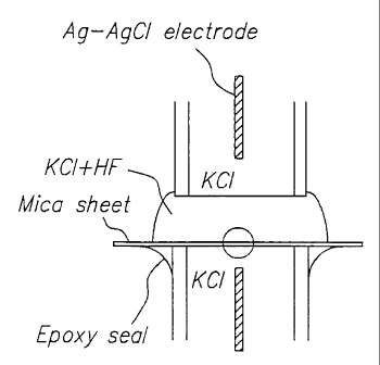
A representative mica nanopore device that may be used in the subject methods
is depicted
in Figures 1 A and 1 B and described in detail in Example I, infra. Briefly,
figures 1 A and B provide
a diagram of a mica sheet being etched to product a nanopore. A thin mica
sheet is cemented to a
glass capillary tube and exposed to 252Cf fission products that on average
product a single nuclear
track through the mica. The tube is then filled with 1.0 M KCl electrolyte and
a silver-silver
chloride electrode is inserted. A second plastic capillary tube filled with
KCl and an electrode is
placed above the mica as shown, and a mixture of 1.0 M KC1-20% hydrofluoric
acid is added to fill
the gap. A voltage of 100 mV is applied. Over a period of several minutes, the
HF etches the track
in the mica, producing a 6 rim diameter nanopore. When the pore is completely
etched through the
mica, an ionic current is measured and the mica is flushed to remove HF. The
device is then ready
for use in the subject methods.
Following contact of the fluid sample with the nanopore, e.g. the cis or trans
side of the
nanopore/barrier structure of the nanopore device, a least a portion of, if
not all of, the nucleic acids
present in the fluid sample are translocated through the nanopore, i.e. are
moved from the cis to the
trans side of the nanopore or vice versa. By "sequentially" is meant that only
one nucleic acid
present in the sample is moved through the nanopore at a time, since the
nanopore is dimensioned
so as to permit passage of only a single nucleic acid at any given time. By at
least a portion is
meant at least about 5, usually at least about 10 and more usually at least
about 15 number % of the
nucleic acids present in the sample. Translocation or movement of the nucleic
acids in the sample
through the nanopore is achieved using any convenient means, where generally
movement is
achieved by applying an electric field to the sample and across the nanopore.
The applied electric
field will be sufficiently strong to move the nucleic acids through the
nanopore. The actual
measured electric field that is applied across a typical nanopore 5 nm in
length is generally from
about 50 to 400 mV and usually 100 to 200 mV. When expressed as volts per cm,
the electric field
that is applied may range from about 50,000 to 500,000 volts per cm, where the
applied electric
5
CA 02384100 2002-03-06
WO 01/18251 PCT/US00/24513
field will typically range from about 100,000 to 400,000 volts per cm and more
usually from about
150,000 to 300,000 volts per cm.
The period of time during which the electric field is applied to the fluid
sample and across
the nanopore varies depending on whether just a portion or substantially all
of the nucleic acids
present in the sample are to be translocated. Generally, the electric field is
applied for at least about
1 ms, usually at least about 1 s and more usually at least about 10 s, where
the electric field may be
applied for 1 min or longer, but will generally not be applied for longer than
about 10 min and
usually will not be applied for longer than about 1 hour.
During the translocation step, the effect over time of the translocation on a
measurable
signal is determined. One convenient signal is the ion current through the
pore. As such, in many
embodiments, the ion current through the pore is measured during the
translocation of the nucleic
acids through the pore. In other words, the current through the pore is
monitored during the
translocation step. The measurements are generally of the amplitude of the
current through the
nanopore. In monitoring the nanopore during the translocation step,
measurements of current
through the pore are typically made at least every I s, usually at least every
0.1 s and more usually
at least every 0.02 s.
In many embodiments, the measured data values, e.g. current amplitudes, are
then
manipulated to produce a current blockade profile or similar output capable of
being compared
against reference outputs such that the nature of the nucleic acid, i.e. the
single or double
strandedness of the nucleic acid passing through the pore can be determined.
By current blockade
profile is meant the collection of current blockade data points plotted versus
a given period of time
upon application of an applied electric field to a nanopore. The given period
of time that a single
nucleic acid molecule is examined is generally at least about 10 microseconds,
usually at least
about 100 microseconds and more usually at least about 250 microseconds and
may be as long as 1
second or longer, but will usually not exceed about 5 milliseconds in length.
The current blockade
data points are derived from the observed change in ionic current through the
nanopore from the cis
to the trans side upon occupancy by the nucleic acid.
Following derivation of the collection of current blockade profiles during the
translocation
procedure, the derived current blockade profiles for a sample are then used to
determine the
presence or absence of double stranded or hybridized nucleic acids in the
sample. By comparing the
observed total current blockade profiles to reference current blockade
profiles of single and double
stranded nucleic acids, the presence of double stranded nucleic acids in a
sample can readily be
determined. In other words, one can look at the current blockade profiles to
identify patterns that
match the current blockade profile generated by translocation of a known
double stranded nucleic
acid through the nanopore. If patterns matching the control pattern are
identified, then one knows
6
CA 02384100 2002-03-06
WO 01/18251 PCTIUSOO/24513
that the sample includes double stranded or hybridized nucleic acids. The
comparison can be done
manually or automatically using computers and appropriate software.
The subject methods, in addition to being useful in determining the presence
of single or
double stranded nucleic acids in a sample, can also be used to determine the
relative amounts of
single and double stranded nucleic acids in a sample. In order to determine
the relative amounts of
single or double stranded nucleic acids in a sample, one can look
qualitatively or quantitatively at
the individual blockade profiles that are measured during translocation and
derive a proportion of
single to double stranded nucleic acids in the sample. The subject methods can
also be used to
quantitatively determine the numbers of single and double stranded nucleic
acids in the sample, e.g.
by counting the number of single stranded blockade profiles and the number of
double stranded
nucleic acid current blockade profiles observed during translocation. Again,
these determinations
may be done manually or using a computer means and appropriate software.
The subject methods find use in a variety of different applications where on
wishes to
determine the presence or absence of double stranded nucleic acids in a
sample. For example, the
subject methods find use in detecting hybridization events in assays where
complementary nucleic
acids are hybridized to each other and are detected. Examples of such
hybridization assays include
assays where one or more probes are combined with target nucleic acid and the
occurrence of
hybridization events in solution is detected. In such assays, unlabeled probe
is contacted with the
target nucleic acid sample. Next, the fluid sample is assayed according to the
subject methods, and
the presence of double-stranded nucleic acids in the sample is determined. The
presence of double-
stranded nucleic acids indicates that hybridization between the probe and
target has occurred.
The following examples are offered by way of illustration and not be way of
limitation.
EXPERIMENTAL
1. Preparation of Mica Nanopore Device
Figure 1 provides a diagram of a mica sheet being etched to produce a
nanopore. A thin
mica sheet is cemented to a glass capillary tube and exposed to 252Cf fission
products that, on
average, produce a single nuclear track through the mica. The tube is then
filled with 1.0 M KC1
electrolyte and a silver-silver chloride electrode is inserted. A second
plastic capillary tube filled
with KCl and an electrode is placed above the mica as shown, and a mixture of
1.0 M KC1-20%
hydrofluoric acid is added to fill the gap. A voltage of 100 mV is applied.
Over a period of several
minutes, the HF etches the track in the mica, producing a 6 nm diameter
nanopore (See e.g. Bean et
al., J. Appl. Phys. (1970) 41:1454-1459). When the pore is completely etched
through the mica an
ionic current is measured and the mica is flushed to remove HF.
7
CA 02384100 2009-09-16
II. Detection of Double Stranded DNA in a Sample
A 10 microliter aliquot of 10 micromolar single stranded 100 mer DNA probe
molecules
prepared on a DNA synthesizer in 1.0 M KCl is placed in two wells, each well
having a 6 nm mica
nanopore as prepared in Example I at the bottom and an electrical circuit so
that a voltage of 100
mV can be applied through the pore. Under these conditions a 6 nm diameter
nanopore will carry a
current of approximately 1.0 nA. As the 100 mer probe molecules are driven
through the nanopore
by the voltage, they produce a series of current blockades, each lasting for a
few hundred
microseconds and reducing the current by 20% (to 0.8 nA) during the blockade:
See Fig. 2A Two
unknown single stranded DNA fragments are then added to the wells, one with a
complementary
base sequence. A new series of higher magnitude blockades is observed in the
well to which the
complementary fragment was added. The higher amplitude blockades are absent in
the well
containing the non-complementary fragments. See Fig. 2B.
It is evident from the above results and discussion that novel methods of
detecting the
presence of duplex nucleic acid molecules, eg. hybridized DNA molecules, are
provided. As such,
new methods of detecting the presence of hybridized probe/target complexes are
provided in which
a detectable label, such as a fluorescent or isotopic label, is not employed
Accordingly, the subject
invention represents a significant contribution to the art
The citation of any publication is for its disclosure prior
to the filing date and should not be construed as an admission that the
present invention is not
entitled to antedate such publication by virtue of prior invention.
Although the foregoing invention has been described in some detail by way of
illustration
and example for purposes of clarity of understanding, it is readily apparent
to those of ordinary skill
in the an in light of the teachings of this invention that certain changes and
modifications may be
made thereto without departing from the spirit or scope of the appended
claims.
8