Note: Descriptions are shown in the official language in which they were submitted.
CA 02385538 2003-05-28
"RECOMBINANT ADENOVIRAL VECTORS AND THEIR UTILITY IN THE
TREATMENT OF VARIOUS TYPES OF FIBROSIS: HEPATIC, RENAL,
PULMONARY, AS WELL AS HYPERTROPHIC SCARS"
TECHNICAL FIELD OF THE INVENTION
The present invention relates to the creation of RECOMBINANT
ADENOVIRAL vectors bearing exogenous genes that encode for therapeutic
proteins
useful in the treatment of HEPATIC cirrhosis and generalized FIBROSIS, such as
renal FIBROSIS, pulmonary FIBROSIS, HYPERTROPHIC scars and keloid of the
skin, and/or in other target organs susceptible to suffer from it. It also
relates to a
mechanism of tissue-specific recognition of the affected cells by means of
delivery of
therapeutic genes to cirrhotic organs.
Moreover, the invention provides an effective way for the treatment of
fibrosis through the employment of recombinant adenoviral vectors which are
claimed
here, as well as the process to prepare these vectors, the pharmaceutical
composition that contains them, and their therapeutic uses in the treatment of
several
fibrosis, which has great commercial expectancy in the pharmaceutical industry
and
also presents an important alternative as gene therapy for the treatment of
chronic-
degenerative diseases characterized by fibrosis, with great therapeutic
application in
the field of Medicine.
INTRODUCTION
PHYSIOPATHOLOGY OF HEPATIC CIRRHOSIS
Hepatic cirrhosis is a disease resulting from hepatic chronic damage.
Damage might be toxic (chronic ingestion of alcohol), infectious (viral
hepatitis,
mainly by hepatitis B and/or C virus), immunological, (primary biliary
cirrhosis), by
biliary obstruction, (secondary biliary cirrhosis), metabolic (Wilson's
disease). All
forms of cirrhosis have characteristics in common: synthesis and excessive
deposition of proteins of extracellular matrix (ECM), mainly collagen I and to
a lesser
extent collagens IV and III), and consequently the formation of nodules of
hepatocytes, abnormal vascularization and portal hypertension (Antoni PP,
Ishak KG,
Nayak NC, Poulsen HE, Scheuer PJ, Sobin LH. The morphology of cirrhosis:
definition, nomenclature, and classification. Bulletin of the World Health
Organization. 1977; 55:521-540). The morphology of cirrhosis: definition,
nomenclature, and classification. Bulletin of the World Health Organization.
1977; 55:521-540 y Scott L. Friedman The cellular basis of hepatic fibrosis:
Mechanisms and treatment strategies. The New England Journal of Medicine
CA 02385538 2003-05-28
2
1993, vol. 328 No. 25:1828-1835. These physiopathological processes lead to an
alteration in the blood supply and in consequence in the nutrition of hepatic
cells.
Regardless of the ethiological agent and morphologic differences, all forms of
cirrhosis have as a common end, hepatic failure causing the patient's death.
As a consequence of the excessive deposition of collagen proteins in the
sub-endothelial space of the sinusoids (Space of Disse), various changes occur
in
the hepatic microenvironment: loss of hepatocyte villi, formation of a
basement
membrane composed by collagens IV and I covering the sinusoids, and loss of
the
fenestration of endotheiial cells which forms the sinusoids. All this process
is known
as "capillarization" of the sinusoids. (Scott L. Friedman The cellular basis
of hepatic
fibrosis: Mechanisms and treatment strategies. The New England Journal of
Medicine
1993, vol. 328 No. 25:1828-1835). Thus, the liver is not able to maintain the
physiologic concentration of solutes in the terminal hepatic vein, in other
words,
HEPATIC failure sets in. This capillarization, with the formation of the
continuous
endothelia (collagen of basement membrane) and the accumulation of other
collagenic proteins, represents a barrier to the normal and bi-directional
exchange of
molecules between the piasma and hepatocytes, as can be appreciated in Figure
1,
where hepatic cirrhosis is characterized by the accumulation in the liver of
type I
collagen. With an excessive deposition of this protein, the free exchange of
nutrients
between blood and liver cells is impeded and the inactivation of toxic agents
by this
organ can not be carried out, thus becoming the main cause of the
pathophysiology
of the disease. To date, no therapeutic agent has been described which is able
to
revert and/or prevent the progressive accumulation of hepatic coliagen with
100%
effectiveness.
Such physiopathological alterations presented in hepatic cirrhosis are
constant and common for the organs that also undergo fibrosis, such as, lung,
heart,
kidney, skin, among others, which should be not considered as limitations of
the
scope of protection of this invention. Therefore, the methodology presented
here for
the treatment of hepatic cirrhosis could be applied also to those organs that
are
susceptible to, or are affected by fibrosis.
Viral vectors and hepatic gene therapy
This technology can be implemented with viral or non-viral vectors.
Previous studies have been designed using plasmids and liposomes (DOTMA),
cationic and anionic, etc. Among the methods employing viral vectors, the most
commonly used include the use of retrovirus and adenovirus.
CA 02385538 2003-05-28
3
In a number of protocols, retroviral vectors have been used to introduce
genes in hepatocytes (JT, and Curiel DT, Adenoviruses as Vectors for gene
Therapy.
Science and Medicine/1997 44-53). However, precautions have to be taken since
these vectors can generate potential replication-competent viruses. Among the
advantages of these vectors is their ability to integrate their genome in a
stable way
in the chromosomes of the guest cell, which confers the possibility of
expression, in
an indefinite way, of the therapeutic transgene cloned in the retrovirus. On
the other
hand, up to date, no study has reported incidences of mutagenesis by insertion
or
activation of oncogenes by the incorporation of the replication-deficient
retrovirus.
Nevertheless, the use of retroviral vectors to transduce genes to the liver is
limited for
the following considerations: 1) these vectors infect only cells which
actively divide
and 2) very low viral particles titers are obtained in the packing cell lines
used to
amplify these viruses (Graham FL, and Van Der Eb AJ. A New Technique for the
Assay of Infectivity of Human Adenovirus 5 DNA. Virology 1973, 52:456-467).
These
two limitations have been successfully overcome in other Gene Therapy
protocols
through the induction of hepatocytes proliferation "in vivo", through the use
Hepatic
Growth Factors and through partial hepatectomy, surgical procedure by which
the
removal of 70% of liver mass induces division of the remaining hepatic cells
"in vivo".
The use of Lentiviral vectors has permitted to overcome partially said
limitations,
2o because they are able to transduce cells which are not actually dividing.
BACKGROUND OF THE INVENTION
Hepatic cirrhosis is a chronic illness of the liver, where diffuse cell
necrosis and a limited regeneration of parenchymal hepatic cells result in
diffuse
percentage increase of connective tissue, causing the distortion of lobular
hepatic
architecture and inducing hemodynamic alterations. Therefore, some strategies
for
the treatment of hepatic cirrhosis could include the prevention and/or
reversion of the
"fibrogenic process", stimulation of hepatic mitosis and re-arrangement of the
architecture of hepatic tissue. The documents of the state of the art related
to the
present invention are mentioned hereinafter only as references.
U.S. patent No. 5,240,846 refers to the use of gene therapy called
"CFTR", which induces a stable correction of the regulation of the chlorine
channel.
This defect is present in epithelial cells. In said invention, adenoviral
recombinant
vectors are used as well as plasmidic vectors. However, it does not have any
association with the therapeutics genes of the present invention. Likewise,
U.S.
patent No. 5,910,487, describes the use of plasmidic vectors for sending
therapeutic
CA 02385538 2003-05-28
4
molecules, but there is no association with the delivery of genes of
inetalloproteases
MMP-8 latent and/or active, MMP-1, MMP-2, MMP-9, MMP-13; or NuPA (wild type
uPA and/or its modified versions) or "Smad7" or the truncated receptors for
transforming growth factor-P (TGF-R type II) as presented here. U.S. patent
No.
5,827,703 refers to the use of adenoviral vector and modified adenoviral
vector to
send genes, however none of these vectors contain the genes used in the
present
invention for the treatment of fibrosis.
U.S. patent No. 5,770,442 claims the use of a recombinant adenovirus
that contains one gene directing the expression of a protein called "fiber" or
a protein
lo called "Fiber-chimera", however said patent does not specifically mention,
which one
is the therapeutic gene. Also, a method of gene therapy involving the use of
such
adenovirus and a vector of transference for the generation of such recombinant
adenovirus is presented. However, nothing is mentioned with regard to the use
of
therapeutic genes cloned and inserted in recombinant adenoviral vectors used
in this
invention in fibrotic livers, or to other target organs such as kidney, lung,
and
hypertrophic scars and others. These therapeutic genes are the gene that codes
for
human metalloproteases MMP-8, latent and/or active, MMP-1, MMP-2, MMP-9 and
MMP-13; human urokinase Plasminogen Activator (wild type and/or modified
huPA),
Smad7, and the truncated receptor for TGF-P type II, claimed herein. Other
members
of the family of genes represented are also included.
U.S. patent No. 5,166,320 refers to the use of a targeted delivery system
to introduce exogenous genes in mammalian hepatic cells. But there is no
association with putative genes directly sent to cirrhotic livers or to
fibrotic kidney or
lungs.
U.S. patent No. 5,872,154, describes a method to reduce the immune
response induced by an adenoviral recombinant vector and a selected immune
modulator, which functions by inhibiting the formation of neutralizing
antibodies
and/or reducing the death of the virally infected cells.
U.S. patent No. 5,871,982, is directed to a hybrid vector, in which a
portion of an adenovirus is included, together with a portion of an adeno-
associated
viral vector that contains a selected transgene. A hybrid virus consisting of
the union
of a conjugate with a polycation to a gene mesh of the adeno-associated viral
vector
to form a simple particle is also described. This is contrary to the present
invention in
which no hybrid viruses are employed, only adenoviral vectors. Besides, in the
above-mentioned patent the gene, transgene or therapeutic gene used is not
stated.
CA 02385538 2003-05-28
U.S. patent No. 5,856,152 is directed to the creation of a hybrid vector
which contains the portion of an adenoviral vector in combination with an
adeno-
associated virus and a selected gene. Through it large quantities of
recombinant
vectors are produced, but they are not carrying cloned therapeutic genes as is
5 described in this invention, in which specific therapeutic genes for the
treatment of
renal and hepatic fibrosis and hypertrophic scars are used.
U.S. patent No. 5,547,932 claims a compound of complexes of nucleic
acids for transfecting eucaryotic cells. These complexes are formed by nucleic
acids
and another substance with affinity for nucleic acids and optionally an
internalizing
lo factor, such as a virus or a component of the virus that can be conjugated.
It also
uses components of specific adenoviral vectors or specific viruses such as Ad2
or
Ad5, but does not mention the genes that are internalized in the cell
cytoplasm and
eventually in the nucleus of these eucaryotic cells. Similarly, U.S. patent No
5,521,291, is related to conjugated adenovirus bound through an antibody to a
substance with affinity to nucleic acids. In this way recombinant genes are
transported to the interior of eucaryotic cells. These conjugated complexes
and
nucleic acids are internalized in the cell, but the genes that can be sent are
not
specifically mentioned. In said patent, contrary to what is described in the
instant
invention, the use of such adenovirus to treat fibrosis or hepatic cirrhosis
or any
another type of fibrosis is not mentioned.
U.S. patent No. 5,585,362, relates to an improved adenoviral vector and
methods to obtain and use such vectors. The use of adenoviral vectors is not
mentioned in said patent. However the adenoviral vectors described in the
present
invention were used like vectors for sending therapeutic genes.
U.S. patent No. 5,756,086, claims an adenovirus, which is represented by
a protein called "fiber", the adenovirus also includes a ligand, that is
specific for a
receptor located in a specific cell type. This adenovirus can have at least a
portion of
this protein called "fiber" and it can be removed and replaced with a ligand,
which is
specific for a receptor in specific cells of the economy, such as hepatocytes.
This
adenovirus can include a gene that codes for a therapeutic agent. Based on the
previous statement, the outstanding technical difference of the instant
invention
compared to the state of the art, is the specificity of the therapeutic agent
as human
metalloproteases MMP-8 active and latent, MMP-1, MMP-2, MMP-9 and MMP-13;
human uPA (urokinase Plasminogen Activator , wild type and/or modified), the
truncated receptor for TGF-P type II and "Smad7" for the treatment of various
fibrosis.
CA 02385538 2003-05-28
6
U.S. patent No. 5,895,759 claims a tissue-specific vector (liver) for gene
therapy that can be used to send genes to a damaged liver. These vectors are
chemically or enzyme coupled to a promoter and can also be coupled to an
antibody
packaged in a polypeptidic envelope. Besides, the vector or the virus to be
assayed
is the hepatitis B virus. Thus the sending of genes to damaged livers
described in this
patent makes use of a system completely different from the one of this
invention, and
there is no relation with the process of fibrosis or cirrhosis to be treated.
U.S. patent No. 5, 559,099 describes an adenoviral recombinant vector
that contains a chimeric protein from the adenovirus called pentona, which
includes a
non-pentona sequence and a therapeutic gene to develop a gene therapy method
involving the use of such adenovirus, transference adenoviral vectors for the
recombination of such adenoviral vectors containing a therapeutic gene.
U.S. patent No. 5,885,808 claims also the use of adenovirus with bonding
molecules of adenovirus to different cells, the molecules of which have been
modified, as in U.S patents No. 5,846,782 and 5,712,136, in which adenoviral
vectors
are employed, which have been modified to contain different peptidic domains.
Finally, U.S. patent No. 5,670,488 relates to vectors for gene therapy,
which are especially useful for cystic fibrosis and also mentions the
development of
methods for the use of these vectors. The possible relation of the instant
invention to
the mentioned state of the art refers to the use of adenoviral vectors, that
can be
modified, as well as the use of inducible promoters driving the expression of
genes to
be inserted in these adenoviral vectors. However, the technical
characteristics of the
present invention are focused on the specific use of therapeutic genes to
treat fibrosis
of different kinds: hepatic, renal and pulmonary fibrosis, as well as
hypertrophic
scars.
The importance of the present invention, contrary to the state of the art
described in the above-mentioned documents, is based on the technical
characteristics of the invention itself, as well as on the additional
advantages derived
from the same, which are described with more details below.
ADENOVIRAL VECTORS
In the instant invention, the use of adenoviral vectors was determined
based on several considerations: 1) these vectors can be generated to very
high
titers of infectious particles per ml.: (109-1010); 2) they infect a great
variety of cells,
however, when they are administered i.v., most of them are located in the
hepatic
organ; 3) they transfer efficiently genes to cells that are not dividing, and
4) they are
CA 02385538 2003-05-28
7
seldom integrated in the guest genome, which avoids the risk of cellular
transformation by insertional mutagenesis (Douglas JT, and Curiel DT.
Adenoviruses
as Vectors for gene Therapy. Science and medicine, March/April 1997. 44-53 and
Zern AM, and Kresina TF. Hepatic Drug delivery and Gene Therapy. Hepatology
1997, Vol. 25, No. 2, 484-491).
Adenovirus are probably the most promising vehicles or vectors for the
delivery of genes in the protocols of gene therapy in human beings, since they
possess a unique attribute that provides them great stability when they are
administered into the bloodstream. This specific characteristic permits them
to be
lo efficiently used in clinical trials with a comfortable i.v. administration
for the patient.
(Douglas JT, and Curiel DT. Adenoviruses as vectors for Gene Therapy. Science
and
Medicine, March/April, 1997, 44-53).
Adenoviruses are double stranded DNA viruses. They have an
icosahaedric structure, infect a great variety of mammalian cell types, and
support the
ubiquitous expression of a specific receptor in the cell surface not yet
identified. Its
union to cells occurs by means of the protein component of the capside and the
virus
enters into the cell by receptor-mediated endocytosis.
More than 40 different human serotypes of adenovirus have been
identified, of which type 2 (Ad2) and 5(Ad5) have been more extensively
studied and,
therefore, more widely used as vectors for gene therapy. A very important
characteristic of these two Ad serotypes is that they have never been
associated with
malignant human processes.
The strategy for the creation of recombinant adenovirus is based on the
organization of the adenoviral genome. The expression of the adenoviral genes
occurs in two phases, early and late, that are defined by the time of
replication of the
adenoviral genome. The early genes encode themselves in 4 distinct
transcriptional
units: El, E2 and E4 encode for essential regulatory proteins that induce the
replication of the adenoviral DNA. The gene E3 is a non-essential gene. The
products
of the late genes include the main proteins of the capside, which are
transcribed
from a unique promoter. (Graham FL, and Van Der Eb AJ. A new technique for the
assay of infectivity of human adenovirus 5 DNA. Virology 1973, 52:456-467).
The recombinant adenoviruses are generated by introduction of the
exogenous gene or sequence of DNA of interest in substitution of the
adenoviral
genome regions required for the replication of the virus. The adenoviral
recombinant
vectors present deletions in El and E3 genome regions. Recombinant adenovirus
generation is conducted both through the replacement of El or E3 regions or
through
CA 02385538 2003-05-28
8
the insertion of the exogenous gene between the E4 region and the right
extreme of
the adenoviral genome. Vectors based on the insertion of the exogenous gene at
the
right extreme of the adenoviral genome or by the replacement of the E3 region
maintain their replication capability. On the contrary, the substitution of
early region
El produces a faulty vector in its replication capability, that, therefore,
can spread
only in a cell line that supplies in "trans" the absent functions of the
replaced
adenoviral region, or in presence of a collaborator virus. Of these, the most
commonly used as gene transference vectors are the replication-deficient
adenovirus
(Douglas JT, and Curiel DT. Adenoviruses as vectors for Gene Therapy. Science
and
to Medicine, March/April, 1997, 44-53).
The creation of adenoviral vectors, as well as their application for the
treatment of fibrosis, are shown in the examples described hereinafter.
SUMMARY OF THE INVENTION
The use of gene therapy for the treatment of different kinds of fibrosis in
human beings is disclosed. The purpose is the use of "therapeutic2 genes
specifically
directed to target organs to revert and/or prevent the deveiopment of the
fibrosis
process.
The potential application of gene therapy to patients with fibrosis and/or
cirrhosis will depend to a large extent on the successful delivery of genes
which
encode for therapeutic proteins to livers with severe fibrosis and that these
genes
which encode for proteins human MMP-8 active and latent, MMP-1, MMP-2, MMP-9
and MMP-13; human uPA wild type and/or modified (or its truncated version),
the
truncated receptor for TGF-R type II and Smad-7 can be directed by adenovirus
and/or other recombinant vectors that cannot transduce (infect) others organs.
The
recombinant adenoviruses (AdR) are vectors highly efficient for the
transduction of
therapeutic genes to diverse target cells. We have proved that they can carry
genes
to cirrhotic livers.
The delivery of therapeutic genes through such adenoviral vectors and
other recombinant vectors could also be performed using cationic and anionic
liposomes (DOTMA).
Therefore, we propose the use of this patent to be applied in the same
manner to:
*Renal fibrosis
*Pulmonary fibrosis
*Hypertrophic and keloid scars (skin fibrosis), and
CA 02385538 2003-05-28
9
*Other kinds of fibrosis.
OBJECTS OF THE INVENTION
Hereinafter, the objects and advantages derived from this invention are
presented.
An object of the present invention is to provide a procedure to prepare
recombinant adenoviral vectors pAdGFP-MMP-8, by means of the cloning of the
reporter genes: lac-7 and GFP and the therapeutic gene of collagenase or
metalloprotease MMP-8 in its latent and/or active forms.
Another object of the invention is to provide an adenoviral recombinant
vector with an exogenous gene or DNA sequence of interest that encodes for
therapeutic proteins useful in the treatment of the generalized fibrosis, in
target
organs susceptible to suffer from it. Such genes are, but are not limited to
MMP-8
active and latent, MMP-1, MMP-2, MMP-9 and MMP-13; and uPA (wild type and/or
modified).
Also, in the present invention, pharmaceutical compositions are provided
which contain the recombinant adenoviral vectors in quantities therapeutically
effective of viral particles for the treatment of generalized fibrosis; as
well as their
uses and therapeutic applications in the treatment of fibrosis.
An advantage of greater importance in the treatment of the generalized
fibrosis, particularly of hepatic cirrhosis, is that the delivery of
therapeutic genes is
carried out through tissue-specific recognition by the way of administration
employed.
Another advantage of the therapeutic uses of the invention, which is
directed initially to revert hepatic cirrhosis, is the treatment of
generalized fibrosis in
other target organs susceptible to suffer from it, including, without
limitation, the
treatment of fibrosis in lung, heart, skin, kidney, among others, in mammalian
animals, including human beings.
Another object is the design of a technology to send genes efficiently to
livers of animals affected by cirrhosis that resemble two types of cirrhosis
that usually
affect human beings (Alcoholic cirrhosis and Primary Biliary Cirrhosis).
Another advantage resulting from the fibrosis treatment is that
recombinant adenovirus does not induce lethal toxicity in none of the injected
animals
with the vectors.
Another objective of the invention allows us to discriminate the
modification of the staining reaction with X-Gal between the endogenous tissue
R-
CA 02385538 2003-05-28
galactosidase activity and the bacterial (3-galactosidase induced by the
infectious
action of the adenoviral vector. The use of the green fluorescent protein
permits us to
verify the in vivo transduction of different organs in rats to verify if the
vector
administration was appropriate, if the expression remains, and besides not
killing the
5 animals it is possible to conduct follow up observation after surgery.
Finally, all this evidence let us suggest that our system comprises an
efficient vehicle to deliver therapeutic genes such as human metalloproteases
MMP-8
active and latent; MMP-1, MMP-2, MMP-9 and MMP-13; collagenase which degrade
the deposited collagen excess and/or genes which encode for promoters of
hepatic
10 regeneration such as human uPA (urokinase Plasminogen Activator, modified
and
wild type), Hepatocite Grow Factor (HGF); the truncated receptor for TGF-R
type II
and Smad 7 to livers of cirrhotic rats, with the purpose to re-establish
normal liver
functions or normal functions of other organs affected by the same pathology.
Thus, in the present invention a process of preparation is given, through
which adenoviral recombinant vectors, pharmaceutical compounds and therapeutic
uses for the fibrosis treatment, especially for the treatment of hepatic
cirrhosis.
BRIEF DESCRIPTION OF THE DRAWINGS
Other particularities and advantages of this invention will be evident in the
following detailed description of the preferred objects and embodiments, from
the
enclosed claims and from the drawings or shapes attached, in which:
Figure 1 shows the cellular physiopathology of hepatic cirrhosis;
Figure 2 shows the proof of concept on how gene therapy works by
reverting the cirrhosis process;
Figure 3 is the schematic representation, which shows the cloning and
production of the adenoviral vector Ad5(3-gal;
Figure 4 shows the schematic development of the AdEasyTM system to
generate recombinant adenoviruses, specifically the pAdGFP-MMP-8;
Figure 5 shows the analysis of the expression of (3-galactosidase in
cultured cells.
Figure 6 shows the expression determination of green fluorescent protein
(GFP) expression in cultured cells;
Figure 7 shows the expression of R-galactosidase in different organs after
the infusion with Ad5(3-gal through the iliac vein.
CA 02385538 2003-05-28
11
Figure 8 shows the analysis of the tropism of the vector Ad5(3-ga1 to
different organs of cirrhotic experiment animals by chronic intoxication with
CC14,
demonstrating that the main target organ is the liver;
Figure 9 shows the analysis of the tropism of vector Ad5(3-gal to different
organs of cirrhotic experiment animals. Cirrhosis was induced by bile duct
ligation
and it was demonstrated that the main target organ is the liver.
Figure 10 shows histological sections of representative images of the in
vivo efficiency transduction assays of the vector Ad5R-gal in cirrhotic rats
with chronic
administration of CCI4;
Figure 11 shows histological sections of representative images of the in
vivo efficiency transduction assays of the vector Ad5p-gal in cirrhotic rats
by common
bile duct ligation;
Figure 12 shows the in vivo determination of the expression of the green
fluorescent protein;
Figure 13 shows the cloning strategy of the latent MMP-8 and active
MMP-8;
Figure 14 shows the mechanisms of complex formation with DNA of
MMP-8s for in vitro transfection essays in cells of hepatic origin (HepG2);
Figure 15 shows the verification through electrophoresis in agarose gels
of the success of cloning of MMP-8 cDNAs in the appropriate plasmids;
Figure 16 shows the transfection efficiency in HepG2 cells (Cells of
hepatic origin) with the plasmids of R-galactosidase and cDNA-MMP-8;
Figure 17 shows the analysis by polymerase chain reaction associated to
reverse transcriptase (RT-PCR) of MMP-8 messenger RNAs;
Figure 18 shows analysis of the collagenolytic activity in the protein
secreted to the culture medium by HepG2 cells after transfection with cDNAs
for
latent MMP-8 and active MMP-8;
Figure 19 shows the hormonal regulation of the MMP-8 gene expression
under the transcriptional control of the regulable promoter PEPCK and,
Figure 20 shows the dose-response assay of the different doses used to
determine the best response of "in vivo" hepatic transduction with the (3-
galactosidase
reporter gene.
DETAILED DESCRIPTION OF THE INVENTION
There are many reports showing that through systemic administration of
recombinant adenoviral vectors (AdR) into healthy experiment animals, a
specific
homing and highly preferential tropism of these vectors into the liver is
observed. Up
CA 02385538 2003-05-28
12
to now, it was not known whether the AdR were able to transduce cirrhotic rat
livers.
As previously mentioned, hepatic cirrhosis is characterized by an increase of
fibrosis
in the entire liver parenchyma, mainly around the central and portal veins,
creating a
barrier which hampers the exchange of macromolecules between the sinusoid and
the hepatocytes (Antoni PP, Hishack KG, Nayak NC, Poulsen HE, Scheuer PJ,
Sobin
LH. The morphology of cirrhosis: Definition, nomenclature and classification.
Bulletin
of the World Health Organization. 1977; 55:521-540; and Scott L. Friedman: The
cellular basis of hepatic fibrosis: Mechanisms and treatment strategies, The
New
England Journal of Medicine, 1993, Vol.328, No. 25:1828-1835), and this
protocol
io was designed to verify if even in presence of this barrier, the exogenous
genes could
be systemically delivered to the cirrhotic liver.
Therefore, our hypothesis is that AdRs containing LacZ and GFP (green
fluorescent protein) reporter genes are capable of transducing livers of
cirrhotic rats
even if the lobular architecture of the liver is distorted.
Thus, we could sent to these livers therapeutic genes such as human
metalloproteases or collagenases human MMP-8 active and latent, MMP-1, MMP-2,
MMP-9 and MMP-3; human Urokinase Plasminogen Activator (uPA wild type and/or
modified); the truncated receptor for TGF-P type II and Smad 7, which degrade
the
excess of collagenic proteins deposited and/or prevent the exacerbated
synthesis of
collagenic proteins, as it is shown in Figures 2 and 18; and/or genes which
encode
for proteins stimulating hepatic regeneration such as uPA, in order to re-
establish the
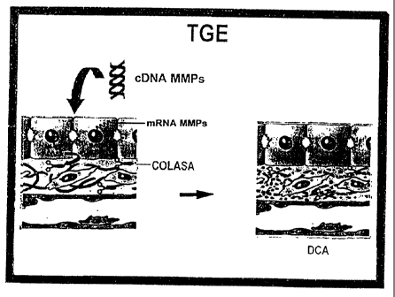
normal functioning of the liver, as is shown in Figure 2.
The current invention initiates a research line to carry out gene therapy as
an alternative for the treatment of chronic degenerative disease, specifically
of
hepatic cirrhosis in human beings, through the establishment of an efficient
vehicle to
send genes to the liver which will produce therapeutic proteins to help re-
establish
the normal functions of the liver, see Fig. 2. Fig. 2 shows how sending
efficiently a
therapeutic gene to the liver, in this case, a collagenase (metalloproteases
of matrix,
MMPs), it is possible to promote degradation of collagen through the over-
expression
of these metalloproteases.
In Figure 3, the strategy for the cloning and production of an adenoviral
vector is shown. The plasmid pDeltaElsplB contains adenovirus Ad5 sequences,
in
which the bacterial gene Lac-z was inserted. This plasmid was recombined with
the
pBHG10 to obtain complete viral particles after co-transfection in the cell
line 293.
The vector pAdGFP was obtained as follows: the MMP-8 gene (coming from the
plasmid PEPCK-MMP-8) was cloned in the vehicle vector, pAdTrack-CMV, the
CA 02385538 2003-05-28
13
resultant plasmid is linearized with the restriction endonuclease Pme I, and
is then
transformed in E. coli (BJ5183) with the plasmid pAdEASY-1. The recombinant
colonies were selected through kanamicine resistance, and the recombination is
confirmed by restriction analysis with endonucleases. Finally, the recombinant
plasmid linearized is transfected in the packaging cell line (293 cells), the
recombinant adenoviruses are obtained within 7 to 12 days as illustrated in
Figures 3
and 4 (Tong Chuan H., Shibin Z., Luis T. Jian Y, Kenneth W. and Volgestein
Bert: A
simplified system for generating recombinant adenoviruses. Prod. Natl. Acad.
Sci.USA Vol. 95: 2509-2514, March 1998). To evaluate the grade of transduction
in
vitro liver HepG2 cell line and peritoneal macrophages isolated from mouse
were
used. In Figure 5 the expression of 0-galactosidase in cultured cells is
shown. A), B)
and C) correspond to HepG2 cells (320X); D), E) and F), are mouse peritoneal
macrophages (100X). In C) and F) the transduced cells are shown with I X 108
viral
particleslml from the Ad5R-Gal vector. Three techniques were conducted to
compare
the degree of incorporation of the reporter gene Lac-Z which was administered
to
each culture dish in the form of plasmidic DNA PGKR-Gal, through precipitation
with
Ca++ phosphate (Chen C, and Okayama H. Calcium Phosphate mediated gene
transfer, a highly efficient system to establish transforming cells with
plasmidic DNA.
Biotechniques 1988, 6:632-638), DNA complexes-polylysine-Lactose (Martinez-
Fong
D., Mullersman JE, Purchio AF, Armendariz-Borunda J., and Martinez-Hernandez
A.,
Non enzymatic glycosylation of poly-L-lysine: A new tool for targeted gene
delivery.
Hepatology, Vol. 20, No. 6: 1602-1608), with the vectors Ad5p-gal and pAdGFP-
MMP8. The visualization of the activity of (3-Gal was verified with the
reactive Xgal
and the GFP in a microscope-stereoscope of fluorescence. For the in vivo
assay, (3-
gal staining was standardized using different pHs of the suspension with the
reactive
Xgal (Weiss DJ, Ligitt D., and Clark JG. In situ photochemical detection of R-
galactosidase activity in lung: assessment of Xgal reagent in distinguished
Lac-Z
gene expression and endogenous R-galactosidase activity. Human being therapy,
September 1, 1997, 8:1545-1554).
The models of experimental hepatic cirrhosis used are: a) Chronic
intoxication caused by carbon tetrachloride (CCI4), in which hepatic cirrhosis
is
established starting from the 8th week of peritoneal administration (Mion F,
Geloen A,
Agosto E. and Minaire Y. Carbon tetrachloride induced cirrhosis in rats:
influence of
the acute effects of the toxin on glucose metabolism. Hepatology 1996, Vol.
23, No.
2:582-587); and B), ligation of the bile duct (LCB) in which cirrhosis is
observed after
the fourth week of surgery (Lee S, Giraud C., Draillon A., HADengue A., and
Lebec
CA 02385538 2003-05-28
14
D., Hemodynamic characterization of chronic bile duct ligated rats; effect of
pentobarbital sodium. AM Journal fisiol. 1986; 251:176-180; Nakano S.,
Harakane J.
and Hashimoto H., Alteration in peribiliary ducts microcirculation in rats
after common
bile duct ligation. Hepatology, 1995, Vol. 21, No. 5: 1380-1995; Dumas Walla
R.,
Belcowitz D., and H. Eubi JE. Adaptive response of the Enterohepatic
circulation of
bile acid to extra hepatic. Cholestiasis Hepatology 1996, Vol. 23, No. 3: 623-
629 and
Poo J.L., Stanes A., Pedraza-Chaverri J., Cruz C., Perez C., Huberman A. and
Uribe
M: Cronologia de Ia Hipertension Portal, Disminucion de la Excrecibn de sodio
y
activacion del sistema renina-angiotensina en cirrosis biliar experimental.
Rev.,
Invest Clin, 49:15-23,1997).
Ad5(3-gal was administered at the same time and from the same lot to
control rats without cirrhosis. Rats with 5 and 8 weeks of CCI4 intoxication
and rats
with 2 and 4 weeks of bile duct ligation (BDL) were sacrificed 72 hrs after
administration of recombinant adenovirus for the histological analysis and
determination of the expression of the (3-galactosidase protein (R-gal)
encoded by the
AdR. For this purpose liver, spleen, heart, lungs, kidneys and brain were
extracted,
tissue sections were cut in cube shapes of 5 to 6 mm., which were absorbed in
freeze
medium Tissue-Tek O.C.T.T"', the tissues were frozen at -30 C and they were
cut
with a cryostat to obtain 8 m sections. These sections were placed on
silanized glass
slides and fixed with formaline, pH 8.5, during 15-30 minutes and were exposed
to
Xgal for 16-18 hours, being counterstained with Neutral Red stain. (Weiss DJ.
Ligitt
D. and Clark JG. In situ Hiti Chemical Detection of P-galactosidase activity
in lung:
assessment of Xgal reagent in distinguishing 1AC-Z Gene expression and
endogenous P-galactosidase activity. Human Gene Therapy, September 1, 1997,
8:1545-1554). The percentage of positive cells was determined by morphometric
analysis in multiple fields of the same size and calculating the average.
Besides, liver
sections of cirrhotic rats were obtained and tissues absorbed in paraffin were
cut and
stained with Sirius red which specifically stains collagenic proteins
(Armendariz-
Borunda J., and Rojkind M., A simple quantitative method for collagen typing
in tissue
samples: Its application to Human liver with schistosomiasis. Collagen Rel.
Res 1984,
Vol. 4, 35-47). Through this technique we can verify clearly the degree of
fibrosis and
the increase of bile ducts in the hepatic parenchyma. To verify the in vivo
transduction of cells with GFP, we used healthy Wistar rats that received
pAdGFP-
MMP-8 vector. 72 hours later, a laparotomy was performed and the exposed
organs
were visualised in the microscope of fluorescence, closing the wound
afterwards to
keep the animal alive.
CA 02385538 2003-05-28
The previous results that are presented here regarding the study of the
physiopathology of experimental hepatic cirrhosis are summarized in Figure 2.
Said
figure shows the role of pro-inflammatory and pro-fibrogenic cytokines
produced in
vivo by Kupffer cells which, in turn, activate the hepatic stellate cells
(HSC) to have
5 them produce excess collagens deposited in the subendothelial space,
obstructing
the exchange between hepatocytes and sinusoids (Armendariz-Borunda J.,
Katayama K., and Seyer J.M.: Transcriptional mechanisms of type I collagen
gene
expression are differentially regulated by IL-1beta, TNFalfa and TGF-0 into
cells. J.
Biol. Chem. 267:14316-14321, 1992; Armendariz-Borunda J:, Katai H., Jones C.M.
10 Seyer J.M. Kang A.H. and Raghow R.: Transforming growth factor beta is
transiently
enhanced at a critical stage during liver regeneration following CCL4
treatment.
Laboratory Investigation. 69:283294, 1993 and Armendariz-Borunda J., Roy N.,
Simjewish C., Raghow R. Seyer J.M. and Kang A.H.; activation of Ito cells
involves
regulation of API collagen Gene Expression. Biochemical Jounal 304:817-824,
1994).
is The degree of incorporation of Lac-z gene in cultured cells showed visible
differences
between techniques of Calcium-Phosphate, DNA-polilysine-lactose complexes and
with the recombinant adenoviral vector in HepG2 and PMM (Peritoneal mouse
macrophages). The degree of transduction with adenovirus reaches 100% and with
the other two techniques about 1% as shown in Figure 5. Figure 6 shows the
expression of green fluorescent protein (GFP) in cultured cells. A).
Peritoneal mouse
Macrophage transduced with the adenoviral vector pAdGFP-MMP8, 72 hours after
its
administration (50X), B).HepG2 cells transduced with the adenoviral vector
pAdGFP-
MMP8, 72 hours after its administration (50X) and C). HepG2 cells without the
adenoviral vector. All the pictures were taken in a microscope stereoscope of
fluorescence. It is necessary to point out that in the development to identify
(3-
galactosidase activity, the cells must be fixed and they die. In the GFP
assay, the
cells are still intact and alive.
Figure 7 shows the expression of P-gal in different organs after infusion
with Ad5p-gal by iliac vein. Fixation, washing and Xgal solutions using
different pHs
were used to discriminate among the endogenous expression and the bacterial
exogenous (3-galactosidase. In figure A, a pH 7.0 was used and in Figure B the
pH
was 8.5. This is the summary of the results of the assays of the different
experimental
conditions and it can be appreciated that the tissue exposition to Xgal
solution with a
pH 8.5 allowed us to eliminate the expression of endogenous P-galactosidase.
We
obtained frozen tissue sections from different organs: liver, kidney, lung,
heart, brain
and spleen from normal rats and intoxicated with CC14 for five and eight
weeks. As
CA 02385538 2003-05-28
16
represented in Figure 8, the graphics show clearly that the main target organ
is the
liver, both in healthy rats as well as in rats with chronic administration of
CCI4. A) 5
weeks of CCI4 administration and B) 8 weeks of CCI4 administration. Spleen and
lung
present a degree of transduction below 1%, and thus this is not evident from
the
graphs. Rats received doses of 3X10¹¹ viral particles/mi of Ad5(3-gal vector.
The
healthy control rats presented a total of 70% of hepatocytes transduced, while
spleen
and lung showed less than 1% transduction. In the other organs no transduction
was
found. Tissue sections were obtained from healthy rats as described before and
compared with tissues from rats with 2 and 4 weeks of BDL. Figure 9 clearly
shows
io how the main target organ is the liver, both in healthy rats as well as in
BDL rats. A) 2
weeks of LCB and B) 4 weeks of BDL. The spleen and the lung present a
transduction grade lower than 1 % , and thus it is hardly noticeable in
graphs. With a
dose of 3X10" viral particles/mi of the AD5P-gal vector, BDL rats present a
total of
10% transduced hepatocytes. Besides liver, spleen and lung presented less than
1%
transduction. The other organs showed no transduction. In Figure 10,
histological
results are shown with the hepatic cirrhosis model induced by the chronic
administration of CCI4i where A) represents a liver section of a normal rat,
72 hours
after the administration of Ad5(3-gal, by iliac vein (one representative cut
of the
experiments of a total of 5 rats). More than 70% of the hepatocytes are
positive to the
expression of 0-gal (200X); D) The same liver as in Figure A, but stained with
Sirius
Red to observe collagen synthesis and deposition (200X); B) liver with 5 weeks
of
chronic intoxication with CCI4. About 30-40% of the hepatocytes were
successfully
transduced; E). The same livers as in B, but stained with Sirius Red, the
increase in
the amount of collagen is notable and the liver architecture begins to distort
(200X);
C) rat liver after 8 weeks of chronic intoxication with CCI4 to cause
cirrhosis, again
more than 40% of liver cells were positive for G3gal expression and F) the
same livers
as in C, but stained with Sirius Red. Large deposits of collagen formed
between the
central and portal veins (200X) are characteristic. In Figure 11, results
obtained in the
model of biliar duct ligation (BDL) induced cirrhosis are shown. A) shows a
liver
section of a normal rat 72 hours after the administration of Ad5(3-gal, by
iliac vein
(one representative cut of the experiments of a total of 5 rats). More than
70% of the
hepatocytes are positive to the expression of R-gal (200X); D) the same liver
as in
Figure A, but stained with Sirius Red to observe collagen (200X); B) rat liver
after 2
weeks of BDL. (3-gal assay was conducted 72 hours after Ad5(3GaI
administration, via
iliac vein. About 10% of the hepatocytes were successfully transduced with the
reporter gene; E) the same livers as in B, but stained with Sirius Red. Liver
CA 02385538 2003-05-28
17
architecture begins to distort due to colestasis-induced fibrosis as well as
to the
important increase of biliar ducts (200X); C) rat liver after 4 weeks of BDL
to cause
cirrhosis. (3-gal essay was conducted 72 hours after the administration of
Ad5RGal,
via iliac vein. Again, 10% of hepatocytes were successfully transduced and F)
the
same livers as in C, but stained with Sirius Red. Observe the large deposit of
collagen proteins formed as well as the proliferation of biliar ducts (200X).
Figure 12
shows a laparotomy of a healthy Wistar rat that received pAdGFP-MMP-8 vector.
The
expression of the GFP is clearly seen in the liver and in insignificant
amounts in the
spleen. A very important fact is that the injection of adenoviral vectors did
not induce
lethal toxicity in experiment animals, both healthy and controls.
The preferred way to apply the present invention is through endovenous
administration of the recombinant adenoviral vectors of this invention or the
pharmaceutical compound which contains them, in which therapeutically
effective
amount is administered with an unitary dose regimen convenient to an
individual with
fibrosis. This regimen can be adjusted according to the affliction degree.
Generally,
unitary doses of about 10' to 1014 viral particles for individual are
employed. The
preparation of a pharmaceutical compound including the adenoviral recombinant
vectors of this invention can be conducted through the employment of standard
techniques very well known by the persons skilled in the art, in combination
with any
of the pharmaceutically acceptable carriers described in the state of the art,
including
without limitation, starch, glucose, lactose, sacharose, gel, malt, rice,
wheat flour,
chalk, silica-gel, magnesium stearate, sodium stearate, powder of glyceril
monostearate, NaCI, glycerol, propilene glycol, water, ethanol, and similar.
These
compounds can take the pharmaceutical form of solutions, suspensions, pills,
tablets,
capsules, powders and slow release formula, and similar.
The above description and the following examples have the purpose to
illustrate particular embodiments of the invention and they should not be
considered
as limitations of the scope of this patent.
Examples:
Example 1)
Methodology to demonstrate the activity of Metalloprotease or Collagenase
(MMP-8) and how to regulate its function
CA 02385538 2003-05-28
18
a) Cell culture. HepG2 cells is a cell line of parenchymal origin derived from
a
human hepatoma, and were cultured in 60 mm culture dish, 37 C in a wet
atmosphere, with 95% air and CO2 5% atmosphere in Eagle's medium modified by
Dulbecco (DMEM), supplemented with 10% fetal bovine serum, 2mM L-Glutamax
and antibiotics (100 U/mI penicillin and 100 pg/mI. streptomycin).
b) Vectors of Expression of latent and active MMP-8 aenes
Two plasmids were used with 2 kinds of MMP-8 genes to transfect the
hepatic cells: The plasmid pcDNA-MMP-8 which contains the cDNA which encodes
for latent MMP-8 (pro-MMP-8) together with the strong viral promoter of
cytomegalovirus (CMV); and the plasmid pcDNA3MMP-8 containing the cDNA which
encodes for the active MMP-8, together with the CMV promoter. This last one
was
created through subclonation using pcDNA3 and PETIIa-HNC plasmids, cutting
with
the restriction enzymes BamHl and Xbal and inserting the PCR product coding
for
the MMP-8 catalytic domain (which lacks the propeptide and carboxy-terminal
fragments), as shown in Figure 13, the delivery of latent and active MMP-8
genes.
Two types of plasmids with the MMP-8 gene were used to be delivered to hepatic
cells in culture: 1) PcDNA3-MMP-8, plasmid with the strong viral promoter of
the
cytomegalovirus (CMV) and the cDNA which encodes for the collagenase in its
active
form.
As a reporter gene pSV2-(3-gal plasmid was used. Said plasmid has the
gene which encodes the enzyme P-galactosidase inserted adjacent to the SV40
virus
promoter.
c) Plasmid Transformation, Amplification and Purification
To obtain a large enough quantity of each one of the plasmids to be used in
the
various assays, each plasmid was introduced to E. coli DH5aTM, (this process
is
known as transformation), according to the instructions of the supplier. (Life
Technologies, Gaithersburg, MD): in a reaction tube 50N1 of the competent
strain
DH5a were used and 2N1 of plasmids (1-10 ng of DNA) were added. After mixing,
it
was incubated on ice during 30 minutes, a thermal shock (37 C for 20 seconds)
was
applied and it was immediately chilled on ice for 2 minutes. At the end of
this period
of time, 0.95 ml of the bacterial culture medium Luria Base (LB) was added and
it
was stirred at 225 rpm during one hour to 37 C to allow plasmid expression.
After the
expression, 50pl of the reaction mix were taken and seeded onto an Agar plate
with
100 Ng/mI of ampiciline and it was incubated to 37 C overnight. The colonies
that
grow after this period are those which contain the plasmid of interest,
because of the
resistance against the antibiotic.
CA 02385538 2003-05-28
19
To amplify the plasmid, two colonies were taken from the Agar plate and
grown in a liter of LB medium containing 100Ng/ml of ampiciline during 24
hours at
37 C, with constant stirring at 225 rpm. Once the optic density of the culture
was 0.6,
it is centrifuged to 6,000 rpm for 20 minutes to recollect the bacterial
pellet. From this
bacterial pellet, plasmidic DNA was separated from the genomic DNA of the
bacteria
using a kit of plasmids purification (Monster-prep, BIO101, Vista, CA), which
is based
on the alkaline lysis of the bacterial wall, the liberation of the plasmid in
its interior
and the separation of this DNA through a particular resin. The quantification
of the
plasmidic DNA was performed measuring spectrophotometrically the resultant
absorbance at k = 260 nm.
d) Transfection of cultured cells
One of the most commonly used methods to introduce genes to
eucaryotic cells, is DNA transfection with calcium phosphate, in which the
exogenous
DNA is precipitated as a fine complex on the cell surface, to be later
incorporated by
the cell and transiently integrated in the chromosomal DNA. To deliver the DNA
with
more selectivity to the hepatic cells, DNA is used in the form of complex with
polylysine-lactose, because of hepatic cells have a specific receptor for
Galactose in
their cell membrane. For this, HepG2 cells were cultured at 70-80% confluence
and
then transfected with plasmids pcDNA-MMP-8, pcDNA3-MMP-8 and pSV2-0-
galactosidase. Transfection was carried out by DNA precipitation with calcium
phosphate (Graham FL, And Van Der Eb AJ. A New Technique for the Assay of
Infectivity of Human Adenovirus 5 DNA. Virilogy 1973, 52:456-467; Chen C, And
Okayama H. Calcium Phosphate-Mediated Gene Transfer, a Highly Efficient System
for Stably Transforming Cells with Plasmid DNA. Biotechniques 1988, 6:632-638)
and
by complex formation with polylysine-lactose (Martinez-Fong D, Mullersman JE,
Phurgio AF, Armendariz-borunda J, And Martinez-Hernandez A. Nonenzimatic
Glicosylation of Poly-L-lysine: A new Tool for Targeted Gene Delivery.
Hepatology,
1994 Vol. 20, No. 6:1602-1608). Briefly, cultured cells were added with the
newly
formed precipitate, product of the addition to plasmidic DNA of a solution of
DNA with
CaCI2 2M, in buffer solution HEPES pH 7.12 in case of the transfection with
calcium
phosphate or DNA complex with polylysine-lactose is added. Cells are incubated
from 4-16 hours to allow the precipitate to appear to the cell surface, and
later the
DNA can be endocyted and introduced transiently to the nucleus. At the end of
this
time, the culture medium is replaced for a fresh one, see Figure 14, where
HepG2
cells are cultured with DMEM medium with 10% bovine fetal serum. When 60-80%
confluency is reached, 10 mg of plasmid with MMP-8 gen is added in its latent
form,
CA 02385538 2003-05-28
as well as in the active or mature form. At the same time, the prokaryotic
gene of (3-
galactosidase ((3-ga!), is added to monitor the transfection and expression
efficiency.
MMP-8 gene was sent in different forms: naked, in complex with CaPO4 or in
complex with polylysine-lactose.
5
e) Formation of complexes polylysine-lactose and DNA: polylvsine-lactose
(DNA:PL)
The polylysine-lactose complex is formed when 14.8 mg of poly-L-Lysine
(0.1 N) react with 200pI of a-lactose 0.5 N (lactose-polylysine ratio: 1.0 N).
Then, 20
10 mg of reducing agent sodium cyanoborohydride 3 M is added and it is
incubated at
37 C for 48 hours with constant stirring at 225 rpm. Then, the reaction goes
through a
desaiting column (BioRad 10-DG) previously conditioned with phosphate buffer
(PBS
pH 7.2), which is eluted with the same buffer. Carbohydrate content is
determined to
the eluted fractions by the method of DuBois M, Gilles KA, Hamilton JK, Rebers
PA,
15 Smith F. Colorimetric method for determination of sugars and related
substances.
Anal Chem 1956, 28:350-6 to analyze the degree of lactosylation of the complex
and
the contents of polylysine according the method of Shen WC, Yang D. Ryser HJP.
Colorimetric determination of microgram quantities of polylysine by trypan
blue
precipitation. Anal Biochem. 1984, 142:521-4, which is considered as a base to
20 evaluate the final concentration of the PL complex. The fraction with a
mayor
concentration of PL is used for its further reaction with the DNA of the
plasmid
containing the gene of interest, as is shown in Figures 14 and 16.
To evaluate the optimal molar ratio of DNA: PL to be used in transfection
assays, the DNA was made to react with several concentrations of PL. At the
end of
one hour of incubation, samples were applied to a 1% retardation agarose gel
and
submitted to electrophoresis of DNA (60 millivoltios, 1.5h), in which the DNA:
PL
complex with the largest PL contents runs a shorter distance than the one run
by the
free plasmid (0% retardation). The DNA: PL ratio which causes 80 to 90% of
retardation of migration in the agarose gel was used as shown in Figure 16 to
obtain
an efficient expression of exogenous genes of (3-galactosidase and pcDNA-MMP-8
delivered to HepG2 cells in complexes with CaPO4 and polylysine-lactose.
f). Assays of transient expression using the reporter gen system of t3-
galactosidase (a-gal)
This system determines the activity of the (3-galactosidase enzyme as a
measure of the level of expression of the transfected gene of interest along
with Lac
Z gene which encodes for this enzyme. The (3-galactosidase is a bacterial
enzyme
CA 02385538 2003-05-28
21
which catalyzes the conversion of the uncolored substrate X-gal to a product
of blue
coloration. Because of this, the P-galactosidase activity observed in
eucaryotic cells
subjected to transfection will indicate the successful incorporation of the
gene of
interest associated to the bacterial gene.
The assay of (3-gal for the stain of cells in culture dish consists in the
fixation of cells
at 4 C during 5 minutes with 2% p-formaldehyde, the subsequent wash with PBS
(3X) and the addition of one ml of a stain solution in PBS containing 20 mM
potassium ferricianide, 20mM potassium ferrocianide and 2mM Magnesium Chloride
followed by the addition of the substrate Xgal in a final concentration of 0.5
mg/ml.
After incubation at 4 C overnight (18 hours) blue cells are identified under
the
microscope (Ausubel F, Brent R, Kingston RE, Moore DD, Seidman JG, Smith JA,
Struhl K (eds.). Short protocols in molecular biology, 3a edicion. 1995. John
Wiley &
sons, Inc., New York).
g) RNA extraction
48 hours after transfection, cells are recollected to extract RNA by the
Method of Chomczynsky P, Sacchi N. Single-step method of RNA isolation by acid
guanidinium thiocyanate-phenol-choroform extraction. Anal Biochem 1987,
162:156-9
using the reactive of TrizolT'", as described hereinafter: to each one of the
cell dishes
one ml of PBS solution was added and cells were recollected by scraping them
from
the dish and transferred to an Eppendorf tube. It was then centrifuged at 1000
rpm for
one minute and the cell pellet was treated with 500 pl of Trizol, homogenized
and
incubated for 5 minutes at 4 C. One hundred l of chloroform were added, and
incubation was conducted during 5 minutes at 4 C. After this, it was
centrifuged at
12,000 g for 15 minutes at 4 C and the aqueous upper phase was transferred to
a
clean tube in which an equal volume of isopropanol is added and incubated at -
70 C
during 15 minutes to precipitate the extracted RNA. Then, it is centrifuged at
12,000 g
during 15 minutes at 4 C, the supernatant is eliminated through decantation
and
drying the tube with clean and sterile paper. Then, 500 NI of 75% ethanol were
added
and it was centrifuged at 12,000 g during 10 minutes to 40 C. Finally, the RNA
pellet
was resuspended with 20 to 50 pl of deionized water treated with
diethylpirocarbonate (DEPC) and RNA concentration was quantified by
spectrophotometry at ~,=260 nm.
h) Analysis of expression of MMP-8 gene by the Polymerase Chain Reaction
(PCR) associated to the reaction of reverse transcriptase (RT-PCR).
To determine the degree of expression of the exogenous gene of MMP-8
incorporated to the cell, complementary DNA was obtained (cDNA) starting from
RNA
CA 02385538 2003-05-28
22
previously extracted and then amplifying the expression signal by the
Polymerase
Chain Reaction.
To obtain the cDNA, the following procedure was used: 2 pg of total RNA
were taken to a volume of 8pI with deionized, sterilized water and incubated
at -70 C
for 10 minutes. Then, the sample was stirred in iced water during 5 minutes
and still
in the ice, the following reagents were added: 4 pl of 5X buffer for the RT
enzyme, 4
NI dNTP's mix 2.5 mM, 1 NI random primers (1 iag/pl), 1 pl inhibitor of RNAase
(one
U/NI ) and finally 2 pl of the Reverse Transcriptase enzyme (200 U/pl). The
reaction
mix was incubated at room temperature for 10 minutes and then at 37 C for one
hour.
At the end of this time, it was placed immediately in a temperature of 95 C
for 10
minutes, and then it was placed on iced water during 5 minutes with constant
stirring
and it was stored at -70 C until its further use.
To analyze the specific expression of MMP-8 gen , a PCR reaction was
set up using the primers or oligonucleotides specific for this gene according
to the
experimental conditions described hereinafter: in a reaction tube containing 2
pl of
cDNA 5 NI of 2.5 mM MgCI2, 5 pl 5X buffer for the polimerase enzyme, from
leukemia
murine virus of Moloney (MMLV), 2pl of 2.5 mM dNTPs, 5 NI of the sense primer
3
M, 5N1 of the antisense primer 3 M, 1 pl of the polymerase enzyme (1 U/pl) and
it is
taken to a final volume of 50 pl with deionized water (Innis M, Gelfand DH,
Sninsky
JJ, and White TJ., (eds.) 1990. PCR Protocols: A Guide to methods and
applications,
Academic Press, San Diego CA). The oligonucleotide sense primer specific for
MMP-
8 is 5'-AGCTGTCAGAGGCTGGAGGTAGAAA-3', and the antisense primer is 5'-
CCTGAAAGCATAGTTGGGATACAT-3' (Cole AA, Chubinskaya S, Schumacher B,
Huch K, Cs-Szabo G, Yao J, Mickecz K, Hasty K, Kuettner KE, Chondrocyte matrix
metalloproteinase-8. J Biol Chem 1996, 271:11023-6). After the addition of
these
reagents, the mix was placed in a thermalcycler during 30 cycles according to
the
following program: denaturation (94 C, 5 min), annealing (60 C, 1 min.) and
extension (72 C, 1.5 min). Then, PCR products are submitted to electrophoresis
(60
mV, 1.5 h) in a 1.5% agarose gel.
i) Assay of Collagenase activity.
The analysis of enzymatic activity of collagenase was performed to
determine the functionality of the enzyme produced, because this protein could
be
found enzymatically inactive, even when RNA expression was positive. Cells are
cultured in serum-free medium for 24 hours, culture medium is recollected and
CA 02385538 2003-05-28
23
activity of collagenase secreted by the cells is determined by a modified
method of
Hasty KA, Hibss MS, Kang AH, Mainardi CL. Secreted forms of human neutrophil
collagenase. J Biol Chem 1986; 261:5645-50 to identify products of degradation
of
specific collagen substrate through 8% polyacrylamide gel electrophoresis .
Briefly: cell supernatants containing 1-1.5 pg of protein were incubated at
27 C during 18 hours with 5 pg of native collagen type I and 60 NI of the
incubation
buffer: 50mM Tris-HCI, 5 mM CaCl200.02% NaN3, 50 mM arginine, 1% Triton X-
100T"" and in absence or presence of 1 mM APMA, pH 7.6. Finally, 30 pl of
product of
reaction were mixed with 30 pl of sample buffer for proteins and
electrophoresis in
SDS-polyacrilamide gels (7.5%) was run to identify the degradation products
a1A and
a2A of collagen type 1.
Example 2:
Results to demostrate the activity of Metalloprotease or collagenase (MMP-
8) and therefore to regulate its function
Subcloning permitted to incorporate MMP-8 cDNA encoding for the fully
functional enzyme was subcloned in a vector appropriate to our needs. Thus,
Figure
15 shows an electrophoresis of the DNA fragments released by cutting MMP-8
plasmids with restriction enzymes. Lane A). Marker of bp of 1 Kb DNA ladder
(Gibco
BRL); B). Perfect DNA marker (Novagen, Inc.); 1) pcDNA-MMP-8 cutting with
BamHl
and Xbal; 2) pcDNA3-MMP-8 cutting with BamHI and Xbal; C) ~X174 marker (Gibco
BRL); a, Hind III Marker (Gibco BRL), in which the latent MMP-8 cDNA (lane 1)
and
the mature MMP (lane 2); were successfully subcloned in the expression vectors
pcDNA and pcDNA3. The released inserts are observed after treatment with
restriction enzymes BamHl and Xbal. The bands stained with ethydium bromide
correspond to each of cDNA (between 506 and 560 base pairs) for mature and
latent
MMP-8 cDNA, respectively. To evaluate the efficiency of incorporation of the
cDNA
for MMP-8 delivered to HepG2 cells in form of complex with CaPO4 and with
polylysine-lactose, the co-transfection of this plasmid was realized along
with the
reporter gene of (3-galactosidase. In this way, cells observed in the
microscope with
blue staining, indicate indirectly that they have also incorporated to the
plasmid of
interest. Figure 16 shows the expression of (3-galactosidase in HepG2 cells,
co-
transfected with free plasmid, in form of complex with CaPO4 , or in its form
of
complex with polylysine-lactose. This figure shows that the DNA binding with
polylysine-lactose was accomplished because the higher the polylysine
CA 02385538 2003-05-28
24
concentration, the clearer the retardation of (3-gal plasmid. The ratio
selected to
transfect the cells was the one that delayed 80% of plasmid migration.
Once demonstrated that the cells in culture are capable of incorporate
and express genes that have been transfected, it was necessary to corroborate
that
such genes were transcribed by the machinery of host cells by means of RT-PCR
assays. Figure 17 shows an analysis by RT-PCR of messenger RNA for MMP-8 and
MMP-13. (This plasmid was used as a further positive control of transfection);
in
which a DNA electrophoresis of PCR amplified products, of the cDNA for MMP-8
delivered as a complex with CaPO4 and polylysine-lactose, has been transcribed
for
both cases in transfected HepG2 cells. It is observed that product signal of
PCR of
MMP-8 (359 base pairs), was more intense when plasmid was delivered as a
complex with polylysine-lactose.
To demonstrate that MMP-8 transcripts expressed by HepG2 cells was
translated into a functional protein, the assay for enzymatic activity was
conducted,
using collagen type I as substrate. Figure 18 shows the enzymatic activity of
type I
collagen degradation of the protein secreted in the culture medium, which was
observed in the transfected cells with the gene of latent MMP-8. With previous
activation with the mercurial agent APMA (lane 7) and with the gene of active
MMP-8
complexed with CaPO4 (lane 9) and with polylysine-lactose (lane 10), and its
specific
inhibition with EDTA 2mM. Negative controls: type I coliagen without addition
of
supernatants of cells (lane 1) and with addition of Trypsin (lane 3), collagen
with
supernatants of cells without transfection (lane 2). Positive controls: type I
collagen
with supernatant of human leukocytes (lane 3), type I collagen with addition
of
0.015% bacterial collagenase (lane 4); and degradation products of native type
I
collagen, separated in a 6% polyacrylamide gel, after it was incubated with
supernatant of transfected cells with latent and active MMP-8 genes. It was
observed
how in both cases the collagenolytic activity is clear in presence of APMA in
the case
of latent MMP-8, and its inhibition for EDTA for both latent and active MMP-8.
This
fact shows that this proteolytic activity corresponds to a metalloprotease of
interstitial
matrix. The incubation of native type I collagen with trypsin did not show
degradation.
So, this experiment clearly shows that MMP-8 action was specific considering
the
intact nature of the collagen molecule.
Figure 19 shows evidence that activities of the enzymes that specifically
degrade collagen can be controlled (turned off and/or turned on) through the
cloning
of its respective cDNAs that are themselves under the transcriptional control
of
promoters of regulable genes, such as the PEPCK (Phosphoenol-piruvate
CA 02385538 2003-05-28
carboxikinase) gene. It is clear that both the stimulation of cells in culture
with
Glucagon (lanes 5 and 6), and cyclic AMP (lanes 7 and 8), up-regulate their
production of messenger RNA that codes for MMP-8. It is also clear that
insulin
lowers said production (lanes 9 and 10).
5 The observations regarding the activity of endogenous (3-galactosidase
suggest that this activity is usually granular and weaker in color than the
dark blue as
a result of the activity of exogenous enzyme (Shimohama S., Rosenbergh MB.
Fagan
AM, Wolff JA, Short MP, Bradfielf XO, Friedman T., and Gage FH: Genetically
Modified Cells into the rat brain: Characteristics of E. coli-Galactosidase as
a reporter
10 gene. Brain Res. 5:271-278, 1989). Many modifications have been described
to
increase the specificity in the determination of exogenous Lac Z gene essay.
Thus,
according to previous information by Weiss, DJ, Ligitt D., and Clark JG. In
situ
histochemical detection of beta-galactosidase activity in lung. Assessment of
Xgal
reagent in distinguishing Lac Z gene Expression and endogenous 0-galactosidase
15 activity. Human Gene Therapy, September 1, 1997, 8:1545-1554; in the
present
invention a solution of X-gal, with a pH 8.5 was used; in this way, the
activity of
exogenous P-gal was demonstrated, minimizing the endogenous activity in vivo.
One of the indicators actually used for in vivo monitoring the efficiency
and location of transduced cells with recombinant adenoviruses, is the
detection of
20 green fluorescent protein (GFP) expression. For this purpose, the gene
which
encodes for this protein is subcloned in adenoviral vectors, and then through
the use
of a fluorescent microscope, the fluorescence given by GFP can be observed
directly
without sacrificing the experiment animal which received the vector (Rojas-
Martinez,
A, Wyde PR, Montgomery CA, Chen SH, Woo SLC and Aguilar-Cordova E.:
25 Distribution toxicity and lack of replication of an E1A-recombinant
adenoviral vector
after systemic delivery in the cotton rat. Cancer Gene Ther. 1998, y TongChuan
H.,
Shibin Z., Luis T., Jian Y., Kenneth W., and Vogelstein Berth: A simplified
system for
generating recombinant adenoviruses. Proc. Natl. Acad. Sci. USA Vol. 95:2509-
2514,
March 1998). A large body of data has been obtained that shows that, after the
i.v.
administration of adenoviruses in healthy animals, the main target cells were
hepatocytes. This has been observed in mice, rabbits, dogs and primates (Zern
AM.
and Kresina TF, Hepatic drug delivery and gene Therapy. Hepatology 1997, vol.
25,
No. 2, 484-491), but not in cirrhotic rats. Probably, the injection in portal
vein could be
more efficient to get to the target cells in the liver, providing them a
favorable
innoculum of viral particles to the entire liver before being diluted into the
bloodstream. This route is efficient, but it has the disadvantage that it
requires a
CA 02385538 2003-05-28
26
laparotomy. On the other hand, peritoneal administration is a faster and
simpler
infusion, but it does not promote hepatocyte transduction. The results of the
present
invention show that the injection of 3 X 10¹¹ viral particles by iliac vein in
normal
Wistar rats of approximately 200 g. produces a very high level of expression
(70% of
transduced hepatocytes). Our results are consistent with a previous report in
which
specific delivery of reporter genes in primates by saphenous vein produced
almost
the same level of transduction and expression of the transgene in the liver,
as
compared with infusion through portal vein (Marie Jean TFD, Poeters V., Lieber
A.,
Perkins J., and Kay MA. Methods for multiple portal vein infusion in mice:
Quantitation of adenovirus-mediated hepatic gene transfer. Biotechniques
February
1996, 20; 278-285 and Zhu G. Nicholson AG. Zheng X., Strom TB, and Sukhame VP.
Adenovirus mediated 0-galactosidase gene delivery to the liver leads to
protein
deposition in kidney glomeruli. Kidney international, 1997, Vol. 52, 992-999).
Furthermore, the expression of the reporter gene in our animals with cirrhosis
ts induced by chronic administration of CCI4 was surprisingly almost as high
as the
normal rats (40% of transduced hepatocytes). These results are very exciting
because our cirrhotic animals could hardly survive the surgical procedure
required to
administrate the adenovirus by the portal vein. This is due to altered
functional
hepatic tests, and elevated prothrombin time as well as important bleeding.
Although
rats with bile duct ligation showed a substantial reduction in the number of
transduced hepatocytes (5-10%), it is also important the number of
hepatocytes,
which eventually could be transduced with therapeutic genes, such as
metalloproteases (MMP-8) and/or genes which encode for stimulating proteins
for
hepatic regeneration such as uPA (Urokinase Plasminogen Activator) and Smad 7.
Other embodiments will be evident for people skilled in the art based on
the present description. Said embodiments are included within the true scope
and
spirit of the invention.
CA 02385538 2003-05-28
27
*The definitions of the symbols used in the figures corresponding to the
present
invention, are shown below:
Figure 1:
CEH= stellate hepatic cell.
CES= Endothelial sinusoidal cell.
CK= Kupffer Cell.
ESET= Subendothelial space.
HE= Hepatocytes.
HIDC= Liver with chronic damage.
HN= Normal liver
SINU= Sinusoid
Figure 2:
COLASA= Collagenase
DCA= Degradation of coliagen.
TGE= Experimental gene therapy
MMPs= Metalloproteases
Figure 3:
CT293= Co-transfection in cells 293
PG CsCl= Purification with CsCI gradients
Figure 4:
BD= Right arm.
B1= Left arm.
CTBK= Co-transfection in bacteria and selection in Kanamicine.
CUL= Culture
LI Pacl= Linearize with Pac I.
LI Pmel= Linearize with Pme I
PV= Viral particles
T293= 293 Cell transfection
GENADR= Generation of recombinant adenovirus.
Figure 7:
B= Spleen.
CA 02385538 2003-05-28
28
CE= Brain
CO= Heart
%CT= Percent of transduced cells
H= Liver
P= Lung
R= Kidney
SAd(3-gal= Without the Ad(3-gal vector
CAdR-gal= With the Ad(3-gal vector
X-GAL7= Reactive X-gal, pH 7.0
1o X-GAL 8.5= Reactive X-gal, pH 8.5
Figure 8:
B= Spleen.
CC145= 5 weeks of intoxication with CCL4
CC148= 8 weeks of intoxication with CCL4
CE= Brain
C0= Heart
%CT= % of transduced cells
H= Liver
P= Lung
R= Kidney
PV= Viral particles
HN= Normal Liver
Figure 9:
B= Spleen
CE= Brain
CO= Heart
%CT= % of transduced cells
H= Liver
LCB2S= 2 weeks of bile duct ligature
LCB4S= 4 weeks of bile duct ligature
P= Lung
R= Kidney
PV= Viral particles
HN= Normal Liver
CA 02385538 2003-05-28
29
Figure 13:
PROT= Protein
APMA: MERCURIAL AMONOPHENYL ACETATE
Figure 14:
ACTP-ga1=p-galactocidase activity
CES= Cells
EAC= Enzymatic Activity
PL= Polylysine
PROT= Protein
RGAL= Galactose residues
SNAD= Supernatant
Figure 16:
ADND= Naked DNA.
GELRADN-PL= Retardation gel for polylysine
Figure 18:
CA= With APMA.
CACE= With APMA and EDTA.
CaPO4= Phosphate.
CE= With EDTA.
COB= Bacterial Collagenase.
COL1= Type I Collagen
PL= Polylysine
SA= Without APMA
SNL= Leucocyte Supernatant
ST= No-transfected
TRIP= Trypsin
Figure 20:
%CT= % of transduced cells.
PV= Viral particles.