Note: Descriptions are shown in the official language in which they were submitted.
CA 02385670 2002-03-22
WO 02/07603 PCT/ITO1/00387
_1 _
"A needle of the biopsy type or for taking other samples from human or animal
organs"
DESCRIPTION
The present invention relates to a needle of the biopsy type or for taking
other samples from human or animal organs. In general; such needles
comprise an external cannula and a stem that can slide within the cannula
and can be controlled independenfily of the latter. The cannula has one end
fixed to a manipulating element, whilst the other end is shaped like the
mouthpiece of a flute, with a sharp-edged mouth designed to penetrate into
tissue. The stem with its end that is distal with respect to the grip is
designed
to closer the mouth of the cannula. from inside. ,It moreover Chas :a recessed
.
area developed for a certain stretch in the vicinity of the aforesaid end in
order
to contain a specimen of. tissue taken from the organ concerned by means of
a well-known manipulation technique.
The aforesaid needles present thedrawback that the amount of tissue',
sampled is small in so far as the aforementioned recessed area occupies only
one part of the section of the stem, in general one half. Consequently, in
order
to take a sufficient amount of tissue, it is necessary to use a needle of
considerable thickness. In addition, the tissue, which in general has a
~ gelatinous consistency may easily get lost or contaminated during
manipulation of the elements of the needle.
The document SU 1537232 describes a biopsy needle comprising an
external cannula having a rectangular cross section, and a flexible closing
element, which can slide inside the cannula. At a point corresponding to the
tip of the cannula, the latter has lateral guide grooves in which said closing
element engages, so as to be deflected and thus close the opening of the
needle both to enable the first step of penetration of the needle into the
body
of the patient without collecting any material and to enclose a specimen of
tissue before extraction of the needle. The above conformation does not,
however, afford the possibility of extracting the tissue specimen from the
needle while still leaving the external cannula of the needle itself in situ
in the
body of the patient, for checking the quality .of the tissue sampled and
CA 02385670 2002-03-22
WO 02/07603 PCT/ITO1/00387
-2-
possibly repeating the sampling operation at a greater depth.
One purpose of the present invention is to overcome the above-
mentioned drawbacks; other advantages will emerge from the ensuing
description and claims.
A biopsy needle according to the invention comprises a cannula with a
sharpened front end shaped like the mouthpiece of a flute, and a stem
designed ~to close the mouth of the cannula in a known way. The cannula has,
for a certain stretch in the vicinity of the mouth, a first portion of
relatively
small thickness and a second portion - in an area corresponding to the mouth
- of a larger thickness, the two portions forming between them, inside the
cartnuia, ~a step-like. portion developed according to an inclined plane with
,
respect to the axis of the cannula. Between the cannula and the stem there is
inserted an axially slidable element for withholding the tissue specimen, said
slidable element being controllable independently of the stem and of the
cannula. Said element has, in the vicinity of said internal step-like portion
of
the cannula, an end provided with a tab-like extension which, by means of the
relative sliding between . the slidable element and the cannula and in the
direction of the mouth of the latter, is designed to engage at the front with
the
step-like portion in order to bend, so closing the, mouth of the cannula, thus
cutting the tissue specimen that is being sampled and withholding it inside
the
withholding element during extraction of the latter and .possibly also of the
needle. In this way, the specimen has a right cross section equal to that of
the
mouth of the cannula and is withheld securely inside the latter both during
extraction of the needle from the organ concerned and afterwards. The tab-
like extension preferably has a restricted area of connection with the
withholding element in order to facilitate bending thereof, the said bending
occurring beyond the elastic limit of the ab itself and thus generally being
permanent.
In a particular embodiment of the invention, the tab-like extension has
side walls that are designed to contain the tissue specimen laterally. .
The external cannula may have a polygonal cross section, for example a
square cross section, or even a circular or elliptical cross section.
CA 02385670 2002-03-22
WO 02/07603 PCT/ITO1/00387
_3_
The arrangement according to the invention can be applied , both to
needles for taking samples of soft tissue and to needles for taking infra-
osseous samples.
In particular embodiments of the invention, there may be provided,
between the withholding element and the stem for closing the needle, a
further axially slidable element, generally of a tubular shape. This
additional
element is configured so that it can be set in contrast with 'said tab of the
withholding element, once the tab is bent, then to re-open the tab in the
specimen-extraction step, so as to facilitate extraction of the tissue
specimen
without damaging it, as will be described in greater detail in what follows.
A better understanding of the present invention will be provided by the
ensuing description and by the attached drawings, which illustrate non-
limiting
examples.of the invention. In the drawings:
Figs. 1, 1 bis and 2 are partial side views of a biopsy needle according to
the invention; with the components of the needle in two different
arrarigements;
Figs: 3 and 4 respectively illustrate a view of the needle taken according
to the plane indicated by 111-II1 in Fig. 2 and a cross section of the needle
taken according to the plane indicated by IV-IV in ,Fig. 1;
Fig. 5 is an exploded perspective partial view of the needle of Fig. 1;
Figs. 6 and 7 are views similar to those of Fig. 1 and Fig. 2 of a biopsy
needle according to another embodiment of the invention;
Fig. 8 is a cross-sectional view according to the plane indicated by VIII-
VIII of Fig. 6;
' Fig. 9 is a perspective view of an element of the needle, according to the
arrow F1 of Fig. 6;
Figs. 10, 11 ~ and 12 are views. similar to those of Figs. 1, 4 and 5,
respectively, according to another embodiment of the invention;
Figs. 13, 14 and 15 are views similar to those of Figs. 1, 4 and 2,
respectively, according to a further embodiment of the invention;
Fig, 16 is a partial perspective view of elements of the needle of Fig. 13;
and
CA 02385670 2002-03-22
WO 02/07603 PCT/ITO1/00387
-4-
Figs. 17 and 18 illustrate views of a biopsy needle, axially sectioned,
according to yet another embodiment of the invention and in different
arrangements of the component elements.
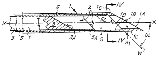
With reference to Figs. 1 to 5, the needle comprises an external cannula
1 of square cross section, one end of which (not illustrated in the drawing)
is
fixed to a manipulation element, such as a grip, whilst the other end 1A is
shaped so that it is inclined like the mouthpiece of a flute and has a mouth 1
B
with its edges sharpened for penetrating into the tissue. The needle also
comprises a stem 3, which can slide inside the cannula, and the end 3A of
which (visible in the drawing) is also shaped like the mouthpiece of a flute
and
has the same inclination as the end 1A of the cannula 1 and a cross section
designed to penetrate with minimal play into the mouth 1 B of the cannula 1 so
as to close it. The stem 3 is controllable independently of the cannula 1 so
that it can be displaced from one completely advanced closing arrangement
(Fig. 1 bis), in which the inclined face 3A of the stem is flush with the face
1 A
of the mouth of the cannula, and a second arrangement, illustrated in Fig. 1,
in which the stem 3 is retracted, arid the face 3A defines, in the cannula 1 !
a
chamber Z (see Fig. 1 ) designed to contain a tissue specimen that is to be
sampled. The cannula 1 has, at least in the stretch shown in the drawing, a
uniform thickness (s) of wall, except for one thicker portion 1 C having a
thickness (s1) in an area corresponding to the mouthlB. Said thicker portion
is delimited, towards the inside of the needle, by a plane W inclined at
approximately 45° with respect to the axis X-X of the needle. Said
plane W
defines an internal edge 1 D which forms a step that separates the two areas
of different thickness of the cannula 1 from one another. Between the cannula
1 and the stem 3 there is inserted a withholding element 5 of a tubular shape
and having a square cross section, said withholding element being axially
slidable both inside the external cannula 1 and about the stem 3. The tubular
element 5 is controllable independently of the stem 3 and of the cannula 1 for
axial sliding and has one end 5A - set facing the mouth 1 B of the cannula 1 -
shaped like the mouthpiece of a flute with the same inclination as the plane
W. Said end 5A is provided at the top (see Figs. 1 and 2) with a tab-like
CA 02385670 2002-03-22
WO 02/07603 PCT/ITO1/00387
_5_
extension 5C. The tab 5C slides laterally with minimal play inside the cannula
1 and is connected to the tubular element 5 by means of .a stretch' SD of
reduced width (see Fig. 5) so as to be easily bendable with respect to the
element 5 itself.
Operation of the needle is described in what follows. The components of
the needle are set together in one first penetration arrangement (Fig. 1 bis)
designed to bring , the needle up to the tissue to be sampled. In this
arrangement, the external cannula 1 and the tubular element 5 are positioned,
with respect to one another, as shown in Fig. 1, i.e., with the tab 5C brought
up to the wall of the cannula 1 and moved away from the thicker side 1 C with
respect to the free section of the mouth 1B. In,this arrangement the stem 3,
unlike in the case illustrated in Fig. 1, is in a position for closing the
mouth 1 B,
with its own face 3A flush with the face 1 A of the cannula. Once the needle
has been made to penetrate, in this first arrangement, up to the tissue to be
sampled, the stem 3 is drawn back with respect to the other components of
the needle in order to form the chamber Z in the front part of the cannula 1,
the components being arranged with respect to one another according a
second arrangement of penetration illustrated in. Fig. 1. The needle is made
to
advance, in this second arrangement, into the tissue that is to be sampled,
where the mouth 1 B of the cannula, with its own cutting edges, cuts a
specimen from the tissue, said specimen entering the chamber Z. Once
advance of the cannula 1 is completed, the element 5 is made to advance
with respect to the cannula 1. In this movement, the front edge of the tab 5C
slides on the edges 1 D inclined according. to the plane W and undergoes
deflection in the thinned portion 5D, bending generally in a permanent way
and enclosing, within the end of the element 5 itself, the majority of the
tissue
specimen that was in the chamber Z. The elements of the needle have thus
now reached an extraction arrangement (Fig. 2), and the needle can be
extracted from the patient, and the specimen can be easily discharged by
extracting the stem 3 and the tubular element 5 from the cannula 1. Since the
thickness 1 C of the mouth of the cannula 1, albeit greater than that of the
remaining part of the cannula, is relatively small if compared to the
thickness
CA 02385670 2002-03-22
WO 02/07603 PCT/ITO1/00387
-g-
of the needle, it follows that the cross section of the specimen, which
corresponds to the cross section of the mouth 1 B, is very close to the total
section of the needle, so enabling use of needles of small thickness as
compared to traditional needles.
According to a second embodiment of the invention, the needle has an
external cannula 101 (Figs. 6 to 8) and an internal stem 103, which are
similar
to the analogous components illustrated in Fig. 1. An intermediate tubular
element 105 is provided which, unlike the analogous component 5 of Fig. 1,
has its front end cut at right angles to the axis Xi-X1. The intermediate
tubular
element 105 is 'provided with a front tab 105C which has a thinned portion
105D (see Fig. 9) to facilitate bending thereof; as in the previous case, and
two side flaps 105E designed to enclose the specimen laterally so as to
facilitate extraction of the latter from the cannula 101.
Figs. 10, 11 and 12 show a biopsy needle according to another
embodiment of the invention. The needle has a circular cross section and,
otherwise, is altogether similar to the needle shown in Figs. 1, 4 and 5. Also
in
this case, the external cannula 201 has its front part 201 C which is
thickened
and forms an internal step 201 D according to an inclined plane, where the
inclination is approximately 45° with respect to the axis of the
needle.
Moreover provided are a closing stem 203, designed to close the mouth of the
needle during penetration of the latter into the body of the patient; and a
withholding element 205 having a tubular shape and beirig provided with a
tab-like extension 205C, which is tile-shaped according to' an elliptical plan
and is connected to the element 205 by means of a restricted part 205D.
Operation of this needle is altogether similar to that of the needle of Fig.
1.
Figs. 13, 14, 15 and 16 show a biopsy needle according to another
embodiment of the invention, which is on the whole similar to the one shown
in Fig. 10. In the present case, the tab 305 is carried by a rectilinear stem
305E which has a rectangular cross section and is guided so that it can slide
between a corresponding internal slot of the cannula 301 and a tubular
element 307, which is also axially slidable. The tubular element 307 has, in
the direction of the tab 305C, an end truncated at right angles with respect
to
CA 02385670 2002-03-22
WO 02/07603 PCT/ITO1/00387
the axis of the needle. Once the specimen that is being taken has entered the
cavity Z3 by means of manoeuvres of the type already described in the
previous cases, and once the stem 305E has been made to advance towards
the tip of the cannula, bending the tab 305C against the inclined plane 301 D
for~withholding the specimen, and finally once the needle has been extracted
from the body of the patient, the arrangement described enables re-opening
of the tab 3050 by pulling the stem 305E in the direction indicated by the
arrow Fi and by pushing against the end of the tubular element 307. Opening
of the tab 305C can therefore take place without interfering with the
specimen,
and hence preventing any possibility of the latter getting damaged. The above
arrangement is particularly ,suited for taking samples of soft, semi-
gelatinous,
tissue.
Figs. 17 and 18 illustrate, a needle according to a further embodiment of
the invention, which is particularly suited for taking bone-marrow samples.
The needle has an external cannula 401, the end 401 A of which is truncated
at right angles with respect to the axis of the cannula and is serrated at the
front, and a closing stem 403, the end of,which is shaped so that it has a
faceted tip. For penetration, the needle is set in the arrangement illustrated
in
Fig. 17, and when it encounters osseous parts, it .can be made to rotate about
its own axis. The needle also comprises a tab 405C, which co-operates with
an internal step 401 D of the cannula 401, and a tubular element 407 for re-
opening the tab 405C in a way similar to the one described for the needle
illustrated in Fig. 13.
For extraction of the specimen from the cannula of the needle, it is
possible to resort to a pushing means after prior re-opening of the
withholding
tab. Alternatively, it is possible to proceed with, a system of extraction by
aspiration, i.e., by suction pressure, from behind the tip 1A of the needle.
In
this case, a vacuum source is connected to a closed element for collecting the
specimen, and from this element a suction pipe is connected to the end of the
needle opposite to the tip 1 A that is shaped like the mouthpiece of a flute
or
the like. When the vacuum source is opened, the specimen is sucked in and
collected in the aforesaid closed element.
CA 02385670 2002-03-22
WO 02/07603 PCT/ITO1/00387
_8_
Before penetration of the needle into the tissue, the cannula 1 may be
coated with a sheath - made of a ,synthetic resin, such as Teflon~ or the like
-
having a sufficient consistency for it to remain in the tissue without
collapsing
after extraction of the needle. In this way, haemorrhage can be prevented,
and an access is maintained for draining, medication, topical treatment, or
other operations. Subsequently, using substances of a collagen type, it is
possible to proceed to plugging the access cavity and to extracting said
sheath.
It is understood that the drawings only illustrate a possible
exemplification of the invention given purely to provide a practical
demonstration..of said invention, which may vary in its embodiments and
arrangements without thereby departing from the scope of the underlying
idea. The possible presence of reference numbers in the attached claims has
the purpose' of facilitating reading thereof in the light of the foregoing
description and in no way limits the scope of protection represented by the
claims.