Note: Descriptions are shown in the official language in which they were submitted.
CA 02385879 2002-03-21
WO 01/21799 PCT/LTS00/40987
NOVEL TRAF4 ASSOCIATED CELL CYCLE PROTEINS, COMPOSITIONS
AND METHODS OF USE
FIELD OF THE INVENTION
The present invention is directed to compositions involved in cell cycle
regulation and methods of
use. More particularly, the present invention is directed to genes encoding
proteins and proteins
involved in cell cycle regulation. Methods of use include use in assays
screening for modulators of
the cell cycle and use as therapeutics.
BACKGROUND OF THE INVENTION
Cells cycle through various stages of growth, starting with the M phase, where
mitosis and
cytoplasmic division (cytokinesis) occurs. The M phase is followed by the G1
phase, in which the
cells resume a high rate of biosynthesis and growth. The S phase begins with
DNA synthesis, and
ends when the DNA content of the nucleus has doubled. The cell then enters G2
phase, which
ends when mitosis starts, signaled by the appearance of condensed chromosomes.
Terminally
differentiated cells are arrested in the G1 phase, and no longer undergo cell
division.
The hallmark of a malignant cell is uncontrolled proliferation. This phenotype
is acquired through
the accumulation of gene mutations, the majority of which promote passage
through the cell cycle.
Cancer cells ignore growth regulatory signals and remain committed to cell
division. Classic
oncogenes, such as ras, lead to inappropriate transition from G1 to S phase of
the cell cycle,
mimicking proliferative extracellular signals. Cell cycle checkpoint controls
ensure faithful
replication and segregation of the genome. The loss of cell cycle checkpoint
control results in
genomic instability, greatly accelerating the accumulation of mutations which
drive malignant
transformation. Thus, modulating cell cycle checkpoint pathways and other such
pathways with
therapeutic agents could exploit the differences between normal and tumor
cells, both improving
the selectivity of radio- and chemotherapy, and leading to novel cancer
treatments. As another
example, it would be useful to control entry into apoptosis.
On the other hand, it is also sometimes desirable to enhance proliferation of
cells in a controlled
CA 02385879 2002-03-21
WO 01/21799 PCT/US00/40987
manner. For example, proliferation of cells is useful in wound healing and
where growth of tissue is
desirable. Thus, identifying modulators which promote, enhance or deter the
inhibition of
proliferation is desirable.
Despite the desirability of identifying cell cycle components and modulators,
there is a deficit in the
field of such compounds. Accordingly, it would be advantageous to provide
compositions and
methods useful in screening for modulators of the cell cycle. It would also be
advantageous to
provide novel compositions which are involved in the cell cycle.
SUMMARY OF THE INVENTION
The present invention provides cell cycle proteins and nucleic acids which
encode such proteins.
Also provided are methods for screening for a bioactive agent capable of
modulating the cell cycle.
The method comprises combining a cell cycle protein and a candidate bioactive
agent and a cell or
a population of cells, and determining the effect on the cell in the presence
and absence of the
candidate agent. Therapeutics for regulating or modulating the cell cycle are
also provided.
In one aspect, a recombinant nucleic acid encoding a cell cycle protein of the
present invention
comprises a nucleic acid that hybridizes under high stringency conditions to a
sequence
complementary to that set forth in Figure 1. In a preferred embodiment, the
cell cycle protein
provided herein binds to Traf4.
In one embodiment, a recombinant nucleic acid is provided which comprises a
nucleic acid
sequence as set forth in Figure 1. In another embodiment, a recombinant
nucleic acid encoding a
cell cycle protein is provided which comprises a nucleic acid sequence having
at least 85%
sequence identity to a sequence as set forth in Figure 1. In a further
embodiment, provided herein
is a recombinant nucleic acid encoding an amino acid sequence as depicted in
Figure 2.
Preferably, the amino acid sequence has 808 amino acids as encoded from the
start to the stop
codon indicated in Figure 1. Preferably, fragments of the protein have at
least about amino acids
14 through about 314 using the numbering starting from the first "M" shown in
Figure 2.
In another aspect of the invention, expression vectors are provided. The
expression vectors
comprise one or more of the recombinant nucleic acids provided herein operably
linked to
regulatory sequences recognized by a host cell transformed with the nucleic
acid. Further provided
herein are host cells comprising the vectors and recombinant nucleic acids
provided herein.
Moreover, provided herein are processes for producing a cell cycle protein
comprising culturing a
host cell as described herein under conditions suitable for expression of the
cell cycle protein. In
one embodiment, the process includes recovering the cell cycle protein.
Also provided herein are recombinant cell cycle proteins encoded by the
nucleic acids of the
2
CA 02385879 2002-03-21
WO 01/21799 PCT/US00/40987
present invention. In one aspect, a recombinant polypeptide is provided herein
which comprises
an amino acid sequence having at least 80% sequence identity with a sequence
as set forth in
Figure 2. In one embodiment, a recombinant cell cycle protein is provided
which comprises an
amino acid sequence as set forth in Figure 2.
In another aspect, the present invention provides isolated polypeptides which
specifically bind to a
cell cycle protein as described herein. Examples of such isolated polypeptides
include antibodies.
Such an antibody can be a monoclonal antibody. In one embodiment, such an
antibody reduces or
eliminates the biological function of said cell cycle protein.
Further provided herein are methods for screening for a bioactive agent
capable of binding to a cell
cycle protein. In one embodiment the method comprises combining a cell cycle
protein and a
candidate bioactive agent, and determining the binding of said candidate
bioactive agent to said
cell cycle protein.
In another aspect, provided herein is a method for screening for a bioactive
agent capable of
interfering with the binding of a cell cycle protein and a Traf4 protein. In
one embodiment, such a
method comprises combining a cell cycle protein, a candidate bioactive agent
and a Traf4 protein,
and determining the binding of the cell cycle protein and the Traf4 protein.
If desired, the cell cycle
protein and the Traf4 protein can be combined first.
Further provided herein are methods for screening for a bioactive agent
capable of modulating the
activity of cell cycle protein. In one embodiment the method comprises adding
a candidate
bioactive agent to a cell comprising a recombinant nucleic acid encoding a
cell cycle protein, and
determining the effect of the candidate bioactive agent on the cell. In a
preferred embodiment, a
library of candidate bioactive agents is added to a plurality of cells
comprising a recombinant
nucleic acid encoding a cell cycle protein.
Other aspects of the invention will become apparent to the skilled artisan by
the following
description of the invention.
BRIEF DESCRIPTION OF THE DRAWINGS
Figures 1A and 1B show the nucleic acid sequence of SEQ ID N0:1, encoding a
cell cycle protein
MKinase, wherein the start codon and stop codon are bolded and underlined.
Figure 2 shows the amino acid sequence of SEQ ID N0:2 which includes the
sequence of a cell
cycle protein Mkinase. The kinase domain and nuclear localization signal are
underlined.
Figure 3 shows the mRNA expression pattern of Mkinase wherein actin is used as
a control.
3
CA 02385879 2002-03-21
WO 01/21799 PCT/US00/40987
associated with microtubules and/or cell cycling; and cell cycle protein
activity as described herein.
The homology to such kinases can be found as described below. In one
embodiment, homology is
found using the following database and parameters. Database:Non-redundant
GenBank CDS
translations+PDB+SwissProt+SPupdate+PIR; Lambda of 0.316, K of 0.133 and H of
0; Gapped
Lambda of 0.27, K of 0.047, and H of 4.94e-324; Matrix is BLOSUM62; Gap
Penalities: Existence:
11, Extension: 1.
In one embodiment, the cell cycle protein is termed Mkinase herein. The
characteristics described
below can apply to any of the cell cycle proteins provided herein, however,
MKinase is used for
illustrative purposes. Mkinase has similarity to proteins belonging to a
family of kinases and has a
kinase domain in its N-terminal. Preferably, Radh binds to members of the
tumor necrosis factor
receptor associated factor (TRAF) family, preferably Traf4. Traf4 expression
may be observed
during embryogenesis, mostly in the central nervous system and peripheral
nervous system, and
remain expressed through adulthood, primarily in the hippocampus and olfactory
bulb. Masson, et
al., Mech. Dev., 71 (1-2):187-91 (1998). Studies have also reported that Traf4
expression exists in
normal epithelial stem cells and expression of such ceases upon
differentiation and malignant
transformation of cells. Moreover, Traf4 expression can also be found in
breast carcinomas.
Krajewska, et al., Am J. Pathol., 152(6):1549-61 (1998), Tomasetto, et al., Am
J Pathol.,
153(6):2007-8 (1998).
Furthermore, regulation of CD40 signaling through multiple TRAF binding sites
and TRAF hetero-
oligomerization is described in, e.g., Pullen, et al., Biochemistry,
37(34):11836-45 (1998); Pullen, et
al., J Biol Chem., 274(20):14246-54 (1999); Ishida, et al., PNAS USA,
93(18):9437-42 (1996);
Kashiwada, et al., J Exp Med, 187(2):237-44(1998). Additionally, cell cycle
and apoptosis-related
proteins, kinases, and carcinomas are described in Muzio, et al., J Dent Res.,
78(7):1345-53
(1999); Jimenez, et al., Nature, 400(6739):81-83 (1999); and Hsieh, Int J
Oncol., 15(2):245-252
(1999).
In a preferred embodiment, Mkinase has a kinase domain in its N-terminal.
Preferably, Mkinase
shares homology with map kinase families and CDK families. Most preferably,
Mkinase shares
homology with protein kinases associated with microtubules and cell cycling.
The novel cell cycle
proteins provided herein share greater homology with the sequences in the
figures than do the
kinases described below or other known proteins. A study reports on MARK, a
novel family of
protein kinases that phosphorylate microtubule-associated proteins and trigger
microtubule
disruption. Drewes, et al., Cell, 89(2):297-308 (1997). Moreover, studies
report on lack of elevated
MAP (Erk) activity in pancreatic carcinomas, blockage of MAP kinase pathways
suppress colon
tumors and MAP involvement in apoptosis and cell activation. Yip-Schneider,
Int J Oncol.,
15(2):271-279 (1999); Sebolt-Leopold, Nat Med., 5(7):810-6 (1999); and
Birkenkamp, et al.,
Leukemia, 13(7):1037-45 (1999). Regarding MAP, also see, Nguyen and Shiozaki,
Genes Dev.,
13(13):1653-1663 (1999). Moreover, regarding cdc2-related kinases, see,
Kinnaird, et al., Mol
5
CA 02385879 2002-03-21
WO 01/21799 PCT/US00/40987
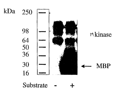
Figure 4 shows the results of an in vitro kinase assay wherein myelin basis
protein (MBP) is used
as the substrate.
Figure 5 shows figures involving a full-length (FL) Mkinase and an N-terminal
deleted (ND)
Mkinase. Particularly, Figures 5A and 5B indicate the approximate kinase
domain and nuclear
localization signal (NLS) of Mkinase wherein 5A is FL and 5B is ND. 5C shows
the results of an in
vitro kinase activity using ND, FL or a control vector and 5D shows the
results of a Western blot
indicating the presence of ND and FL used in 5C.
Figure 6 shows the localization of Mkinase in Hela cells wherein 6A and 6B
show staining with an
anti-flag and Figures 6C and 6D show staining with DAPI.
Figure 7A shows the approximate location of the domain in Mkinase that has
homology to other
known kinases. Figure 7B shows a multiple sequence alignment of Mkinase from
amino acids 1
through 233 against corresponding regions of other known kinases.
DETAILED DESCRIPTION OF THE INVENTION
The present invention provides cell cycle proteins and nucleic acids which
encode such proteins.
Also provided are methods for screening for a bioactive agent capable of
modulating the cell cycle.
The method comprises combining a cell cycle protein and a candidate bioactive
agent and a cell or
a population of cells, and determining the effect on the cell in the presence
and absence of the
candidate agent. Other screening assays including binding assays are also
provided herein as
described below. Therapeutics for regulating or modulating the cell cycle are
also provided and
described herein. Diagnostics, as further described below, are also provided
herein.
A cell cycle protein of the present invention may be identified in several
ways. "Protein" in this
sense includes proteins, polypeptides, and peptides. The cell cycle proteins
of the invention fall
into two general classes: proteins that are completely novel, i.e. are not
part of a public database
as of the time of discovery, although they may have homology to either known
proteins or peptides
encoded by expressed sequence tags (ESTs). Alternatively, the cell cycle
proteins are known
proteins, but that were not known to be involved in the cell cycle; i.e. they
are identified herein as
having a novel biological function. Accordingly, a cell cycle protein may be
initially identified by its
association with a protein known to be involved in the cell cycle. Wherein the
cell cycle proteins
and nucleic acids are novel, compositions and methods of use are provided
herein. In the case
that the cell cycle proteins and nucleic acids were known but not known to be
involved in cell cycle
activity as described herein, methods of use, i.e. functional screens, are
provided.
In one embodiment provided herein, a cell cycle protein as defined herein has
one or more of the
following characteristics: binding to Traf4; homology to protein kinases,
preferably protein kinases
4
CA 02385879 2002-03-21
WO 01/21799 PCT/IJS00/40987
Microbiol., 22(2):293-302 (1996).
In one embodiment, cell cycle nucleic acids or cell cycle proteins are
initially identified by
substantial nucleic acid and/or amino acid sequence identity or similarity to
the sequences)
provided herein. In a preferred embodiment, cell cycle nucleic acids or cell
cycle proteins have
sequence identity or similarity to the sequences provided herein as described
below and one or
more of the cell cycle protein bioactivities as further described below. Such
sequence identity or
similarity can be based upon the overall nucleic acid or amino acid sequence.
In a preferred embodiment, a protein is a "cell cycle protein" as defined
herein if the overall
sequence identity of the amino acid sequence of Figure 2 is preferably greater
than about 75%,
more preferably greater than about 80%, even more preferably greater than
about 85% and most
preferably greater than 90%. In some embodiments the sequence identity will be
as high as about
93 to 95 or 98%.
In another preferred embodiment, a cell cycle protein has an overall sequence
similarity with the
amino acid sequence of Figure 2 of greater than about 80%, more preferably
greater than about
85%, even more preferably greater than about 90% and most preferably greater
than 93%. In
some embodiments the sequence identity will be as high as about 95 to 98 or
99%.
As is known in the art, a number of different programs can be used to identify
whether a protein (or
nucleic acid as discussed below) has sequence identity or similarity to a
known sequence.
Sequence identity and/or similarity is determined using standard techniques
known in the art,
including, but not limited to, the local sequence identity algorithm of Smith
& Waterman, Adv. Appl.
Math. 2:482 (1981 ), by the sequence identity alignment algorithm of Needleman
& Wunsch, J. Mol.
Biool. 48:443 (1970), by the search for similarity method of Pearson & Lipman,
PNAS USA 85:2444
(1988), by computerized implementations of these algorithms (GAP, BESTFIT,
FASTA, and
TFASTA in the Wisconsin Genetics Software Package, Genetics Computer Group,
575 Science
Drive, Madison, WI), the Best Fit sequence program described by Devereux et
al., NucL Acid Res.
72:387-395 (1984), preferably using the default settings, or by inspection.
Preferably, percent
identity is calculated by FastDB based upon the following parameters: mismatch
penalty of 1; gap
penalty of 1; gap size penalty of 0.33; and joining penalty of 30, "Current
Methods in Sequence
Comparison and Analysis," Macromolecule Sequencing and Synthesis, Selected
Methods and
Applications, pp 127-149 (1988), Alan R. Liss, Inc.
An example of a useful algorithm is PILEUP. PILEUP creates a multiple sequence
alignment from
a group of related sequences using progressive, pairwise alignments. It can
also plot a tree
showing the clustering relationships used to create the alignment. PILEUP uses
a simplification of
the progressive alignment method of Feng & Doolittle, J. Mol. Evol. 35:351-360
(1987); the method
is similar to that described by Higgins & Sharp CABIOS 5:151-153 (1989).
Useful PILEUP
6
CA 02385879 2002-03-21
WO 01/21799 PCT/US00/40987
parameters including a default gap weight of 3.00, a default gap length weight
of 0.10, and
weighted end gaps.
Another example of a useful algorithm is the BLAST algorithm, described in
Altschul et al., J. Mol.
Biol. 275, 403-410, (1990) and Karlin et al., PNAS USA 90:5873-5787 (1993). A
particularly useful
BLAST program is the WU-BLAST-2 program which was obtained from Altschul et
al., Methods in
Enzymology, 266: 460-480 (1996); http://blast.wustl/edu/blast/ README.html].
WU-BLAST-2 uses
several search parameters, most of which are set to the default values. The
adjustable parameters
are set with the following values: overlap span =1, overlap fraction = 0.125,
word threshold (T) _
11. The HSP S and HSP S2 parameters are dynamic values and are established by
the program
itself depending upon the composition of the particular sequence and
composition of the particular
database against which the sequence of interest is being searched; however,
the values may be
adjusted to increase sensitivity.
An additional useful algorithm is gapped BLAST as reported by Altschul et al.
Nucleic Acids Res.
25:3389-3402. Gapped BLAST uses BLOSUM-62 substitution scores; threshold T
parameter set
to 9; the two-hit method to trigger ungapped extensions; charges gap lengths
of k a cost of 10+k;
X~ set to 16, and X9 set to 40 for database search stage and to 67 for the
output stage of the
algorithms. Gapped alignments are triggered by a score corresponding to -22
bits.
A % amino acid sequence identity value is determined by the number of matching
identical
residues divided by the total number of residues of the "longer" sequence in
the aligned region.
The "longer" sequence is the one having the most actual residues in the
aligned region (gaps
introduced by WU-Blast-2 to maximize the alignment score are ignored).
In a similar manner, "percent (%) nucleic acid sequence identity" with respect
to the coding
sequence of the polypeptides identified herein is defined as the percentage of
nucleotide residues
in a candidate sequence that are identical with the nucleotide residues in the
coding sequence of
the cell cycle protein. A preferred method utilizes the BLASTN module of WU-
BLAST-2 set to the
default parameters, with overlap span and overlap fraction set to 1 and 0.125,
respectively.
The alignment may include the introduction of gaps in the sequences to be
aligned. In addition, for
sequences which contain either more or fewer amino acids than the protein
encoded by the
sequences in the Figures, it is understood that in one embodiment, the
percentage of sequence
identity will be determined based on the number of identical amino acids in
relation to the total
number of amino acids. Thus, for example, sequence identity of sequences
shorter than that
shown in the Figure, as discussed below, will be determined using the number
of amino acids in
the shorter sequence, in one embodiment. In percent identity calculations
relative weight is not
assigned to various manifestations of sequence variation, such as, insertions,
deletions,
substitutions, etc.
7
CA 02385879 2002-03-21
WO 01/21799 PCT/US00/40987
In one embodiment, only identities are scored positively (+1 ) and all forms
of sequence variation
including gaps are assigned a value of "0", which obviates the need for a
weighted scale or
parameters as described below for sequence similarity calculations. Percent
sequence identity can
be calculated, for example, by dividing the number of matching identical
residues by the total
number of residues of the "shorter" sequence in the aligned region and
multiplying by 100. The
°'longer" sequence is the one having the most actual residues in the
aligned region.
As will be appreciated by those skilled in the art, the sequences of the
present invention may
contain sequencing errors. That is, there may be incorrect nucleosides,
frameshifts, unknown
nucleosides, or other types of sequencing errors in any of the sequences;
however, the correct
sequences will fall within the homology and stringency definitions herein.
Cell cycle proteins of the present invention may be shorter or longer than the
amino acid sequence
encoded by the nucleic acid shown in the Figure. Thus, in a preferred
embodiment, included within
the definition of cell cycle proteins are portions or fragments of the amino
acid sequence encoded
by the nucleic acid sequence provided herein. In one embodiment herein,
fragments of cell cycle
proteins are considered cell cycle proteins if a) they share at least one
antigenic epitope; b) have at
least the indicated sequence identity; c) and preferably have cell cycle
biological activity as further
defined herein. In some cases, where the sequence is used diagnostically, that
is, when the
presence or absence of cell cycle protein nucleic acid is determined, only the
indicated sequence
identity is required. The nucleic acids of the present invention may also be
shorter or longer than
the sequence in the Figure. The nucleic acid fragments include any portion of
the nucleic acids
provided herein which have a sequence not exactly previously identified;
fragments having
sequences with the indicated sequence identity to that portion not previously
identified are provided
in an embodiment herein.
In addition, as is more fully outlined below, cell cycle proteins can be made
that are longer than
those depicted in the Figure; for example, by the addition of epitope or
purification tags, the
addition of other fusion sequences, or the elucidation of additional coding
and non-coding
sequences. As described below, the fusion of a cell cycle peptide to a
fluorescent peptide, such as
Green Fluorescent Peptide (GFP), is particularly preferred.
Cell cycle proteins may also be identified as encoded by cell cycle nucleic
acids which hybridize to
the sequence depicted in the Figure, or the complement thereof, as outlined
herein. Hybridization
conditions are further described below.
In a preferred embodiment, when a cell cycle protein is to be used to generate
antibodies, a cell
cycle protein must share at least one epitope or determinant with the full
length protein. By
"epitope" or "determinant" herein is meant a portion of a protein which will
generate and/or bind an
antibody. Thus, in most instances, antibodies made to a smaller cell cycle
protein will be able to
8
CA 02385879 2002-03-21
WO 01/21799 PCT/US00/40987
bind to the full length protein. In a preferred embodiment, the epitope is
unique; that is, antibodies
generated to a unique epitope show little or no cross-reactivity. The term
"antibody" includes
antibody fragments, as are known in the art, including Fab Fabz, single chain
antibodies (Fv for
example), chimeric antibodies, etc., either produced by the modification of
whole antibodies or
those synthesized de novo using recombinant DNA technologies.
In a preferred embodiment, the antibodies to a cell cycle protein are capable
of reducing or
eliminating the biological function of the cell cycle proteins described
herein, as is described below.
That is, the addition of anti-cell cycle protein antibodies (either polyclonal
or preferably monoclonal)
to cell cycle proteins (or cells containing cell cycle proteins) may reduce or
eliminate the cell cycle
activity. Generally, at least a 25% decrease in activity is preferred, with at
least about 50% being
particularly preferred and about a 95-100% decrease being especially
preferred.
The cell cycle antibodies of the invention specifically bind to cell cycle
proteins. In a preferred
embodiment, the antibodies specifically bind to cell cycle proteins. By
"specifically bind" herein is
meant that the antibodies bind to the protein with a binding constant in the
range of at least 10~-
10-6 M-', with a preferred range being 10'' - 10'9 M''. Antibodies are further
described below.
In the case of the nucleic acid, the overall sequence identity of the nucleic
acid sequence is
commensurate with amino acid sequence identity but takes into account the
degeneracy in the
genetic code and codon bias of different organisms. Accordingly, the nucleic
acid sequence
identity may be either lower or higher than that of the protein sequence. Thus
the sequence
identity of the nucleic acid sequence as compared to the nucleic acid sequence
of the Figure is
preferably greater than 75%, more preferably greater than about 80%,
particularly greater than
about 85% and most preferably greater than 90%. In some embodiments the
sequence identity will
be as high as about 93 to 95 or 98%.
In a preferred embodiment, a cell cycle nucleic acid encodes a cell cycle
protein. As will be
appreciated by those in the art, due to the degeneracy of the genetic code, an
extremely large
number of nucleic acids may be made, all of which encode the cell cycle
proteins of the present
invention. Thus, having identified a particular amino acid sequence, those
skilled in the art could
make any number of different nucleic acids, by simply modifying the sequence
of one or more
codons in a way which does not change the amino acid sequence of the cell
cycle protein.
In one embodiment, the nucleic acid is determined through hybridization
studies. Thus, for
example, nucleic acids which hybridize under high stringency to the nucleic
acid sequence shown
in the Figure, or its complement is considered a cell cycle nucleic acid. High
stringency conditions
are known in the art; see for example Maniatis et al., Molecular Cloning: A
Laboratory Manual, 2d
Edition, 1989, and Short Protocols in Molecular Biology, ed. Ausubel, et al.,
both of which are
hereby incorporated by reference. Stringent conditions are sequence-dependent
and will be
9
CA 02385879 2002-03-21
WO 01/21799 PCT/US00/40987
different in different circumstances. Longer sequences hybridize specifically
at higher
temperatures. An extensive guide to the hybridization of nucleic acids is
found in Tijssen,
Techniques in Biochemistry and Molecular Biology--Hybridization with Nucleic
Acid Probes,
"Overview of principles of hybridization and the strategy of nucleic acid
assays" (1993). Generally,
stringent conditions are selected to be about 5-10'C lower than the thermal
melting point (Tm) for
the specific sequence at a defined ionic strength pH. The Tm is the
temperature (under defined
ionic strength, pH and nucleic acid concentration) at which 50% of the probes
complementary to
the target hybridize to the target sequence at equilibrium (as the target
sequences are present in
excess, at Tm, 50% of the probes are occupied at equilibrium). Stringent
conditions will be those in
which the salt concentration is less than about 1.0 sodium ion, typically
about 0.01 to 1.0 M sodium
ion concentration (or other salts) at pH 7.0 to 8.3 and the temperature is at
least about 30'C for
short probes (e.g. 10 to 50 nucleotides) and at least about 60'C for long
probes (e.g. greater than
50 nucleotides). Stringent conditions may also be achieved with the addition
of destabilizing
agents such as formamide.
In another embodiment, less stringent hybridization conditions are used; for
example, moderate or
low stringency conditions may be used, as are known in the art; see Maniatis
and Ausubel, supra,
and Tijssen, supra.
The cell cycle proteins and nucleic acids of the present invention are
preferably recombinant. As
used herein and further defined below, "nucleic acid" may refer to either DNA
or RNA, or molecules
which contain both deoxy- and ribonucleotides. The nucleic acids include
genomic DNA, cDNA
and oligonucleotides including sense and anti-sense nucleic acids. Such
nucleic acids may also
contain modifications in the ribose-phosphate backbone to increase stability
and half life of such
molecules in physiological environments.
The nucleic acid may be double stranded, single stranded, or contain portions
of both double
stranded or single stranded sequence. As will be appreciated by those in the
art, the depiction of a
single strand ("Watson") also defines the sequence of the other strand
("Crick"); thus the
sequences depicted in the Figures also include the complement of the sequence.
By the term
"recombinant nucleic acid" herein is meant nucleic acid, originally formed in
vitro, in general, by the
manipulation of nucleic acid by endonucleases, in a form not normally found in
nature. Thus an
isolated cell cycle nucleic acid, in a linear form, or an expression vector
formed in vitro by ligating
DNA molecules that are not normally joined, are both considered recombinant
for the purposes of
this invention. It is understood that once a recombinant nucleic acid is made
and reintroduced into
a host cell or organism, it will replicate non-recombinantly, i.e. using the
in vivo cellular machinery
of the host cell rather than in vitro manipulations; however, such nucleic
acids, once produced
recombinantly, although subsequently replicated non-recombinantly, are still
considered
recombinant for the purposes of the invention.
CA 02385879 2002-03-21
WO 01/21799 PCT/US00/40987
Similarly, a "recombinant protein" is a protein made using recombinant
techniques, i.e. through the
expression of a recombinant nucleic acid as depicted above. A recombinant
protein is
distinguished from naturally occurring protein by at least one or more
characteristics. For example,
the protein may be isolated or purified away from some or all of the proteins
and compounds with
which it is normally associated in its wild type host, and thus may be
substantially pure. For
example, an isolated protein is unaccompanied by at least some of the material
with which it is
normally associated in its natural state, preferably constituting at least
about 0.5%, more preferably
at least about 5% by weight of the total protein in a given sample. A
substantially pure protein
comprises at least about 75% by weight of the total protein, with at least
about 80% being
preferred, and at least about 90% being particularly preferred. The definition
includes the
production of a cell cycle protein from one organism in a different organism
or host cell.
Alternatively, the protein may be made at a significantly higher concentration
than is normally seen,
through the use of a inducible promoter or high expression promoter, such that
the protein is made
at increased concentration levels. Alternatively, the protein may be in a form
not normally found in
nature, as in the addition of an epitope tag or amino acid substitutions,
insertions and deletions, as
discussed below.
In one embodiment, the present invention provides cell cycle protein variants.
These variants fall
into one or more of three classes: substitutional, insertional or deletional
variants. These variants
ordinarily are prepared by site specific mutagenesis of nucleotides in the DNA
encoding a cell cycle
protein, using cassette or PCR mutagenesis or other techniques well known in
the art, to produce
DNA encoding the variant, and thereafter expressing the DNA in recombinant
cell culture as
outlined above. However, variant cell cycle protein fragments having up to
about 100-150 residues
may be prepared by in vitro synthesis using established techniques. Amino acid
sequence variants
are characterized by the predetermined nature of the variation, a feature that
sets them apart from
naturally occurring allelic or interspecies variation of the cell cycle
protein amino acid sequence.
The variants typically exhibit the same qualitative biological activity as the
naturally occurring
analogue, although variants can also be selected which have modified
characteristics as will be
more fully outlined below.
While the site or region for introducing an amino acid sequence variation is
predetermined, the
mutation per se need not be predetermined. For example, in order to optimize
the performance of
a mutation at a given site, random mutagenesis may be conducted at the target
codon or region
and the expressed cell cycle variants screened for the optimal combination of
desired activity.
Techniques for making substitution mutations at predetermined sites in DNA
having a known
sequence are well known, for example, M13 primer mutagenesis and PCR
mutagenesis.
Screening of the mutants is done using assays of cell cycle protein
activities.
Amino acid substitutions are typically of single residues; insertions usually
will be on the order of
from about 1 to 20 amino acids, although considerably larger insertions may be
tolerated.
11
CA 02385879 2002-03-21
WO 01/21799 PCT/CTS00/40987
Deletions range from about T to about 20 residues, although in some cases
deletions may be much
larger.
Substitutions, deletions, insertions or any combination thereof may be used to
arrive at a final
derivative. Generally these changes are done on a few amino acids to minimize
the alteration of
the molecule. However, larger changes may be tolerated in certain
circumstances. When small
alterations in the characteristics of the cell cycle protein are desired,
substitutions are generally
made in accordance with the following chart:
Chart I
Original Residue Exemplary Substitutions
10Ala Ser
Arg Lys
Asn Gln, His
Asp Glu
Cys Ser
15Gln Asn
Glu Asp
Gly Pro
His Asn, Gln
Ile Leu, Val
20Leu Ile, Val
Lys Arg, Gln, Glu
Met Leu, Ile
Phe Met, Leu, Tyr
Ser Thr
25Thr Ser
Trp Tyr
Tyr Trp, Phe
Val Ile, Leu
Substantial changes in function or immunological identity are made by
selecting substitutions that
30 are less conservative than those shown in Chart I. For example,
substitutions may be made which
more significantly affect: the structure of the polypeptide backbone in the
area of the alteration, for
example the alpha-helical or beta-sheet structure; the charge or
hydrophobicity of the molecule at
the target site; or the bulk of the side chain. The substitutions which in
general are expected to
produce the greatest changes in the polypeptide's properties are those in
which (a) a hydrophilic
35 residue, e.g. seryl or threonyl, is substituted for (or by) a hydrophobic
residue, e.g. leucyl, isoleucyl,
phenylalanyl, valyl or alanyl; (b) a cysteine or proline is substituted for
(or by) any other residue; (c)
a residue having an electropositive side chain, e.g. lysyl, arginyl, or
histidyl, is substituted for (or
12
CA 02385879 2002-03-21
WO 01/21799 PCT/LJS00/40987
by) an electronegative residue, e.g. glutamyl or aspartyl; or (d) a residue
having a bulky side chain,
e.g. phenylalanine, is substituted for (or by) one not having a side chain,
e.g. glycine.
The variants typically exhibit the same qualitative biological activity and
will elicit the same immune
response as the naturally-occurring analogue, although variants also are
selected to modify the
characteristics of the cell cycle proteins as needed. Alternatively, the
variant may be designed
such that the biological activity of the cell cycle protein is altered. For
example, glycosylation sites
may be altered or removed.
Covalent modifications of cell cycle polypeptides are included within the
scope of this invention.
One type of covalent modification includes reacting targeted amino acid
residues of a cell cycle
polypeptide with an organic derivatizing agent that is capable of reacting
with selected side chains
or the N-or C-terminal residues of a cell cycle polypeptide. Derivatization
with bifunctional agents
is useful, for instance, for crosslinking cell cycle to a water-insoluble
support matrix or surface for
use in the method for purifying anti-cell cycle antibodies or screening
assays, as is more fully
described below. Commonly used crosslinking agents include, e.g., 1,1-
bis(diazoacetyl)-2-
phenylethane, glutaraldehyde, N-hydroxysuccinimide esters, for example, esters
with 4-azido-
salicylic acid, homobifunctional imidoesters, including disuccinimidyl esters
such as 3,3'-dithiobis-
(succinimidylpropionate), bifunctional maleimides such as bis-N-maleimido-1,8-
octane and agents
such as methyl-3-[(p-azidophenyl)dithio]propioimidate.
Other modifications include deamidation of glutaminyl and asparaginyl residues
to the
corresponding glutamyl and aspartyl residues, respectively, hydroxylation of
proline and lysine,
phosphorylation of hydroxyl groups of seryl or threonyl residues, methylation
of the "-amino groups
of lysine, arginine, and histidine side chains [T.E. Creighton, Proteins:
Structure and Molecular
Properties, W.H. Freeman & Co., San Francisco, pp. 79-86 (1983)], acetylation
of the N-terminal
amine, and amidation of any C-terminal carboxyl group.
Another type of covalent modification of the cell cycle polypeptide included
within the scope of this
invention comprises altering the native glycosylation pattern of the
polypeptide. "Altering the native
glycosylation pattern" is intended for purposes herein to mean deleting one or
more carbohydrate
moieties found in native sequence cell cycle polypeptide, and/or adding one or
more glycosylation
sites that are not present in the native sequence cell cycle polypeptide.
Addition of glycosylation sites to cell cycle polypeptides may be accomplished
by altering the amino
acid sequence thereof. The alteration may be made, for example, by the
addition of, or substitution
by, one or more serine or threonine residues to the native sequence cell cycle
polypeptide (for O-
linked glycosylation sites). The cell cycle amino acid sequence may optionally
be altered through
changes at the DNA level, particularly by mutating the DNA encoding the cell
cycle polypeptide at
preselected bases such that codons are generated that will translate into the
desired amino acids.
13
CA 02385879 2002-03-21
WO 01/21799 PCT/US00/40987
Another means of increasing the number of carbohydrate moieties on the cell
cycle polypeptide is
by chemical or enzymatic coupling of glycosides to the polypeptide. Such
methods are described
in the art, e.g., in WO 87/05330 published 11 September 1987, and in Aplin and
Wriston, CRC Crit.
Rev. Biochem., pp. 259-306 (1981 ).
Removal of carbohydrate moieties present on the cell cycle polypeptide may be
accomplished
chemically or enzymatically or by mutational substitution of codons encoding
for amino acid
residues that serve as targets for glycosylation. Chemical deglycosylation
techniques are known in
the art and described, for instance, by Hakimuddin, et al., Arch. Biochem.
Bioohys., 259:52 (1987)
and by Edge et al., Anal. Biochem., 118:131 (1981 ). Enzymatic cleavage of
carbohydrate moieties
on polypeptides can be achieved by the use of a variety of endo-and exo-
glycosidases as
described by Thotakura et al., Meth. Enzymol., 138:350 (1987).
Another type of covalent modification of cell cycle comprises linking the cell
cycle polypeptide to
one of a variety of nonproteinaceous polymers, e.g., polyethylene glycol,
polypropylene glycol, or
polyoxyalkylenes, in the manner set forth in U.S. Patent Nos. 4,640,835;
4,496,689; 4,301,144;
4,670,417; 4,791,192 or4,179,337.
Cell cycle polypeptides of the present invention may also be modified in a way
to form chimeric
molecules comprising a cell cycle polypeptide fused to another, heterologous
polypeptide or amino
acid sequence. In one embodiment, such a chimeric molecule comprises a fusion
of a cell cycle
polypeptide with a tag polypeptide which provides an epitope to which an anti-
tag antibody can
selectively bind. The epitope tag is generally placed at the amino-or carboxyl-
terminus of the cell
cycle polypeptide. The presence of such epitope-tagged forms of a cell cycle
polypeptide can be
detected using an antibody against the tag polypeptide. Also, provision of the
epitope tag enables
the cell cycle polypeptide to be readily purified by affinity purification
using an anti-tag antibody or
another type of affinity matrix that binds to the epitope tag. In an
alternative embodiment, the
chimeric molecule may comprise a fusion of a cell cycle polypeptide with an
immunoglobulin or a
particular region of an immunoglobulin. For a bivalent form of the chimeric
molecule, such a fusion
could be to the Fc region of an IgG molecule as discussed further below.
Various tag polypeptides and their respective antibodies are well known in the
art. Examples
include poly-histidine (poly-his) or poly-histidine-glycine (poly-his-gly)
tags; the flu HA tag
polypeptide and its antibody 12CA5 [Field et al., Mol. Cell. Biol., 8:2159-
2165 (1988)]; the c-myc
tag and the 8F9, 3C7, 6E10, G4, B7 and 9E10 antibodies thereto [Evan et al.,
Molecular and
Cellular Biology, 5:3610-3616 (1985)]; and the Herpes Simplex virus
glycoprotein D (gD) tag and
its antibody [Paborsky et al., Protein Engineering, 3(6):547-553 (1990)].
Other tag polypeptides
include the Flag-peptide [Hopp et al., BioTechnolocty, 6:1204-1210 (1988)];
the KT3 epitope
peptide [Martin et al., Science, 255:192-194 (1992)]; tubulin epitope peptide
[Skinner et al., J. Biol.
Chem., 266:15163-15166 (1991 )]; and the T7 gene 10 protein peptide tag [Lutz-
Freyermuth et al.,
14
CA 02385879 2002-03-21
WO 01/21799 PCT/US00/40987
Proc. Natl. Acad. Sci. USA, 87:6393-6397 (1990)].
In an embodiment herein, cell cycle proteins of the cell cycle family and cell
cycle proteins from
other organisms are cloned and expressed as outlined below. Thus, probe or
degenerate
polymerase chain reaction (PCR) primer sequences may be used to find other
related cell cycle
proteins from humans or other organisms. As will be appreciated by those in
the art, particularly
useful probe and/or PCR primer sequences include the unique areas of the cell
cycle nucleic acid
sequence. As is generally known in the art, preferred PCR primers are from
about 15 to about 35
nucleotides in length, with from about 20 to about 30 being preferred, and may
contain inosine as
needed. The conditions for the PCR reaction are well known in the art. It is
therefore also
understood that provided along with the sequences in the sequences listed
herein are portions of
those sequences, wherein unique portions of 15 nucleotides or more are
particularly preferred.
The skilled artisan can routinely synthesize or cut a nucleotide sequence to
the desired length.
Once isolated from its natural source, e.g., contained within a plasmid or
other vector or excised
therefrom as a linear nucleic acid segment, the recombinant cell cycle nucleic
acid can be further-
used as a probe to identify and isolate other cell cycle nucleic acids. It can
also be used as a
"precursor" nucleic acid to make modified or variant cell cycle nucleic acids
and proteins.
Using the nucleic acids of the present invention which encode a cell cycle
protein, a variety of
expression vectors are made. The expression vectors may be either self-
replicating
extrachromosomal vectors or vectors which integrate into a host genome.
Generally, these
expression vectors include transcriptional and translational regulatory
nucleic acid operably linked
to the nucleic acid encoding the cell cycle protein. The term "control
sequences" refers to DNA
sequences necessary for the expression of an operably linked coding sequence
in a particular host
organism. The control sequences that are suitable for prokaryotes, for
example, include a
promoter, optionally an operator sequence, and a ribosome binding site.
Eukaryotic cells are
known to utilize promoters, polyadenylation signals, and enhancers.
Nucleic acid is "operably linked" when it is placed into a functional
relationship with another nucleic
acid sequence. For example, DNA for a presequence or secretory leader is
operably linked to
DNA for a polypeptide if it is expressed as a preprotein that participates in
the secretion of the
polypeptide; a promoter or enhancer is operably linked to a coding sequence if
it affects the
transcription of the sequence; or a ribosome binding site is operably linked
to a coding sequence if
it is positioned so as to facilitate translation. As another example, operably
linked refers to DNA
sequences linked so as to be contiguous, and, in the case of a secretory
leader, contiguous and in
reading phase. However, enhancers do not have to be contiguous. Linking is
accomplished by
ligation at convenient restriction sites. If such sites do not exist, the
synthetic oligonucleotide
adaptors or linkers are used in accordance with conventional practice. The
transcriptional and
translational regulatory nucleic acid will generally be appropriate to the
host cell used to express
CA 02385879 2002-03-21
WO 01/21799 PCT/US00/40987
the cell cycle protein; for example, transcriptional and translational
regulatory nucleic acid
sequences from Bacillus are preferably used to express the cell cycle protein
in Bacillus.
Numerous types of appropriate expression vectors, and suitable regulatory
sequences are known
in the art for a variety of host cells.
In general, the transcriptional and translational regulatory sequences may
include, but are not
limited to, promoter sequences, ribosomal binding sites, transcriptional start
and stop sequences,
translational start and stop sequences, and enhancer or activator sequences.
In a preferred
embodiment, the regulatory sequences include a promoter and transcriptional
start and stop
sequences.
Promoter sequences encode either constitutive or inducible promoters. The
promoters may be
either naturally occurring promoters or hybrid promoters. Hybrid promoters,
which combine
elements of more than one promoter, are also known in the art, and are useful
in the present
invention.
In addition, the expression vector may comprise additional elements. For
example, the expression
vector may have two replication systems, thus allowing it to be maintained in
two organisms, for
example in mammalian or insect cells for expression and in a procaryotic host
for cloning and
amplification. Furthermore, for integrating expression vectors, the expression
vector contains at
least one sequence homologous to the host cell genome, and preferably two
homologous
sequences which flank the expression construct. The integrating vector may be
directed to a
specific locus in the host cell by selecting the appropriate homologous
sequence for inclusion in the
vector. Constructs for integrating vectors are well known in the art.
In addition, in a preferred embodiment, the expression vector contains a
selectable marker gene to
allow the selection of transformed host cells. Selection genes are well known
in the art and will
vary with the host cell used.
A preferred expression vector system is a retroviral vector system such as is
generally described in
PCT/US97/01019 and PCT/US97/01048, both of which are hereby expressly
incorporated by
reference.
Cell cycle proteins of the present invention are produced by culturing a host
cell transformed with
an expression vector containing nucleic acid encoding a cell cycle protein,
under the appropriate
conditions to induce or cause expression of the cell cycle protein. The
conditions appropriate for
cell cycle protein expression will vary with the choice of the expression
vector and the host cell,
and will be easily ascertained by one skilled in the art through routine
experimentation. For
example, the use of constitutive promoters in the expression vector will
require optimizing the
growth and proliferation of the host cell, while the use of an inducible
promoter requires the
16
CA 02385879 2002-03-21
WO 01/21799 PCT/US00/40987
appropriate growth conditions for induction. In addition, in some embodiments,
the timing of the
harvest is important. For example, the baculoviral systems used in insect cell
expression are lytic
viruses, and thus harvest time selection can be crucial for product yield.
Appropriate host cells include yeast, bacteria, archebacteria, fungi, and
insect and animal cells,
including mammalian cells. Of particular interest are Drosophila melangaster
cells,
Saccharomyces cerevisiae and other yeasts, E. coli, Bacillus subtilis, SF9
cells, C129 cells, 293
cells, Neurospora, BHK, CHO, COS, and HeLa cells, fibroblasts, Schwanoma cell
lines,
immortalized mammalian myeloid and lymphoid cell lines and tumor cell lines.
In a preferred embodiment, the cell cycle proteins are expressed in mammalian
cells. Mammalian
expression systems are also known in the art, and include retroviral systems.
A mammalian
promoter is any DNA sequence capable of binding mammalian RNA polymerase and
initiating the
downstream (3') transcription of a coding sequence for cell cycle protein into
mRNA. A promoter
will have a transcription initiating region, which is usually placed proximal
to the 5' end of the coding
sequence, and a TATA box, using a located 25-30 base pairs upstream of the
transcription
initiation site. The TATA box is thought to direct RNA polymerase II to begin
RNA synthesis at the
correct site. A mammalian promoter will also contain an upstream promoter
element (enhancer
element), typically located within 100 to 200 base pairs upstream of the TATA
box. An upstream
promoter element determines the rate at which transcription is initiated and
can act in either
orientation. Of particular use as mammalian promoters are the promoters from
mammalian viral
genes, since the viral genes are often highly expressed and have a broad host
range. Examples
include the SV40 early promoter, mouse mammary tumor virus LTR promoter,
adenovirus major
late promoter, herpes simplex virus promoter, and the CMV promoter.
Typically, transcription termination and polyadenylation sequences recognized
by mammalian cells
are regulatory regions located 3' to the translation stop codon and thus,
together with the promoter
elements, flank the coding sequence. The 3' terminus of the mature mRNA is
formed by site-
specific post-translational cleavage and polyadenylation. Examples of
transcription terminator and
polyadenlytion signals include those derived form SV40.
The methods of introducing exogenous nucleic acid into mammalian hosts, as
well as other hosts,
is well known in the art, and will vary with the host cell used. Techniques
include dextran-mediated
transfection, calcium phosphate precipitation, polybrene mediated
transfection, protoplast fusion,
electroporation, viral infection, encapsulation of the polynucleotide(s) in
liposomes, and direct
microinjection of the DNA into nuclei.
In a preferred embodiment, cell cycle proteins are expressed in bacterial
systems. Bacterial
expression systems are well known in the art.
17
CA 02385879 2002-03-21
WO 01/21799 PCT/US00/40987
A suitable bacterial promoter is any nucleic acid sequence capable of binding
bacterial RNA
polymerase and initiating the downstream (3') transcription of the coding
sequence of cell cycle
protein into mRNA. A bacterial promoter has a transcription initiation region
which is usually placed
proximal to the 5' end of the coding sequence. This transcription initiation
region typically includes
an RNA polymerase binding site and a transcription initiation site. Sequences
encoding metabolic
pathway enzymes provide particularly useful promoter sequences. Examples
include promoter
sequences derived from sugar metabolizing enzymes, such as galactose, lactose
and maltose, and
sequences derived from biosynthetic enzymes such as tryptophan. Promoters from
bacteriophage
may also be used and are known in the art. In addition, synthetic promoters
and hybrid promoters
are also useful; for example, the fac promoter is a hybrid of the trp and lac
promoter sequences.
Furthermore, a bacterial promoter can include naturally occurring promoters of
non-bacterial origin
that have the ability to bind bacterial RNA polymerase and initiate
transcription.
In addition to a functioning promoter sequence, an efficient ribosome binding
site is desirable. In E.
coli, the ribosome binding site is called the Shine-Delgarno (SD) sequence and
includes an
initiation codon and a sequence 3-9 nucleotides in length located 3 - 11
nucleotides upstream of
the initiation codon.
The expression vector may also include a signal peptide sequence that provides
for secretion of
the cell cycle protein in bacteria. The signal sequence typically encodes a
signal peptide
comprised of hydrophobic amino acids which direct the secretion of the protein
from the cell, as is
well known in the art. The protein is either secreted into the growth media
(gram-positive bacteria)
or into the periplasmic space, located between the inner and outer membrane of
the cell (gram-
negative bacteria).
The bacterial expression vector may also include a selectable marker gene to
allow for the
selection of bacterial strains that have been transformed. Suitable selection
genes include genes
which render the bacteria resistant to drugs such as ampicillin,
chloramphenicol, erythromycin,
kanamycin, neomycin and tetracycline. Selectable markers also include
biosynthetic genes, such
as those in the histidine, tryptophan and leucine biosynthetic pathways.
These components are assembled into expression vectors. Expression vectors for
bacteria are
well known in the art, and include vectors for Bacillus subtilis, E. coli,
Streptococcus cremoris, and
Streptococcus lividans, among others.
The bacterial expression vectors are transformed into bacterial host cells
using techniques well
known in the art, such as calcium chloride treatment, electroporation, and
others.
In one embodiment, cell cycle proteins are produced in insect cells.
Expression vectors for the
transformation of insect cells, and in particular, baculovirus-based
expression vectors, are well
18
CA 02385879 2002-03-21
WO 01/21799 PCT/US00/40987
known in the art.
In a preferred embodiment, cell cycle protein is produced in yeast cells.
Yeast expression systems
are well known in the art, and include expression vectors for Saccharomyces
cerevisiae, Candida
albicans and C. maltosa, Hansenula polymorpha, Kluyveromyces fragilis and K.
lactis, Pichia
guillerimondii and P. pastoris, Schizosaccharomyces pombe, and Yarrowia
lipolytica. Preferred
promoter sequences for expression in yeast include the inducible GAL1,10
promoter, the
promoters from alcohol dehydrogenase, enolase, glucokinase, glucose-6-
phosphate isomerase,
glyceraldehyde-3-phosphate-dehydrogenase, hexokinase, phosphofructokinase, 3-
phosphoglycerate mutase, pyruvate kinase, and the acid phosphatase gene. Yeast
selectable
markers include ADE2, HIS4, LEU2, TRP1, and ALG7, which confers resistance to
tunicamycin;
the neomycin phosphotransferase gene, which confers resistance to 6418; and
the CUP1 gene,
which allows yeast to grow in the presence of copper ions.
The cell cycle protein may also be made as a fusion protein, using techniques
well known in the
art. Thus, for example, for the creation of monoclonal antibodies, if the
desired epitope is small,
the cell cycle protein may be fused to a carrier protein to form an immunogen.
Alternatively, the
cell cycle protein may be made as a fusion protein to increase expression, or
for other reasons.
For example, when the cell cycle protein is a cell cycle peptide, the nucleic
acid encoding the
peptide may be linked to other nucleic acid for expression purposes.
Similarly, cell cycle proteins
of the invention can be linked to protein labels, such as green fluorescent
protein (GFP), red
fluorescent protein (RFP), blue fluorescent protein (BFP), yellow fluorescent
protein (YFP), etc.
In one embodiment, the cell cycle nucleic acids, proteins and antibodies of
the invention are
labeled. By "labeled" herein is meant that a compound has at least one
element, isotope or
chemical compound attached to enable the detection of the compound. In
general, labels fall into
three classes: a) isotopic labels, which may be radioactive or heavy isotopes;
b) immune labels,
which may be antibodies or antigens; and c) colored or fluorescent dyes. The
labels may be
incorporated into the compound at any position.
In a preferred embodiment, the cell cycle protein is purified or isolated
after expression. Cell cycle
proteins may be isolated or purified in a variety of ways known to those
skilled in the art depending
on what other components are present in the sample. Standard purification
methods include
electrophoretic, molecular, immunological and chromatographic techniques,
including ion
exchange, hydrophobic, affinity, and reverse-phase HPLC chromatography, and
chromatofocusing.
For example, the cell cycle protein may be purified using a standard anti-cell
cycle antibody
column. Ultrafiltration and diafiltration techniques, in conjunction with
protein concentration, are
also useful. For general guidance in suitable purification techniques, see
Scopes, R., Protein
Purification, Springer-Verlag, NY (1982). The degree of purification necessary
will vary depending
on the use of the cell cycle protein. In some instances no purification will
be necessary.
19
CA 02385879 2002-03-21
WO 01/21799 PCT/US00/40987
Once expressed and purified~if necessary, the cell cycle proteins and nucleic
acids are useful in a
number of applications.
The nucleotide sequences (or their complement) encoding cell cycle proteins
have various
applications in the art of molecular biology, including uses as hybridization
probes, in chromosome
and gene mapping and in the generation of anti-sense RNA and DNA. Cell cycle
protein nucleic
acid will also be useful for the preparation of cell cycle proteins by the
recombinant techniques
described herein.
The full-length native sequence cell cycle protein gene, or portions thereof,
may be used as
hybridization probes for a cDNA library to isolate other genes (for instance,
those encoding
naturally-occurring variants of cell cycle protein or cell cycle protein from
other species) which have
a desired sequence identity to the cell cycle protein coding sequence.
Optionally, the length of the
probes will be about 20 to about 50 bases. The hybridization probes may be
derived from the
nucleotide sequences herein or from genomic sequences including promoters,
enhancer elements
and introns of native sequences as provided herein. By way of example, a
screening method will
comprise isolating the coding region of the cell cycle protein gene using the
known DNA sequence
to synthesize a selected probe of about 40 bases. Hybridization probes may be
labeled by a
variety of labels, including radionucleotides such as 32P or 35S, or enzymatic
labels such as alkaline
phosphatase coupled to the probe via avidin/biotin coupling systems. Labeled
probes having a
sequence complementary to that of the cell cycle protein gene of the present
invention can be used
to screen libraries of human cDNA, genomic DNA or mRNA to determine which
members of such
libraries the probe hybridizes.
Nucleotide sequences encoding a cell cycle protein can also be used to
construct hybridization
probes for mapping the gene which encodes that cell cycle protein and for the
genetic analysis of
individuals with genetic disorders. The nucleotide sequences provided herein
may be mapped to a
chromosome and specific regions of a chromosome using known techniques, such
as in situ
hybridization, linkage analysis against known chromosomal markers, and
hybridization screening
with libraries.
Nucleic acids which encode cell cycle protein or its modified forms can also
be used to generate
either transgenic animals or "knock out" animals which, in turn, are useful in
the development and
screening of therapeutically useful reagents. A transgenic animal (e.g., a
mouse or rat) is an
animal having cells that contain a transgene, which transgene was introduced
into the animal or an
ancestor of the animal at a prenatal, e.g., an embryonic stage. A transgene is
a DNA which is
integrated into the genome of a cell from which a transgenic animal develops.
In one embodiment,
cDNA encoding a cell cycle protein can be used to clone genomic DNA encoding a
cell cycle
protein in accordance with established techniques and the genomic sequences
used to generate
transgenic animals that contain cells which express the desired DNA. Methods
for generating
CA 02385879 2002-03-21
WO 01/21799 PCT/US00/40987
transgenic animals, particularly animals such as mice or rats, have become
conventional in the art
and are described, for example, in U.S. Patent Nos. 4,736,866 and 4,870,009.
Typically, particular
cells would be targeted for the cell cycle protein transgene incorporation
with tissue-specific
enhancers. Transgenic animals that include a copy of a transgene encoding a
cell cycle protein
introduced into the germ line of the animal at an embryonic stage can be used
to examine the
effect of increased expression of the desired nucleic acid. Such animals can
be used as tester
animals for reagents thought to confer protection from, for example,
pathological conditions
associated with its overexpression. In accordance with this facet of the
invention, an animal is
treated with the reagent and a reduced incidence of the pathological
condition, compared to
untreated animals bearing the transgene, would indicate a potential
therapeutic intervention for the
pathological condition.
Alternatively, non-human homologues of the cell cycle protein can be used to
construct a cell cycle
protein "knock out" animal which has a defective or altered gene encoding a
cell cycle protein as a
result of homologous recombination between the endogenous gene encoding a cell
cycle protein
and altered genomic DNA encoding a cell cycle protein introduced into an
embryonic cell of the
animal. For example, cDNA encoding a cell cycle protein can be used to clone
genomic DNA
encoding a cell cycle protein in accordance with established techniques. A
portion of the genomic
DNA encoding a cell cycle protein can be deleted or replaced with another
gene, such as a gene
encoding a selectable marker which can be used to monitor integration.
Typically, several
kilobases of unaltered flanking DNA (both at the 5' and 3' ends) are included
in the vector [see e.g.,
Thomas and Capecchi, Cell, 51:503 (1987) for a description of homologous
recombination
vectors]. The vector is introduced into an embryonic stem cell line (e.g., by
electroporation) and
cells in which the introduced DNA has homologously recombined with the
endogenous DNA are
selected [see e.g., Li et al., Cell, 69:915 (1992)]. The selected cells are
then injected into a
blastocyst of an animal (e.g., a mouse or rat) to form aggregation chimeras
[see e.g., Bradley, in
Teratocarcinomas and Embryonic Stem Cells: A Practical Approach, E. J.
Robertson, ed. (IRL,
Oxford, 1987), pp. 113-152]. A chimeric embryo can then be implanted into a
suitable
pseudopregnant female foster animal and the embryo brought to term to create a
"knock out"
animal. Progeny harboring the homologously recombined DNA in their germ cells
can be identified
by standard techniques and used to breed animals in which all cells of the
animal contain the
homologously recombined DNA. Knockout animals can be characterized for
instance, for their
ability to defend against certain pathological conditions and for their
development of pathological
conditions due to absence of the cell cycle protein.
It is understood that the models described herein can be varied. For example,
"knock-in" models
can be formed, or the models can be cell-based rather than animal models.
Nucleic acid encoding the cell cycle polypeptides, antagonists or agonists may
also be used in
gene therapy. In gene therapy applications, genes are introduced into cells in
order to achieve in
21
CA 02385879 2002-03-21
WO 01/21799 PCT/US00/40987
vivo synthesis of a therapeutically effective genetic product, for example for
replacement of a
defective gene. "Gene therapy" includes both conventional gene therapy where a
lasting effect is
achieved by a single treatment, and the administration of gene therapeutic
agents, which involves
the one time or repeated administration of a therapeutically effective DNA or
mRNA. Antisense
RNAs and DNAs can be used as therapeutic agents for blocking the expression of
certain genes in
vivo. It has already been shown that short antisense oligonucleotides can be
imported into cells
where they act as inhibitors, despite their low intracellular concentrations
caused by their restricted
uptake by the cell membrane. (Zamecnik et al., Proc. Natl. Acad. Sci. USA 83,
4143-4146 [1986]).
The oligonucleotides can be modified to enhance their uptake, e.g. by
substituting their negatively
charged phosphodiester groups by uncharged groups.
There are a variety of techniques available for introducing nucleic acids into
viable cells. The
techniques vary depending upon whether the nucleic acid is transferred into
cultured cells in vitro,
or in vivo in the cells of the intended host. Techniques suitable for the
transfer of nucleic acid into
mammalian cells in vitro include the use of liposomes, electroporation,
microinjection, cell fusion,
DEAE-dextran, the calcium phosphate precipitation method, etc. The currently
preferred in vivo
gene transfer techniques include transfection with viral (typically
retroviral) vectors and viral coat
protein-liposome mediated transfection (Dzau et al., Trends in Biotechnolocw
11, 205-210 [1993]).
In some situations it is desirable to provide the nucleic acid source with an
agent that targets the
target cells, such as an antibody specific for a cell surface membrane protein
or the target cell, a
ligand for a receptor on the target cell, etc. Where liposomes are employed,
proteins which bind to
a cell surface membrane protein associated with endocytosis may be used for
targeting and/or to
facilitate uptake, e.g. capsid proteins or fragments thereof tropic for a
particular cell type,
antibodies for proteins which undergo internalization in cycling, proteins
that target intracellular
localization and enhance intracellular half-life. The technique of receptor-
mediated endocytosis is
described, for example, by Wu et al., J. Biol. Chem. 262, 4429-4432 (1987);
and Wagner et al.,
Proc. Natl. Acad. Sci. USA 87, 3410-3414 (1990). For review of gene marking
and gene therapy
protocols see Anderson et al., Science 256, 808-813 (1992).
In a preferred embodiment, the cell cycle proteins, nucleic acids, variants,
modified proteins, cells
and/or transgenics containing the said nucleic acids or proteins are used in
screening assays.
Identification of the cell cycle protein provided herein permits the design of
drug screening assays
for compounds that bind or interfere with the binding to the cell cycle
protein and for compounds
which modulate cell cycle activity.
The assays described herein preferably utilize the human cell cycle protein,
although other
mammalian proteins may also be used, including rodents (mice, rats, hamsters,
guinea pigs, etc.),
farm animals (cows, sheep, pigs, horses, etc.) and primates. These latter
embodiments may be
preferred in the development of animal models of human disease. In some
embodiments, as
outlined herein, variant or derivative cell cycle proteins may be used,
including deletion cell cycle
22
CA 02385879 2002-03-21
WO 01/21799 PCT/US00/40987
proteins as outlined above.
In a preferred embodiment, the methods comprise combining a cell cyle protein
and a candidate
bioactive agent, and determining the binding of the candidate agent to the
cell cycle protein. In
other embodiments, further discussed below, binding interference or
bioactivity is determined.
The term "candidate bioactive agent" or "exogeneous compound" as used herein
describes any
molecule, e.g., protein, small organic molecule, carbohydrates (including
polysaccharides),
polynucleotide, lipids, etc. Generally a plurality of assay mixtures are run
in parallel with different
agent concentrations to obtain a differential response to the various
concentrations. Typically, one
of these concentrations serves as a negative control, i.e., at zero
concentration or below the level
of detection. In addition, positive controls, i.e. the use of agents known to
alter cell cycling, may be
used. For example, p21 is a molecule known to arrest cells in the G1 cell
phase, by binding G1
cyclin-CDK complexes.
Candidate agents encompass numerous chemical classes, though typically they
are organic
molecules, preferably small organic compounds having a molecular weight of
more than 100 and
less than about 2,500 daltons. Candidate agents comprise functional groups
necessary for
structural interaction with proteins, particularly hydrogen bonding, and
typically include at least an
amine, carbonyl, hydroxyl or carboxyl group, preferably at least two of the
functional chemical
groups. The candidate agents often comprise cyclical carbon or heterocyclic
structures and/or
aromatic or polyaromatic structures substituted with one or more of the above
functional groups.
Candidate agents are also found among biomolecules including peptides,
saccharides, fatty acids,
steroids, purines, pyrimidines, derivatives, structural analogs or
combinations thereof. Particularly
preferred are peptides.
Candidate agents are obtained from a wide variety of sources including
libraries of synthetic or
natural compounds. For example, numerous means are available for random and
directed
synthesis of a wide variety of organic compounds and biomolecules, including
expression of
randomized oligonucleotides. Alternatively, libraries of natural compounds in
the form of bacterial,
fungal, plant and animal extracts are available or readily produced.
Additionally, natural or
synthetically produced libraries and compounds are readily modified through
conventional
chemical, physical and biochemical means. Known pharmacological agents may be
subjected to
directed or random chemical modifications, such as acylation, alkylation,
esterification,
amidification to produce structural analogs.
In a preferred embodiment, a library of different candidate bioactive agents
are used. Preferably,
the library should provide a sufficiently structurally diverse population of
randomized agents to
effect a probabilistically sufficient range of diversity to allow binding to a
particular target.
Accordingly, an interaction library should be large enough so that at least
one of its members will
23
CA 02385879 2002-03-21
WO 01/21799 PCT/US00/40987
have a structure that gives it affinity for the target. Although it is
difficult to gauge the required
absolute size of an interaction library, nature provides a hint with the
immune response: a diversity
of 10'-108 different antibodies provides at least one combination with
sufficient affinity to interact
with most potential antigens faced by an organism. Published in vitro
selection techniques have
also shown that a library size of 10' to 108 is sufficient to find structures
with affinity for the target.
A library of all combinations of a peptide 7 to 20 amino acids in length, such
as generally proposed
herein, has the potential to code for 20' (109) to 202° . Thus, with
libraries of 10' to 108 different
molecules the present methods allow a "working" subset of a theoretically
complete interaction
library for 7 amino acids, and a subset of shapes for the 202° library.
Thus, in a preferred
embodiment, at least 106, preferably at least 10', more preferably at least
108 and most preferably
at least 109 different sequences are simultaneously analyzed in the subject
methods. Preferred
methods maximize library size and diversity.
In a preferred embodiment, the candidate bioactive agents are proteins. By
"protein" herein is
meant at least two covalently attached amino acids, which includes proteins,
polypeptides,
oligopeptides and peptides. The protein may be made up of naturally occurring
amino acids and
peptide bonds, or synthetic peptidomimetic structures. Thus "amino acid", or
"peptide residue", as
used herein means both naturally occurring and synthetic amino acids. For
example, homo-
phenylalanine, citrulline and noreleucine are considered amino acids for the
purposes of the
invention. "Amino acid" also includes imino acid residues such as proline and
hydroxyproline.
The side chains may be in either the (R) or the (S) configuration. In the
preferred embodiment, the
amino acids are in the (S) or L-configuration. If non-naturally occurring side
chains are used, non-
amino acid substituents may be used, for example to prevent or retard in vivo
degradations.
Chemical blocking groups or other chemical substituents may also be added.
In a preferred embodiment, the candidate bioactive agents are naturally
occurring proteins or
fragments of naturally occurring proteins. Thus, for example, cellular
extracts containing proteins,
or random or directed digests of proteinaceous cellular extracts, may be used.
In this way libraries
of procaryotic and eukaryotic proteins may be made for screening in the
systems described herein.
Particularly preferred in this embodiment are libraries of bacterial, fungal,
viral, and mammalian
proteins, with the latter being preferred, and human proteins being especially
preferred.
In a preferred embodiment, the candidate bioactive agents are peptides of from
about 5 to about 30
amino acids, with from about 5 to about 20 amino acids being preferred, and
from about 7 to about
15 being particularly preferred. The peptides may be digests of naturally
occurring proteins as is
outlined above, random peptides, or "biased" random peptides. By "randomized"
or grammatical
equivalents herein is meant that each nucleic acid and peptide consists of
essentially random
nucleotides and amino acids, respectively. Since generally these random
peptides (or nucleic
acids, discussed below) are chemically synthesized, they may incorporate any
nucleotide or amino
acid at any position. The synthetic process can be designed to generate
randomized proteins or
24
CA 02385879 2002-03-21
WO 01/21799 PCT/US00/40987
nucleic acids, to allow the formation of all or most of the possible
combinations over the length of
the sequence, thus forming a library of randomized candidate bioactive
proteinaceous agents.
In one embodiment, the library is fully randomized, with no sequence
preferences or constants at
any position. In a preferred embodiment, the library is biased. That is, some
positions within the
sequence are either held constant, or are selected from a limited number of
possibilities. For
example, in a preferred embodiment, the nucleotides or amino acid residues are
randomized within
a defined class, for example, of hydrophobic amino acids, hydrophilic
residues, sterically biased
(either small or large) residues, towards the creation of cysteines, for cross-
linking, prolines for SH-
3 domains, serines, threonines, tyrosines or histidines for phosphorylation
sites, etc., or to purines,
1 0 etc.
In a preferred embodiment, the candidate bioactive agents are nucleic acids.
By "nucleic acid" or
"oligonucleotide" or grammatical equivalents herein means at least two
nucleotides covalently
linked together. A nucleic acid of the present invention will generally
contain phosphodiester
bonds, although in some cases, as outlined below, nucleic acid analogs are
included that may
have alternate backbones, comprising, for example, phosphoramide (Beaucage, et
al.,
Tetrahedron, 49(10):1925 (1993) and references therein; Letsinger, J. 0r4.
Chem., 35:3800
(1970); Sprinzl, et al., Eur. J. Biochem., 81:579 (1977); Letsinger, et al.,
Nucl. Acids Res., 14:3487
(1986); Sawai, et al., Chem. Lett., 805 (1984), Letsinger, et al., J. Am.
Chem. Soc., 110:4470
(1988); and Pauwels, et al., Chemica Scripts, 26:141 (1986)), phosphorothioate
(Mag, et al.,
Nucleic Acids Res., 19:1437 (1991 ); and U.S. Patent No. 5,644,048),
phosphorodithioate (Briu, et
aL, J. Am. Chem. Soc., 111:2321 (1989)), O-methylphophoroamidite linkages (see
Eckstein,
Oligonucleotides and Analogues: A Practical Approach, Oxford University
Press), and peptide
nucleic acid backbones and linkages (see Egholm, J. Am. Chem. Soc., 114:1895
(1992); Meier, et
al., Chem. Int. Ed. Engl., 31:1008 (1992); Nielsen, Nature, 365:566 (1993);
Carlsson, et al., Nature,
380:207 (1996), all of which are incorporated by reference)). Other analog
nucleic acids include
those with positive backbones (Denpcy, et al., Proc. Natl. Acad. Sci. USA,
92:6097 (1995)); non-
ionic backbones (U.S. Patent Nos. 5,386,023; 5,637,684; 5,602,240; 5,216,141;
and 4,469,863;
Kiedrowshi, et al., Anaew. Chem. Intl. Ed. English, 30:423 (1991 ); Letsinger,
et al., J. Am. Chem.
Soc., 110:4470 (1988); Letsinger, et al., Nucleoside & Nucleotide, 13:1597
(1994); Chapters 2 and
3, ASC Symposium Series 580, "Carbohydrate Modifications in Antisense
Research", Ed. Y.S.
Sanghui and P. Dan Cook; Mesmaeker, et al., Bioorctanic & Medicinal Chem.
Lett., 4:395 (1994);
Jeffs, et al., J. Biomolecular NMR, 34:17 (1994); Tetrahedron Lett., 37:743
(1996)) and non-ribose
backbones, including those described in U.S. Patent Nos. 5,235,033 and
5,034,506, and Chapters
6 and 7, ASC Symposium Series 580, "Carbohydrate Modifications in Antisense
Research", Ed.
Y.S. Sanghui and P. Dan Cook. Nucleic acids containing one or more carbocyclic
sugars are also
included within the definition of nucleic acids (see Jenkins, et al., Chem.
Soc. Rev., (1995) pp. 169-
176). Several nucleic acid analogs are described in Rawls, C & E News, June 2,
1997, page 35.
All of these references are hereby expressly incorporated by reference. These
modifications of the
CA 02385879 2002-03-21
WO 01/21799 PCT/US00/40987
ribose-phosphate backbone may be done to facilitate the addition of additional
moieties such as
labels, or to increase the stability and half-life of such molecules in
physiological environments. In
addition, mixtures of naturally occurring nucleic acids and analogs can be
made. Alternatively,
mixtures of different nucleic acid analogs, and mixtures of naturally
occurring nucleic acids and
analogs may be made. The nucleic acids may be single stranded or double
stranded, as specified,
or contain portions of both double stranded or single stranded sequence. The
nucleic acid may be
DNA, both genomic and cDNA, RNA or a hybrid, where the nucleic acid contains
any combination
of deoxyribo- and ribo-nucleotides, and any combination of bases, including
uracil, adenine,
thymine, cytosine, guanine, inosine, xathanine hypoxathanine, isocytosine,
isoguanine, etc.
As described above generally for proteins, nucleic acid candidate bioactive
agents may be
naturally occurring nucleic acids, random nucleic acids, or "biased" random
nucleic acids. For
example, digests of procaryotic or eukaryotic genomes may be used as is
outlined above for
proteins.
In a preferred embodiment, the candidate bioactive agents are organic chemical
moieties, a wide
variety of which are available in the literature.
In a preferred embodiment, the candidate bioactive agents are linked to a
fusion partner. By
"fusion partner" or "functional group" herein is meant a sequence that is
associated with the
candidate bioactive agent, that confers upon all members of the library in
that class a common
function or ability. Fusion partners can be heterologous (i.e. not native to
the host cell), or
synthetic (not native to any cell). Suitable fusion partners include, but are
not limited to: a)
presentation structures, which provide the candidate bioactive agents in a
conformationally
restricted or stable form; b) targeting sequences, which allow the
localization of the candidate
bioactive agent into a subcellular or extracellular compartment; c) rescue
sequences which allow
the purification or isolation of either the candidate bioactive agents or the
nucleic acids encoding
them; d) stability sequences, which confer stability or protection from
degradation to the candidate
bioactive agent or the nucleic acid encoding it, for example resistance to
proteolytic degradation; e)
dimerization sequences, to allow for peptide dimerization; or f) any
combination of a), b), c), d), and
e), as well as linker sequences as needed.
In one embodiment of the methods described herein, portions of cell cycle
proteins are utilized; in a
preferred embodiment, portions having cell cycle activity are used. Cell cycle
activity is described
further below and includes binding activity to a TRAF protein or cell cycle
protein modulators as
further described below. In addition, the assays described herein may utilize
either isolated cell
cycle proteins or cells comprising the cell cycle proteins.
Generally, in a preferred embodiment of the methods herein, for example for
binding assays, the
cell cycle protein or the candidate agent is non-diffusibly bound to an
insoluble support having
26
CA 02385879 2002-03-21
WO 01/21799 PCT/US00/40987
isolated sample receiving areas (e.g. a microtiter plate, an array, etc.). The
insoluble supports
may be made of any composition to which the compositions can be bound, is
readily separated
from soluble material, and is otherwise compatible with the overall method of
screening. The
surface of such supports may be solid or porous and of any convenient shape.
Examples of
suitable insoluble supports include microtiter plates, arrays, membranes and
beads. These are
typically made of glass, plastic (e.g., polystyrene), polysaccharides, nylon
or nitrocellulose,
teflonT"', etc. Microtiter plates and arrays are especially convenient because
a large number of
assays can be carried out simultaneously, using small amounts of reagents and
samples. In some
cases magnetic beads and the like are included. The particular manner of
binding of the
composition is not crucial so long as it is compatible with the reagents and
overall methods of the
invention, maintains the activity of the composition and is nondiffusable.
Preferred methods of
binding include the use of antibodies (which do not sterically block either
the ligand binding site or
activation sequence when the protein is bound to the support), direct binding
to "sticky" or ionic
supports, chemical crosslinking, the synthesis of the protein or agent on the
surface, etc. In some
embodiments, Traf4 can be used. Following binding of the protein or agent,
excess unbound
material is removed by washing. The sample receiving areas may then be blocked
through
incubation with bovine serum albumin (BSA), casein or other innocuous protein
or other moiety.
Also included in this invention are screening assays wherein solid supports
are not used; examples
of such are described below.
In a preferred embodiment, the cell cycle protein is bound to the support, and
a candidate bioactive
agent is added to the assay. Alternatively, the candidate agent is bound to
the support and the cell
cycle protein is added. Novel binding agents include specific antibodies, non-
natural binding
agents identified in screens of chemical libraries, peptide analogs, etc. Of
particular interest are
screening assays for agents that have a low toxicity for human cells. A wide
variety of assays may
be used for this purpose, including labeled in vitro protein-protein binding
assays, electrophoretic
mobility shift assays, immunoassays for protein binding, functional assays
(phosphorylation
assays, etc.) and the like.
The determination of the binding of the candidate bioactive agent to the cell
cycle protein may be
done in a number of ways. In a preferred embodiment, the candidate bioactive
agent is labelled,
and binding determined directly. For example, this may be done by attaching
all or a portion of the
cell cycle protein to a solid support, adding a labelled candidate agent (for
example a fluorescent
label), washing off excess reagent, and determining whether the label is
present on the solid
support. Various blocking and washing steps may be utilized as is known in the
art.
By "labeled" herein is meant that the compound is either directly or
indirectly labeled with a label
which provides a detectable signal, e.g. radioisotope, fluorescers, enzyme,
antibodies, particles
such as magnetic particles, chemiluminescers, or specific binding molecules,
etc. Specific binding
molecules include pairs, such as biotin and streptavidin, digoxin and
antidigoxin etc. For the
27
CA 02385879 2002-03-21
WO 01/21799 PCT/US00/40987
specific binding members, the complementary member would normally be labeled
with a molecule
which provides for detection, in accordance with known procedures, as outlined
above. The label
can directly or indirectly provide a detectable signal.
In some embodiments, only one of the components is labeled. For example, the
proteins (or
proteinaceous candidate agents) may be labeled at tyrosine positions using
'251, or with
tluorophores. Alternatively, more than one component may be labeled with
different labels; using
'251 for the proteins, for example, and a fluorophor for the candidate agents.
In a preferred embodiment, the binding of the candidate bioactive agent is
determined through the
use of competitive binding assays. In this embodiment, the competitor is a
binding moiety known
to bind to the target molecule (i.e. cell cycle protein), such as an antibody,
peptide, binding partner,
ligand, etc. In a preferred embodiment, the competitor is Traf4. Under certain
circumstances, there
may be competitive binding as between the bioactive agent and the binding
moiety, with the
binding moiety displacing the bioactive agent. This assay can be used to
determine candidate
agents which interfere with binding between cell cycle proteins and Traf4.
"Interference of binding"
as used herein means that native binding of the cell cycle protein differs in
the presence of the
candidate agent. The binding can be eliminated or can be with a reduced
affinity. Therefore, in
one embodiment, interference is caused by, for example, a conformation change,
rather than direct
competition for the native binding site.
In one embodiment, the candidate bioactive agent is labeled. Either the
candidate bioactive agent,
or the competitor, or both, is added first to the protein for a time
sufficient to allow binding, if
present. Incubations may be performed at any temperature which facilitates
optimal activity,
typically between 4 and 40°C. Incubation periods are selected for
optimum activity, but may also
be optimized to facilitate rapid high through put screening. Typically between
0.1 and 1 hour will be
sufficient. Excess reagent is generally removed or washed away. The second
component is then
added, and the presence or absence of the labeled component is followed, to
indicate binding.
In a preferred embodiment, the competitor is added first, followed by the
candidate bioactive agent.
Displacement of the competitor is an indication that the candidate bioactive
agent is binding to the
cell cycle protein and thus is capable of binding to, and potentially
modulating, the activity of the
cell cycle protein. In this embodiment, either component can be labeled. Thus,
for example, if the
competitor is labeled, the presence of label in the wash solution indicates
displacement by the
agent. Alternatively, if the candidate bioactive agent is labeled, the
presence of the label on the
support indicates displacement.
In an alternative embodiment, the candidate bioactive agent is added first,
with incubation and
washing, followed by the competitor. The absence of binding by the competitor
may indicate that
the bioactive agent is bound to the cell cycle protein with a higher affinity.
Thus, if the candidate
28
CA 02385879 2002-03-21
WO 01/21799 PCT/US00/40987
bioactive agent is labeled, the presence of the label on the support, coupled
with a lack of
competitor binding, may indicate that the candidate agent is capable of
binding to the cell cycle
protein.
In a preferred embodiment, the methods comprise differential screening to
identity bioactive agents
that are capable of modulating the activity of the cell cycle proteins. Such
assays can be done with
the cell cycle protein or cells comprising said cell cycle protein. In one
embodiment, the methods
comprise combining an cell cycle protein and a competitor in a first sample. A
second sample
comprises a candidate bioactive agent, an cell cycle protein and a competitor.
The binding of the
competitor is determined for both samples, and a change, or difference in
binding between the two
samples indicates the presence of an agent capable of binding to the cell
cycle protein and
potentially modulating its activity. That is, if the binding of the competitor
is different in the second
sample relative to the first sample, the agent is capable of binding to the
cell cycle protein.
Alternatively, a preferred embodiment utilizes differential screening to
identify drug candidates that
bind to the native cell cycle protein, but cannot bind to modified cell cycle
proteins. The structure of
the cell cycle protein may be modeled, and used in rational drug design to
synthesize agents that
interact with that site. Drug candidates that affect cell cycle bioactivity
are also identified by
screening drugs for the ability to either enhance or reduce the activity of
the protein.
Positive controls and negative controls may be used in the assays. Preferably
all control and test
samples are performed in at least triplicate to obtain statistically
significant results. Incubation of all
samples is for a time sufficient for the binding of the agent to the protein.
Following incubation, all
samples are washed free of non-specifically bound material and the amount of
bound, generally
labeled agent determined. For example, where a radiolabel is employed, the
samples may be
counted in a scintillation counter to determine the amount of bound compound.
A variety of other reagents may be included in the screening assays. These
include reagents like
salts, neutral proteins, e.g. albumin, detergents, etc which may be used to
facilitate optimal
protein-protein binding and/or reduce non-specific or background interactions.
Also reagents that
otherwise improve the efficiency of the assay, such as protease inhibitors,
nuclease inhibitors,
anti-microbial agents, etc., may be used. The mixture of components may be
added in any order
that provides for the requisite binding.
Screening for agents that modulate the activity of cell cycle may also be
done. In a preferred
embodiment, methods for screening for a bioactive agent capable of modulating
the activity of cell
cycle comprise the steps of adding a candidate bioactive agent to a sample of
a cell cycle protein
(or cells comprising a cell cycle protein) and determining an alteration in
the biological activity of
the cell cycle protein. "Modulating the activity of a cell cycle protein"
includes an increase in
activity, a decrease in activity, or a change in the type or kind of activity
present. Thus, in this
29
CA 02385879 2002-03-21
WO 01/21799 PCT/US00/40987
embodiment, the candidate agent should both bind to cell cycle (although this
may not be
necessary), and alter its biological or biochemical activity as defined
herein. The methods include
both in vitro screening methods, as are generally outlined above, and in vivo
screening of cells for
alterations in the presence, distribution, activity or amount of cell cycle
protein.
Thus, in this embodiment, the methods comprise combining an cell cycle sample
and a candidate
bioactive agent, and evaluating the effect on the cell cycle. By "cell cycle
protein activity" or
grammatical equivalents herein is meant one or more of the cell cycle
protein's biological activities,
including, but not limited to, its ability to affect the cell cycle, bind to a
TRAF protein, phosphorylate
proteins associated with microtubules and/or cell cycling, affect cell
proliferation, mediate
apoptosis, mediate cellular activation, and/or modulate pathways involved in
tumor growth.
In a preferred embodiment, the activity of the cell cycle protein is
decreased; in another preferred
embodiment, the activity of the cell cycle protein is increased. Thus,
bioactive agents that are
antagonists are preferred in some embodiments, and bioactive agents that are
agonists may be
preferred in other embodiments.
In a preferred embodiment, the invention provides methods for screening for
bioactive agents
capable of modulating the activity of an cell cycle protein. The methods
comprise adding a
candidate bioactive agent, as defined above, to a cell comprising cell cycle
proteins. Preferred cell
types include almost any cell. The cells contain a recombinant nucleic acid
that encodes an cell
cycle protein. In a preferred embodiment, a library of candidate agents are
tested on a plurality of
cells.
Detection of cell cycle regulation may be done as will be appreciated by those
in the art. In one
embodiment, indicators of the cell cycle are used. There are a number of
parameters that may be
evaluated or assayed to allow the detection of alterations in cell cycle
regulation, including, but not
limited to, cell viability assays, assays to determine whether cells are
arrested at a particular cell
cycle stage ("cell proliferation assays"), and assays to determine at which
cell stage the cells have
arrested ("cell phase assays"). By assaying or measuring one or more of these
parameters, it is
possible to detect not only alterations in cell cycle regulation, but
alterations of different steps of
the cell cycle regulation pathway. This may be done to evaluate native cells,
for example to
quantify the aggressiveness of a tumor cell type, or to evaluate the effect of
candidate drug agents
that are being tested for their effect on cell cycle regulation. In this
manner, rapid, accurate
screening of candidate agents may be performed to identify agents that
modulate cell cycle
regulation.
Thus, the present compositions and methods are useful to elucidate bioactive
agents that can
cause a cell or a population of cells to either move out of one growth phase
and into another, or
arrest in a growth phase. In some embodiments, the cells are arrested in a
particular growth
CA 02385879 2002-03-21
WO 01/21799 PCT/L1S00/40987
phase, and it is desirable to either get them out of that phase or into a new
phase. Alternatively, it
may be desirable to force a cell to arrest in a phase, for example G1, rather
than continue to move
through the cell cycle. Similarly, it may be desirable in some circumstances
to accelerate a non-
arrested but slowly moving population of cells into either the next phase or
just through the cell
cycle, or to delay the onset of the next phase. For example, it may be
possible to alter the
activities of certain enzymes, for example kinases, phosphatases, proteases or
ubiquitination
enzymes, that contribute to initiating cell phase changes.
In a preferred embodiment, the methods outlined herein are done on cells that
are not arrested in
the G1 phase; that is, they are rapidly or uncontrollably growing and
replicating, such as tumor
cells. In this manner, candidate agents are evaluated to find agents that can
alter the cell cycle
regulation, i.e. cause the cells to arrest at cell cycle checkpoints, such as
in G1 (although arresting
in other phases such as S, G2 or M are also desirable). Alternatively,
candidate agents are
evaluated to find agents that can cause proliferation of a population of
cells, i.e. that allow cells that
are generally arrested in G1 to start proliferating again; for example,
peripheral blood cells,
terminally differentiated cells, stem cells in culture, etc.
Accordingly, the invention provides methods for screening for alterations in
cell cycle regulation of
a population of cells. By "alteration" or "modulation" (used herein
interchangeably), is generally
meant one of two things. In a preferred embodiment, the alteration results in
a change in the cell
cycle of a cell, i.e. a proliferating cell arrests in any one of the phases,
or an arrested cell moves
out of its arrested phase and starts the cell cycle, as compared to another
cell or in the same cell
under different conditions. Alternatively, the progress of a cell through any
particular phase may be
altered; that is, there may be an acceleration or delay in the length of time
it takes for the cells to
move thorough a particular growth phase. For example, the cell may be normally
undergo a G1
phase of several hours; the addition of an agent may prolong the G1 phase.
The measurements can be determined wherein all of the conditions are the same
for each
measurement, or under various conditions, with or without bioactive agents, or
at different stages
of the cell cycle process. For example, a measurement of cell cycle regulation
can be determined
in a cell or cell population wherein a candidate bioactive agent is present
and wherein the
candidate bioactive agent is absent. In another example, the measurements of
cell cycle
regulation are determined wherein the condition or environment of the cell or
populations of cells
differ from one another. For example, the cells may be evaluated in the
presence or absence or
previous or subsequent exposure of physiological signals, for example
hormones, antibodies,
peptides, antigens, cytokines, growth factors, action potentials,
pharmacological agents including
chemotherapeutics, radiation, carcinogenics, or other cells (i.e. cell-cell
contacts). In another
example, the measurements of cell cycle regulation are determined at different
stages of the cell
cycle process. In yet another example, the measurements of cell cycle
regulation are taken
wherein the conditions are the same, and the alterations are between one cell
or cell population
31
CA 02385879 2002-03-21
WO 01/21799 PCT/LTS00/40987
and another cell or cell population.
By a "population of cells" or "library of cells" herein is meant at least two
cells, with at least about
103 being preferred, at least about 106 being particularly preferred, and at
least about 108 to 109
being especially preferred. The population or sample can contain a mixture of
different cell types
from either primary or secondary cultures although samples containing only a
single cell type are
preferred, for example, the sample can be from a cell line, particularly tumor
cell lines, as outlined
below. The cells may be in any cell phase, either synchronously or not,
including M, G1, S, and
G2. In a preferred embodiment, cells that are replicating or proliferating are
used; this may allow
the use of retroviral vectors for the introduction of candidate bioactive
agents. Alternatively, non-
replicating cells may be used, and other vectors (such as adenovirus and
lentivirus vectors) can be
used. In addition, although not required, the cells are compatible with dyes
and antibodies.
Preferred cell types for use in the invention include, but are not limited to,
mammalian cells,
including animal (rodents, including mice, rats, hamsters and gerbils),
primates, and human cells,
particularly including tumor cells of all types, including breast, skin, lung,
cervix, colonrectal,
leukemia, brain, etc.
In a preferred embodiment, the methods comprise assaying one or more of
several different cell
parameters, including, but not limited to, cell viability, cell proliferation,
and cell phase. Other
parameters include TRAF protein binding, phosphorylation activity,
particularly of those proteins
associated with microtubules and/or cell cycling, apoptosis inducement or
regulation, cellular
activation, and/or involvement in pathways involved in tumor growth. In
another embodiment,
Mkinase may act to recruit a protein having phosphorylation (kinase) activity.
For example,
Mkinase may complex, associate or induce a protein which has kinase activity.
In yet another embodiment, Mkinase binds to any one of the following: Homo
sapiens mRNA for
CDC23- cell division cycle protein; RCC1- antioxidant protein 1 (AOP1 ); Novel
associated with
ARF; Human helicase II (RAD54L); Apurinic/apyrimidinic endonuclease (HAP);
Novel with
homology to ELK1 at DNA level; Homo sapiens lipoprotein receptor-related
protein- AM2 receptor;
LDL-receptorlow density lipoprotein-related protein; Homo sapiens putative G
protein-coupled
receptor; Human glycoprotein receptor gp330 precursor; A novel transmembrane
protein in
prostate cancer, TENB2; MOAT-D multidrug resistance-associate; evectin-1-
contains PH domain;
Homo sapiens nel- PKC binding protein; TPRD1- contains tetratricopeptide
repeat; Homo sapiens
GAGA-B1 a receptor; Homo sapiens mRNA for nuclear receptor co-repressor (hN-
CoR); Ig-like
protein- CD33L2; von Willebrand factor; Homo sapiens RIG-like 7-1 mRNA; Novel
Zn finger
protein associated with p27; Human menin (MEN1) gene- multiple endocrine
neoplasia-type 1;
KIAA1097- contains ubiquitin protease domain; Human family of notch2; Human
Notch3; Human
adenylate kinase 1 (hAK1 ); Human phosphate cytidylytransferase; H.sapiens
mRNA for
epithelin/granulin; Human c-erb-B-2 mRNA; Homo sapiens WD40 protein Ciao1;
Novel GTP-
32
CA 02385879 2002-03-21
WO 01/21799 PCT/US00/40987
binding protein; KIAA0618; KIAA0863; KIAA1064; and KIAA0275.
In a preferred embodiment, cell viability is assayed, to ensure that a lack of
cellular change is due
to experimental conditions (i.e. the introduction of a candidate bioactive
agent) not cell death.
There are a variety of suitable cell viability assays which can be used,
including, but not limited to,
light scattering, viability dye staining, and exclusion dye staining.
In a preferred embodiment, a light scattering assay is used as the viability
assay, as is well known
in the art. For example, when viewed in the FACS, cells have particular
characteristics as
measured by their forward and 90 degree (side) light scatter properties. These
scatter properties
represent the size, shape and granule content of the cells. These properties
account for two
parameters to be measured as a readout for the viability. Briefly, the DNA of
dying or dead cells
generally condenses, which alters the 90' scatter; similarly, membrane
blebbing can alter the
forward scatter. Alterations in the intensity of light scattering, or the cell-
refractive index indicate
alterations in viability.
Thus, in general, for light scattering assays, a live cell population of a
particular cell type is
evaluated to determine it's forward and side scattering properties. This sets
a standard for
scattering that can subsequently be used.
In a preferred embodiment, the viability assay utilizes a viability dye. There
are a number of known
viability dyes that stain dead or dying cells, but do not stain growing cells.
For example, annexin V
is a member of a protein family which displays specific binding to
phospholipid (phosphotidylserine)
in a divalent ion dependent manner. This protein has been widely used for the
measurement of
apoptosis (programmed cell death) as cell surface exposure of
phosphatidylserine is a hallmark
early signal of this process. Suitable viability dyes include, but are not
limited to, annexin, ethidium
homodimer-1, DEAD Red, propidium iodide, SYTOX Green, etc., and others known
in the art; see
the Molecular Probes Handbook of Fluorescent Probes and Research Chemicals,
Haugland, Sixth
Edition, hereby incorporated by reference; see Apoptosis Assay on page 285 in
particular, and
Chapter 16.
Protocols for viability dye staining for cell viability are known, see
Molecular Probes catalog, supra.
In this embodiment, the viability dye such as annexin is labeled, either
directly or indirectly, and
combined with a cell population. Annexin is commercially available, i.e., from
PharMingen, San
Diego, California, or Caltag Laboratories, Millbrae, California. Preferably,
the viability dye is
provided in a solution wherein the dye is in a concentration of about 100
ng/ml to about 500 ng/ml,
more preferably, about 500 ng/ml to about 1 Ng/ml, and most preferably, from
about 1 Ng/ml to
about 5 Ng/ml. In a preferred embodiment, the viability dye is directly
labeled; for example, annexin
may be labeled with a fluorochrome such as fluorecein isothiocyanate (FITC),
Alexa dyes, TRITC,
AMCA, APC, tri-color, Cy-5, and others known in the art or commercially
available. In an alternate
33
CA 02385879 2002-03-21
WO 01/21799 PCT/US00/40987
preferred embodiment, the viability dye is labeled with a first label, such as
a hapten such as biotin,
and a secondary fluorescent label is used, such as fluorescent streptavidin.
Other first and second
labeling pairs can be used as will be appreciated by those in the art.
Once added, the viability dye is allowed to incubate with the cells for a
period of time, and washed,
if necessary. The cells are then sorted as outlined below to remove the non-
viable cells.
In a preferred embodiment, exclusion dye staining is used as the viability
assay. Exclusion dyes
are those which are excluded from living cells, i.e. they are not taken up
passively (they do not
permeate the cell membrane of a live cell). However, due to the permeability
of dead or dying
cells, they are taken up by dead cells. Generally, but not always, the
exclusion dyes bind to DNA,
for example via intercalation. Preferably, the exclusion dye does not
fluoresce, or fluoresces
poorly, in the absence of DNA; this eliminates the need for a wash step.
Alternatively, exclusion
dyes that require the use of a secondary label may also be used. Preferred
exclusion dyes
include, but are not limited to, ethidium bromide; ethidium homodimer-1;
propidium iodine; SYTOX
green nucleic acid stain; Calcein AM, BCECF AM; fluorescein diacetate; TOTO~
and TO-PROT"'
(from Molecular Probes; supra, see chapter 16) and others known in the art.
Protocols for exclusion dye staining for cell viability are known, see the
Molecular Probes catalog,
supra. In general, the exclusion dye is added to the cells at a concentration
of from about 100
ng/ml to about 500 ng/ml, more preferably, about 500 ng/ml to about 1 Ng/ml,
and most preferably,
from about 0.1 ~!g/ml to about 5 Ng/ml, with about 0.5 ~g/ml being
particularly preferred. The cells
and the exclusion dye are incubated for some period of time, washed, if
necessary, and then the
cells sorted as outlined below, to remove non-viable cells from the
population.
In addition, there are other cell viability assays which may be run, including
for example enzymatic
assays, which can measure extracellular enzymatic activity of either live
cells (i.e. secreted
proteases, etc.), or dead cells (i.e. the presence of intracellular enzymes in
the media; for example,
intracellular proteases, mitochondria) enzymes, etc.). See the Molecular
Probes Handbook of
Fluorescent Probes and Research Chemicals, Haugland, Sixth Edition, hereby
incorporated by
reference; see chapter 16 in particular.
In a preferred embodiment, at least one cell viability assay is run, with at
least two different cell
viability assays being preferred, when the fluors are compatible. When only 1
viability assay is run,
a preferred embodiment utilizes light scattering assays (both forward and side
scattering). When
two viability assays are run, preferred embodiments utilize light scattering
and dye exclusion, with
light scattering and viability dye staining also possible, and all three being
done in some cases as
well. Viability assays thus allow the separation of viable cells from non-
viable or dying cells.
In addition to a cell viability assay, a preferred embodiment utilizes a cell
proliferation assay. By
34
CA 02385879 2002-03-21
WO 01/21799 PCT/US00/40987
"proliferation assay" herein is meant an assay that allows the determination
that a cell population is
either proliferating, i.e. replicating, or not replicating.
In a preferred embodiment, the proliferation assay is a dye inclusion assay. A
dye inclusion assay
relies on dilution effects to distinguish between cell phases. Briefly, a dye
(generally a fluorescent
dye as outlined below) is introduced to cells and taken up by the cells. Once
taken up, the dye is
trapped in the cell, and does not diffuse out. As the cell population divides,
the dye is
proportionally diluted. That is, after the introduction of the inclusion dye,
the cells are allowed to
incubate for some period of time; cells that lose fluorescence over time are
dividing, and the cells
that remain fluorescent are arrested in a non-growth phase.
Generally, the introduction of the inclusion dye may be done in one of two
ways. Either the dye
cannot passively enter the cells (e.g. it is charged), and the cells must be
treated to take up the
dye; for example through the use of a electric pulse. Alternatively, the dye
can passively enter the
cells, but once taken up, it is modified such that it cannot diffuse out of
the cells. For example,
enzymatic modification of the inclusion dye may render it charged, and thus
unable to diffuse out of
the cells. For example, the Molecular Probes CeIITrackerT"' dyes are
fluorescent chloromethyl
derivatives that freely diffuse into cells, and then glutathione S-transferase-
mediated reaction
produces membrane impermeant dyes.
Suitable inclusion dyes include, but are not limited to, the Molecular Probes
line of CeIITrackerT""
dyes , including, but not limited to CeIITrackerT"' Blue, CeIITrackerT"'
Yellow-Green, CeIITrackerT""
Green, CeIITrackerT'°' Orange, PKH26 (Sigma), and others known in the
art; see the Molecular
Probes Handbook, supra; chapter 15 in particular.
In general, inclusion dyes are provided to the cells at a concentration
ranging from about
100 ng/ml to about 5 ~g/ml, with from about 500 ng/ml to about 1 Ng/ml being
preferred. A wash
step may or may not be used. In a preferred embodiment, a candidate bioactive
agent is combined
with the cells as described herein. The cells and the inclusion dye are
incubated for some period of
time, to allow cell division and thus dye dilution. The length of time will
depend on the cell cycle
time for the particular cells; in general, at least about 2 cell divisions are
preferred, with at least
about 3 being particularly preferred and at least about 4 being especially
preferred. The cells are
then sorted as outlined below, to create populations of cells that are
replicating and those that are
not. As will be appreciated by those in the art, in some cases, for example
when screening for anti-
proliferation agents, the bright (i.e. fluorescent) cells are collected; in
other embodiments, for
example for screening for proliferation agents, the low fluorescence cells are
collected. Alterations
are determined by measuring the fluorescence at either different time points
or in different cell
populations, and comparing the determinations to one another or to standards.
In a preferred embodiment, the proliferation assay is an antimetabolite assay.
In general,
CA 02385879 2002-03-21
WO 01/21799 PCT/US00/40987
antimetabolite assays find the most use when agents that cause cellular arrest
in G1 or G2 resting
phase is desired. In an antimetabolite proliferation assay, the use of a toxic
antimetabolite that will
kill dividing cells will result in survival of only those cells that are not
dividing. Suitable
antimetabolites include, but are not limited to, standard chemotherapeutic
agents such as
methotrexate, cisplatin, taxol, hydroxyurea, nucleotide analogs such as AraC,
etc. In addition,
antimetabolite assays may include the use of genes that cause cell death upon
expression.
The concentration at which the antimetabolite is added will depend on the
toxicity of the particular
antimetabolite, and will be determined as is known in the art. The
antimetabolite is added and the
cells are generally incubated for some period of time; again, the exact period
of time will depend on
the characteristics and identity of the antimetabolite as well as the cell
cycle time of the particular
cell population. Generally, a time sufficient for at least one cell division
to occur.
In a preferred embodiment, at least one proliferation assay is run, with more
than one being
preferred. Thus, a proliferation assay results in a population of
proliferating cells and a population
of arrested cells. Moreover, other proliferation assays may be used, i.e.,
colorimetric assays
known in the art.
In a preferred embodiment, either after or simultaneously with one or more of
the proliferation
assays outlined above, at least one cell phase assay is done. A "cell phase"
assay determines at
which cell phase the cells are arrested, M, G1, S, or G2.
In a preferred embodiment, the cell phase assay is a DNA binding dye assay.
Briefly, a DNA
binding dye is introduced to the cells, and taken up passively. Once inside
the cell, the DNA
binding dye binds to DNA, generally by intercalation, although in some cases,
the dyes can be
either major or minor groove binding compounds. The amount of dye is thus
directly correlated to
the amount of DNA in the cell, which varies by cell phase; G2 and M phase
cells have twice the
DNA content of G1 phase cells, and S phase cells have an intermediate amount,
depending on at
what point in S phase the cells are. Suitable DNA binding dyes are permeant,
and include, but are
not limited to, Hoechst 33342 and 33258, acridine orange, 7-AAD, LDS 751,
DAPI, and SYTO 16,
Molecular Probes Handbook, supra; chapters 8 and 16 in particular.
In general, the DNA binding dyes are added in concentrations ranging from
about 1 Ng/ml to about
5 ~!g/ml. The dyes are added to the cells and allowed to incubate for some
period of time; the
length of time will depend in part on the dye chosen. In one embodiment,
measurements are taken
immediately after addition of the dye. The cells are then sorted as outlined
below, to create
populations of cells that contain different amounts of dye, and thus different
amounts of DNA; in
this way, cells that are replicating are separated from those that are not. As
will be appreciated by
those in the art, in some cases, for example when screening for anti-
proliferation agents, cells with
the least fluorescence (and thus a single copy of the genome) can be separated
from those that
36
CA 02385879 2002-03-21
WO 01/21799 PCT/CJS00/40987
are replicating and thus contain more than a single genome of DNA. Alterations
are determined by
measuring the fluorescence at either different time points or in different
cell populations, and
comparing the determinations to one another or to standards.
In a preferred embodiment, the cell phase assay is a cyclin destruction assay.
In this embodiment,
prior to screening (and generally prior to the introduction of a candidate
bioactive agent, as outlined
below), a fusion nucleic acid is introduced to the cells. The fusion nucleic
acid comprises nucleic
acid encoding a cyclin destruction box and a nucleic acid encoding a
detectable molecule. "Cyclin
destruction boxes" are known in the art and are sequences that cause
destruction via the
ubiquitination pathway of proteins containing the boxes during particular cell
phases. That is, for
example, G1 cyclins may be stable during G1 phase but degraded during S phase
due to the
presence of a G1 cyclin destruction box. Thus, by linking a cyclin destruction
box to a detectable
molecule, for example green fluorescent protein, the presence or absence of
the detectable
molecule can serve to identify the cell phase of the cell population. In a
preferred embodiment,
multiple boxes are used, preferably each with a different fluor, such that
detection of the cell phase
can occur.
A number of cyclin destruction boxes are known in the art, for example, cyclin
A has a destruction
box comprising the sequence RTVLGVIGD; the destruction box of cyclin B1
comprises the
sequence RTALGDIGN. See Glotzer et al., Nature 349:132-138 (1991 ). Other
destruction boxes
are known as well: YMTVSIIDRFMQDSCVPKKMLQLVGVT (rat cyclin B);
KFRLLQETMYMTVSIIDRFMQNSCVPKK (mouse cyclin B);
RAILIDWLIOVQMKFRLLQETMYMTVS (mouse cyclin B1 ); DRFLQAQLVCRKKLQVVGITALLLASK
(mouse cyclin B2); and MSVLRGKLQLVGTAAMLL (mouse cyclin A2).
The nucleic acid encoding the cyclin destruction box is operably linked to
nucleic acid encoding a
detectable molecule. The fusion proteins are constructed by methods known in
the art. For
example, the nucleic acids encoding the destruction box is ligated to a
nucleic acid encoding a
detectable molecule. By "detectable molecule" herein is meant a molecule that
allows a cell or
compound comprising the detectable molecule to be distinguished from one that
does not contain
it, i.e., an epitope, sometimes called an antigen TAG, a specific enzyme, or a
fluorescent molecule.
Preferred fluorescent molecules include but are not limited to green
fluorescent protein (GFP), blue
fluorescent protein (BFP), yellow fluorescent protein (YFP), red fluorescent
protein (RFP), and
enzymes including luciferase and ~i-galactosidase. When antigen TAGs are used,
preferred
embodiments utilize cell surface antigens. The epitope is preferably any
detectable peptide which
is not generally found on the cytoplasmic membrane, although in some
instances, if the epitope is
one normally found on the cells, increases may be detected, although this is
generally not
preferred. Similarly, enzymatic detectable molecules may also be used; for
example, an enzyme
that generates a novel or chromogenic product.
37
CA 02385879 2002-03-21
WO 01/21799 PCT/US00/40987
Accordingly, the results of sorting after cell phase assays generally result
in at least two
populations of cells that are in different cell phases.
The proteins and nucleic acids provided herein can also be used for screening
purposes wherein
the protein-protein interactions of the cell cycle proteins can be identified.
Genetic systems have
been described to detect protein-protein interactions. The first work was done
in yeast systems,
namely the "yeast two-hybrid" system. The basic system requires a protein-
protein interaction in
order to turn on transcription of a reporter gene. Subsequent work was done in
mammalian cells.
See Fields et al., Nature 340:245 (1989); Vasavada et al., PNAS USA 88:10686
(1991 ); Fearon et
al., PNAS USA 89:7958 (1992); Dang et al., Mol. Cell. Biol. 11:954 (1991 );
Chien et al., PNAS USA
88:9578 (1991); and U.S. Patent Nos. 5,283,173, 5,667,973, 5,468,614,
5,525,490, and 5,637,463.
a preferred system is described in Serial Nos. 09/050,863, filed March 30,
1998 and 09/359,081
filed July 22, 1999, entitled "Mammalian Protein Interaction Cloning System".
For use in
conjunction with these systems, a particularly useful shuttle vector is
described in Serial No.
09/133,944, filed August 14, 1998, entitled "Shuttle Vectors".
In general, two nucleic acids are transformed into a cell, where one is a
"bait" such as the gene
encoding a cell cycle protein or a portion thereof, and the other encodes a
test candidate. Only if
the two expression products bind to one another will an indicator, such as a
fluorescent protein, be
expressed. Expression of the indicator indicates when a test candidate binds
to the cell cycle
protein and can be identified as an cell cycle protein. Using the same system
and the identified cell
cycle proteins the reverse can be performed. Namely, the cell cycle proteins
provided herein can
be used to identify new baits, or agents which interact with cell cycle
proteins. Additionally, the
two-hybrid system can be used wherein a test candidate is added in addition to
the bait and the cell
cycle protein encoding nucleic acids to determine agents which interfere with
the bait, such as
Traf4, and the cell cycle protein.
In one embodiment, a mammalian two-hybrid system is preferred. Mammalian
systems provide
post-translational modifications of proteins which may contribute
significantly to their ability to
interact. In addition, a mammalian two-hybrid system can be used in a wide
variety of mammalian
cell types to mimic the regulation, induction, processing, etc. of specific
proteins within a particular
cell type. For example, proteins involved in a disease state (i.e., cancer,
apoptosis related
disorders) could be tested in the relevant disease cells. Similarly, for
testing of random proteins,
assaying them under the relevant cellular conditions will give the highest
positive results.
Furthermore, the mammalian cells can be tested under a variety of experimental
conditions that
may affect intracellular protein-protein interactions, such as in the presence
of hormones, drugs,
growth factors and cytokines, radiation, chemotherapeutics, cellular and
chemical stimuli, etc., that
may contribute to conditions which can effect protein-protein interactions,
particularly those
involved in cancer.
38
CA 02385879 2002-03-21
WO 01/21799 PCT/US00/40987
Assays involving binding such as the two-hybrid system may take into account
non-specific binding
proteins (NSB).
Expression in various cell types, and assays for cell cycle activity are
described above. The
activity assays, such as having an effect on cell proliferation or
microtubules, can be performed to
confirm the activity of cell cycle proteins which have already been identified
by their sequence
identity/similarity or binding to Traf4 as well as to further confirm the
activity of lead compounds
identified as modulators of Mkinase.
The components provided herein for the assays provided herein may also be
combined to form
kits. The kits can be based on the use of the protein and/or the nucleic acid
encoding the cell cycle
proteins. In one embodiment, other components are provided in the kit. Such
components include
one or more of packaging, instructions, antibodies, and labels. Additional
assays such as those
used in diagnostics are further described below.
In this way, bioactive agents are identified. Compounds with pharmacological
activity are able to
enhance or interfere with the activity of the cell cycle protein. The
compounds having the desired
pharmacological activity may be administered in a physiologically acceptable
carrier to a host, as
further described below.
The present discovery relating to the role of cell cycle proteins in the cell
cycle thus provides
methods for inducing or preventing cell proliferation in cells. In a preferred
embodiment, the cell
cycle proteins, and particularly cell cycle protein fragments, are useful in
the study or treatment of
conditions which are mediated by the cell cycle proteins, i.e. to diagnose,
treat or prevent cell cycle
associated disorders. Thus, "cell cycle associated disorders" or "disease
state" include conditions
involving both insufficient or excessive cell proliferation including for
example, cancer.
Thus, in one embodiment, cell cycle regulation in cells or organisms are
provided. In one
embodiment, the methods comprise administering to a cell or individual in need
thereof, a cell cycle
protein in a therapeutic amount. Alternatively, an anti-cell cycle antibody
that reduces or eliminates
the biological activity of the endogeneous cell cycle protein is administered.
In another
embodiment, a bioactive agent as identified by the methods provided herein is
administered.
Alternatively, the methods comprise administering to a cell or individual a
recombinant nucleic acid
encoding an cell cycle protein. As will be appreciated by those in the art,
this may be accomplished
in any number of ways. In a preferred embodiment, the activity of cell cycle
is increased by
increasing the amount of cell cycle in the cell, for example by overexpressing
the endogeneous cell
cycle or by administering a gene encoding a cell cycle protein, using known
gene-therapy
techniques, for example. In a preferred embodiment, the gene therapy
techniques include the
incorporation of the exogeneous gene using enhanced homologous recombination
(EHR), for
example as described in PCT/US93/03868, hereby incorporated by reference in
its entirety.
39
CA 02385879 2002-03-21
WO 01/21799 PCT/US00/40987
Without being bound by theory, it appears that cell cycle protein is an
important protein in the cell
cycle. Accordingly, disorders based on mutant or variant cell cycle genes may
be determined. In
one embodiment, the invention provides methods for identifying cells
containing variant cell cycle
genes comprising determining all or part of the sequence of at least one
endogeneous cell cycle
genes in a cell. As will be appreciated by those in the art, this may be done
using any number of
sequencing techniques. In a preferred embodiment, the invention provides
methods of identifying
the cell cycle genotype of an individual comprising determining all or part of
the sequence of at
least one cell cycle gene of the individual. This is generally done in at
least one tissue of the
individual, and may include the evaluation of a number of tissues or different
samples of the same
tissue. The method may include comparing the sequence of the sequenced cell
cycle gene to a
known cell cycle gene, i.e. a wild-type gene.
The sequence of all or part of the cell cycle gene can then be compared to the
sequence of a
known cell cycle gene to determine if any differences exist. This can be done
using any number of
known sequence identity programs, such as Bestfit, etc. In a preferred
embodiment, the presence
of a difference in the sequence between the cell cycle gene of the patient and
the known cell cycle
gene is indicative of a disease state or a propensity for a disease state.
In one embodiment, the invention provides methods for diagnosing a cell cycle
related condition in
an individual. The methods comprise measuring the activity of cell cycle in a
tissue from the
individual or patient, which may include a measurement of the amount or
specific activity of a cell
cycle protein. This activity is compared to the activity of cell cycle from
either a unaffected second
individual or from an unaffected tissue from the first individual. When these
activities are different,
the first individual may be at risk for a cell cycle associated disorder. In
this way, for example,
monitoring of various disease conditions may be done, by monitoring the levels
of the protein or the
expression of mRNA therefor. Similarly, expression levels may correlate to the
prognosis.
In one aspect, the expression levels of cell cycle protein genes are
determined in different patient
samples or cells for which either diagnosis or prognosis information is
desired. Gene expression
monitoring is done on genes encoding cell cycle proteins. In one aspect, the
expression levels of
cell cycle protein genes are determined for different cellular states, such as
normal cells and cells
undergoing apoptosis or transformation. By comparing cell cycle protein gene
expression levels in
cells in different states, information including both up- and down-regulation
of cell cycle protein
genes is obtained, which can be used in a number of ways. For example, the
evaluation of a
particular treatment regime may be evaluated: does a chemotherapeutic drug act
to improve the
long-term prognosis in a particular patient. Similarly, diagnosis may be done
or confirmed by
comparing patient samples. Furthermore, these gene expression levels allow
screening of drug
candidates with an eye to mimicking or altering a particular expression level.
This may be done by
making biochips comprising sets of important cell cycle protein genes, such as
those of the present
invention, which can then be used in these screens. These methods can also be
done on the
CA 02385879 2002-03-21
WO 01/21799 PCT/US00/40987
protein basis; that is, protein expression levels of the cell cycle proteins
can be evaluated for
diagnostic purposes or to screen candidate agents. In addition, the cell cycle
protein nucleic acid
sequences can be administered for gene therapy purposes, including the
administration of
antisense nucleic acids, or the cell cycle proteins administered as
therapeutic drugs.
Cell cycle protein sequences bound to biochips include both nucleic acid and
amino acid
sequences as defined above. In a preferred embodiment, nucleic acid probes to
cell cycle protein
nucleic acids (both the nucleic acid sequences having the sequences outlined
in the Figures and/or
the complements thereof) are made. The nucleic acid probes attached to the
biochip are designed
to be substantially complementary to the cell cycle protein nucleic acids,
i.e. the target sequence
(either the target sequence of the sample or to other probe sequences, for
example in sandwich
assays), such that hybridization of the target sequence and the probes of the
present invention
occurs. As outlined below, this complementarity need not be perfect; there may
be any number of
base pair mismatches which will interfere with hybridization between the
target sequence and the
single stranded nucleic acids of the present invention. However, if the number
of mutations is so
great that no hybridization can occur under even the least stringent of
hybridization conditions, the
sequence is not a complementary target sequence. Thus, by "substantially
complementary" herein
is meant that the probes are sufficiently complementary to the target
sequences to hybridize under
normal reaction conditions, particularly high stringency conditions, as
outlined herein.
A "nucleic acid probe" is generally single stranded but can be partially
single and partially double
stranded. The strandedness of the probe is dictated by the structure,
composition, and properties
of the target sequence. In general, the nucleic acid probes range from about 8
to about 100 bases
long, with from about 10 to about 80 bases being preferred, and from about 30
to about 50 bases
being particularly preferred. In some embodiments, much longer nucleic acids
can be used, up to
hundreds of bases (e.g., whole genes).
As will be appreciated by those in the art, nucleic acids can be attached or
immobilized to a solid
support in a wide variety of ways. By "immobilized" and grammatical
equivalents herein is meant
the association or binding between the nucleic acid probe and the solid
support is sufficient to be
stable under the conditions of binding, washing, analysis, and removal as
outlined below. The
binding can be covalent or non-covalent. By "non-covalent binding" and
grammatical equivalents
herein is meant one or more of either electrostatic, hydrophilic, and
hydrophobic interactions.
Included in non-covalent binding is the covalent attachment of a molecule,
such as, streptavidin to
the support and the non-covalent binding of the biotinylated probe to the
streptavidin. By "covalent
binding" and grammatical equivalents herein is meant that the two moieties,
the solid support and
the probe, are attached by at least one bond, including sigma bonds, pi bonds
and coordination
bonds. Covalent bonds can be formed directly between the probe and the solid
support or can be
formed by a cross linker or by inclusion of a specific reactive group on
either the solid support or
the probe or both molecules. Immobilization may also involve a combination of
covalent and non-
41
CA 02385879 2002-03-21
WO 01/21799 PCT/US00/40987
covalent interactions.
In general, the probes are attached to the biochip in a wide variety of ways,
as will be appreciated
by those in the art. As described herein, the nucleic acids can either be
synthesized first, with
subsequent attachment to the biochip, or can be directly synthesized on the
biochip.
The biochip comprises a suitable solid substrate. By "substrate" or "solid
support" or other
grammatical equivalents herein is meant any material that can be modified to
contain discrete
individual sites appropriate for the attachment or association of the nucleic
acid probes and is
amenable to at least one detection method. As will be appreciated by those in
the art, the number
of possible substrates are very large, and include, but are not limited to,
glass and modified or
functionalized glass, plastics (including acrylics, polystyrene and copolymers
of styrene and other
materials, polypropylene, polyethylene, polybutylene, polyurethanes, TetlonJ,
etc.),
polysaccharides, nylon or nitrocellulose, resins, silica or silica-based
materials including silicon
and modified silicon, carbon, metals, inorganic glasses, plastics, etc. In
general, the substrates
allow optical detection and do not appreciably show fluorescence.
In a preferred embodiment, the surface of the biochip and the probe may be
derivatized with
chemical functional groups for subsequent attachment of the two. Thus, for
example, the biochip is
derivatized with a chemical functional group including, but not limited to,
amino groups, carboxy
groups, oxo groups and thiol groups, with amino groups being particularly
preferred. Using these
functional groups, the probes can be attached using functional groups on the
probes. For
example, nucleic acids containing amino groups can be attached to surfaces
comprising amino
groups, for example using linkers as are known in the art; for example, homo-
or hetero-bifunctional
linkers as are well known (see 1994 Pierce Chemical Company catalog, technical
section on
cross-linkers, pages 155-200, incorporated herein by reference). In addition,
in some cases,
additional linkers, such as alkyl groups (including substituted and
heteroalkyl groups) may be used.
In this embodiment, oligonucleotides, corresponding to the nucleic acid probe,
are synthesized as
is known in the art, and then attached to the surface of the solid support. As
will be appreciated by
those skilled in the art, either the 5' or 3' terminus may be attached to the
solid support, or
attachment may be via an internal nucleoside.
In an additional embodiment, the immobilization to the solid support may be
very strong, yet non-
covalent. For example, biotinylated oligonucleotides can be made, which bind
to surfaces
covalently coated with streptavidin, resulting in attachment.
Alternatively, the oligonucleotides may be synthesized on the surface, as is
known in the art. For
example, photoactivation techniques utilizing photopolymerization compounds
and techniques are
used. In a preferred embodiment, the nucleic acids can be synthesized in situ,
using well known
42
CA 02385879 2002-03-21
WO 01/21799 PCT/US00/40987
photolithographic techniques, such as those described in WO 95/25116; WO
95/35505; U.S.
Patent Nos. 5,700,637 and 5,445,934; and references cited within, all of which
are expressly
incorporated by reference; these methods of attachment form the basis of the
Affimetrix
GeneChipT"" technology.
"Differential expression," or grammatical equivalents as used herein, refers
to both qualitative as
well as quantitative differences in the genes' temporal and/or cellular
expression patterns within
and among the cells. Thus, a differentially expressed gene can qualitatively
have its expression
altered, including an activation or inactivation, in, for example, normal
versus apoptotic cell. That is,
genes may be turned on or turned off in a particular state, relative to
another state. As is apparent
to the skilled artisan, any comparison of two or more states can be made. Such
a qualitatively
regulated gene will exhibit an expression pattern within a state or cell type
which is detectable by
standard techniques in one such state or cell type, but is not detectable in
both. Alternatively, the
determination is quantitative in that expression is increased or decreased;
that is, the expression of
the gene is either upregulated, resulting in an increased amount of
transcript, or downregulated,
resulting in a decreased amount of transcript. The degree to which expression
differs need only be
large enough to quantify via standard characterization techniques as outlined
below, such as by
use of Affymetrix GeneChipTM expression arrays, Lockhart, Nature Biotechnology
14:1675-1680
(1996), hereby expressly incorporated by reference. Other techniques include,
but are not limited
to, quantitative reverse transcriptase PCR, Northern analysis and RNase
protection.
As will be appreciated by those in the art, this may be done by evaluation at
either the gene
transcript, or the protein level; that is, the amount of gene expression may
be monitored using
nucleic acid probes to the DNA or RNA equivalent of the gene transcript, and
the quantification of
gene expression levels, or, alternatively, the final gene product itself
(protein) can be monitored, for
example through the use of antibodies to the cell cycle protein and standard
immunoassays
(ELISAs, etc.) or other techniques, including mass spectroscopy assays, 2D gel
electrophoresis
assays, etc.
In another method detection of the mRNA is performed in situ. In this method
permeabilized cells
or tissue samples are contacted with a detestably labeled nucleic acid probe
for sufficient time to
allow the probe to hybridize with the target mRNA. Following washing to remove
the non-
specifically bound probe, the label is detected. For example a digoxygenin
labeled riboprobe (RNA
probe) that is complementary to the mRNA encoding an cell cycle protein is
detected by binding
the digoxygenin with an anti-digoxygenin secondary antibody and developed with
vitro blue
tetrazolium and 5-bromo-4-chloro-3-indoyl phosphate.
In another preferred method, expression of cell cycle protein is performed
using in situ imaging
techniques employing antibodies to cell cycle proteins. In this method cells
are contacted with from
one to many antibodies to the cell cycle protein(s). Following washing to
remove non-specific
43
CA 02385879 2002-03-21
WO 01/21799 PCT/LTS00/40987
antibody binding, the presence of the antibody or antibodies is detected. In
one embodiment the
antibody is detected by incubating with a secondary antibody that contains a
detectable label. In
another method the primary antibody to the cell cycle proteins) contains a
detectable label. In
another preferred embodiment each one of multiple primary antibodies contains
a distinct and
detectable label. This method finds particular use in simultaneous screening
for a plurality of cell
cycle proteins. The label may be detected in a fluorometer which has the
ability to detect and
distinguish emissions of different wavelengths. In addition, a fluorescence
activated cell sorter
(FACS) can be used in this method. As will be appreciated by one of ordinary
skill in the art,
numerous other histological imaging techniques are useful in the invention and
the antibodies can
be used in ELISA, immunoblotting (Western blotting), immunoprecipitation,
BIACORE technology,
and the like.
In one embodiment, the cell cycle proteins of the present invention may be
used to generate
polyclonal and monoclonal antibodies to cell cycle proteins, which are useful
as described herein.
Similarly, the cell cycle proteins can be coupled, using standard technology,
to affinity
chromatography columns. These columns may then be used to purify cell cycle
antibodies. In a
preferred embodiment, the antibodies are generated to epitopes unique to the
cell cycle protein;
that is, the antibodies show little or no cross-reactivity to other proteins.
These antibodies find use
in a number of applications. For example, the cell cycle antibodies may be
coupled to standard
affinity chromatography columns and used to purify cell cycle proteins as
further described below.
The antibodies may also be used as blocking polypeptides, as outlined above,
since they will
specifically bind to the cell cycle protein.
The anti-cell cycle protein antibodies may comprise polyclonal antibodies.
Methods of preparing
polyclonal antibodies are known to the skilled artisan. Polyclonal antibodies
can be raised in a
mammal, for example, by one or more injections of an immunizing agent and, if
desired, an
adjuvant. Typically, the immunizing agent and/or adjuvant will be injected in
the mammal by
multiple subcutaneous or intraperitoneal injections. The immunizing agent may
include the cell
cycle protein or a fusion protein thereof. It may be useful to conjugate the
immunizing agent to a
protein known to be immunogenic in the mammal being immunized. Examples of
such
immunogenic proteins include but are not limited to keyhole limpet hemocyanin,
serum albumin,
bovine thyroglobulin, and soybean trypsin inhibitor. Examples of adjuvants
which may be
employed include Freund's complete adjuvant and MPL-TDM adjuvant
(monophosphoryl Lipid a,
synthetic trehalose dicorynomycolate). The immunization protocol may be
selected by one skilled
in the art without undue experimentation.
The anti-cell cycle protein antibodies may, alternatively, be monoclonal
antibodies. Monoclonal
antibodies may be prepared using hybridoma methods, such as those described by
Kohler and
Milstein, Nature, 256:495 (1975). In a hybridoma method, a mouse, hamster, or
other appropriate
host animal, is typically immunized with an immunizing agent to elicit
lymphocytes that produce or
44
CA 02385879 2002-03-21
WO 01/21799 PCT/US00/40987
are capable of producing antibodies that wilt specifically bind to the
immunizing agent.
Alternatively, the lymphocytes may be immunized in vitro.
The immunizing agent will typically include the cell cycle protein or a fusion
protein thereof.
Generally, either peripheral blood lymphocytes ("PBLs") are used if cells of
human origin are
desired, or spleen cells or lymph node cells are used if non-human mammalian
sources are
desired. The lymphocytes are then fused with an immortalized cell line using a
suitable fusing
agent, such as polyethylene glycol, to form a hybridoma cell [coding,
Monoclonal Antibodies:
Principles and Practice, Academic Press, (1986) pp. 59-103]. Immortalized cell
lines are usually
transformed mammalian cells, particularly myeloma cells of rodent, bovine and
human origin.
Usually, rat or mouse myeloma cell lines are employed. The hybridoma cells may
be cultured in a
suitable culture medium that preferably contains one or more substances that
inhibit the growth or
survival of the unfused, immortalized cells. For example, if the parental
cells lack the enzyme
hypoxanthine guanine phosphoribosyl transferase (HGPRT or HPRT), the culture
medium for the
hybridomas typically will include hypoxanthine, aminopterin, and thymidine
("HAT medium"), which
substances prevent the growth of HGPRT-deficient cells.
Preferred immortalized cell lines are those that fuse efficiently, support
stable high level expression
of antibody by the selected antibody-producing cells, and are sensitive to a
medium such as HAT
medium. More preferred immortalized cell lines are murine myeloma lines, which
can be obtained,
for instance, from the Salk Institute Cell Distribution Center, San Diego,
California and the
American Type Culture Collection, Rockville, Maryland. Human myeloma and mouse-
human
heteromyeloma cell lines also have been described for the production of human
monoclonal
antibodies [Kozbor, J. Immunol., 133:3001 (1984); Brodeur et al., Monoclonal
Antibody Production
Technioues and Applications, Marcel Dekker, Inc., New York, (1987) pp. 51-63].
The culture medium in which the hybridoma cells are cultured can then be
assayed for the
presence of monoclonal antibodies directed against cell cycle protein.
Preferably, the binding
specificity of monoclonal antibodies produced by the hybridoma cells is
determined by
immunoprecipitation or by an in vitro binding assay, such as radioimmunoassay
(RIA) or enzyme-
linked immunosorbent assay (ELISA). Such techniques and assays are known in
the art. The
binding affinity of the monoclonal antibody can, for example, be determined by
the Scatchard
analysis of Munson and Pollard, Anal. Biochem., 107:220 (1980).
After the desired hybridoma cells are identified, the clones may be subcloned
by limiting dilution
procedures and grown by standard methods [coding, su ra . Suitable culture
media for this
purpose include, for example, Dulbecco's Modified Eagle's Medium and RPMI-1640
medium.
Alternatively, the hybridoma cells may be grown in vivo as ascites in a
mammal.
The monoclonal antibodies secreted by the subclones may be isolated or
purified from the culture
CA 02385879 2002-03-21
WO 01/21799 PCT/US00/40987
medium or ascites fluid by conventional immunoglobulin purification procedures
such as, for
example, protein a-Sepharose, hydroxylapatite chromatography, gel
electrophoresis, dialysis, or
affinity chromatography.
The monoclonal antibodies may also be made by recombinant DNA methods, such as
those
described in U.S. Patent No. 4,816,567. DNA encoding the monoclonal antibodies
of the invention
can be readily isolated and sequenced using conventional procedures (e.g., by
using
oligonucleotide probes that are capable of binding specifically to genes
encoding the heavy and
light chains of murine antibodies). The hybridoma cells of the invention serve
as a preferred
source of such DNA. Once isolated, the DNA may be placed into expression
vectors, which are
then transfected into host cells such as simian COS cells, Chinese hamster
ovary (CHO) cells, or
myeloma cells that do not otherwise produce immunoglobulin protein, to obtain
the synthesis of
monoclonal antibodies in the recombinant host cells. The DNA also may be
modified, for example,
by substituting the coding sequence for human heavy and light chain constant
domains in place of
the homologous murine sequences [U.S. Patent No. 4,816,567; Morrison et al.,
su ra or by
covalently joining to the immunoglobulin coding sequence all or part of the
coding sequence for a
non-immunoglobulin polypeptide. Such a non-immunoglobulin polypeptide can be
substituted for
the constant domains of an antibody of the invention, or can be substituted
for the variable domains
of one antigen-combining site of an antibody of the invention to create a
chimeric bivalent antibody.
The antibodies may be monovalent antibodies. Methods for preparing monovalent
antibodies are
well known in the art. For example, one method involves recombinant expression
of
immunoglobulin light chain and modified heavy chain. The heavy chain is
truncated generally at
any point in the Fc region so as to prevent heavy chain crosslinking.
Alternatively, the relevant
cysteine residues are substituted with another amino acid residue or are
deleted so as to prevent
crosslinking.
In vitro methods are also suitable for preparing monovalent antibodies.
Digestion of antibodies to
produce fragments thereof, particularly, Fab fragments, can be accomplished
using routine
techniques known in the art.
The anti-cell cycle protein antibodies of the invention may further comprise
humanized antibodies
or human antibodies. Humanized forms of non-human (e.g., murine) antibodies
are chimeric
immunoglobulins, immunoglobulin chains or fragments thereof (such as Fv, Fab,
Fab', F(ab')2 or
other antigen-binding subsequences of antibodies) which contain minimal
sequence derived from
non-human immunoglobulin. Humanized antibodies include human immunoglobulins
(recipient
antibody) in which residues from a complementary determining region (CDR) of
the recipient are
replaced by residues from a CDR of a non-human species (donor antibody) such
as mouse, rat or
rabbit having the desired specificity, affinity and capacity. In some
instances, Fv framework
residues of the human immunoglobulin are replaced by corresponding non-human
residues.
46
CA 02385879 2002-03-21
WO 01/21799 PCT/US00/40987
Humanized antibodies may also comprise residues which are found neither in the
recipient
antibody nor in the imported CDR or framework sequences. In general, the
humanized antibody
will comprise substantially all of at least one, and typically two, variable
domains, in which all or
substantially all of the CDR regions correspond to those of a non-human
immunoglobulin and all or
substantially all of the FR regions are those of a human immunoglobulin
consensus sequence.
The humanized antibody optimally also will comprise at least a portion of an
immunoglobulin
constant region (Fc), typically that of a human immunoglobulin [Jones et al.,
Nature, 321:522-525
(1986); Riechmann et al., Nature, 332:323-329 (1988); and Presta, Curr. Op.
Struct. Biol., 2:593-
596 (1992)].
Methods for humanizing non-human antibodies are well known in the art.
Generally, a humanized
antibody has one or more amino acid residues introduced into it from a source
which is non-
human. These non-human amino acid residues are often referred to as "import"
residues, which
are typically taken from an "import" variable domain. Humanization can be
essentially performed
following the method of Winter and co-workers [Jones et al., Nature, 321:522-
525 (1986);
Riechmann et al., Nature, 332:323-327 (1988); Verhoeyen et al., Science,
239:1534-1536 (1988)],
by substituting rodent CDRs or CDR sequences for the corresponding sequences
of a human
antibody. Accordingly, such "humanized" antibodies are chimeric antibodies
(U.S. Patent No.
4,816,567), wherein substantially less than an intact human variable domain
has been substituted
by the corresponding sequence from a non-human species. In practice, humanized
antibodies are
typically human antibodies in which some CDR residues and possibly some FR
residues are
substituted by residues from analogous sites in rodent antibodies.
Human antibodies can also be produced using various techniques known in the
art, including
phage display libraries [Hoogenboom and Winter, J. Mol. Biol., 227:381 (1991
); Marks et al., J. Mol.
Biol., 222:581 (1991 )]. The techniques of Cole et al. and Boerner et al. are
also available for the
preparation of human monoclonal antibodies (Cole et al., Monoclonal Antibodies
and Cancer
Therapy, Alan R. Liss, p. 77 (1985) and Boerner et al., J. Immunol., 147 1 :86-
95 (1991 )].
Similarly, human antibodies can be made by introducing of human immunoglobulin
loci into
transgenic animals, e.g., mice in which the endogenous immunoglobulin genes
have been partially
or completely inactivated. Upon challenge, human antibody production is
observed, which closely
resembles that seen in humans in all respects, including gene rearrangement,
assembly, and
antibody repertoire. This approach is described, for example, in U.S. Patent
Nos. 5,545,807;
5,545,806; 5,569,825; 5,625,126; 5,633,425; 5,661,016, and in the following
scientific publications:
Marks et al., Bio/Technolo4y 10, 779-783 (1992); Lonberg et al., Nature 368
856-859 (1994);
Morrison, Nature 368, 812-13 (1994); Fishwild et al., Nature Biotechnolo4y 14,
845-51 (1996);
Neuberger, Nature Biotechnolo4y 14, 826 (1996); Lonberg and Huszar, Intern.
Rev. Immunol. 13
65-93 (1995).
47
CA 02385879 2002-03-21
WO 01/21799 PCT/US00/40987
Bispecific antibodies are monoclonal, preferably human or humanized,
antibodies that have binding
specificities for at least two different antigens. In the present case, one of
the binding specificities
is for the cell cycle protein, the other one is for any other antigen, and
preferably for a cell-surface
protein or receptor or receptor subunit.
Methods for making bispecific antibodies are known in the art. Traditionally,
the recombinant
production of bispecific antibodies is based on the co-expression of two
immunoglobulin heavy-
chain/light-chain pairs, where the two heavy chains have different
specificities [Milstein and Cuello,
Nature, 305:537-539 (1983)]. Because of the random assortment of
immunoglobulin heavy and
light chains, these hybridomas (quadromas) produce a potential mixture of ten
different antibody
molecules, of which only one has the correct bispecific structure. The
purification of the correct
molecule is usually accomplished by affinity chromatography steps. Similar
procedures are
disclosed in WO 93/08829, published 13 May 1993, and in Traunecker et al.,
EMBO J., 10:3655-
3659 (1991 ).
Antibody variable domains with the desired binding specificities (antibody-
antigen combining sites)
can be fused to immunoglobulin constant domain sequences. The fusion
preferably is with an
immunoglobulin heavy-chain constant domain, comprising at least part of the
hinge, CH2, and CH3
regions. It is preferred to have the first heavy-chain constant region (CH1 )
containing the site
necessary for light-chain binding present in at least one of the fusions. DNAs
encoding the
immunoglobulin heavy-chain fusions and, if desired, the immunoglobulin light
chain, are inserted
into separate expression vectors, and are co-transfected into a suitable host
organism. For further
details of generating bispecific antibodies see, for example, Suresh et al.,
Methods in EnzymoloQV,
121:210 (1986).
Heteroconjugate antibodies are also within the scope of the present invention.
Heteroconjugate
antibodies are composed of two covalently joined antibodies. Such antibodies
have, for example,
been proposed to target immune system cells to unwanted cells [U.S. Patent No.
4,676,980], and
for treatment of HIV infection [VllO 91/00360; WO 92/200373; EP 03089]. It is
contemplated that
the antibodies may be prepared in vitro using known methods in synthetic
protein chemistry,
including those involving crosslinking agents. For example, immunotoxins may
be constructed
using a disulfide exchange reaction or by forming a thioether bond. Examples
of suitable reagents
for this purpose include iminothiolate and methyl-4-mercaptobutyrimidate and
those disclosed, for
example, in U.S. Patent No. 4,676,980.
The anti-cell cycle protein antibodies of the invention have various
utilities. For example, anti-cell
cycle protein antibodies may be used in diagnostic assays for an cell cycle
protein, e.g., detecting
its expression in specific cells, tissues, or serum. Various diagnostic assay
techniques known in
the art may be used, such as competitive binding assays, direct or indirect
sandwich assays and
48
CA 02385879 2002-03-21
WO 01/21799 PCT/US00/40987
immunoprecipitation assays conducted in either heterogeneous or homogeneous
phases [Zola,
Monoclonal Antibodies: a Manual of Techni4ues, CRC Press, Inc. (1987) pp. 147-
158]. The
antibodies used in the diagnostic assays can be labeled with a detectable
moiety. The detectable
moiety should be capable of producing, either directly or indirectly, a
detectable signal. For
example, the detectable moiety may be a radioisotope, such as 3H,'4C, 32P,
ssS, or '251, a
fluorescent or chemiluminescent compound, such as fluorescein isothiocyanate,
rhodamine, or
luciferin, or an enzyme, such as alkaline phosphatase, beta-galactosidase or
horseradish
peroxidase. Any method known in the art for conjugating the antibody to the
detectable moiety
may be employed, including those methods described by Hunter et al., Nature,
144:945 (1962);
David et al., Biochemistry, 13:1014 (1974); Pain et al., J. Immunol. Meth.,
40:219 (1981 ); and
Nygren, J. Histochem. and Cytochem., 30:407 (1982).
Anti-Cell cycle protein antibodies also are useful for the affinity
purification of cell cycle protein from
recombinant cell culture or natural sources. In this process, the antibodies
against cell cycle
protein are immobilized on a suitable support, such a Sephadex resin or filter
paper, using methods
well known in the art. The immobilized antibody then is contacted with a
sample containing the cell
cycle protein to be purified, and thereafter the support is washed with a
suitable solvent that will
remove substantially all the material in the sample except the cell cycle
protein, which is bound to
the immobilized antibody. Finally, the support is washed with another suitable
solvent that will
release the cell cycle protein from the antibody.
The anti-cell cycle protein antibodies may also be used in treatment. In one
embodiment, the
genes encoding the antibodies are provided, such that the antibodies bind to
and modulate the cell
cycle protein within the cell.
In one embodiment, a therapeutically effective dose of an cell cycle protein,
agonist or antagonist is
administered to a patient. By "therapeutically effective dose" herein is meant
a dose that produces
the effects for which it is administered. The exact dose will depend on the
purpose of the
treatment, and will be ascertainable by one skilled in the art using known
techniques. As is known
in the art, adjustments for cell cycle protein degradation, systemic versus
localized delivery, as well
as the age, body weight, general health, sex, diet, time of administration,
drug interaction and the
severity of the condition may be necessary, and will be ascertainable with
routine experimentation
by those skilled in the art.
A "patient" for the purposes of the present invention includes both humans and
other animals,
particularly mammals, and organisms. Thus the methods are applicable to both
human therapy
and veterinary applications. In the preferred embodiment the patient is a
mammal, and in the most
preferred embodiment the patient is human.
The administration of the cell cycle protein, agonist or antagonist of the
present invention can be
49
CA 02385879 2002-03-21
WO 01/21799 PCT/LTS00/40987
done in a variety of ways, including, but not limited to, orally,
subcutaneously, intravenously,
intranasally, transdermally, intraperitoneally, intramuscularly,
intrapulmonary, vaginally, rectally, or
intraocularly. In some instances, for example, in the treatment of wounds and
inflammation, the
composition may be directly applied as a solution or spray. Depending upon the
manner of
introduction, the compounds may be formulated in a variety of ways. The
concentration of
therapeutically active compound in the formulation may vary from about 0.1-100
wt.%.
The pharmaceutical compositions of the present invention comprise an cell
cycle protein, agonist
or antagonist (including antibodies and bioactive agents as described herein)
in a form suitable for
administration to a patient. In the preferred embodiment, the pharmaceutical
compositions are in a
water soluble form, such as being present as pharmaceutically acceptable
salts, which is meant to
include both acid and base addition salts. "Pharmaceutically acceptable acid
addition salt" refers
to those salts that retain the biological effectiveness of the free bases and
that are not biologically
or otherwise undesirable, formed with inorganic acids such as hydrochloric
acid, hydrobromic acid,
sulfuric acid, nitric acid, phosphoric acid and the like, and organic acids
such as acetic acid,
propionic acid, glycolic acid, pyruvic acid, oxalic acid, malefic acid,
malonic acid, succinic acid,
fumaric acid, tartaric acid, citric acid, benzoic acid, cinnamic acid,
mandelic acid, methanesulfonic
acid, ethanesulfonic acid, p-toluenesulfonic acid, salicylic acid and the
like. "Pharmaceutically
acceptable base addition salts" include those derived from inorganic bases
such as sodium,
potassium, lithium, ammonium, calcium, magnesium, iron, zinc, copper,
manganese, aluminum
salts and the like. Particularly preferred are the ammonium, potassium,
sodium, calcium, and
magnesium salts. Salts derived from pharmaceutically acceptable organic non-
toxic bases include
salts of primary, secondary, and tertiary amines, substituted amines including
naturally occurring
substituted amines, cyclic amines and basic ion exchange resins, such as
isopropylamine,
trimethylamine, diethylamine, triethylamine, tripropylamine, and ethanolamine.
The pharmaceutical compositions may also include one or more of the following:
carrier proteins
such as serum albumin; buffers; fillers such as microcrystalline cellulose,
lactose, corn and other
starches; binding agents; sweeteners and other flavoring agents; coloring
agents; and polyethylene
glycol. Additives are well known in the art, and are used in a variety of
formulations.
Combinations of the compositions may be administered. Moreover, the
compositions may be
administered in combination with other therapeutics, including growth factors
or chemotherapeutics
and/or radiation. Targeting agents (i.e. ligands for receptors on cancer
cells) may also be
combined with the compositions provided herein.
In one embodiment provided herein, the antibodies are used for immunotherapy,
thus, methods of
immunotherapy are provided. By "immunotherapy" is meant treatment of cell
cycle protein related
disorders with an antibody raised against a cell cycle protein. As used
herein, immunotherapy can
be passive or active. Passive immunotherapy, as defined herein, is the passive
transfer of
CA 02385879 2002-03-21
WO 01/21799 PCT/L1S00/40987
antibody to a recipient (patient). Active immunization is the induction of
antibody and/or T-cell
responses in a recipient (patient). Induction of an immune response can be the
consequence of
providing the recipient with an cell cycle protein antigen to which antibodies
are raised. As
appreciated by one of ordinary skill in the art, the cell cycle protein
antigen may be provided by
injecting an cell cycle protein against which antibodies are desired to be
raised into a recipient, or
contacting the recipient with an cell cycle protein nucleic acid, capable of
expressing the cell cycle
protein antigen, under conditions for expression of the cell cycle protein
antigen.
In a preferred embodiment, a therapeutic compound is conjugated to an
antibody, preferably an
cell cycle protein antibody. The therapeutic compound may be a cytotoxic
agent. In this method,
targeting the cytotoxic agent to apoptotic cells or tumor tissue or cells,
results in a reduction in the
number of afflicted cells, thereby reducing symptoms associated with
apoptosis, cancer cell cycle
protein related disorders. Cytotoxic agents are numerous and varied and
include, but are not
limited to, cytotoxic drugs or toxins or active fragments of such toxins.
Suitable toxins and their
corresponding fragments include diptheria A chain, exotoxin A chain, ricin A
chain, abrin A chain,
curcin, crotin, phenomycin, enomycin and the like. Cytotoxic agents also
include radiochemicals
made by conjugating radioisotopes to antibodies raised against cell cycle
proteins, or binding of a
radionuclide to a chelating agent that has been covalently attached to the
antibody.
In a preferred embodiment, cell cycle protein genes are administered as DNA
vaccines, either
single nucleic acids or combinations of cell cycle protein genes. Naked DNA
vaccines are
generally known in the art; see Brower, Nature Biotechnology 16:1304-1305
(1998). Methods for
the use of nucleic acids as DNA vaccines are well known to one of ordinary
skill in the art, and
include placing an cell cycle protein gene or portion of an cell cycle protein
nucleic acid under the
control of a promoter for expression in a patient. The cell cycle protein gene
used for DNA
vaccines can encode full-length cell cycle proteins, but more preferably
encodes portions of the cell
cycle proteins including peptides derived from the cell cycle protein. In a
preferred embodiment a
patient is immunized with a DNA vaccine comprising a plurality of nucleotide
sequences derived
from a cell cycle protein gene. Similarly, it is possible to immunize a
patient with a plurality of cell
cycle protein genes or portions thereof, as defined herein. Without being
bound by theory,
following expression of the polypeptide encoded by the DNA vaccine, cytotoxic
T-cells, helper T-
cells and antibodies are induced which recognize and destroy or eliminate
cells expressing cell
cycle proteins.
In a preferred embodiment, the DNA vaccines include a gene encoding an
adjuvant molecule with
the DNA vaccine. Such adjuvant molecules include cytokines that increase the
immunogenic
response to the cell cycle protein encoded by the DNA vaccine. Additional or
alternative adjuvants
are known to those of ordinary skill in the art and find use in the invention.
For illustrative purposes, hela cells were transfected with flag tagged
mkinase. An in vitro kinase
51
CA 02385879 2002-03-21
WO 01/21799 PCT/US00/40987
activity was performed wherein mkinase showed phosphorylation activity or
phosphorylation
recruiting or inducing activity. Moreover, anti-flag staining and DAPI
localization assays were
performed (data shown below).
It is understood that the invention can be varied. All references cited herein
are expressly
incorporated by reference in their entirety. Moreover, all sequences
displayed, cited by reference
or accession number in the references are incorporated by reference herein.
The examples are for
illustrative purposes only and are not meant to limit the invention.
EXAMPLES
EXAMPLE 1-EXPRESSION
Figure 3 shows the mRNA expression pattern of Mkinase wherein actin is used as
a control.
Mkinase is ubiquitously expressed and includes strong expression in the
prostate, testis and
ovaries where one would expect cell cycle activity/regulation to be occurring.
EXAMPLE 2-KINASE ACTIVITY
Figure 4 shows the results of an in vitro kinase assay wherein myelin basis
protein (MBP) is used
as the substrate. The assay was performed using Mkinase in an immuno complex
assay by using
a flag-tag. The results show that the complex has strong kinase activity
indicating that Mkinase
has kinase activity or or is an adaptor protein which recruits a kinase (or
has "pseudo kinase
activity). In a preferred embodiment herein, it is believed that Mkinase has
kinase activity.
Figure 5 shows figures involving a full-length (FL) Mkinase and an N-terminal
deleted (ND)
Mkinase. Particularly, Figures 5A and 5B indicate the approximate kinase
domain and nuclear
localization signal (NLS) of Mkinase wherein 5A is FL and 5B is ND. 5C shows
the results of an in
vitro kinase activity using ND, FL or a control vector and 5D shows the
results of a Western blot
indicating the presence of ND and FL used in 5C. The Mkinase is in an immuno
complex as
discussed above.
EXAMPLE 3-LOCALIZATION
Figure 6 shows the localization of Mkinase in Hela cells wherein 6A and 6B
show staining with an
anti-flag and Figures 6C and 6D show staining with DAPI. The results show
strong localization to
the cytoplasm. Upon division, ubiquitous localization, most likely due to
nuclear membrane
breakdown.
EXAMPLE 4-ASSOCIATION
52
CA 02385879 2002-03-21
WO 01/21799 PCT/US00/40987
Yeast two-hybrid screening was performed with Traf4 as a bait to identify
Mkinase as a protein
which interacts with Traf4. Mkinase was then used as a bait to identify the
following peptides as
peptides which bind to Mkinase: Homo sapiens mRNA for CDC23- cell division
cycle protein;
RCC1- antioxidant protein 1 (AOP1 ); Novel associated with ARF; Human helicase
II (RAD54L);
Apurinic/apyrimidinic endonuclease (HAP); Novel with homology to ELK1 at DNA
level; Homo
sapiens lipoprotein receptor-related protein- AM2 receptor; LDL-receptorlow
density lipoprotein-
related protein; Homo sapiens putative G protein-coupled receptor; Human
glycoprotein receptor
gp330 precursor; A novel transmembrane protein in prostate cancer, TENB2; MOAT-
D multidrug
resistance-associate; evectin-1- contains PH domain; Homo Sapiens nel- PKC
binding protein;
TPRD1- contains tetratricopeptide repeat; Homo sapiens GABA-B1a receptor; Homo
sapiens
mRNA for nuclear receptor co-repressor (hN-CoR); Ig-like protein- CD33L2; von
Willebrand factor;
Homo sapiens RIG-like 7-1 mRNA; Novel Zn finger protein associated with p27;
Human menin
(MEN1) gene- multiple endocrine neoplasia-type 1; KIAA1097- contains ubiquitin
protease
domain; Human family of notch2; Human Notch3; Human adenylate kinase 1 (hAK1
); Human
phosphate cytidylytransferase; H.sapiens mRNA for epithelin/granulin; Human c-
erb-B-2 mRNA;
Homo sapiens WD40 protein Ciao1; Novel GTP-binding protein; KIAA0618;
KIAA0863; KIAA1064;
and KIAA0275.
EXAMPLE 5-CLONING
Figures 1 and 2 show embodiments of the sequence of Mkinase. The start of
Mkinase in Figure 2
is believed to begin at the first methionine as indicated in Figure 7. Figure
7A shows the
approximate location of the domain in Mkinase that has homology to other known
kinases. Figure
7B shows a multiple sequence alignment of Mkinase from amino acids 1 through
233 against
corresponding regions of other known kinases.
53