Note: Descriptions are shown in the official language in which they were submitted.
CA 02386217 2002-03-28
WO 01/23529 PCT/US00/26766
BIOENGINEERED COLLAGEN FIBRILS
Field of the Invention
The invention relates to a class of fiber or strand suspension compositions,
including methods and apparatus for producing such compositions. The
compositions may be processed further into viscoelastic pastes or porous
solids.
Preferred compositions of the invention comprise biologically derived or
biologically compatible materials, such as collagen that can be injected or
implanted for tissue augmentation or repair.
Background of the Invention
Collagen is the principal structural protein in the body and constitutes
approximately one-third of the total body protein. It comprises most of the
organic matter of the skin, tendons, bones and teeth and occurs as fibrous
inclusions in most other body structures. Some of the properties of collagen
are
its high tensile strength; its ion exchanging ability, due in part to the
binding of
electrolytes, metabolites and drugs; its low antigenicity, due to masking of
potential antigenic determinants by the helical structure, and its low
extensibility,
semipermeability, and solubility. Furthermore collagen is a natural substance
for
cell adhesion. These properties make this protein suitable for fabrication of
bioremodelable research products and medical devices such as implantable
prostheses, cell growth substrates, and cellular and acellular tissue
constructs.
Collagen compositions are typically prepared from skin or tendons by
dispersion, digestion or dissolution, or a combination thereof, of the native
tissue
collagen. Dispersion involves mechanically shearing the tissue to produce a
suspension of collagen fibers. Digestion involves enzyme degradation of the
non-
helical telopeptide portions of the collagen molecule, resulting in a solution
of
atelopeptide collagen. Dissolution involves cleavage of acid labile crosslinks
in
newly formed collagen fibers resulting in a solution of collagen monomers and
polymers using procedures involving acid or enzyme extraction. Enzyme
extraction is preferable in many instances because its methodology produces
increased yield and higher purity collagen. However enzyme extraction suffers
the disadvantage of producing partially degraded collagen, i.e., the
extraction
SUBSTITUTE SHEET (RULE 26)
CA 02386217 2002-03-28
WO 01/23529 PCT/US00/26766
enzymes cleave the collagen molecule at the terminal non-helical regions which
contain the inter-molecular cross-linkages.
Injectable formulations have been used in the art as tissue bulking
compositions, particularly in urology and plastic surgery. Upon implantation
to a
patient, however, the volume persistence of previous implants decreases partly
due to the absorption of the aqueous carrier by the body and partly due to the
low
concentration of the collagen. Follow up or "top-off' injections at the site
are
usually necessary with previously developed collagen compositions because the
volume decreases due to the absorption of liquid component of the composition
by
the body. Therefor, volume persistence and shape persistence are desired of an
injectable collagen implant. Higher concentrations of collagen helps to
maintain
volume persistence, but at the same time decreases extrudability and
intrudability
of the composition through a needle and into the patient's tissue.
Besides volume persistence, shape persistence is desired of the injectable
collagen compositions know in the art. When injected, the collagen tends to
migrate through the tissue; therefore, if specific and local tissue
augmentation or
bulking is required, such migration would necessitate subsequent injections.
The present invention describes a collagen composition in the form of
bioengineered collagen fibers, apparatus methods for making bioengineered
collagen fibers and their use as an injectable collagen composition that
overcomes
the drawbacks of injectable collagen compositions known in the art. Other
preferred embodiments directed to bioengineered collagen fibers formed into a
matrix substrate for cell culture and a composition comprising compacted
fibers
for surgical implantation are also disclosed.
SUMMARY OF THE INVENTION
The invention provides bioengineered collagen fibers and injectable
collagen compositions comprising bioengineered collagen fibers and apparatus
and methods for making and using such bioengineered collagen fibers.
The present invention provides injectable collagen compositions having
improved properties over known injectable collagen compositions in the art.
2
SUBSTITUTE SHEET (RULE 26)
CA 02386217 2009-12-15
The present invention provides a method for producing strands comprising the
steps of
continuously extruding a material through a first orifice in a gaseous
environment to form
droplets, extruding the droplets through a second orifice to elongate the
droplets into strands,
and depositing the strands into a coagulation agent that solidifies the
material in that form,
wherein the material is selected from the group consisting of collagen,
hyaluronic acid,
mixtures of collagen and hyaluronic acid, poly-glycolic acid and poly-lactic
acid.
The present invention further provides a method of producing strands
comprising the steps of
continuously extruding a material through a first orifice in a gaseous
environment to form
droplets, and passing the droplets through a second orifice into a coagulation
agent to
elongate the droplets into strands, and solidify them in that form, wherein
the material is
selected from the group consisting of collagen, hyaluronic acid, mixtures of
collagen and
hyaluronic acid, poly-glycolic acid and poly-lactic acid.
It is also provided a method for producing collagen strands comprising the
steps of
continuously extruding a collagen solution through a first orifice in a
gaseous environment to
form droplets, and passing the droplets through a second orifice into a
coagulation agent to
elongate the droplets into collagen strands and solidify them in that form.
It is further provided a method for producing strands comprising the steps of
continuously
extruding material through an orifice submerged in a stream of flowing
coagulation agent that
shears off strands of material to solidify the material in that form, wherein
the material is
selected from the group consisting of collagen, hyaluronic acid, mixtures of
collagen and
hyaluronic acid, poly-glycolic acid and poly-lactic acid, and concentrating
the strands by
forcing a solution of the strands back and forth in an alternating manner
through the lumen of
a tangential flow filter whereby transluminal effluent passes through the
filter resulting in a
more concentrated solution of strands.
It is further provided a closed production system for producing collagen
strands comprising a
production loop and a filtration loop the production loop comprising
circulating coagulation
agent, a needle bridge containing a needle submerged in the coagulation agent,
and a collagen
solution pumped through the needle into the coagulation agent, and the
filtration loop
comprising a tangential flow filter.
2a
CA 02386217 2002-03-28
WO 01/23529 PCT/US00/26766
Preferred injectable collagen compositions prepared in accordance to the
present
invention have a high concentration of collagen. The injectable compositions
are
useful for tissue augmentation, tissue repair and drug delivery. The
bioengineered
collagen fiber compositions may be used to make a matrix substrate for cell
culture or a solid compacted matrix of fibers for implantation. The
bioengineered
collagen fiber compositions have improved characteristics for bioremodeling
than
other known compositions.
DESCRIPTION OF THE FIGURES
Figure 1 is a schematic representation of one apparatus for use in the
methods to produce reconstituted collagen strands.
Figure 2 is a drawing of the preferred embodiment of the needle bridge,
used to deliver collagen axially into the center of a flowing PEG stream in a
closed system.
Figure 3 is a schematic representation of the one dimensional permeation
testing apparatus used to determine the permeability of bioengineered collagen
fiber formulations.
Figure 4 depicts the permeability values for different formulations at
different concentrations.
Figure 5 is a schematic representation of the axisymmetric confined
compression loading device used to determine the creep response of
bioengineered collagen fiber formulations in compression.
Figure 6 shows representative graphs of the load response on
bioengineered collagen fiber compositions. Figure 6a shows low load creep
response; 6b, high load creep response of compacted bioengineered collagen
fibers, and 6c, the recovery response of bioengineered collagen fibers after
compression.
Figure 7 shows the short term compaction of two formulations of
bioengineered collagen fibers in the subcutaneous implant in the rabbit ear.
3
SUBSTITUTE SHEET (RULE 26)
CA 02386217 2002-03-28
WO 01/23529 PCT/US00/26766
Figure 8 demonstrates the persistence of the subcutaneous implant height
in the rabbit ear over 330 days for both long and wide strand bioengineered
collagen fibers and short and thin strand bioengineered collagen fibers.
Figure 9 demonstrates the persistence of the subcutaneous implant height
in the rabbit ear over 84 days for bioengineered collagen fibers made using
Vitrogen 100 collagen.
DETAILED DESCRIPTION OF THE INVENTION
The invention is directed to fibers or strands formed from viscous or
viscoelastic materials, methods for the production of such fibers or strands
formed
from viscous or viscoelastic materials, compositions formed therefrom, and
uses
therefor.
There are no limitations on the starting materials that are used to produce
the strands except that they be extrudable and be able to be induced to become
sufficiently solid by some means after extrusion into a coagulation agent so
that
the extruded strand shape is maintained. The method for producing the
composition of the invention comprises a means for extruding a material
through
an orifice into a coagulation agent to form strands from the material; a
filtration
means to remove the strands from the coagulation agent; and, optionally, a
concentration means for concentrating the strands produced. The method for
producing these strands is repeatable and scalable, and may be performed in a
closed system to maintain aseptic processing conditions.
In the method of the invention, material is passed through and reformed by
an orifice that determines the dimension and shape of the strands produced.
The
size and shape of the orifice may be changed to alter the shape of the
strands.
Upon extrusion of the material from the orifice, the material is contacted
with a
coagulation agent that causes the material to solidify, or at least become
partially
solid as compared to its state prior to contacting the agent. Preferably, the
coagulation agent and the extruded material are generally immiscible. There
are
two fundamental approaches to extrusion production of the strands of the
present
invention: a one step extrusion method and a two step extrusion method.
4
SUBSTITUTE SHEET (RULE 26)
CA 02386217 2002-03-28
WO 01/23529 PCT/USOO/26766
In the one step extrusion method, the material is extruded from an orifice
submerged in a stream of a flowing coagulation agent so that liquid breakup of
the
extruded stream occurs. In other words, material is introduced from the
orifice,
extending therefrom for a length, until the shear forces of the flowing
coagulation
agent around it cause the strand to break at the point near the orifice
opening that
is in contact with the stream. The length of the forming strand at the point
of
liquid breakup can be modulated by varying the different flow rates and flow
characteristics of both the material and the coagulation agent to vary the
resultant
length and shape of the strands. The skilled artisan can determine and
accomplish
alterations to the production configurations by manipulating any one or more
of
the aforementioned parameters in this process to produce the strand material
of the
invention. The process is preferably carried out when the properties of the
extruded material result in a cross-sectional shape and size are similar to
the
extrusion orifice. Coagulation or solidification of the extruded material
takes
place in that strand form so that its formed shape is maintained as it is
carried
downstream in the coagulation agent.
In the two-step extrusion method the material is first extruded into a
gaseous environment to form droplets of material due to surface tension of the
material. Droplet size is determined and controlled by the orifice size,
shearing
gas flow, flow perturbations, or other methods available to those skilled in
the art.
While the droplets of material are still solidifying, they are passed though a
second orifice into the coagulation agent to create the strand shape. In that
shape
coagulation is completed and the strand shape is retained in the final strand
form.
After the coagulation agent has acted on the extruded starting material to
form a
strand, the formed strand is filtered from the coagulation agent.
Filtration of the formed strands from the coagulation agent to collect the
formed strands during production is generally desirable in the production
method.
Filtration means include, but are not limited to: standard macro-filtration,
techniques and apparatus, such as flat bed vacuum filtration, dead end
filtration,
and by other techniques known in the art of filtration. In the one-step
method,
where high flow rates of coagulation buffer are used to effect the shearing
5
SUBSTITUTE SHEET (RULE 26)
CA 02386217 2002-03-28
WO 01/23529 PCTIUS00/26766
formation of the strand shape, continuous tangential flow filtration is a
preferred
means of filtration. In the two-step process the shearing is effected by gas
flow or
extrusion rate alone, therefore continuous tangential flow filtration or other
filtration methods may be employed. Tangential flow filtration requires
continuous pumping of the strand material within the coagulation agent through
the bore of the filter in the filtration loop to avoid caking and clogging the
filter.
Because strands in this invention are generally shear sensitive and would
easily
become entangled in pumping equipment and standard valves, the method
includes an effective filtration technique to filter the coagulation agent
from the
strands using hydrostatic pressure as the driving force and a unique valve
configuration to allow virtually continuous flow of the coagulation agent.
This
technique is not the only way to accommodate filtration, but is the preferred
filtration method for use in the production method of the invention.
After the production and filtration of an amount of strands has been
completed, they are then collected or concentrated to form paste compositions
or
other materials. In the concentration step, any one or number of concentration
means, techniques, and apparatus can be employed, such as: centrifugation,
flatbed filtration, gravity sedimentation, or other techniques known in the
art of
concentration. In some cases it may be desirable to use more than one
concentration method in a multi-step concentration process. In the preferred
method a second tangential flow filtration scheme is used to provide uniform
concentrations and aseptic continuous processing adjacent to the production
loop.
In a more preferred embodiment, the strands formed from collagen
solutions are bioengineered collagen fibers that have an elongated, and
substantially cylindrical shape. In a more preferred method, the invention is
directed to a method for producing collagen fiber compositions for use in
medicine and surgery. The method of the invention is particularly adaptable
for
producing compositions comprising strands of biomaterial comprising
extracellular matrix components such as collagen or hyaluronic acid or
mixtures
thereof and for biocompatable materials such as poly-glycolic acid (PGA) or
poly-
lactic acid (PLA). As collagen is a more preferred starting material for the
6
SUBSTITUTE SHEET (RULE 26)
CA 02386217 2002-03-28
WO 01/23529 PCT/US00/26766
production of extruded strands, the method described below is the preferred
method for producing collagen strands from a solution comprising collagen. In
a
most preferred method of the invention, a closed flow system is employed in
order
to facilitate aseptic production of the collagen strands.
In the more preferred method, collagen strands are made by the method
comprising: extruding a solution containing collagen into a coagulation agent
that
comprises either a dehydration or a pH neutralizing agent, or both; allowing
or
inducing the extruded collagen to form a discrete unitary segment or strand
having
an elongated and somewhat cylindrical shape; allowing or inducing the collagen
to dehydrate or neutralize to solidify to form a strand; and collecting the
formed
strands from the coagulation agent using a filtration means.
In an even more preferred method, an acidic collagen solution is dispensed
from a containing reservoir using a fluid pumping means through an orifice
disposed and immersed in the flow of a coagulation agent to contact the
collagen
solution with the coagulation agent. The collagen is extruded at a rate so
that a
continuous mass of collagen emerges, extends and elongates from the orifice
that
is then allowed or caused to break or be shorn away from the orifice by the
coagulant flow to produce a discrete unitary segment or mass of collagen. As
the
contact with the coagulation agent occurs, the acidic collagen solution
becomes
neutralized or undergo some degree of hydration, or both, causing the
solubilized
collagen molecules to precipitate and become fibrillar within a unitary,
cohesive
strand of collagen. By the precipitation and fibril formation, the collagen
solidifies to become a hydrated viscoelastic solid strand of collagen, a
bioengineered collagen fiber. After the strand has shorn from the orifice and
is
carried by the flowing coagulation agent, the collagen strand continues to
form
and solidify until it is collected by the filtration apparatus until it is
rinsed of
coagulation agent. The method for reducing the strands to a final usable
product
includes the concentration of the collagen strands but any preferred
concentration
method ultimately depends on the qualities of the material desired in its
final
form. The final product may resemble a paste form or the material may he
processed further into a solid form to produce other usable constructs. In
some
7
SUBSTITUTE SHEET (RULE 26)
CA 02386217 2009-12-15
preferred methods the strands may be lyophilized, that is, freeze-dried, to
remove
water from the strand composition, either prior to or after any degree of
concentration. In other preferred methods, the collagen strands are compacted
to
remove excess liquid from the strands to produce a dense fibrillar collagen
matrix.
Engineered collagen fibers may then be terminally sterilized using means
known in the art of medical device sterilization. A preferred method for
sterilization is by contacting the fibers with sterile 0.1% peracetic acid
(PA)
treatment neutralized with a sufficient amount of 10 N sodium hydroxide
(NaOH),
according to U.S. Patent No. 5,460,962,
Decontamination is performed in the concentration loop, in a
sterilization loop integrated with the apparatus, or in a separate container
for up to
about 24 hours. Fibers are then rinsed by contacting them with three volumes
of
sterile water for 10 minutes each rinse. Another preferred sterilization means
is
by gamma irradiation. Collagen strands are packaged in syringes, bags, or
other
containers made from material suitable for gamma irradiation between 25.0 and.
35.0 kGy. Still another preferred sterilization means is electron-beam, or "e-
beam", sterilization where the collagen strand product is subjected to a beam
of
electrons to inactivate any microorganisms present.
Collagen for use in the present invention may be obtained from any
suitable source, typically skin and tendons. Many procedures for obtaining and
purifying collagen, typically involving acid or enzyme extraction, are known
to
practitioners in the art and may be used to prepare collagen for use in the
present
invention. Collagen obtained using acid extraction methods is more preferable
over enzyme extraction methods such as' by pepsin extraction as the non-
helical
telopeptide regions are maintained in the collagen molecule when acid
extraction
methods are used. While not wishing to be bound by theory, it is believed that
the
telopeptide regions play an integral role in collagen fibritlogenesis and -the
fibrillar
nature of collagen composition of the invention is desirable. A preferred
collagen
composition for use in the present invention is acid extracted bovine tendon
collagen, disclosed in U.S. Patent No. 5,106,949,
However, atelopeptide collagen may be desirable due to its lower
8
CA 02386217 2002-03-28
WO 01/23529 PCT/US00/26766
antigenicity. and its widespread use in certain medical techniques. Collagen
solutions comprising collagen for making collagen thread segments by the
methods described herein are generally at a concentration preferably between
about 1 mg/ml to about 10 mg/ml, more preferably from about 2 mg/ml to about 6
mg/ml, most preferably from about 4.5 to about 5.5 mg/ml for telopeptide
collagen and between about 2.0 to about 4.0 mg/ml, more preferably between
about 2.5 to about 3.5 mg/ml for atelopeptide collagen. The preferred pH for
the
collagen solution is a pH of about 2 to about 4. A preferred solvent for the
collagen is dilute acetic acid in water at about 0.05 to about 0.1%, more
preferably
at about 0.05%, pH 3.5. Other dilute acid solvents that can be used are
hydrochloric acid, citric acid, and formic acid. Another preferred collagen
solution is Vitrogen 100 (obtained from Collagen Corp.), a 3 mg/ml purified
solution of pepsin solubilized atelopeptide bovine dermal collagen dissolved
in
0.012N HCl (pH 2.0). In atelopeptide collagen, the non-helical terminals are
not
completely intact and as a result there are less natively cross-banded fibrils
in the
collagen formulation described herein. The collagen solution may optionally
contain substances such as pharmaceuticals; growth factors; hormones; other
extracellular matrix components; other collagen types; or genetic material
such as
vectors or other genetic constructs, or antisense oligonucleotides, or the
like,
included in the solution. When collagen fiber segments are formed with these
substances in the collagen solution, these substances will be incorporated in
the
segments. The presence of these components in the collagen fibers when
implanted will signal patient's cells to draw them to infiltrate the implant
area at a
preferred rate of infiltration or transform the patient's cells with the
vectors so the
cells will synthesize therapeutics to aid in the healing or integration of the
implant
at the implant site.
The coagulation agent is an agent that is capable of solidifying the
extruded material to such a degree that strand shape is maintained. A
preferred
coagulation agent, or coagulant, should be immiscible with the collagen
solution
and be capable of removing the water from the collagen solution so that the
collagen solution is transformed into a concentrated semi-solid mass having a
9
SUBSTITUTE SHEET (RULE 26)
CA 02386217 2002-03-28
WO 01/23529 PCT/US00/26766
shape predetermined by the orifice. When the collagen concentrates, it becomes
more solid and on a molecular level, the collagen molecules are brought into
close
proximity for fibrillogenesis to occur. A preferred coagulation agent
comprises a
dehydrating agent having a higher osmotic pressure than that of the collagen
solution, preferably at least about 250 mOsm and a preferred pH from about 5
to
about 10, and a more preferred pH from about 7 to about 9. The pH of the
coagulation agent is maintained using a buffering agent such as phosphate
buffer,
borate buffer, or citrate buffer. A preferred buffering agent is one that
promotes
fibrillogenesis of collagen molecules to result in fibrillar collagen strands.
The
most preferred buffering agent is phosphate buffer. Preferred dehydrating
agents
include water soluble, neutral, biocompatible polymers such as DEXTRAN and
polyethylene glycol (PEG). Other preferred dehydration agents are isopropyl
alcohol and acetone. In the most preferred embodiment, 20% w/v polyethylene
glycol, MW 8000 (PEG-8000), in phosphate buffer is used. Polyethylene glycol
compositions are available at a range of molecular weights and may be used at
varying concentrations for obtaining the composition of the invention.
For the purpose of illustrating preferred embodiments of the invention
only, and not for the purpose of limiting the same, the apparatus of the
present
invention will be illustrated by describing the preparation of collagen
strands.
Figure 1 shows a schematic diagram of one form of an apparatus that may be
used
to produce bioengineered collagen fibers in accordance with the present
invention.
The apparatus for the production of collagen strands comprises a production
element and a filtration element, and may optionally comprise a concentration
element. These elements are circuits or loops that may be assembled to form a
closed production system to provide aseptic processing of the strand
composition.
The process begins in the production loop of the apparatus. Reservoir
vessels 33, 34, 25, and 43, are filled with a coagulation agent comprising 20%
(w/w) PEG 8000 water with phosphate buffers at inlet port 5, with a
peristaltic
pump 51, through a filter 52. A 5 mg/ml collagen solution is stored at 4 C in
a
refrigerator 17. It is pumped from the collagen reservoir 11, by means of a
peristaltic pump 13, controlled by weight 12. It travels through size 16
tubing 93,
SUBSTITUTE SHEET (RULE 26)
CA 02386217 2002-03-28
WO 01/23529 PCT/US00/26766
through the collagen dampener 14, and a safety valve 15, where it is extruded
through the needle 16, in needle bridge 21 where the collagen enters the
circulating PEG.
The collagen strand emerges and extends from the needle orifice and
eventually breaks away from the orifice at a predetermined length by the shear
force set by the rate of the circulating PEG, and travels through a length of
about 8
feet of size 17 tubing 92, to the sampling bell 22. At the onset, samples are
taken
to ensure the appropriate sized bioengineered collagen fibers are being
produced.
While adjustments to the flow are made, the bioengineered collagen fibers are
deposited in the waste filter 24. Once the appropriate sized bioengineered
collagen fibers are being produced, the stream is diverted to the reservoir
vessel
25. As the reservoir vessel fills, the capacitive proximity switch sensor 61,
senses
the level of the fluid and turns on the pump 31, through relay 63 to send the
formed fibers from the production loop to the filtration loop.
The needle bridge 21 is detailed in Figure 2. In this embodiment, the
needle bridge is a coaxial flow system with collagen flowing in the central
region
and PEG flowing in the annular outer region. The collagen is introduced
through
a medical grade needle that is inserted, through a sealed silicone gasket,
into the
center of the PEG flow that would otherwise be Hagen-Poiseuille flow. This is
the preferred method for introduction of the collagen into the PEG due to
reduced
variability and greater flexibility in methods of flow disruption causing
strand
formation. For example those skilled in the art of liquid jets may facilitate
strand
formation by axial vibration of the collagen flow either in addition to, or in
place
of shearing by the coagulation buffer. However, the collagen may be introduced
to the flow at any angle as long as the component of collagen velocity in the
direction of PEG flow is non-zero.
Specifically, The needle bridge 21 comprises a body 211, a gasket plate
212, a transition plug 213, rubber gasket 215, silicon O-ring gaskets 216,
needle
230. The body 211 is made of polycarbonate or other rigid biocompatible
material and has a and L-shaped cross-section with a first bore 220
communicating both ends of the longer length of the L and a second bore 221
that
11
SUBSTITUTE SHEET (RULE 26)
CA 02386217 2002-03-28
WO 01/23529 PCT/US00/26766
communicates with the short end of the L and the lumen of the first bore. The
needle 230 is inserted into the needle bridge through gasket plate 212 and
rubber
gasket 215 that seal the needle entry and past the transition plug 213 so that
the
needle tip is downstream from the juncture of the first and second bores and
its
length is centered coaxially within the first bore. The needle 230 is
preferably
blunt but may also be angled and sharp and may be at other angles rather than
centrally and coaxially inserted and positioned. The O-ring gasket 216 at the
downstream end secures and seals the tubing juncture where the PEG flow
carries
strands out of the needle bridge at opening 242. PEG flow enters the needle
bridge at opening, travels through the bore 221 turns at the juncture of bore
221
and bore 220 and through bore 220, past needle 230 where collagen emerges at
the needle orifice and is shorn off by the flow. The flowing PEG carries the
forming collagen fiber out opening 242 to the rest of the processing loop.
Throughout production, the bioengineered collagen fibers are then
transferred to the filtration loop to take them out of the production loop.
Referring
again to Figure 1, the fibers are pumped to a first filter reservoir 32 to
remove it
from the production flow. The fibers are exchanged through the filter 34, and
a
second filter reservoir vessel 33, by means of air pressure. The air enters
through
port valve 75 and is then filtered in through a filter 74. The pressure valve
72,
regulates the switching of the pressure from reservoir 33 to reservoir 32 and
is
controlled by weight with the weight controller 71, and platform scale 35. As
reservoir 32 is pressurized, reservoir 33 is depressurized through a filter
73. As
the bioengineered collagen fibers are exchanged, the PEG is recycled from the
filter housing 34, to the PEG reservoir 43. The level of the fluid in the PEG
reservoir 43, is controlled by a capacitive proximity switch sensor 62, which
triggers the relay 64, and opens/closes the valve 41. The flow rate of the
effluent
is monitored by a flowmeter 42, and collected with a datalogger 47. The PEG is
then pumped from the PEG reservoir 43, with a peristaltic pump 44, through a
dampener 45, and flow meter 46, and back to the needle bridge 21.
After production is complete, the bioengineered collagen fibers exchange
through the filtration system for about 12 hours, while soaking in PEG. They
are
12
SUBSTITUTE SHEET (RULE 26)
CA 02386217 2002-03-28
WO 01/23529 PCT/USOO/26766
then concentrated in a 1000 ml volume by allowing effluent flow to continue
after
production flow has stopped. The strands are and rinsed with sterile water
(WFI)
until the percentage of PEG in the composition is less than about 0.02%. Using
filter 52 at input port 5 and filter 2 at output port 23, multiple volume
exchanges
of sterile water are pumped through the system. At this point, the formed
bioengineered collagen fibers may either be removed from the system for use or
further processing outside of the system, or concentrated in a concentration
loop
integral to the filtration loop.
The collagen strands may be concentrated using the same modified
tangential flow filtration method that is employed for filtration. The dilute
suspension of strands in water or buffer is pumped from the filtration
reservoir
into small concentration cylinders. A piston is used to force the strands back
and
forth from one cylinder to the other through a connecting tangential flow
filter.
Slow and controlled release of the water or buffer through the filter causes a
concentration increase in the collagen strands to the point where the
suspension
becomes a paste-like composition. The piston can be driven and controlled by
air
pressure, as described herein, by mechanical means or by other means known in
the art.
The bioengineered collagen fibers are emptied from reservoir 33 through
tubing, in one or more batches, such as in two 400 ml batches, to a first
concentration reservoir 81. They are exchanged through the filter 83, and a
second concentration reservoir 82, by means of air pressure. The air is
filtered in
through a filter 811. The pressure valves 86 and 87, regulate the switching of
the
pressure from reservoir 81 to 82 and is controlled by weight with the weight
controller 88, and platform scale 89. As air in reservoir 81 is pressurized,
air in
reservoir 82 is depressurized through a filter 810. The effluent is removed
from
the filter housing 83, through a filter 813, with a peristaltic pump 85. An
additional pump 84, and filter 814, are available for filling the filter
housing 83.
After concentration, the concentrated bioengineered collagen fibers are
transferred
from reservoir 82 and loaded into a syringe 812.
13
SUBSTITUTE SHEET (RULE 26)
CA 02386217 2002-03-28
WO 01/23529 PCT/USOO/26766
An alternative preferred method for concentration of the composition is to
either remove the composition from the system after filtration and rinsing or
after
partial concentration to about 10-20 mg/ml and then drying the composition,
preferably by lyophilizing (i.e., freeze drying), to remove substantially all
rinse
agent to make a substantially dry composition. Once the composition is dry,
the
mass of the dry composition is easily determinable by weighing. After
determining the weight, the composition may then be reconstituted by adding a
specific amount of liquid carrier agent, preferably an aqueous carrier agent,
to
rehydrate the composition to form a reconstituted collagen fiber composition
having a desired concentration. Composition concentrations beyond the
limitations of the concentration apparatus of the invention may be achieved in
this
manner.
In another preferred embodiment, the delivery of bioengineered collagen
fibers to a patient is performed by injection through a syringe where the
composition is loaded after filtration or concentration into a syringe chamber
and
the composition is lyophilized or dried while in the syringe chamber. The dry
composition may then be terminally sterilized and then stored sterile in the
syringe
for.an extended amount of time until needed. When needed, the dry composition
is then rehydrated by drawing a desired amount of aqueous carrier agent into
the
syringe to reconstitute the composition to form a reconstituted collagen fiber
composition having a desired concentration. This concentration method is the
preferred method due to the flexibility in the control over concentration
levels by
end-users, better dispensing repeatability at lower concentrations, and the
extended product shelf life. For biomedical applications this embodiment would
preferably be carried out as part of the aseptic system accomplished in the
system
described above by attaching a manifold onto an exit port from the
concentration
cylinders.
In dry form, terminal sterilization can be performed in the standard
available methods including but not limited to gamma irradiation, electron
beam
irradiation and ultraviolet irradiation. It may be additionally desirable to
crosslink
the collagen fiber strands. Crosslinking provides strength to the collagen
fibers
14
SUBSTITUTE SHEET (RULE 26)
CA 02386217 2002-03-28
WO 01/23529 PCT/US00/26766
.and regulates bioremodeling of the collagen by patient's cells when implanted
into
a patient. Although crosslinking may be carried out without rinsing the
collagen
fiber strands after production, in preferred embodiments the collagen fiber
strands
are rinsed of coagulant prior to crosslinking. In Figure 1, the crosslinking
agent
may be introduced to the production loop at input port 5.
Non-woven meshes and solid constructs can be made from this material by
continued concentration from paste form to solid form. The result is a
hydrated
porous solid formed from bioengineered collagen fibers. This hydrated porous
solid is accomplished by either mechanical compaction, injection molding using
porous molds or other methods available to those skilled in the art. Different
levels of compaction result in constructs with different mechanical
properties.
In another preferred embodiment, the fiber strands are formed from collagen
and
remain hydrated after rinsing and concentration. Concentrated as an injectable
formulation, the strands may range from 10-100 mg/ml, more preferably 20-60
mg/ml and most preferably 30-40 mg/ml. These levels of concentration can be
achieved in the preferred embodiment of the production, filtration, and
concentration methods and apparatus described above. For soft tissue
constructs,
more concentrated bioengineered collagen fibers are required. Mechanically
forcing the fluid out of the bioengineered collagen fibers creates the desired
construct. This is accomplished by compressing the bioengineered collagen
fibers
in a confined compression configuration using porous platens. Alternatively,
filling a porous mold with bioengineered collagen fibers will accomplish this
result. The force of injection into the mold forces the carrier fluid out
through the
pores that are too small to pass the bioengineered collagen fibers so that the
fibers
become compacted as more material in carrier fluid is forced into the mold. A
less controlled method, although also suitable and desirable for irregular
geometry, is mechanical handling and compression at atmospheric pressure (open
air).
The bioengineered collagen fibers may be crosslinked with a crosslinking
agent, preferably a chemical crosslinking agent that preserves the
bioremodelability of the bioengineered collagen fiber material. Various types
of
SUBSTITUTE SHEET (RULE 26)
CA 02386217 2002-03-28
WO 01/23529 PCT/US00/26766
crosslinking agents are known in the art and can be used such as ribose and
other
sugars, oxidative agents and dehydrothermal (DHT) methods. A preferred
crosslinking agent is 1-ethyl-3-(3-dimethylaminopropyl) carbodiimide
hydrochloride (EDC). In an another preferred method, sulfo-N-
hydroxysuccinimide is added to the EDC crosslinking agent as described by
Staros, J.V., Biochem. 21, 3950-3955, 1982. Removed sentence on layer bonding
and preferred concentrations since this has not been done with ECF. In the
most
preferred method, EDC is solubilized in water at a concentration preferably
between about 0.1 mM to about 100 mM, more preferably between about 1.0 mM
to about 10 mM, most preferably at about 1.0 mM. Besides water, phosphate
buffered saline or (2-[N-morpholino]ethanesulfonic acid) (MES) buffer may be
used to dissolve the EDC. Other agents may be added to the solution, such as
acetone or an alcohol, up to 99% v/v in water, typically 50%, to make
crosslinking
more uniform and efficient. These agents remove water from the matrix fibers
together to promote crosslinking. The ratio of these agents to water in the
crosslinking agent can be used to regulate crosslinking. EDC crosslinking
solution is prepared immediately before use as EDC will lose its activity over
time. To contact the crosslinking agent to the bioengineered collagen fibers,
the
hydrated bioengineered collagen fibers are immersed in crosslinking agent for
between about 30 minutes to about 24 hours, more preferably between 4 to about
16 hours at a temperature between about 4 C to about 20 C. Crosslinking can
be
regulated with temperature: At lower temperatures, crosslinking is more
effective
as the reaction is slowed; at higher temperatures, crosslinking is less
effective as
the EDC is less stable.
Regardless of the starting material used in this process to form the strands,
the removal of the rinse solution or fluid carrier from the strands allows the
further
entanglement and intertwining of the strands to provide a lattice structure
that is
continuously porous. The properties of the resulting material depend on the
concentration of the strands and the strand dimensions. However, in all cases
the
strands go through a transition from a fluid to a viscoelastic fluid, to a
viscoelastic
solid as carrier is removed. The final material has the properties of a porous
16
SUBSTITUTE SHEET (RULE 26)
CA 02386217 2002-03-28
WO 01/23529 PCTIUSOO/26766
matrix demonstrable by creep compliance due to further fluid exudation and
hydraulic permeability. The ability of this material to obtain different
fundamental
characteristics depending on the concentration of the strands is the core of
the
technology. A high degree of concentration to the extent that strand
interactions
(mechanical intertwining) are cohesive enough to provide a more solid like
structure can be done in a number of ways. Some methods described below are
based on the final purpose for the composition.
There are a few essential aspects of this class of material that make it
particularly suited for injectable implantation, soft tissue constructs and
cell
scaffolding, or delivery devices. This material undergoes transitions to
different
levels of structure that are obtained depending on the degree of compaction or
concentration of the material. Also, despite its solid-like behavior in the
compacted state, the material is still a porous material with room for fluid
to flow
through and between the interconnecting strands providing a matrix that is
accessible to host cell infiltration as well as nutrient support for those
cells. These
aspects of the material are borne by its unique response in creep testing and
one-
dimensional permeation tests. These assays apply to porous materials but not
standard viscoelastic materials and the data demonstrates that the response of
this
material in those assays is dependent on the concentration of the material.
As an injectable composition, bioengineered collagen fibers provide a
unique advantage due to its concentration dependent structure. The material
can
be injected as a fluid into the host tissue and the forces of the displaced
tissue act
on the bioengineered collagen fibers forcing the fluid carrier to exude from
the
implant thus, in effect, concentrating the bioengineered collagen fibers into
a
matrix in situ. There is a structural transition that the bioengineered
collagen
fibers undergo as it changes from a fluid to a solid. The degree of in vivo
compaction and solidification of the bioengineered collagen fibers is a
function of
the hydraulic permeability and of the lattice structure (compressive
resistance) of
the bioengineered collagen fibers as described above and the properties of the
surrounding tissue.
17
SUBSTITUTE SHEET (RULE 26)
CA 02386217 2002-03-28
WO 01/23529 PCTIUSOO/26766
As an injectable paste or compacted solid composition, these
bioengineered collagen fibers are useful for implantation into a patient for
repair
or replacement of tissue, tissue augmentation, cell delivery, or delivery of
cytokines, growth factors or genetically modified DNA. The injectable collagen
fiber composition of the invention is useful for tissue augmentation,
particularly
for bulking up the urinary sphincter in incontinent patients.
The following examples are provided to better explain the practice of the
present invention and should not be interpreted in any way to limit the scope
of
the present invention. It will be appreciated that the device design in its
composition, shape, and thickness is to be selected depending on the ultimate
indication for the construct. Those skilled in the art will recognize that
various
modifications can be made to the methods described herein while not departing
from the spirit and scope of the present invention.
EXAMPLES
Example 1: Bioengineered Collagen Fibers Made From Collagen Solutions
This study was carried out to demonstrate the flexibility of the production
method in that it is capable of producing the collagenous strand formulations
from
numerous different types of collagen solutions. The one step extrusion
production
method described in the preferred embodiment of the manufacturing apparatus
described above with 20% PEG (MW 8000) at 700 mOsm as the coagulation
agent was used. In this example the purpose was only to make small batches of
the
material. In this Example, the apparatus of Figure 1 employing the bag filter
24,
was used to collect and remove the formed collagen strands.
We have successfully produced collagen strands from the following
collagen preparations:
Telopeptide intact, acid extracted, bovine tendon collagen Type I in 0.05%
acetic acid solution at pH 3.5 at the following concentrations: I mg/ml, 2
mg/ml, 3
mg/ml, 4 mg/ml, 4.6 mg/ml, 5 mg/ml and 5.5 mg/ml.
18
SUBSTITUTE SHEET (RULE 26)
CA 02386217 2002-03-28
WO 01/23529 PCT/US00/26766
Atelopeptide collagen, pepsin digested collagen Type I from bovine hides
(Vitrogen 1008; Collagen Corporation, Palo Alto, CA) in Hydrochloric Acid at
pH 2Ø
Collagen stand compositions were formed using the apparatus and
collected in the bag 24. Samples of strands were selected from each collagen
preparation, measured and examined under light microscopy. A range of strand
dimensions from 1 mm to 15 mm in length and 0.2 mm to 0.7 mm in width was
achieved with each of the above noted collagen solutions.
Example 2: Bioengineered Collagen Fibers Made Using Various
Coagulation Agents
This example demonstrates the flexibility of the production method in that
it is capable of producing the collagenous strand formulations using different
coagulation buffers. The one step extrusion production method described in the
preferred embodiment of the manufacturing apparatus described above using acid
extracted collagen in 0.05% acetic acid at pH 3.5. Because the purpose of this
study was to make only small batches of the material, the bag filter 24 (shown
in
Figure 1), was used to collect and remove formed collagen strands from the
system and used to contain the sample as it was concentrated by compressing
excess fluid from the sample. Samples of strands were selected from each
collagen preparation, measured and examined under light microscopy.
From this study, collagen strands were produced using the following PEG
based coagulation buffers which vary in the molecular weight, the amount of
PEG
used, and the osmolality of the buffer and its ionic content. The buffer
conditions
are listed below in Table 1. The strand dimensions formed ranged from lmm to
15mm in length and 0.2mm to 0.7mm in width.
Table 1
Buffer # % PEG Mol. Wt. mOsm Buffer Formulation
1 20 8000 700 2000g PEG 8000, 142.00g Na2HPO4, 20.00g
NaH2PO4, fill to 10 liter volume with RODI water
19
SUBSTITUTE SHEET (RULE 26)
CA 02386217 2002-03-28
WO 01/23529 PCT/US00/26766
2 9.2 200 700 1000g PEG 1000, 28.40g Na2HPO4, 4.00g
NaH2PO4, fill to 9.25 liter volume with RODI water
3 0 not 700 85.20g Na2HPO4, 12.00g NaH2PO4, fill to 2 liter
applicable volume with RODI water
4 0 not 1200 160.80g Na2HPO4, 22.60g NaH2PO4, fill to 2 liter
applicable volume with RODI water
20 1000 700 200g PEG 1000, 5.08g Na2HPO4, 0.72g NaH2PO4,
fill to 1 liter volume with RODI water
6 10 8000 700 600g PEG 8000, 159.94g Na2HPO4, 22.60g
NaH2PO4, fill to 6 liter volume with RODI water
7 20 8000 443 1200g PEG 8000, 33.819 Na2HPO4, 4.82g
NaH2PO4, fill to 6 liter volume with RODI water
8 20 8000 355 1200g PEG 8000, 25.008 Na2HPO4, 3.50g
NaH2PO4, fill to 6 liter volume with RODI water
Example 3: Bioengineered Collagen Fibers Made in Varying Dimensions
The collagen strands produced by the one step extrusion production
method described above are repeatably produced both within a batch and in
5 comparisons between batches.
A syringe pump was used to extrude acid extracted collagen at 5.6 mg/ml at a
rate
of 0.8 ml/min through a 20-gauge needle into a closed PEG stream. The
coagulation buffer was polyethylene glycol (PEG) 8000 MW at 20% w/v and 700
mOsm. The PEG flow rate was set at 500 ml/min in '/a" diameter tubing at the
point of collagen extrusion at midstream. Thirteen batches made in this way
had
strands with lengths and widths of 7.7 mm and 0.64 mm respectively on average.
The standard deviations for length and width were 0.35mm and 0.03mm
respectively.
Long thin strands can be produced by using a 25 gauge needle instead of a
20 gauge needle and modifying the collagen and PEG flow rates. The following
Table 2 demonstrates a number of different formulations with the appropriate
flow
SUBSTITUTE SHEET (RULE 26)
CA 02386217 2002-03-28
WO 01/23529 PCT/USOO/26766
rates. The within batch variability is noted for each batch as standard
deviation
(SD) for both length (L) and width (W) in the table:
Table 2
PEG flow Collagen Flow Needle Length SD (L) Width SD (W)
(ml/min) (mi/min)
900 0.05 25 1.77 0.27 0.31 0.08
1200 0.05 25 2.04 0.42 0.26 0.08
495 0.01 25 2.56 0.66 0.27 0.05
705 0.1 25 3.53 0.36 0.28 0.11
1200 0.1 25 2.51 0.46 0.23 0.08
1005 0.1 25 1.3 0.35 0.36 0.05
240 0.2 25 17.81 1.92 0.51 0.14
300 0.2 25 20.03 3.01 0.53 0.12
395 0.2 25 9.52 1.79 0.39 0.07
510 0.2 25 7.59 1.17 0.39 0.09
810 0.2 25 3.88 0.51 0.32 0.08
900 0.2 25 2.91 0.59 0.24 0.09
1155 0.2 25 1.35 0.38 0.34 0.08
1305 0.2 25 1.51 0.44 0.33 0.06
1005 0.2 25 3.03 0.82 0.28 0.1
1005 0.4 25 3.82 0.64 0.34 0.08
1500 0.4 25 2.86 1.23 0.25 0.09
Example 4: Bioengineered Collagen Fibers Prepared as an Injectable
Composition
The needle size and concentration of the collagen strand composition both
effect the force required for extrusion, such as when the composition is
administered to a patient. The ability to inject the material using a syringe
was
evaluated in two ways: (1) A syringe with a needle attached is mounted on an
MTS Bionix testing system and a volume of material was extruded at a constant
21
SUBSTITUTE SHEET (RULE 26)
CA 02386217 2002-03-28
WO 01/23529 PCT/US00/26766
flow rate and the force recorded; and, (2) The material was extruded from a
syringe by hand and the force is recorded from an attached load cell.
The following are specific examples of prepared injectable formulations of
the strands. A material is considered to be injectable if it requires less
than 40 N
of force to extrude the material from a syringe (Wallace, 1989). The collagen
strand formulations described herein were extruded with 15-25 N of force
through
20 gauge needles:
Composition 4A: The length and width of the collagen strands were 11.1
(SD 1.4) mm and 0.57 (SD 0.13) mm respectively and the concentration of
collagen in the composition containing strands and carrier was 49 mg
collagen/ml.
Extrusion through a 20 gauge needle from a 3 ml syringe required 19.2 N (48.1
max) by hand and 21.7 (SD 9.7) N at 5 ml/min on the MTS.
Composition 4B: The length and width were 4.17 (SD 1.28) mm and
0.58 (SD 0.12) mm respectively and the concentration of collagen in the
composition containing strands and carrier was 61 mg collagen/ml. Extrusion
through a 20 gauge needle from a 3 ml syringe required 18.8 N (53.0 N max) by
hand and 22.4 (SD 14.6) N at 5 ml/min on the MTS.
Composition 4C (Effect of concentration and needle gauge): Higher
concentration and needle gauge (smaller diameter bore) increase the force
required for extrusion. This formulation was tested at 50, 70 and 85 mg
collagen/ml on the MTS using 18, 20 and 22 gauge needles. The Table 3 below
indicates the force in Newtons required for extrusion.
Table 3
Collagen Concentration (mg/ml)
Needle Gauge 50 mg/ml 70 mg/ml 85 mg/ml
18 6.8(1.2) 18.5(3) 40.8(1.6)
20 10.1(1.7) 32.4(4.4) 59.9(3.5)
22 20.4(4.4) 55.3(5.4) 118.8(5.8)
22
SUBSTITUTE SHEET (RULE 26)
CA 02386217 2002-03-28
WO 01/23529 PCTIUSOO/26766
Example 5: Bioengineered Collagen Fibers Prepared as a Lattice for Cell
Growth in Culture
ECF was made as described in the preferred embodiment and concentrated
only to approximately 5mg/ml. Mid-sized strands were made for the following
constructs.
Sample 5A: The ECF is poured in dilute homogeneous form into a
transwell. Then the carrier was drawn out of the ECF from the bottom by
capillary
action using an absorbent material. In this way the ECF forms a cohesive layer
at
the base of the transwell. Cells are then added to the surface of the layer
and
infiltrate the layer easily. The cells adhere to the matrix, are viable and
function
normally.
Sample 513: ECF made from NaOH treated collagen was used for this
sample. The material was lyophilized, gamma sterilized and then reconstituted
with 10% (v/v) phosphate buffered saline to a concentration of 12.47mg/ml,
determined via hydroxyproline assay. Sterile 60 mm petri dishes were filled,
wrapped in sterile `blue-wrap', and frozen at -80 C for 2 hours. They were
then
lyophilized for approximately 2 days. The ECF formed a coherent matrix in the
petri dish. Subpassaged human dermal fibroblasts were seeded to the matrix in
culture medium comprising DMEM containing newborn calf serum. The cell
growth on the construct was healthy with evidence of cell ingrowth into the
construct.
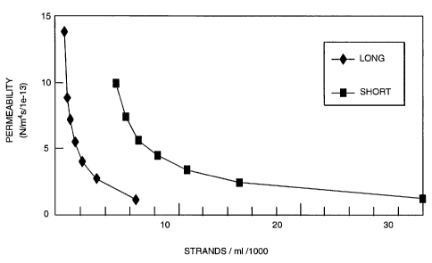
Example 6: Permeability of Bioengineered Collagen Fibers
One-dimensional permeation experiments were conducted on ECF made
from both long wide strands (LW) and short thin strands (ST). An axially
symmetric apparatus was designed to hold ECF between two porous filter discs
at
a prescribed and adjustable thickness (t) (Figure 3). Water is forced through
the
ECF using a syringe pump and the pressure is recorded on an in-line pressure
transducer coupled to a data logger. After '/2 ml of ECF is loaded into the
chamber providing an initial thickness (t), t= 4 mm, the pressure is increased
to 5
psi using a flow rate (Q) of lml/min. The flow is then reduced to 0.1ml/min
and
23
SUBSTITUTE SHEET (RULE 26)
CA 02386217 2002-03-28
WO 01/23529 PCT/US00/26766
the pressure level (P) is monitored until a plateau is reached. This pressure
is used
to calculate the apparent permeability as: kaPP= (Q=t) / (P=A), where A is
cross-
sectional area of the filters. The thickness of the ECF was then reduced in
this
experiment in increments of 0.8mm, by mechanically compressing the sample
between the porous filters. Dilatation of ECF in this way is equivalent to
concentration of the ECF paste. The permeability at each point was used to
determine the strain dependant permeability (or concentration dependence of
permeability) by fitting the data to the exponential law established by Lai et
al
(1980) for soft tissues, kaPP = k0exp(-M=e), where e is the dilatation of the
matrix.
The results show that at a specific concentration there is a substantial
decrease in permeability due to a structural change in the ECF material as the
individual strands intertwine and provide a cohesive matrix. At this
transition the
strand concentrations were 13,000 and 80,000 strands/ml for the LW and ST
formulations respectively. The permeability values at that point were
comparable
at 13.8e-13 m4/Ns (LW) and 12.8e-13 m4/Ns (ST). Compaction of the ECF
decreased the permeability in a non-linear fashion as seen in Figure 4. The
transition concentration (and thickness) for each formulation was used as the
starting point to calculate dilatation in the strain dependent permeability
analysis.
Both formulations fit well to the model with R2 = 0.991 in both cases. The fit
parameters were ko=13.4 m4/Ns and M=2.3 for the LW material and k0=13.0
m4/Ns and M=1.9 for the ST material.
The permeation results demonstrate the continuity of the porous structure
and the suitability in that regard as a soft tissue implant. It also
demonstrates that
the structure of ECF changes with the dimensions of it component strands and
their concentration. It follows that ECF could be modified to provide implants
of
different structures to suit the needs of a particular application. It is
clear from a
mechanical standpoint that the permeability of the structure is a function of
the
ECF dimensions and concentration.
Example 7: Creep Compression Evaluation of Bioengineered Collagen
Fibers
24
SUBSTITUTE SHEET (RULE 26)
CA 02386217 2002-03-28
WO 01/23529 PCTIUSOO/26766
Samples of ECF (0.8m1) were subjected to uniaxial confined compression
between a solid piston and a porous filter. The apparatus that was used is
schematically shown in Figure 5. A locking pin was used to initiate step
loading
and fix displacement for material equilibration as needed. A laser micrometer
recorded both reference heights and displacements of the piston over time. The
loading protocol was as follows: a tare load of 0.77 kPa was applied and
allowed
to equilibrate for 10 minutes. This was considered as the reference loading
state.
Subsequently, a step load of 3.9 kPa was applied and the specimen creep was
monitored for one hour. The piston was then locked for ten minutes to allow
for
any further equilibration of the sample. This was followed by a step load of
15.6
kPa applied to the same sample for three hours. Again the piston was locked in
place and the sample was allowed to equilibrate. Next the load was removed and
the recovery of the sample was monitored for one hour still under the tare
load.
For each of the three phases strains were calculated as displacement relative
to the
height of the sample at the start of the phase. Empirical models were
evaluated to
determine the best description of the creep and recovery data as a means for
comparing the LW and ST formulations and the creep versus recovery response
for a given formulation.
A log-linear model, = M*ln(t) - c, was successfully fit to the
experimental data for the first stage of creep even for the very early time
points
(figure 6a). For both the second loading phase and the recovery phase of the
test a
power law model, c = bta, fit the data very well (Figures 6b and 6c). The
results
from these empirical fits are shown in the Table 4 below.
Table 4
SUBSTITUTE SHEET (RULE 26)
CA 02386217 2002-03-28
WO 01/23529 PCT/US00/26766
N=5 M* a Recovery* b Recover
a b*
Longer Wider 0.033 0.002 0.338 0.088 0.355 0.050 0.023 0.022 0.004
0.(02
(8mm)
Shorter Thinner 0.040 004 0.358 0.042 0.231 0.072** 0.012 0.004 0.014 0107
(2mm)
* Significant difference between formulations using student's t-test (p<0.05).
** Significant difference between compression and recovery using student's t-
test (p<0.05).
By using these models the LW and ST formulations of ECF can be
distinguished in their creep and recovery responses. The initial compression
rate
is faster (larger M, p<0.05) and Recovery rate is slower (smaller (X (alpha)
p<0.05)
for shorter thinner strands compared with longer wider strands. Also, the
recovery
rate is slower (smaller (x (alpha) p<0.05) than the compression rate for the
ST
material.
This assay demonstrates the continuously porous structure of the ECF
matrix. Concentration and the dimensions of its component strands influence
that
matrix's compressive response kinetics.
Example 8: Mechanical Compaction of Bioengineered Collagen Fibers
Bioengineered collagen fibers were loaded (0.8ml) into a confined
compression system (Figure 5) for an initial height of about 1/4". A
polycarbonate piston was lowered onto the material and then weights were added
to the top of the piston. Adding 200 g to the piston (15.9 kPa) resulted in a
compressed, compacted construct of bioengineered collagen fibers with a height
less than 1/4" within 24 hours. The construct was removed from the interior of
the
compression system for testing and evaluation. The compacted bioengineered
collagen fiber construct maintained its shape and was mechanically stable even
when agitated in water for several days.
Example 9: Short Term Compaction And Long Term Persistence Of
Bioengineered Collagen Fibers In A Rabbit Subcutaneous Ear Model.
26
SUBSTITUTE SHEET (RULE 26)
CA 02386217 2002-03-28
WO 01/23529 PCT/USO0/26766
The collagen matrix made as described above was injected subcutaneously
into the ears of New Zealand white rabbits using a 20g needle. Prior to
injection
the height of the implant site was measured using a micrometer caliper.
Immediately after injection the height of the implants were measured. All
implants
were 0.5m1. Persistence was defined as the height of the implant at the time
of
measurement (hi) relative to the initial height of the implant (ho). In one
experiment short term compaction was investigated while a second experiment
focused on the long term persistence of implants made from different size
strands.
In the short term experiment the height of the implant was measured at 1 hour
(ho), 4 hours, 3days. In the long term experiment the implants were measured
at
1(ho), 21, 42, 84, 180, and 330 days. The animals were sacrificed at those
times
(except day 1) and the implants were cut from the surrounding tissue, fixed in
formalin, and stained with hemotoxylin and eosin for histological evaluation.
During the first 3 days the height of the implant is reduced by 15-25%
(Figure 7). This is due to the initial compaction or concentration of the
strands
from an injectable paste to a viscoelastic solid, by the surrounding tissue
forces.
The permeability and elasticity of the formulation, the surrounding tissue in
situ,
and the volume and concentration of the implanted material determine the
degree
of fluid exudation from the implant. Persistence of the implant height over
330
days for both LW and ST materials is shown in (Figure 8). The implant is
palpable and measurable over the entire period and still retains 50% of its
height
at 330 days. Histological evaluation indicates vascularization of the implants
and
fibroblast ingrowth as well as substantial new collagen deposition by 3
months.
At 330 days there is still a substantial amount of the initial implant at the
site of
implantation. There is no evidence that the ECF strands are dispersed to
surrounding areas.
Example 10: Long Term Persistence Of Bioengineered Collagen Fibers In
A Rabbit Intramuscular Model.
The collagen matrix made as described above in the preferred embodiment
was injected into the hind leg muscle of New Zealand 15 white rabbits using a
20g
27
SUBSTITUTE SHEET (RULE 26)
CA 02386217 2002-03-28
WO 01/23529 PCT/US00/26766
needle. All implants were 0.5m1. Persistence and migration of the material was
assessed from histological sections. The implants were sacrificed at 21, 42,
84,
180, and 330 days, three at each time point, and the implants were cut from
the
surrounding tissue, fixed in formalin, and stained with hemotoxylin and eosin
for
histological evaluation. The implants were found to integrate well over time
with
adjacent muscle tissue. There was some host cell infiltration without a lot of
remodeling. The implants were well contained within the muscle with very
little
spreading. Also, rabbit sera were tested and found to be negative for collagen
specific antibodies.
Example 11: Bioengineered Collagen Fibers Made From Atelopeptide
Collagen In A Rabbit Ear Persistence Model
An injectable collagen composition made according to the method
described above was produced using Vitrogen 100 as the starting collagen
solution. Eight rabbits were injected in both ears with the material and
persistence
was measured at 4, 7, 14, 21, 42, 63 and 84 days. Half of he animals were
sacrificed at 21 days and the rest at 84 days, for histological evaluation.
The
persistence relative to day 1 was maintained at almost 80% for some implants
for
84 days Data is shown in Figure 9. Histological evaluation indicated a very
dormant host response to the material. Also the material remained localized at
the
implantation site. Rabbit sera were tested and found to be negative for
collagen
specific antibodies.
Example 12: Additional Formulations Of Bioengineered Collagen Fibers In The
Rabbit Models.
The models described in the previous examples were used to evaluate the
following formulations of the material as well: (1) material that had not been
terminally sterilized with peracetic acid (but produced under aseptic
conditions);
(2) material that had undergone lyophilization prior to implantation; (3)
material
that had undergone lyophilization and terminal sterilization by gamma
irradiation;
and, (4) a formulation produced using PEG with a low osmolality (430 mOsm).
28
SUBSTITUTE SHEET (RULE 26)
CA 02386217 2002-03-28
WO 01/23529 PCTIUS00/26766
All formulations were successfully injected into the rabbit ear model and
compacted to varying degrees. The compositions stayed in the targeted location
in both the muscle and the ear models and were measurable at three months.
Example 13: Bioengineered Collagen Fibers Used in a Minipig Model for
Wound Filling
Collagen fiber compositions was made in an equivalent system to that
described above. In this example the coagulation buffer used for production
was a
low osmolality (430 mOsm), 20% w/v polyethylene glycol (PEG) buffer. In this
example the material was lyophilized after concentration, gamma irradiated for
terminal sterilization and reconstituted with phosphate buffered saline before
use.
Three minipigs were used in this example, one for each time point: 5, 10
and 21 days. Each animal received 14 wounds on its back (2 rows of seven
parallel to the spine) made by biopsy punch lcm in diameter. Ten wounds were
filled with the collagen fiber composition and 4 were left untreated. All were
dressed only with an occlusive spray on dressing (Op-Site).
The endpoints criteria included vascularization, epithelial advance,
epithelial projections (rete pegs), and matrix density. Vascularization was
assessed by manual counting of blood vessels within the wound under a light
microscope. This data was collected only for day 21. Epithelial advance was
determined as a percent of total wound width on histological sections. These
measurements were made manually and for all time points. The number of rete
pegs per unit length was counted as a measure of the quality of wound closure
at
21 days. Matrix density both within and adjacent to the wound was
quantitatively
measured using image analysis techniques. These assessments were made using
Picro Sirius Red (PSR) staining, which shows only matrix, and polarized light
microscopy. The cellular response was evaluated using hemotoxilin and eosin
stained sections.
The results indicated showed the treated wounds to be rich in fibrin after 5
days and there was an extensive fibroblast proliferation accompanied by
collagen
deposition. There was notably no ECF remaining in the wounds indicating its
29
SUBSTITUTE SHEET (RULE 26)
CA 02386217 2002-03-28
WO 01/23529 PCT/US00/26766
apparent dissolution/degradation. After five days there was not statistical
difference in epithelial advance between the ECF treated and the untreated
wounds. Both were about 22-23% covered. However, after 10 days the ECF
treated wounds were 85% covered while the untreated wounds were 72% covered
and this difference was statistically significant (p<0.001).
At 10 days in normal pig wounds there was a fair amount of
epithelialization and the granulation tissue was rich in proliferating
fibroblasts.
There was conspicuously denser collagen deposition in the granulation tissue
of
ECF treated wounds compared with the untreated wounds. In some sections the
tissue was similar to that seen in control sections at 21 days. There was no
foreign
body response.
At 21 days the degree of number of blood vessels per unit area within the
ECF treated wounds (0.50 0.09) was lower than in the untreated wounds (0.34
0.03) (p<0.001). Also, the matrix density within the wound was significantly
closer to the matrix density of the adjacent tissue (p=0.038) in the ECF
treated
wounds with a difference of 23.1 8.5 compared with the untreated controls at
33.9 5.1. Although there was not statistical difference (p>0.5) for the rete
pegs
parameter, it did appear that at the edges of the treated wounds there were
some
rete pegs while there were none in the controls.
Although the foregoing invention has been described in some detail by
way of illustration and example for purposes of clarity and understanding, it
will
be obvious to one of skill in the art that certain changes and modifications
may be
practiced within the scope of the appended claims.
SUBSTITUTE SHEET (RULE 26)