Note: Descriptions are shown in the official language in which they were submitted.
CA 02386858 2006-12-18
36P6D5: SECRETED TUMOR ANTIGEN
FIELD OF THE INVENTION
The invention described herein relates to a gene and its encoded tumor
antigen,
termed 36P6D5, and to diagnostic and therapeutic methods and compositions
useful in
the management of various cancers that express 36P6D5 gene products.
BACKGROUND OF THE INVENTION
Cancer is the second leading cause of human death next to coronary disease.
Worldwide, millions of people die from cancer every year. In the United States
alone,
cancer causes the death of well over a half-million people each year, with
some 1.4
million new cases diagnosed per year. While deaths from heart disease have
been
declining significandy, those resulting from cancer generally are on the rise.
In the early
part of the next century, cancer is predicted to become the leading cause of
death.
Worldwide, several cancers stand out as the leading killers. In particular,
carcinomas of the lung, prostate, breast, colon, pancreas, and ovary represent
the primary
causes of cancer death. These and virtually all other carcinomas share a
common lethal
feature. With very few exceptions, metastatic disease from a carcinoma is
fatal.
Moreover, even for those cancer patients who initially survive their primary
cancers,
common experience has shown that their lives are dramatically altered. Many
cancer
patients experience strong anxieties driven by the awareness of the potential
for
recurrence or treatment failure. Many cancer patients experience physical
debilitations
following treatment. Many cancer patients experience a recurrence.
Generally speaking, the fundamental problem in the management of the deadliest
cancers is the lack of effective and non-toxic systemic therapies. Molecular
medicine,
still very much in its infancy, promises to redefine the ways in which these
cancers are
managed. Unquestionably, there is an intensive worldwide effort aimed at the
development of novel molecular approaches to cancer diagnosis and treatment.
For
example, there is a great interest in identifying truly tumor-specific genes
and proteins
1
CA 02386858 2002-04-08
WO 01/31015 PCT/US00/29894
that could be used as diagnostic and prognostic markers and/or therapeutic
targets or
agents. Research efforts in these areas are encouraging, and the increasing
availability of
useful molecular technologies has accelerated the acquisition of meaningful
knowledge
about cancer. Nevertheless, progress is slow and generally uneven.
As discussed below, the management of prostate cancer serves as a good example
of the limited extent to which molecular biology has translated into real
progress in the
clinic. With limited exceptions, the situation is more or less the same for
the other major
carcinomas mentioned above.
Worldwide, prostate cancer is the fourth most prevalent cancer in men. In
North
America and Northern Europe, it is by far the most common male cancer and is
the
second leading cause of cancer death in men. In the United States alone, well
over
40,000 men die annually of this disease - second only to lung cancer. Despite
the
magnitude of these figures, there is still no effective treatment for
metastatic prostate
cancer. Surgical prostatectomy, radiation therapy, hormone ablation therapy,
and
chemotherapy remain fixed as the main treatment modalities. Unfortunately,
these
treatments are ineffective for many and are often associated with significant
undesirable
consequences.
On the diagnostic front, the lack of a prostate tumor marker that can
accurately
detect early-stage, localized tumors remains a significant limitation in the
management of
this disease. Although the serum PSA assay has been a very useful tool, its
specificity
and general utility is widely regarded as lacking in several important
respects, as further
discussed below. Most prostate cancers initially occur in the peripheral zone
of the
prostate gland, away from the urethra. Tumors within this zone may not produce
any
symptoms and, as a result, most men with early-stage prostate cancer will not
present
clinical symptoms of the disease until significant progression has occurred.
Tumor
progression into the transition zone of the prostate may lead to urethral
obstruction, thus
producing the first symptoms of the disease. However, these clinical symptoms
are
indistinguishable from the common non-malignant condition of benign prostatic
hyperplasia (BPH). Early detection and diagnosis of prostate cancer currently
relies on
digital rectal examinations (DRE), prostate specific antigen (PSA)
measurements,
transrectal ultrasonography (TRUS), and transrectal needle biopsy (TRNB). At
present,
serum PSA measurement in combination with DRE represent the leading tool used
to
2
CA 02386858 2002-04-08
WO 01/31015 PCT/US00/29894
detect and diagnose prostate cancer. Both have major limitations which have
fueled
intensive research into finding better diagnostic markers of this disease.
Similarly, there is no available marker that can predict the emergence of the
typically fatal metastatic stage of prostate cancer. Diagnosis of metastatic
stage is
presently achieved by open surgical or laparoscopic pelvic lymphadenectomy,
whole
body radionuclide scans, skeletal radiography, and/or bone lesion biopsy
analysis.
Clearly, better imaging and other less invasive diagnostic methods offer the
promise of
easing the difficulty those procedures place on a patient, as well as
improving diagnostic
accuracy and opening therapeutic options. A similar problem is the lack of an
effective
prognostic marker for determining which cancers are indolent and which ones
are or will
be aggressive. PSA, for example, fails to discriminate accurately between
indolent and
aggressive cancers. Until there are prostate tumor markers capable of reliably
identifying
early-stage disease, predicting susceptibility to metastasis, and precisely
imaging tumors,
the management of prostate cancer will continue to be extremely difficult.
PSA is the most widely used tumor marker for screening, diagnosis, and
monitoring prostate cancer today. In particular, several immunoassays for the
detection
of serum PSA are in widespread clinical use. Recently, a reverse transcriptase-
polymerase
chain reaction (RT-PCR) assay for PSA mRNA in serum has been developed.
However,
PSA is not a disease-specific marker, as elevated levels of PSA are detectable
in a large
percentage of patients with BPH and prostatitis (25-86%)(Gao et al., 1997,
Prostate 31:
264-281), as well as in other nonmalignant disorders and in some normal men, a
factor
which significantly limits the diagnostic specificity of this marker. For
example,
elevations in serum PSA of between 4 to 10 ng/ml are observed in BPH, and even
higher values are observed in prostatitis, particularly acute prostatitis. BPH
is an
extremely common condition in men. Further confusing the situation is the fact
that
serum PSA elevations may be observed without any indication of disease from
DRE, and
visa-versa. Moreover, it is now recognized that PSA is not prostate-specific
(Gao et al.,
supra, for review).
Various methods designed to improve the specificity of PSA-based detection
have been described, such as measuring PSA density and the ratio of free vs.
complexed
PSA. However, none of these methodologies have been able to reproducibly
distinguish
benign from malignant prostate disease. In addition, PSA diagnostics have
sensitivities
3
CA 02386858 2002-04-08
WO 01/31015 PCT/US00/29894
of between 57-79% (Cupp & Osterling, 1993, Mayo Clin Proc 68:297-306), and
thus miss
identifying prostate cancer in a significant population of men with the
disease.
There are some known markers which are expressed predominantly in prostate,
such as prostate specific membrane antigen (PSM), a hydrolase with 85%
identity to a rat
neuropeptidase (Carter et al., 1996, Proc. Natl. Acad. Sci. USA 93: 749;
Bzdega et al.,
1997, J. Neurochem. 69: 2270). However, the expression of PSM in small
intestine and
brain (Israeli et al., 1994, Cancer Res. 54: 1807), as well its potential role
in neuropeptide
catabolism in brain, raises concern of potential neurotoxicity with anti-PSM
therapies.
Preliminary results using an Indium-111 labeled, anti-PSM monoclonal antibody
to image
recurrent prostate cancer show some promise (Sodee et al., 1996, Clin Nuc Med
21: 759-
766). More recently identified prostate cancer markers include PCTA-1 (Su et
al., 1996,
Proc. Natl. Acad. Sci. USA 93: 7252) and prostate stem cell antigen (PSCA)
(Reiter et al.,
1998, Proc. Natl. Acad. Sci. USA 95: 1735). PCTA-1, a novel galectin, is
largely secreted
into the media of expressing cells and may hold promise as a diagnostic serum
marker
for prostate cancer (Su et al., 1996). PSCA, a GPI-linked cell surface
molecule, was
cloned from LAPC-4 cDNA and is unique in that it is expressed primarily in
basal cells
of normal prostate tissue and in cancer epithelia (Reiter et al., 1998).
Vaccines for
prostate cancer are also being actively explored with a variety of antigens,
including PSM
and PSA.
While previously identified markers such as PSA, PSM, PCTA and PSCA have
facilitated efforts to diagnose and treat prostate cancer, there is need for
the identification
of additional markers and therapeutic targets for prostate and related cancers
in order to
further improve diagnosis and therapy.
SUMMARY OF THE INVENTION
The present invention relates to a gene and protein designated 36P6D5. In
normal individuals, 36P6D5 protein appears to be predominantly expressed in
pancreas,
with lower levels of expression detected in prostate and small intestine. The
36P6D5
gene is also expressed in several human cancer xenografts and cell lines
derived from
prostate, breast, ovarian and colon cancers, in some cases at high levels.
Over-expression
of 36P6D5, relative to normal, is observed in prostate cancer xenografts
initially derived
from a prostate cancer lymph node metastasis and passaged intratibially and
4
CA 02386858 2002-04-08
WO 01/31015 PCT/US00/29894
subcutaneously in SCID mice. Extremely high level expression of 36P6D5 is
detected in
the breast cancer cell line DU4475, a cell line that was initially derived
from a mammary
gland carcinoma (Langlois et al., 1979, Cancer Res. 39: 2604). The 36P6D5 gene
is also
expressed in tumor patient samples derived from bladder, kidney, colon and
lung
cancers, in some cases at high levels.
A full length 36P6D5 cDNA of 931 bp (SEQ ID NO: 1) provided herein
encodes a 235 amino acid open reading frame (SEQ ID NO: 2) with significant
homology to the 2-19 protein precursor (Genbank P98173) as well as a gene
previously
cloned from human osteoblasts (Q92520). The predicted 235 amino acid 36P6D5
protein also contains an N-terminal signal sequence, indicating that the
36P6D5 protein
is secreted. The 36P6D5 gene therefore encodes a secreted tumor antigen which
may be
useful as a diagnostic, staging and/or prognostic marker for, and/or may serve
as a target
for various approaches to the treatment of, prostate, breast, colon,
pancreatic, and
ovarian cancers expressing 36P6D5. The predicted molecular weight of the
36P6D5
protein is approximately 26 kD and its' pI is 8.97.
Expression analysis demonstrates high levels of 36P6D5 expression in several
prostate and other cancer cell lines as well as prostate cancer patient
samples and tumor
xenografts. The expression profile of 36P6D5 in normal adult tissues, combined
with
the over-expression observed in cancer cells such as bladder, colon, kidney,
breast, lung,
ovary and prostate cancer cell lines and/or cancer patient samples, provides
evidence that
36P6D5 is aberrantly expressed in at least some cancers, and can serve as a
useful
diagnostic and/or therapeutic target for such cancers.
The invention provides polynucleotides corresponding or complementary to all
or part of the 36P6D5 genes, mRNAs, and/or coding sequences, preferably in
isolated
form, including polynucleotides encoding 36P6D5 proteins and fragments
thereof, DNA,
RNA, DNA/RNA hybrid, and related molecules, polynucleotides or
oligonucleotides
complementary to the 36P6D5 genes or mRNA sequences or parts thereof, and
polynucleotides or oligonucleotides that hybridize to the 36P6D5 genes, mRNAs,
or to
36P6D5-encoding polynucleotides. Also provided are means for isolating cDNAs
and the
genes encoding 36P6D5. Recombinant DNA molecules containing 36P6D5
polynucleotides, cells transformed or transduced with such molecules, and host-
vector
systems for the expression of 36P6D5 gene products are also provided. The
invention
5
CA 02386858 2002-04-08
WO 01/31015 PCT/US00/29894
further provides 36P6D5 proteins and polypeptide fragments thereof. The
invention
further provides antibodies that bind to 36P6D5 proteins and polypeptide
fragments
thereof, including polyclonal and monoclonal antibodies, marine and other
mammalian
antibodies, chimeric antibodies, humanized and fully human antibodies, and
antibodies
labeled with a detectable marker.
The invention further provides methods for detecting the presence and status
of
36P6D5 polynucleotides and proteins in various biological samples, as well as
methods for
identifying cells that express 36P6D5. A typical embodiment of this invention
provides
methods for monitoring 36P6D5 gene products in a tissue sample having or
suspected of
having some form of growth dysregulation such as cancer.
The invention further provides various therapeutic compositions and strategies
for
treating cancers that express 36P6D5 such as prostate cancers, including
therapies aimed at
inhibiting the transcription, translation, processing or function of 36P6D5 as
well as cancer
vaccines.
BRIEF DESCRIPTION OF THE FIGURES
FIGS. 1A-1B. Nucleotide (SEQ ID NO: 1) and deduced amino acid (SEQ ID
NO: 2) sequences of 36P6D5 cDNA. The start methionine and consensus Kozak
sequence are indicated in bold and the putative N-terminal signal sequence is
underlined.
FIGS. 2A-2B. Amino acid sequence alignment of the 36P6D5 ORF with 2-19
protein precursor (2A) (SEQ ID NO: 15) and GS3786 protein (osteoblast protein)
(2B)
(SEQ ID NO: 16). Percent sequence identities are indicated on the figure.
FIG. 3. Northern blot analysis of human 36P6D5 expression in various normal
tissues showing predominant expression in pancreas and low level expression in
prostate
and small intestine.
FIGS. 4A-4B. Northern blot analysis of human 36P6D5 mRNA expression in a
panel of prostate cancer xenografts and various other human cancer cell lines.
FIGS. 5A-5B. Secretion of 36P6D5 protein from transfected 293T cells and
detection with anti-36P6D5 polyclonal antibody. 293T cells were transiently
transfected
with either pCDNA 3.1 MYC/HIS 36P6D5, pTag5 36P6D5, in which the natural
signal
sequence of 36P6D5 is replaced with an immunoglobulin signal sequence, or
pAPTag5
36P6D5 which encodes a fusion protein composed of 36P6D5 and alkaline
phosphatase
6
CA 02386858 2006-12-18
Various embodiments of this invention provide a method for obtaining an
indication
of the presence of bladder, colon, kidney, breast, lung, ovarian, pancreatic
or prostate cancer
in an individual, which method comprises measuring the level of a protein or
mRNA
expressed by cells in a test sample from the individual and comparing the
level so measured
to the level of said protein or mRNA expressed in a corresponding normal
sample, wherein
the presence of elevated protein or mRNA in the test sample relative to the
normal sample
provides an indication of the presence of said cancer, and wherein said
protein has an amino
acid sequence that is at least 90% identical to SEQ ID NO:2 over its entire
length and said
mRNA encodes said protein.
Other embodiments of this invention provide a method for obtaining an
indication of
aberrant cellular growth in a test sample of bladder, colon, kidney, breast,
lung, ovary,
pancreas or prostate tissue from an individual comprising measuring the level
of expression
of a mRNA or a protein in the test sample and comparing the level so
determined to the level
of expression of the mRNA or protein in a corresponding normal sample, wherein
the
presence of an elevated level of said mRNA or protein expression in the test
sample relative
to the normal sample provides an indication of aberrant cell growth within
said tissue,
wherein said protein has an amino acid sequence that is at least 90% identical
to SEQ ID
NO:2 over its entire length and said mRNA encodes said protein.
Other embodiments of this invention provide a method of examining a biological
sample from an individual for evidence of aberrant cellular growth in bladder,
colon, kidney,
breast, lung, ovary, pancreas or prostate tissue of said individual,
comprising comparing the
level of expression of a protein or mRNA in the biological sample to the level
of expression
of said mRNA or protein in a corresponding normal sample, wherein elevation in
said level
in the biological sample as compared to the normal sample is associated with
aberrant
cellular growth in said tissue, wherein said protein has an amino acid
sequence that is at least
90% identical to SEQ ID NO:2 over its entire length and said mRNA encodes said
protein.
Other embodiments of this invention provide a method of detecting an
indication of
the presence of cancer in an individual comprising: (a) measuring the level of
a mRNA
expressed in a test sample obtained from bladder, colon, kidney, breast, lung,
ovarian,
pancreatic or prostate tissue of the individual; and (b) comparing the level
so determined to
the level of said mRNA expressed in a comparable known normal sample, wherein
the
presence of elevated mRNA expression in the test sample relative to the normal
sample
provides an indication of the presence of cancer in said tissue, and wherein
said mRNA
6a
CA 02386858 2007-09-24
encodes an amino acid sequence that is at least 90% identical to SEQ ID NO:2
over its entire
length.
Other embodiments of this invention provide a method of detecting an
indication of
the presence of cancer in an individual comprising: (a) measuring the level of
a protein
expressed in a test sample obtained from bladder, colon, kidney, breast, lung,
ovarian,
pancreatic or prostate tissue of the individual; and (b) comparing the level
so determined to
the level of said protein expressed in a comparable known normal sample,
wherein the
presence of elevated protein in the test sample relative to the normal sample
provides an
indication of the presence of cancer in said tissue, and wherein said protein
has an amino acid
sequence that is at least 90% identical to SEQ ID NO:2 over its entire length.
Other embodiments of this invention provide a polynucleotide selected from the
group consisting of: (a) a polynucleotide comprising the sequence as shown in
SEQ ID
NO:1, wherein T can also be U; (b) a polynucleotide comprising the sequence as
shown in
SEQ ID NO: 1, from about nucleotide residue number 59 through about nucleotide
residue
number 763, wherein T can also be U; (c) a polynucleotide encoding a protein
having the
amino acid sequence shown in SEQ ID NO:2; (d) a polynucleotide that is a
fragment of the
polynucleotide of (a), (b) or (c) that is at least 20 nucleotide bases in
length; (e) a
polynucleotide that is fully complementary to a polynucleotide of any one of
(a) to (d); and
(f) a polynucleotide that selectively hybridizes under stringent conditions to
the
polynucleotide of any one of (a) to (d), wherein the stringent conditions
comprise
hybridization in the presence of 50% (v/v) formamide with 0.1% bovine serum
albumin,
0.1% Ficoll, 0.1% polyvinylpyrrolidone, and 50 mM sodium phosphate buffer at
pH 6.5 with
750 mM sodium chloride and 75 mM sodium citrate at 42 C, and wherein the
polynucleotide
is labelled with a detectable marker, for use in detecting or measuring levels
of a mRNA
which is associated which is associated with aberrant cellular growth or
cancer in bladder or
prostate tissue.
Other embodiments of this invention provide a polynucleotide that enc des a
polypeptide that is at least 90% identical to the amino acid sequence shown in
EQ ID NO:2
over its entire length and wherein the polynucleotide is labelled with a
detecta le marker, for
use in detecting or measuring levels of a mRNA which is associated which is
sociated with
aberrant cellular growth or cancer in bladder or prostate tissue.
Other embodiments of this invention provide a polynucleotide that selectively
hybridizes under stringent conditions to a polynucleotide that encodes a
polypeptide that is at
6b
CA 02386858 2007-09-24
least 90% identical to the amino acid sequence shown in SEQ ID NO;2 over its
entire length,
for use in detecting or measuring levels of a mRNA which is associated with
aberrant cellular
growth or cancer in bladder or prostate tissue, wherein the stringent
conditions comprise
hybridization in the presence of 50% (v/v) formamide with 0.1% bovine serum
albumin,
0.1% Ficoll, 0.1% polyvinylpyrrolidone, and 50 mM sodium phosphate buffer at
pH 6.5 with
750 mM sodium chloride and 75 mM sodium citrate at 42 C.
Other embodiments of this invention provide a polynucleotide labelled with a
detectable marker that encodes a polypeptide associated with aberrant cellular
growth or
cancer in bladder or prostate tissue, wherein the polypeptide includes one or
more amino acid
sequences selected from the group consisting of. NVTA at residues 120-123 of
SEQ ID
NO:2; NHSD at residues 208-211 of SEQ ID NO:2; SIR at residues 43-45 of SEQ ID
NO:2;
STR at residues 160-162 of SEQ ID NO:2; SIGE at residues 46-49 of SEQ ID NO:2;
TYDD
at residues 155-158 of SEQ ID NO:2; GGGRSK at residues 81-86 of SEQ ID NO:2;
GINIAI
at residues 108-113 of SEQ ID NO:2; GNVTAT at residues 119-124 of SEQ ID NO:2;
AGGLLKVVFVVFASLCAWYSGYLLAELIPDAP at residues 5-36 of SEQ ID NO:2;
GLLKVVFVV (SEQ ID NO:19); LMGEQLGNV (SEQ ID NO:20); LLAELIPDA (SEQ ID
NO:21); FIAAKGL (SEQ ID NO:22); VVFVVFASL (SEQ ID NO:23); GGLLKVVFV
(SEQ ID NO:24); FIAAKGLEL (SEQ ID NO:25); KICFEDNLL (SEQ ID NO:26);
INIAIVNYV (SEQ ID NO:27); and, NMKFRSSWV (SEQ ID NO:28).
Other embodiments of this invention provide a polypeptide or a composition
comprising the polypeptide and a pharmaceutically acceptable carrier or
excipient, wherein
the polypeptide comprises at least 15 contiguous amino acids of a polypeptide
that is at least
90% identical to the amino acid sequence shown in SEQ ID NO:2 over its entire
length, for
use in eliciting an immune response to an antigen of the polypeptide at least
90% identical to
the amino acid sequence shown in SEQ ID NO:2 that is associated with aberrant
cellular
growth or cancer in bladder or prostate tissue.
Other embodiments of this invention provide a polypeptide or a composition
comprising the polypeptide and a pharmaceutically acceptable carrier or
excipient for use in
eliciting an immune response against an antigen of a polypeptide at least 90%
identical to the
amino acid sequence shown in SEQ ID NO:2 that is associated with aberrant
cellular growth
or cancer in bladder or prostate tissue, wherein the polypeptide comprises a
sequence selected
from the group consisting of: GLLKVVFVV (SEQ ID NO: 19); LMGEQLGNV (SEQ ID
NO:20); LLAELIPDA (SEQ ID NO:21); FIAAKGL (SEQ ID NO:22); VVFVVFASL (SEQ
6c
CA 02386858 2007-09-24
ID NO:23); GGLLKVVFV (SEQ ID NO:24); FIAAKGLEL (SEQ ID NO:25);
KICFEDNLL (SEQ ID NO:26); INIAIVNYV (SEQ ID NO:27); and, NMKFRSSWV (SEQ
ID NO:28).
Other embodiments of this invention provide an antibody or antibody fragment
specific for a polypeptide as defined in this invention for use in binding an
antigen of a
polypeptide at least 90% identical to the amino acid sequence shown in SEQ ID
NO:2 that is
associated with aberrant cellular growth or cancer in bladder or prostate
tissue.
Other embodiments of this invention provide a vector encoding a single chain
monoclonal antibody of this invention, wherein the vector is for use in
expressing the
monoclonal antibody to bind to an antigen associated with aberrant cellular
growth or cancer
in bladder or prostate tissue.
BRIEF DESCRIPTION OF THE FIGURES
FIGS. IA-1B. Nucleotide (SEQ ID NO: I) and deduced amino acid (SEQ ID NO:2)
sequences of 36P6D5 cDNA. The start methionine and consensus Kozak sequence
are
indicated in bold and the putative N-terminal signal sequence is underlined.
FIGS. 2A-2B. Amino acid sequence alignment of the 36P6D5 ORF with 2-19
protein precursor (2A) (SEQ ID NO:15) and GS3786 protein (osteoblast protein)
(2B) (SEQ
ID NO: 16). Percent sequence identities are indicated on the figure.
FIG. 3. Northern blot analysis of human 36P6D5 expression in various normal
tissues showing predominant expression in pancreas and low level expression in
prostate and
small intestine.
FIGS. 4A-4B. Northern blot analysis of human 36P6D5 mRNA expression in a
panel of prostate cancer xenografts and various other human cancer cell lines.
FIGS. 5A-5B. Secretion of 36P6D5 protein from transfected 293T cells and
detection with anti-36P6D5 polyclonal antibody. 293T cells were transiently
transfected with
either pCDNA 3.1 MYC/HIS 36P6D5, pTag5 36P6D5, in which the natural signal
sequence
of 36P6D5 is replaced with an immunoglobulin signal sequence, or pAPTag5
36P6D5 which
encodes a fusion protein composed of 36P6D5 and alkaline phosphatase.
6d
CA 02386858 2002-04-08
WO 01/31015 PCT/US00/29894
(also containing the Ig signal sequence). Conditioned media or whole cell
lysates were
subjected to Western blotting with either rabbit anti-His pAb (Santa Cruz,
Biotechnology, Inc., 1:2,500 dilution, left panel) or with affinity purified
rabbit anti-
36P6D5 pAb (1 ug/ml, right panel). Anti-His and anti-36P6D5 pAb reactive bands
were
visualized by incubation of the blots with anti-rabbit-HRP conjugated
secondary
antibody followed by enhanced chemiluminescence detection.
FIG. 6. Expression of 36P6D5 in cancer patient tumors. RT-PCR analysis of
36P6D5 mRNA expression in bladder cancer, kidney cancer, colon cancer, and
lung
cancer patient tumors.
FIG. 7. 36P6D5 expression in bladder cancer and their matched normal tissues
was tested by Northern blot analysis. For this figure, 10 g of total RNA were
loaded for
each sample. Overexpression of 36P6D5 expression was detected in 3 out of 4
cancers
tested (lanes 3,5,7 and 8). No expression was seen in bladder tissue isolated
from a
normal individual (lane 1).
FIG. 8. Binding of 36P6D5 to LAPC9 AD. A single cell suspension of LAPC9
AD xenograft cells were allowed to adhere overnight to a 6 well plate. The
cells were
incubated in the presence of control or 36P6D5-AP fusion protein. The alkaline
phosphate
substrate, BM purple, was used for detection.
FIGS. 9A-9B. Human cancer cells express and secrete 36P6D5 protein.
Conditioned media and/or cell lysates from a variety of cancer cell lines
representing
cancers derived from prostate (LAPC4 xenograft), colon (Colo 205, CaCo-1),
breast
(Du4475), and pancreatic (Capan-1) tissues, as well as PC3 prostate cancer
cells
engineered to overexpress 36P6D5 protein, were subjected to Western analysis
using an
anti-36P6D5 murine pAb. The specific anti-36P6D5 immunoreactive bands
representing
endogenous 36P6D5 protein are indicated with arrows and run approximately
between
and 40 kD. The molecular weight of 36P6D5 calculated from the amino acid
sequence is 26 kD suggesting that endogenous 36P6D5 protein is post-
translationally
modified, possibly by glycosylation.
FIG 10. A sensitive and specific capture ELISA detects 36P6D5 protein in
30 supernatants of human cancer cell lines. A capture ELISA was developed
using protein
G purified murine anti-36P6D5 pAb as capture Ab and a biotinylated form of the
same
pAb as detection Ab. Shown is the standard curve generated using the Tag5-
36P6D5
7
CA 02386858 2002-04-08
WO 01/31015 PCT/US00/29894
protein and specific detection and quantitation of 36P6D5 present in
supernatants
derived from PC-3-Neo transfected cells (O.D.=0.023, 36P6D5 protein
concentration in
ng/ml=N.D.), PC-3 cells overexpressing 36P6D5 (O.D.=0.186, 36P6D5 protein
concentration in ng/m1=1.48) and endogenous 36P6D5 protein secreted by Du4475
breast cancer cells (O.D.=0.085, 36P6D5 protein concentration in ng/ml=0.50).
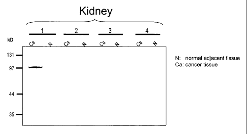
FIGS. 11A-11B. Detection of 36P6D5 expression in human cancers. Cell lysates
from Colon, breast and kidney cancer tissues (Ca), as well as their normal
matched
adjacent tissues (N) were subjected to Western analysis using an anti-36P6D
antibody.
The specific anti-36P6D5 immunoreactive bands represent a monomeric form of
the
36P6D5 protein, which runs approximately between 35 and 40 kD, and multimeric
forms of the protein, which run approximately at 90 and 120 kD.
DETAILED DESCRIPTION OF THE INVENTION
Unless otherwise defined, all terms of art, notations and other scientific
terminology used herein are intended to have the meanings commonly understood
by
those of skill in the art to which this invention pertains. In some cases,
terms with
commonly understood meanings are defined herein for clarity and/or for ready
reference, and the inclusion of such definitions herein should not necessarily
be
construed to represent a substantial difference over what is generally
understood in the
art. The techniques and procedures described or referenced herein are
generally well
understood and commonly employed using conventional methodology by those
skilled in
the art, such as, for example, the widely utilized molecular cloning
methodologies
described in Sambrook et al., Molecular Cloning: A Laboratory Manual 2nd.
edition
(1989) Cold Spring Harbor Laboratory Press, Cold Spring Harbor, N.Y. As
appropriate,
procedures involving the use of commercially available kits and reagents are
generally
carried out in accordance with manufacturer defined protocols and/or
parameters unless
otherwise noted.
As used herein, the terms "advanced prostate cancer", "locally advanced
prostate
cancer", "advanced disease" and "locally advanced disease" mean prostate
cancers which
have extended through the prostate capsule, and are meant to include stage C
disease
under the American Urological Association (AUA) system, stage C1 - C2 disease
under
the Whitmore Jewett system, and stage T3 - T4 and N+ disease under the TNM
(tumor,
8
CA 02386858 2002-04-08
WO 01/31015 PCT/US00/29894
node, metastasis) system. In general, surgery is not recommended for patients
with
locally advanced disease, and these patients have substantially less favorable
outcomes
compared to patients having clinically localized (organ-confined) prostate
cancer. Locally
advanced disease is clinically identified by palpable evidence of induration
beyond the
lateral border of the prostate, or asymmetry or induration above the prostate
base.
Locally advanced prostate cancer is presently diagnosed pathologically
following radical
prostatectomy if the tumor invades or penetrates the prostatic capsule,
extends into the
surgical margin, or invades the seminal vesicles.
As used herein, the terms "metastatic prostate cancer" and "metastatic
disease"
mean prostate cancers which have spread to regional lymph nodes or to distant
sites, and
are meant to include stage D disease under the AUA system and stage TxNxM+
under
the TNM system. As is the case with locally advanced prostate cancer, surgery
is
generally not indicated for patients with metastatic disease, and hormonal
(androgen
ablation) therapy is the preferred treatment modality. Patients with
metastatic prostate
cancer eventually develop an androgen-refractory state within 12 to 18 months
of
treatment initiation, and approximately half of these patients die within 6
months
thereafter. The most common site for prostate cancer metastasis is bone.
Prostate cancer
bone metastases are, on balance, characteristically osteoblastic rather than
osteolytic (i.e.,
resulting in net bone formation). Bone metastases are found most frequently in
the
spine, followed by the femur, pelvis, rib cage, skull and humerus. Other
common sites
for metastasis include lymph nodes, lung, liver and brain. Metastatic prostate
cancer is
typically diagnosed by open or laparoscopic pelvic lymphadenectomy, whole body
radionuclide scans, skeletal radiography, and/or bone lesion biopsy.
As used herein, the term "polynucleotide" means a polymeric form of
nucleotides
of at least about 10 bases or base pairs in length, either ribonucleotides or
deoxynucleotides or a modified form of either type of nucleotide, and is meant
to include
single and double stranded forms of DNA.
As used herein, the term "polypeptide" means a polymer of at least about 6
amino acids. Throughout the specification, standard three letter or single
letter
designations for amino acids are used.
As used herein, the terms "hybridize", "hybridizing", "hybridizes" and the
like,
used in the context of polynucleotides, are meant to refer to conventional
hybridization
9
CA 02386858 2002-04-08
WO 01/31015 PCT/US00/29894
conditions, preferably such as hybridization in 50% formamide/6XSSC/0.1%
SDS/100
g/ml ssDNA, in which temperatures for hybridization are above 37 degrees C and
temperatures for washing in 0.1X SSC/0.1% SDS are above 55 degrees C, and most
preferably to stringent hybridization conditions.
"Stringency" of hybridization reactions is readily determinable by one of
ordinary
skill in the art, and generally is an empirical calculation dependent upon
probe length,
washing temperature, and salt concentration. In general, longer probes require
higher
temperatures for proper annealing, while shorter probes need lower
temperatures.
Hybridization generally depends on the ability of denatured DNA to reanneal
when
complementary strands are present in an environment below their melting
temperature.
The higher the degree of desired homology between the probe and hybridizable
sequence, the higher the relative temperature that can be used. As a result,
it follows that
higher relative temperatures would tend to make the reaction conditions more
stringent,
while lower temperatures less so. For additional details and explanation of
stringency of
hybridization reactions, see Ausubel et al., Current Protocols in Molecular
Biology, Wiley
Interscience Publishers, (1995).
"Stringent conditions" or "high stringency conditions", as defined herein, may
be
identified by those that: (1) employ low ionic strength and high temperature
for washing,
for example 0.015 M sodium chloride/0.0015 M sodium citrate/0.1% sodium
dodecyl
sulfate at 50 C; (2) employ during hybridization a denaturing agent, such as
formamide,
for example, 50% (v/v) formamide with 0.1% bovine serum albumin/0.1%
Ficoll/0.1%
polyvinylpyrrohdone/50mM sodium phosphate buffer at pH 6.5 with 750 mM sodium
chloride, 75 mM sodium citrate at 42 C; or (3) employ 50% formamide, 5 x SSC
(0.75 M
NaCl, 0.075 M sodium citrate), 50 mM sodium phosphate (pH 6.8), 0.1% sodium
pyrophosphate, 5 x Denhardt's solution, sonicated salmon sperm DNA (50 g/ml),
0.1% SDS, and 10% dextran sulfate at 42 C, with washes at 42 C in 0.2 x SSC
(sodium
chloride/ sodium. citrate) and 50% formamide at 55 C, followed by a high-
stringency
wash consisting of 0.1 x SSC containing EDTA at 55 C.
"Moderately stringent conditions" may be identified as described by Sambrook
et
al., 1989, Molecular Cloning: A Laboratory Manual, New York: Cold Spring
Harbor
Press, and include the use of washing solution and hybridization conditions
(e.g.,
temperature, ionic strength and %SDS) less stringent than those described
above. An
CA 02386858 2006-12-18
example of moderately stringent conditions is overnight incubation at 37"C in
a solution
comprising: 20% formamide, 5 x SSC (150 mM NaCl, 15 rnM trisodium citrate), 50
mM
sodium phosphate (pH 7.6), 5 x Denhardt's solution, 10% dextran sulfate, and
20
mg/mL denatured sheared salmon sperm DNA, followed by washing the filters in 1
x
SSC at about 37-50 C. The skilled artisan will recognize how to adjust the
temperature,
ionic strength, etc. as necessary to accommodate factors such as probe length
and the
like.
In the context of amino acid sequence comparisons, the term "identity" is used
to express the percentage of amino acid residues at the same relative
positions that are
the same. Also in this context, the term "homology" is used to express the
percentage of
amino acid residues at the same relative positions that are either identical
or are similar,
using the conserved amino acid criteria of BLAST analysis, as is generally
understood in
the art. For example, % identity values may be generated by WU-BLAST-2
(Altschul et
al., 1996, Methods in Enzymology 266:460-480).
Further details regarding amino acid
substitutions, which are considered conservative under such criteria, are
provided below.
Additional definitions are provided throughout the subsections that follow.
MOLECULAR BIOLOGY OF AND USES FOR 36P6D5
As is further described in the Examples below, the 36P6D5 gene and protein
have been characterized using a number of analytical approaches. For example,
analyses
of nucleotide coding and amino acid sequences were conducted in order to
identify
potentially related molecules, as well as recognizable structural domains,
topological
features, and other elements within the 36P6D5 mRNA and protein structure.
Northern
blot analyses of 36P6D5 mRNA expression were conducted in order to establish
the
range of-normal and cancerous tissues expressing 36P6D5 message.
The 36P6D5 protein is predicted to be initially translated into a 235 amino
acid
precursor containing a signal sequence which, during post-translational
processing, is
cleaved to yield a mature 211 or 212 amino acid secreted protein (FIG. 1). The
36P6D5
protein has a predicted molecular weight=25.9 and pI=8.97. 36P6D5 is predicted
to be a
membrane associated protein, with the first 28 amino acids being
intracellular. Based on
the Signal Peptide algorithm, the intracellular portion of the protein seems
to be cleaved
11
CA 02386858 2002-04-08
WO 01/31015 PCT/US00/29894
off between as 29 and 30, releasing the rest of the molecule as a soluble
protein. The
36P6D5 gene is normally expressed predominantly in pancreas (FIG. 3), but is
also
expressed or over-expressed in several human cancers, including cancers of the
prostate,
breast, pancreas, colon, lung, bladder, kidney and ovary (FIG. 4, 6 and 7).
The 36P6D5
protein structure shows significant homology to two previously described
protein
sequences, 2-19 protein precursor (Genbank P98173) (SEQ ID NO: 15) and an
osteoblast protein designated GS3786 (Q92520) (SEQ ID NO: 16) (FIG. 2). 36P6D5
has
similarities to a predicted osteoblast protein, NP-055703.1, with 36% identity
over its
entire sequence, with most of the 54% homology occurring between as 74 and as
235.
It is also weakly similar to protein GS3786. The fact that 36P6D5 has
similarities to
bone derived proteins is not surprising as it was discovered using SSH
comparing
LAPC4AD(IT) and LAPC4AD(SQ) RNA. Given its homology to the osteoblast
protein, it is possible that the 36P6D5 protein may function as a secreted
factor that
stimulates the proliferation of cancer cells (such as the 36P5D6 cancer cells
shown in
figures 4, 6 and 7) in bone.
As disclosed herein, 36P6D5 exhibits specific properties that are analogous to
those found in a family of genes whose polynucleotides, polypeptides and anti-
polypeptide antibodies are used in well known diagnostic assays directed to
examining
conditions associated with dysregulated cell growth such as cancer, in
particular prostate
cancer (see e.g. both its highly specific pattern of tissue expression as well
as its
overexpression in prostate cancers as described for example in Example 3). The
best
known member of this class is PSA, the archetypal marker that has been used by
medical
practitioners for years to identify and monitor the presence of prostate
cancer (see e.g.
Merrill et al., J. Urol. 163(2): 503-5120 (2000); Polascik et al., J. Urol.
Aug;162(2):293-306
(1999) and Fortier et al., J. Nat. Cancer Inst. 91(19): 1635-1640(1999)). A
variety of
other diagnostic markers are also used in this context including p53 and K-ras
(see e.g.
Tulchinsky et al., Int J Mol Med 1999 Jul;4(1):99-102 and Minimoto et al.,
Cancer Detect
Prey 2000;24(1):1-12). Consequently, this disclosure of the 36P6D5
polynucleotides and
polypeptides (as well as the 36P6D5 polynucleotide probes and anti-36P6D5
antibodies
used to identify the presence of these molecules) and their properties allows
skilled
artisans to utilize these molecules in methods that are analogous to those
used, for
12
CA 02386858 2002-04-08
WO 01/31015 PCT/USOO/29894
example, in a variety of diagnostic assays directed to examining conditions
associated
with cancer.
Typical embodiments of diagnostic methods which utilize the 36P6D5
polynucleotides, polypeptides and antibodies described herein are analogous to
those
methods from well established diagnostic assays which employ PSA
polynucleotides,
polypeptides and antibodies. For example, just as PSA polynucleotides are used
as
probes (for example in Northern analysis, see e.g. Sharief et al., Biochem.
Mol. Biol. Int.
33(3):567-74(1994)) and primers (for example in PCR analysis, see e.g. Okegawa
et al., J.
Urol. 163(4): 1189-1190 (2000)) to observe the presence and/or the level of
PSA
mRNAs in methods of monitoring PSA overexpression or the metastasis of
prostate
cancers, the 36P6D5 polynucleotides described herein can be utilized in the
same way to
detect 36P6D5 overexpression or the metastasis of prostate and other cancers
expressing
this gene. Alternatively, just as PSA polypeptides are used to generate
antibodies specific
for PSA which can then be used to observe the presence and/or the level of PSA
proteins in methods of monitoring PSA protein overexpression (see e.g. Stephan
et al.,
Urology 55(4):560-3 (2000)) or the metastasis of prostate cells (see e.g.
Alanen et al.,
Pathol. Res. Pract. 192(3):233-7 (1996)), the 36P6D5 polypeptides described
herein can
be utilized to generate antibodies for use in detecting 36P6D5 overexpression
or the
metastasis of prostate cells and cells of other cancers expressing this gene.
Specifically,
because metastases involves the movement of cancer cells from an organ of
origin (such
as the bladder, kidney or prostate gland etc.) to a different area of the body
(such as a
lymph node), assays which examine a biological sample for the presence of
cells
expressing 36P6D5 polynucleotides and/or polypeptides can be used to provide
evidence of metastasis, for example, when a biological sample from tissue that
does not
normally contain 36P6D5 expressing cells (lymph node) is found to contain
36P6D5
expressing cells. Alternatively 36P6D5 polynucleotides and/or polypeptides can
be used
to provide evidence of cancer, for example, when a cells in biological sample
that do not
normally express 36P6D5 or express 36P6D5 at a different level (such as
kidney, bladder,
lung and prostate cells etc.) are found to express 36P6D5 or have an increased
expression of 36P6D5. In such assays, artisans may further wish to generate
supplementary evidence of metastasis by testing the biological sample for the
presence of
13
CA 02386858 2002-04-08
WO 01/31015 PCT/US00/29894
a second tissue restricted marker (in addition to 36P6D5) such as PSA, PSCA
etc. (see
e.g. Alanen et al., Pathol. Res. Pract. 192(3): 233-237 (1996)).
Just as PSA polynucleotide fragments and polynucleotide variants are employed
by skilled artisans for use in methods of monitoring this molecule, 36P6D5
polynucleotide fragments and polynucleotide variants can also be used in an
analogous
manner. In particular, typical PSA polynucleotides used in methods of
monitoring this
molecule are probes or primers which consist of fragments of the PSA cDNA
sequence.
Illustrating this, primers used to PCR amplify a PSA polynucleotide must
include less
than the whole PSA sequence to function in the polymerase chain reaction. In
the
context of such PCR reactions, skilled artisans generally create a variety of
different
polynucleotide fragments that can be used as primers in order to amplify
different
portions of a polynucleotide of interest or to optimize amplification
reactions (see e.g.
Caetano-Anolles, G. Biotechniques 25(3): 472-476, 478-480 (1998); Robertson et
al.,
Methods Mol. Biol. 98:121-154 (1998)). An additional illustration of the
utility of such
fragments is provided in Example 3, where a 36P6D5 polynucleotide fragment is
used as
a probe to show the overexpression of 36P6D5 mRNAs in cancer cells. In
addition, in
order to facilitate their use by medical practitioners, variant polynucleotide
sequences are
typically used as primers and probes for the corresponding mRNAs in PCR and
Northern analyses (see e.g. Sawai et al., Fetal Diagn. Ther. 1996 Nov-
Dec;11(6):407-13
and Current Protocols In Molecular Biology, Volume 2, Unit 2, Frederick M.
Ausubul et
al. eds., 1995)). Polynucleotide fragments and variants are typically useful
in this context
as long as they have the common attribute or characteristic of being capable
of binding
to a target polynucleotide sequence (e.g. the 36P6D5 polynucleotide shown in
SEQ ID
NO: 1) under conditions of relatively high stringency.
Just as PSA polypeptide fragments and polypeptide variants are employed by
skilled artisans for use in methods of monitoring this molecule, 36P6D5
polypeptide
fragments and polypeptide variants can also be used in an analogous manner. In
particular, typical PSA polypeptides used in methods of monitoring this
molecule are
fragments of the PSA protein which contain an epitope that can be recognized
by an
antibody which will specifically bind to the PSA protein. This practice of
using
polypeptide fragments or polypeptide variants used to generate antibodies
(such as anti-
PSA antibodies) is typical in the art with a wide variety of systems such as
fusion proteins
14
CA 02386858 2002-04-08
WO 01/31015 PCT/US00/29894
being used by practitioners (see e.g. Current Protocols In Molecular Biology,
Volume 2,
Unit 16, Frederick M. Ausubul et al. eds., 1995). In this context, each of the
variety of
epitopes in a protein of interest functions to provide the architecture upon
which the
antibody is generated. Typically, skilled artisans generally create a variety
of different
polypeptide fragments that can be used in order to generate antibodies
specific for
different portions of a polypeptide of interest (see e.g. U.S. Patent No.
5,840,501 and
U.S. Patent No. 5,939,533). For example it may be preferable to utilize a
polypeptide
comprising one of the 36P6D5 biological motifs discussed below. Polypeptide
fragments and variants are typically useful in this context as long as they
have the
common attribute or characteristic of having an epitope capable of generating
an
antibody specific for a target polypcptide sequence (e.g. the 36P6D5
polypeptide shown
in SEQ ID NO: 2).
As shown herein, the 36P6D5 polynucleotides and polypeptides (as well as the
36P6D5 polynucleotide probes and anti-36P6D5 antibodies used to identify the
presence
of these molecules) exhibit specific properties that make them useful in
diagnosing
cancers of the prostate. The described diagnostic assays that measures the
presence of
36P6D5 gene products, in order to evaluate the presence or onset of the
particular
disease conditions described herein such as prostate cancer are particularly
useful in
identifying potential candidates for preventive measures or further
monitoring, as has
been done so successfully with PSA. Moreover, these materials satisfy a need
in the art
for molecules having similar characteristics to PSA in situations where, for
example, a
definite diagnosis of metastasis of prostatic origin cannot be made on the
basis of a
testing for PSA alone (see e.g. Alanen et al., Pathol. Res. Pract. 192(3): 233-
237 (1996)),
and consequently, materials such as 36P6D5 polynucleotides and polypeptides
(as well as
the 36P6D5 polynucleotide probes and anti-36P6D5 antibodies used to identify
the
presence of these molecules) must be employed to confirm metastases of
prostatic origin.
Finally, in addition to their use in diagnostic assays, the 36P6D5
polynucleotides
disclosed herein have a number of other specific utilities such as their use
in the
identification of oncogenetic associated chromosomal abnormalities in 21g22.2-
22.3.
Moreover, in addition to their use in diagnostic assays, the 36P6D5
polypeptides and
polynucleotides disclosed herein have other utilities such as their use in the
forensic
CA 02386858 2002-04-08
WO 01/31015 PCT/USOO/29894
analysis of tissues of unknown origin (see e.g. Takahama K Forensic Sci Int
1996 Jun
28;80(1-2): 63-9).
As discussed in detail below, 36P6D5 function can be assessed in mammalian
cells using a variety of techniques that are well known in the art. For
mammalian
expression, 36P6D5 can be cloned into several vectors, including pcDNA 3.1 myc-
His-
tag (Invitrogen)and the retroviral vector pSRcctkneo (Muller et al., 1991, MCB
11:1785).
Using these expression vectors, 36P6D5 can be expressed in several cell lines,
including
PC-3, NIH 3T3, LNCaP and 293T. Expression of 36P6D5 can be monitored using
northern blot analysis. The mammalian cell lines expressing 36P6D5 can be
tested in
several in vitro and in vivo assays, including cell proliferation in tissue
culture, activation
of apoptotic signals, tumor formation in SCID mice, and in vitro invasion
using a
membrane invasion culture system (MICS) (Welch et al. Int. J. Cancer 43: 449-
457). The
36P6D5 cell phenotype can be compared to the phenotype of cells that lack
expression
of 36P6D5.
36P6D5 POLYNUCLEOTIDES
One aspect of the invention provides polynucleotides corresponding or
complementary to all or part of a 36P6D5 gene, mRNA, and/or coding sequence,
preferably in isolated form, including polynucleotides encoding a 36P6D5
protein and
fragments thereof, DNA, RNA, DNA/RNA hybrid, and related molecules,
polynucleotides or oligonucleotides complementary to a 36P6D5 gene or mRNA
sequence or a part thereof, and polynucleotides or oligonucleotides that
hybridize to a
36P6D5 gene, mRNA, or to a 36P6D5 encoding polynucleotide (collectively,
"36P6D5
polynucleotides"). As used herein, the 36P6D5 gene and protein is meant to
include the
36P6D5 genes and proteins specifically described herein and the genes and
proteins
corresponding to other 36P6D5 proteins and structurally similar variants of
the
foregoing. Such other 36P6D5 proteins and variants will generally have coding
sequences that are highly homologous to the 36P6D5 coding sequence, and
preferably
will share at least about 50% amino acid identity and at least about 60% amino
acid
homology (using BLAST criteria), more preferably sharing 70% or greater
homology
(using BLAST criteria).
16
CA 02386858 2002-04-08
WO 01/31015 PCT/US00/29894
A 36P6D5 polynucleotide may comprise a polynucleotide having the nucleotide
sequence of human 36P6D5 as shown in FIG. 1 (SEQ ID NO: 1), wherein T can also
be
U; a polynucleotide which encodes all or part of the 36P6D5 protein; a
sequence
complementary to the foregoing; or a polynucleotide fragment of any of the
foregoing.
Another embodiment comprises a polynucleotide having the sequence as shown in
FIG.
1 (SEQ ID NO: 1), from nucleotide residue number 59 through nucleotide residue
number 763 or 766, wherein T can also be U. Another embodiment comprises a
polynucleotide having the sequence as shown in FIG. 1 (SEQ ID NO: 1), from
nucleotide residue number 131 through nucleotide residue number 763 or 766,
wherein
T can also be U. Another embodiment comprises a polynucleotide encoding a
36P6D5
polypeptide whose sequence is encoded by the cDNA contained in the plasmid as
deposited on April 9, 1999 with American Type Culture Collection, 10801
University
Boulevard, Manassas, Virginia 20110-2209, USA (ATCC) as Accession No. 207197.
Another embodiment comprises a polynucleotide which is capable of hybridizing
under
stringent hybridization conditions to the human 36P6D5 cDNA shown in FIG. 1
(SEQ
ID NO: 1).
Typical embodiments of the invention disclosed herein include 36P6D5
polynucleoddes containing specific portions of the 36P6D5 mRNA sequence (and
those
which are complementary to such sequences) such as those that encode the
protein and
fragments thereof. For example, representative embodiments of the invention
disclosed
herein include: polynucleotides encoding about amino acid 1 to about amino
acid 10 of
the 36P6D5 protein shown in SEQ ID NO: 2, polynucleotides encoding about amino
acid 20 to about amino acid 30 of the 36P6D5 protein shown in SEQ ID NO: 2,
polynucleotides encoding about amino acid 30 to about amino acid 40 of the
36P6D5
protein shown in SEQ ID NO: 2, polynucleotides encoding about amino acid 40 to
about amino acid 50 of the 36P6D5 protein shown in SEQ ID NO: 2,
polynucleotides
encoding about amino acid 50 to about amino acid 60 of the 36P6D5 protein
shown in
SEQ ID NO: 2, polynucleotides encoding about amino acid 60 to about amino acid
70
of the 36P6D5 protein shown in SEQ ID NO: 2, polynucleotides encoding about
amino
acid 70 to about amino acid 80 of the 36P6D5 protein shown in SEQ ID NO: 2,
polynucleotides encoding about amino acid 80 to about amino acid 90 of the
36P6D5
protein shown in SEQ ID NO: 2 and polynucleotides encoding about amino acid 90
to
17
CA 02386858 2002-04-08
WO 01/31015 PCT/US00/29894
about amino acid 100 of the 36P6D5 protein shown in SEQ ID NO: 2, etc.
Following
this scheme, polynucleotides encoding portions of the amino acid sequence of
amino
acids 100-235 of the 36P6D5 protein are typical embodiments of the invention.
Polynucleotides encoding larger portions of the 36P6D5 protein are also
contemplated.
For example polynucleotides encoding from about amino acid 1 (or 20 or 30 or
40 etc.)
to about amino acid 20, (or 30, or 40 or 50 etc.) of the 36P6D5 protein shown
in SEQ
ID NO: 2 may be generated by a variety of techniques well known in the art.
Additional illustrative embodiments of 36P6D5 polynucleotides include
embodiments consisting of a polynucleotide having the sequence as shown in
FIG. 1
(SEQ ID NO: 1) from about nucleotide residue number 1 through about nucleotide
residue number 250, from about nucleotide residue number 250 through about
nucleotide residue number 500 and from about nucleotide residue number 500
through
about nucleotide residue number 750 and from about nucleotide residue number
750
through about nucleotide residue number 913. These polynucleotide fragments
can
include any portion of the 36P6D5 sequence as shown in FIG. 1 (SEQ ID NO: 1),
for
example a polynucleotide having the 235 amino acid ORF within the
polynucleotide
sequence as shown in FIG. 1 (SEQ ID NO: 1), e.g. from about nucleotide residue
number 59 through about nucleotide residue number 763.
Additional illustrative embodiments of the invention disclosed herein include
36P6D5 polynucleotide fragments encoding one or more of the biological motifs
contained within the 36P6D5 protein sequence. Typical polynucleotide fragments
of the
invention include those that encode one or more of the 36P6D5 N-glycosylation
sites,
casein kinase II phosphorylation sites, the protein kinase c phosphorylation
sites, the
amino acid permeases signature or n-myristoylation sites as disclosed in
greater detail in
the text discussing the 36P6D5 protein and polypeptides below.
The polynucleotides of the preceding paragraphs have a number of different
specific uses. For example, because the human 36P6D5 gene maps to chromosome
21g22.2-22.3, polynucleotides encoding different regions of the 36P6D5 protein
can be
used to characterize cytogenetic abnormalities on chromosome 21, bands q22.2-
22.3 that
have been identified as being associated with various cancers. In particular,
a variety of
chromosomal abnormalities in 21g22.2-22.3have been identified as frequent
cytogenetic
abnormalities in a number of different cancers (see, e.g., Babu et al., Cancer
Genet
18
CA 02386858 2002-04-08
WO 01/31015 PCT/USOO/29894
Cytogenet. 1989 Mar;38(1):127-9 and Ho et al., Blood. 1996 Jun 15;87(12):5218-
24).
Consequently, polynucleotides encoding specific regions of the 36P6D5 protein
provide
new tools that can be used to delineate with a greater precision than
previously possible,
the specific nature of the cytogenetic abnormalities in this region of
chromosome 21 that
may contribute to the malignant phenotype. In this context, these
polynucleotides satisfy
a need in the art for expanding the sensitivity of chromosomal screening in
order to
identify more subtle and less common chromosomal abnormalities (see, e.g.,
Evans et al.,
1994, Am. J. Obstet. Gynecol. 171(4):1055-1057).
Alternatively, as 36P6D5 is shown to be highly expressed in prostate cancers
(see
e.g. FIG. 4), these polynucleotides may be used in methods assessing the
status of
36P6D5 gene products in normal versus cancerous tissues. Typically,
polynucleotides
encoding specific regions of the 36P6D5 protein may be used to assess the
presence of
perturbations (such as deletions, insertions, point mutations etc.) in
specific regions (such
as regions containing the RNA binding sequences) of the 36P6D5 gene products.
Exemplary assays include both RT-PCR assays as well as single-strand
conformation
polymorphism (SSCP) analysis (see, e.g., Marrogi et al., 1999, J. Cutan.
Pathol. 26(8): 369-
378), both of which utilize polynucleotides encoding specific regions of a
protein to
examine these regions within the protein.
Other specifically contemplated embodiments of the invention disclosed herein
are
genomic DNA, cDNAs, ribozymes, and antisense molecules, as well as nucleic
acid
molecules based on an alternative backbone or including alternative bases,
whether derived
from natural sources or synthesized. For example, antisense molecules can be
RNAs or
other molecules, including peptide nucleic acids (PNAs) or non-nucleic acid
molecules
such as phosphorothioate derivatives, that specifically bind DNA or RNA in a
base pair-
dependent manner. A skilled artisan can readily obtain these classes of
nucleic acid
molecules using the 36P6D5 polynucleotides and polynucleotide sequences
disclosed
herein.
Antisense technology entails the administration of exogenous oligonucleotides
that bind to a target polynucleotide located within the cells. The term
"antisense" refers
to the fact that such oligonucleotides are complementary to their
intracellular targets, e.g.,
36P6D5. See for example, Jack Cohen, 1988, OLIGODEOXYNUCLEOTIDES,
Antisense Inhibitors of Gene Expression, CRC Press; and Synthesis 1:1-5
(1988). The
19
CA 02386858 2002-04-08
WO 01/31015 PCT/US00/29894
36P6D5 antisense oligonucleotides of the present invention include derivatives
such as S-
oligonucleotides (phosphorothioate derivatives or S-oligos, see, Jack Cohen,
supra),
which exhibit enhanced cancer cell growth inhibitory action. S-oligos
(nucleoside
phosphorothioates) are isoelectronic analogs of an oligonucleotide (0-oligo)
in which a
nonbridging oxygen atom of the phosphate group is replaced by a sulfur atom.
The S-
oligos of the present invention may be prepared by treatment of the
corresponding 0-
oligos with 3H-1,2-benzodithiol-3-one-1,1-dioxide, which is a sulfur transfer
reagent.
See Iyer, R. P. et al, 1990, J. Org. Chem. 55:4693-4698; and lyer, R. P. et
al., 1990, J. Am.
Chem. Soc. 112:1253-1254, the disclosures of which are fully incorporated by
reference
herein. Additional 36P6D5 antisense oligonucleotides of the present invention
include
morpholino antisense oligonucleotides known in the art (see e.g. Partridge et
al., 1996,
Antisense & Nucleic Acid Drug Development 6: 169-175).
The 36P6D5 antisense oligonucleotides of the present invention typically may
be
RNA or DNA that is complementary to and stably hybridizes with the first 100 N-
terminal codons or last 100 C-terminal codons of the 36P6D5 genomic sequence
or the
corresponding mRNA. While absolute complementarity is not required, high
degrees of
complementarity are preferred. Use of an oligonucleotide complementary to this
region
allows for the selective hybridization to 36P6D5 mRNA and not to mRNA
specifying
other regulatory subunits of protein kinase. Preferably, the 36P6D5 antisense
oligonucleotides of the present invention are a 15 to 30-mer fragment of the
antisense
DNA molecule having a sequence that hybridizes to 36P6D5 mRNA. Optionally,
36P6D5 antisense oligonucleotide is a 30-mer oligonucleotide that is
complementary to a
region in the first 10 N-terminal codons and last 10 C-terminal codons of
36P6D5.
Alternatively, the antisense molecules are modified to employ ribozymes in the
inhibition
of 36P6D5 expression (L. A. Couture & D. T. Stinchcomb, 1996, Trends Genet.
12:
510-515).
Further specific embodiments of this aspect of the invention include primers
and
primer pairs, which allow the specific amplification of the polynucleotides of
the
invention or of any specific parts thereof, and probes that selectively or
specifically
hybridize to nucleic acid molecules of the invention or to any part thereof.
Probes may
be labeled with a detectable marker, such as, for example, a radioisotope,
fluorescent
compound, bioluminescent compound, a chemiluminescent compound, metal chelator
CA 02386858 2002-04-08
WO 01/31015 PCT/US00/29894
or enzyme. Such probes and primers can be used to detect the presence of a
36P6D5
polynucleotide in a sample and as a means for detecting a cell expressing a
36P6D5 protein.
Examples of such probes include polynucleotides comprising all or part of the
human 36P6D5 cDNA sequences shown in SEQ ID NO: 1. Examples of primer pairs
capable of specifically amplifying 36P6D5 mRNAs are also described in the
Examples that
follow. As will be understood by the skilled artisan, a great many different
primers and
probes may be prepared based on the sequences provided herein and used
effectively to
amplify and/or detect a 36P6D5 mRNA.
As used herein, a polynucleotide is said to be "isolated" when it is
substantially
separated from contaminant polynucleotides that correspond or are
complementary to
genes other than the 36P6D5 gene or that encode polypeptides other than 36P6D5
gene
product or fragments thereof. A skilled artisan can readily employ nucleic
acid isolation
procedures to obtain an isolated 36P6D5 polynucleotide.
The 36P6D5 polynucleotides of the invention are useful for a variety of
purposes, including but not limited to their use as probes and primers for the
amplification and/or detection of the 36P6D5 gene(s), mRNA(s), or fragments
thereof;
as reagents for the diagnosis and/or prognosis of prostate cancer and other
cancers; as
coding sequences capable of directing the expression of 36P6D5 polypeptides;
as tools
for modulating or inhibiting the expression of the 36P6D5 gene(s) and/or
translation of
the 36P6D5 transcript(s); and as therapeutic agents.
ISOLATION OF 36P6D5-ENCODING NUCLEIC ACID MOLECULES
The 36P6D5 cDNA sequences described herein enable the isolation of other
polynucleotides encoding 36P6D5 gene product(s), as well as the isolation of
polynucleotides encoding 36P6D5 gene product homologs, alternatively spliced
isoforms,
allelic variants, and mutant forms of the 36P6D5 gene product. Various
molecular cloning
methods that can be employed to isolate full length cDNAs encoding a 36P6D5
gene are
well known (See, e.g., Sambrook, J. et al., 1989, Molecular Cloning: A
Laboratory Manual,
2d ed., Cold Spring Harbor Press, New York; Ausubel et al., eds., 1995,
Current Protocols
in Molecular Biology, Wiley and Sons). For example, lambda phage cloning
methodologies
may be conveniently employed, using commercially available cloning systems
(e.g., Lambda
ZAP Express, Stratagene). Phage clones containing 36P6D5 gene cDNAs may be
21
CA 02386858 2002-04-08
WO 01/31015 PCT/US00/29894
identified by probing with a labeled 36P6D5 cDNA or a fragment thereof. For
example, in
one embodiment, the 36P6D5 cDNA (SEQ ID NO: 1) or a portion thereof can be
synthesized and used as a probe to retrieve overlapping and full length cDNAs
corresponding to a 36P6D5 gene. The 36P6D5 gene itself may be isolated by
screening
genomic DNA libraries, bacterial artificial chromosome libraries (BACs), yeast
artificial
chromosome libraries (YACs), and the like, with 36P6D5 DNA probes or primers.
RECOMBINANT DNA MOLECULES AND HOST-VECTOR SYSTEMS
The invention also provides recombinant DNA or RNA molecules containing a
36P6D5 polynucleotide, including but not limited to phages, plasmids,
phagemids, cosmids,
YACs, BACs, as well as various viral and non-viral vectors well known in the
art, and cells
transformed or transfected with such recombinant DNA or RNA molecules. As used
herein, a recombinant DNA or RNA molecule is a DNA or RNA molecule that has
been
subjected to molecular manipulation in vitro. Methods for generating such
molecules are
well known (see, e.g., Sambrook et al, 1989, supra).
The invention further provides a host-vector system comprising a recombinant
DNA molecule containing a 36P6D5 polynucleotide within a suitable prokaryotic
or
eukaryotic host cell. Examples of suitable eukaryotic host cells include a
yeast cell, a
plant cell, or an animal cell, such as a mammalian cell or an insect cell
(e.g., a baculovirus-
infectible cell such as an Sf9 or HighFive cell). Examples of suitable
mammalian cells
include various prostate cancer cell lines such as PrEC, LNCaP and TsuPrl,
other
transfectable or transducible prostate cancer cell lines, as well as a number
of mammalian
cells routinely used for the expression of recombinant proteins (e.g., COS,
CHO, 293,
293T cells). More particularly, a polynucleotide comprising the coding
sequence of
36P6D5 may be used to generate 36P6D5 proteins or fragments thereof using any
number
of host-vector systems routinely used and widely known in the art.
A wide range of host-vector systems suitable for the expression of 36P6D5
proteins
or fragments thereof are available (see, e.g., Sambrook et al., 1989, supra;
Current Protocols
in Molecular Biology, 1995, supra). Preferred vectors for mammalian expression
include
but are not limited to pcDNA 3.1 myc-His-tag (Invitrogen) and the retroviral
vector
pSRatkneo (Muller et al., 1991, MCB 11:1785). Using these expression vectors,
36P6D5
22
CA 02386858 2002-04-08
WO 01/31015 PCT/USOO/29894
may be preferably expressed in several prostate cancer and non-prostate cell
lines,
including for example 293, 293T, rat-1, NIH 3T3 and TsuPrl. The host-vector
systems
of the invention are useful for the production of a 36P6D5 protein or fragment
thereof.
Such host-vector systems may be employed to study the functional properties of
36P6D5
and 36P6D5 mutations.
Recombinant human 36P6D5 protein may be produced by mammalian cells
transfected with a construct encoding 36P6D5. In an illustrative embodiment
described
in the Examples, 293T cells can be transfected with an expression plasmid
encoding
36P6D5, the 36P6D5 protein is expressed in the 293T cells, and the recombinant
36P6D5 protein can be isolated using standard purification methods (e.g.,
affinity
purification using anti-36P6D5 antibodies). In another embodiment, also
described in
the Examples herein, the 36P6D5 coding sequence is subcloned into the
retroviral vector
pSRaMSVtkneo and used to infect various mammalian cell lines, such as NIH 3T3,
TsuPrl, 293 and rat-1 in order to establish 36P6D5 expressing cell lines.
Various other
expression systems well known in the art may also be employed. Expression
constructs
encoding a leader peptide joined in frame to the 36P6D5 coding sequence may be
used
for the generation of a secreted form of recombinant 36P6D5 protein.
Proteins encoded by the 36P6D5 genes, or by fragments thereof, will have a
variety of uses, including but not limited to generating antibodies and in
methods for
identifying ligands and other agents and cellular constituents that bind to a
36P6D5 gene
product. Antibodies raised against a 36P6D5 protein (like 36P6D5
polynucleotides) or
fragment thereof may be useful in diagnostic and prognostic assays, and
imaging
methodologies in the management of human cancers characterized by expression
of
36P6D5 protein, including but not limited to cancers of the kidney lung,
colon, prostate,
brain, bladder, pancreas, ovaries, lung, and breast (see e.g. Figs. 4, 6 and
7). Such
antibodies may be expressed intracellularly and used in methods of treating
patients with
such cancers. Various immunological assays useful for the detection of 36P6D5
proteins
are contemplated, including but not limited to FAGS analysis, various types of
radioimmunoassays, enzyme-linked immunosorbent assays (ELISA), enzyme-linked
immunofluorescent assays (ELIFA), immunocytochemical methods, and the like.
Such
antibodies may be labeled and used as immunological imaging reagents capable
of detecting
23
CA 02386858 2002-04-08
WO 01/31015 PCT/US00/29894
36P6D5 expressing cells (e.g., in radioscintigraphic imaging methods). 36P6D5
proteins
may also be particularly useful in generating cancer vaccines, as further
described below.
36P6D5 POLYPEPTIDES
Another aspect of the present invention provides 36P6D5 proteins and
polypeptide
fragments thereof. The 36P6D5 proteins of the invention include those
specifically
identified herein, as well as allelic variants, conservative substitution
variants and homologs
that can be isolated/generated and characterized without undue experimentation
following
the methods outlined below. Fusion proteins that combine parts of different
36P6D5
proteins or fragments thereof, as well as fusion proteins of a 36P6D5 protein
and a
heterologous polypeptide are also included. Such 36P6D5 proteins will be
collectively
referred to as the 36P6D5 proteins, the proteins of the invention, or 36P6D5.
As used
herein, the term "36P6D5 polypeptide" refers to a polypeptide fragment or a
36P6D5
protein of at least 6 amino acids, preferably at least 15 amino acids.
Specific embodiments of 36P6D5 proteins comprise a polypeptide having the
amino acid sequence of human 36P6D5 as shown in SEQ ID NO: 2. Alternatively,
embodiments of 36P6D5 proteins comprise variant polypeptides having
alterations in the
amino acid sequence of human 36P6D5 as shown in SEQ ID NO: 2.
In general, naturally occurring allelic variants of human 36P6D5 will share a
high
degree of structural identity and homology (e.g., 90% or more identity).
Typically, allelic
variants of the 36P6D5 proteins will contain conservative amino acid
substitutions within
the 36P6D5 sequences described herein or will contain a substitution of an
amino acid from
a corresponding position in a 36P6D5 homologue. One class of 36P6D5 allelic
variants will
be proteins that share a high degree of homology with at least a small region
of a particular
36P6D5 amino acid sequence, but will further contain a radical departure from
the
sequence, such as a non-conservative substitution, truncation, insertion or
frame shift.
Conservative amino acid substitutions can frequently be made in a protein
without altering either the conformation or the function of the protein. Such
changes
include substituting any of isoleucine (I), valine (V), and leucme (L) for any
other of these
hydrophobic amino acids; aspartic acid (D) for glutamic acid (E) and vice
versa;
glutamine (Q) for asparagine (N) and vice versa; and serine (S) for threonine
(T) and vice
versa. Other substitutions can also be considered conservative, depending on
the
24
CA 02386858 2002-04-08
WO 01/31015 PCT/US00/29894
environment of the particular amino acid and its role in the three-dimensional
structure
of the protein. For example, glycine (G) and alanine (A) can frequently be
interchangeable, as can alanine (A) and valine (V). Methionine (M), which is
relatively
hydrophobic, can frequently be interchanged with leucine and isoleucine, and
sometimes
with valine. Lysine (K) and arginine (R) are frequently interchangeable in
locations in
which the significant feature of the amino acid residue is its charge and the
differing pK's
of these two amino acid residues are not significant. Still other changes can
be
considered "conservative" in particular environments.
Embodiments of the invention disclosed herein include a wide variety of art
accepted variants of 36P6D5 proteins such as polypeptides having amino acid
insertions,
deletions and substitutions. 36P6D5 variants can be made using methods known
in the
art such as site-directed mutagenesis, alanine scanning, and PCR mutagenesis.
Site-
directed mutagenesis (Carter et al., 1986, Nucl. Acids Res. 13:4331; Zoller et
al., 1987,
Nucl. Acids Res. 10:6487), cassette mutagenesis (Wells et al., 1985, Gene
34:315),
restriction selection mutagenesis (Wells et al., 1986, Philos. Trans. R. Soc.
London Ser. A,
317:415) or other known techniques can be performed on the cloned DNA to
produce
the 36P6D5 variant DNA. Scanning amino acid analysis can also be employed to
identify one or more amino acids along a contiguous sequence. Among the
preferred
scanning amino acids are relatively small, neutral amino acids. Such amino
acids include
alanine, glycine, serine, and cysteine. Alanine is typically a preferred
scanning amino acid
among this group because it eliminates the side-chain beyond the beta-carbon
and is less
likely to alter the main-chain conformation of the variant. Alanine is also
typically
preferred because it is the most common amino acid. Further, it is frequently
found in
both buried and exposed positions (Creighton, The Proteins, (W.H. Freeman &
Co.,
N.Y.); Chothia, 1976, J. Mol. Biol., 150:1). If alanine substitution does not
yield adequate
amounts of variant, an isosteric amino acid can be used.
As defined herein, 36P6D5 variants have the distinguishing attribute of having
at
least one epitope in common with a 36P6D5 protein having the amino acid
sequence of
SEQ ID NO: 2, such that an antibody that specifically binds to a 36P6D5
variant will
also specifically bind to the 36P6D5 protein having the amino acid sequence of
SEQ ID
NO: 2. A polypeptide ceases to be a variant of the protein shown in SEQ ID NO:
2
when it no longer contains an epitope capable of being recognized by an
antibody that
CA 02386858 2002-04-08
WO 01/31015 PCT/US00/29894
specifically binds to a 36P6D5 protein. Those skilled in the art understand
that
antibodies that recognize proteins bind to epitopes of varying size, and a
grouping of the
order of about six amino acids, contiguous or not, is regarded as a typical
number of
amino acids in a minimal epitope. See e.g. Hebbes et al., Mol Immunol (1989)
26(9):865-73; Schwartz et al., j Immunol (1985) 135(4):2598-608. As there are
approximately 20 amino acids that can be included at a given position within
the minimal
6 amino acid epitope, an approximation of the odds of such an epitope
occurring by
chance are about 206 or about 1 in 64 million. Another specific class of
36P6D5 protein
variants shares 90% or more identity with the amino acid sequence of SEQ ID
NO: 2.
Another specific class of 36P6D5 protein variants comprises one or more of the
36P6D5
biological motifs described below.
As discussed above, embodiments of the claimed invention include polypeptides
containing less than the 235 amino acid sequence of the 36P6D5 protein shown
in SEQ
ID NO: 2 (and the polynucleotides encoding such polypeptides). For example,
representative embodiments of the invention disclosed herein include
polypeptides
consisting of about amino acid 1 to about amino acid 10 of the 36P6D5 protein
shown
in SEQ ID NO: 2, polypeptides consisting of about amino acid 20 to about amino
acid
30 of the 36P6D5 protein shown in SEQ ID NO: 2, polypeptides consisting of
about
amino acid 30 to about amino acid 40 of the 36P6D5 protein shown in SEQ ID NO:
2,
polypeptides consisting of about amino acid 40 to about amino acid 50 of the
36P6D5
protein shown in SEQ ID NO: 2, polypeptides consisting of about amino acid 50
to
about amino acid 60 of the 36P6D5 protein shown in SEQ ID NO: 2, polypeptides
consisting of about amino acid 60 to about amino acid 70 of the 36P6D5 protein
shown
in SEQ ID NO: 2, polypeptides consisting of about amino acid 70 to about amino
acid
80 of the 36P6D5 protein shown in SEQ ID NO: 2, polypeptides consisting of
about
amino acid 80 to about amino acid 90 of the 36P6D5 protein shown in SEQ ID NO:
2
and polypeptides consisting of about amino acid 90 to about amino acid 100 of
the
36P6D5 protein shown in SEQ ID NO: 2, etc. Following this scheme, polypeptides
consisting of portions of the amino acid sequence of amino acids 100-235 of
the 36P6D5
protein are typical embodiments of the invention. Polypeptides consisting of
larger
portions of the 36P6D5 protein are also contemplated. For example polypeptides
consisting of about amino acid 1 (or 20 or 30 or 40 etc.) to about amino acid
20, (or 30,
26
CA 02386858 2002-04-08
WO 01/31015 PCT/US00/29894
or 40 or 50 etc.) of the 36P6D5 protein shown in SEQ ID NO: 2 may be generated
by a
variety of techniques well known in the art.
Additional illustrative embodiments of the invention disclosed herein include
36P6D5 polypeptides containing the amino acid residues of one or more of the
biological motifs contained within the 36P6D5 polypeptide sequence as shown in
SEQ
ID NO: 2. In one embodiment, typical polypeptides of the invention can contain
one or
more of the 36P6D5 N-glycosylation sites such as NVTA at residues 120-123
and/or
NHSD at residues 208-211 (SEQ ID NO: 2). In another embodiment, typical
polypeptides of the invention can contain one or more of the 36P6D5 protein
kinase C
phosphorylation sites such as SIR at residues 43-45 and/or STR at residues 160-
162
(SEQ ID NO: 2). In another embodiment, typical polypeptides of the invention
can
contain one or more of the 36P6D5 casein kinase II phosphorylation sites such
as SIGE
at residues 46-49 and/or TYDD at residues 155-158 (SEQ ID NO: 2). In another
embodiment, typical polypeptides of the invention can contain one or more of
the N-
myristoylation sites such as GGGRSK at residues 81-86, GINIAI at residues 108-
113
and/or GNVTAT at residues 119-124 (SEQ ID NO: 2). In another embodiment,
typical polypeptides of the invention can contain the amino acid permease
signature
AGGLLKVVFVVFASLCAWYSGYLLAELIPDAP at residues 5-36 (SEQ ID NO: 2).
In another embodiment, typical polypeptides of the invention can contain the
signal
sequence shown in Figure 1. In yet another embodiment, typical polypeptides of
the
invention can contain one or more immunogenic epitopes identified by a process
described herein such as those shown in Table 1. Related embodiments of these
inventions include polypeptides containing combinations of the different
motifs
discussed above with preferable embodiments being those which contain no
insertions,
deletions or substitutions either within the motifs or the intervening
sequences of these
polypeptides. In addition, embodiments which include a number of either N-
terminal
and/or C-terminal amino acid residues on either side of these motifs may be
desirable
(to, for example, include a greater portion of the polypeptide architecture in
which the
motif is located). Typically the number of N-terminal and/or C-terminal amino
acid
residues on either side of a motif is between about 5 to about 50 amino acid
residues.
Illustrative examples of such embodiments includes a polypeptide having one or
more motifs selected from the group consisting of SIR and/or SIGE and/or NHSD
27
CA 02386858 2002-04-08
WO 01/31015 PCT/US00/29894
(SEQ ID NO: 2). Alternatively polypeptides having other combinations of the
biological
motifs disclosed herein are also contemplated such as a polypeptide having SIR
and any
one of the other biological motifs such as SIGE or a polypeptide having TYDD
and any
one of the other biological motifs such as GGGRSK etc. (SEQ ID NO: 2).
Polypeptides consisting of one or more of the 36P6D5 motifs discussed above
are useful in elucidating the specific characteristics of a malignant
phenotype in view of
the observation that the 36P6D5 motifs discussed above are associated with
growth
dysregulation and because 36P6D5 is overexpressed in cancers (FIGS. 4, 6 and
7).
Casein kinase II and protein kinase C for example are enzymes known to be
associated
with the development of the malignant phenotype (see e.g. Chen et al., 1998,
Lab Invest.,
78(2):165-174; Gaiddon et al., 1995, Endocrinology 136(10):4331-4338; Hall et
al., 1996,
Nucleic Acids Research 24(6):1119-1126; Peterziel et al., 1999, Oncogene
18(46):6322-
6329; and O'Brian, 1998, Oncol. Rep. 5(2): 305-309). Moreover, both
glycosylation and
myristoylation are protein modifications also associated with cancer and
cancer
progression (see e.g. Dennis et al., 1999, Biochim. Biophys. Acta 1473(1):21-
34; Raju et
al., 1997, Exp. Cell Res. 235(1):145-154).
The polypeptides of the preceding paragraphs have a number of different
specific
uses. As 36P6D5 is shown to be expressed in a variety of cancers including
kidney,
prostate, bladder, ovarian, breast, pancreas, colon and lung cancer cell lines
and/or
patient samples (see e.g. FIGS. 4, 6 and 7), these polypeptides may be used in
methods
assessing the status of 36P6D5 gene products in normal versus cancerous
tissues and
elucidating the malignant phenotype. Typically, polypeptides encoding specific
regions
of the 36P6D5 protein may be used to assess the presence of perturbations
(such as
deletions, insertions, point mutations etc.) in specific regions of the 36P6D5
gene
products (such as regions containing the RNA binding motifs). Exemplary assays
can
utilize antibodies targeting a 36P6D5 polypeptide containing the amino acid
residues of
one or more of the biological motifs contained within the 36P6D5 polypeptide
sequence
in order to evaluate the characteristics of this region in normal versus
cancerous tissues.
Alternatively, 36P6D5 polypeptides containing the amino acid residues of one
or more of
the biological motifs contained within the 36P6D5 polypeptide sequence can be
used to
screen for factors that interact with that region of 36P6D5.
28
CA 02386858 2006-12-18
As discussed above, redundancy in the genetic code permits variation in 36P6D5
gene sequences. In particular, one skilled in the art will recognize specific
codon
preferences by a specific host species and can adapt the disclosed sequence as
preferred
for a desired host. For example, preferred codon sequences typically have rare
codons
(i.e., codons having a usage frequency of less than about 20% in known
sequences of the
desired host) replaced with higher frequency codons. Codon preferences for a
specific
organism may be calculated, for example, by utilizing codon usage tables
available on the
Internet.
Nucleotide sequences that have been optimized for a particular host species by
replacing
any codons having a usage frequency of less than about 20% are referred to
herein as
"codon optimized sequences."
Additional sequence modifications are known to enhance protein expression in a
cellular host. These include elimination of sequences encoding spurious
polyadenylation
signals, exon/intron splice site signals, transposon-like repeats, and/or
other such well-
characterized sequences that may be deleterious to gene expression. The GC
content of
the sequence may be adjusted to levels average for a given cellular host, as
calculated by
reference to known genes expressed in the host cell. Where possible, the
sequence may
also be modified to avoid predicted hairpin secondary mRNA structures. Other
useful
modifications include the addition of a translational initiation consensus
sequence at the
start of the open reading frame, as described in Kozak, 1989, Mol. Cell Biol.,
9:5073-
5080. Nucleotide sequences that have been optimized for expression in a given
host
species by elimination of spurious polyadenylation sequences, elimination of
exon/intron
splicing signals, elimination of transposon-like repeats and/or optimization
of GC
content in addition to codon optimization are referred to herein as an
"expression
enhanced sequence."
36P6D5 proteins may be embodied in many forms, preferably in isolated form.
As used herein, a protein is said to be "isolated" when physical, mechanical
or chemical
methods are employed to remove the 36P6D5 protein from cellular constituents
that are
normally associated with the protein. A skilled artisan can readily employ
standard
purification methods to obtain an isolated 36P6D5 protein. A purified 36P6D5
protein
molecule will be substantially free of other proteins or molecules that impair
the binding
of 36P6D5 to antibody or other ligand. The nature and degree of isolation and
29
CA 02386858 2002-04-08
WO 01/31015 PCT/USOO/29894
purification will depend on the intended use. Embodiments of a 36P6D5 protein
include
a purified 36P6D5 protein and a functional, soluble 36P6D5 protein. In one
form, such
functional, soluble 36P6D5 proteins or fragments thereof retain the ability to
bind
antibody or other ligand.
The invention also provides 36P6D5 polypeptides comprising biologically active
fragments of the 36P6D5 amino acid sequence, such as a polypeptide
corresponding to
part of the amino acid sequence for 36P6D5 as shown in SEQ ID NO: 2. Such
polypeptides of the invention exhibit properties of the 36P6D5 protein, such
as the
ability to elicit the generation of antibodies that specifically bind an
epitope associated
with the 36P6D5 protein.
36P6D5 polypeptides can be generated using standard peptide synthesis
technology
or using chemical cleavage methods well known in the art based on the amino
acid
sequences of the human 36P6D5 proteins disclosed herein. Alternatively,
recombinant
methods can be used to generate nucleic acid molecules that encode a
polypeptide fragment
of a 36P6D5 protein. In this regard, the 36P6D5-encoding nucleic acid
molecules described
herein provide means for generating defined fragments of 36P6D5 proteins.
36P6D5
polypeptides are particularly useful in generating and characterizing domain
specific
antibodies (e.g., antibodies recognizing an extracellular or intracellular
epitope of a 36P6D5
protein), in identifying agents or cellular factors that bind to 36P6D5 or a
particular
structural domain thereof, and in various therapeutic contexts, including but
not limited to
cancer vaccines.
36P6D5 polypeptides containing particularly interesting structures can be
predicted
and/or identified using various analytical techniques well known in the art,
including, for
example, the methods of Chou-Fasman, Gamier-Robson, Kyte-Doolittle, Eisenberg,
Karplus-Schultz or Jameson-Wolf analysis, or on the basis of immunogenicity.
Fragments
containing such structures are particularly useful in generating subunit
specific anti-36P6D5
antibodies or in identifying cellular factors that bind to 36P6D5.
In an embodiment described in the examples that follow, 36P6D5 can be
conveniently expressed in cells (such as 293T cells) transfected with a
commercially
available expression vector such as a CMV-driven expression vector encoding
36P6D5
with a C-terminal 6XHis and MYC tag (pcDNA3.1/mycHIS, Invitrogen or Tag5,
GenHunter Corporation, Nashville TN). The Tag5 vector provides an IgGK
secretion
CA 02386858 2002-04-08
WO 01/31015 PCT/US00/29894
signal that can be used to facilitate the production of a secreted 36P6D5
protein in
transfected cells. The secreted HIS-tagged 36P6D5 in the culture media may be
purified
using a nickel column using standard techniques.
Modifications of 36P6D5 such as covalent modifications are included within the
scope of this invention. One type of covalent modification includes reacting
targeted
amino acid residues of a 36P6D5 polypeptide with an organic derivatizing agent
that is
capable of reacting with selected side chains or the N- or C- terminal
residues of the
36P6D5. Another type of covalent modification of the 36P6D5 polypeptide
included
within the scope of this invention comprises altering the native glycosylation
pattern of
the polypeptide. "Altering the native glycosylation pattern" is intended for
purposes
herein to mean deleting one or more carbohydrate moieties found in native
sequence
36P6D5 (either by removing the underlying glycosylation site or by deleting
the
glycosylation by chemical and/or enzymatic means), and/or adding one or more
glycosylation sites that are not present in the native sequence 36P6D5. In
addition, the
phrase includes qualitative changes in the glycosylation of the native
proteins, involving a
change in the nature and proportions of the various carbohydrate moieties
present.
Another type of covalent modification of 36P6D5 comprises linking the 36P6D5
polypeptide to one of a variety of nonproteinaceous polymers, e.g.,
polyethylene glycol
(PEG), polypropylene glycol, or polyoxyalkylenes, in the manner set forth in
U.S. Patent
Nos. 4,640,835; 4,496,689; 4,301,144; 4,670,417; 4,791,192 or 4,179,337.
The 36P6D5 of the present invention may also be modified in a way to form a
chimeric molecule comprising 36P6D5 fused to another, heterologous polypeptide
or
amino acid sequence. In one embodiment, such a chimeric molecule comprises a
fusion
of the 36P6D5 with a polyhistidine epitope tag, which provides an epitope to
which
immobilized nickel can selectively bind. The epitope tag is generally placed
at the amino-
or carboxyl- terminus of the 36P6D5. In an alternative embodiment, the
chimeric
molecule may comprise a fusion of the 36P6D5 with an immunoglobulin or a
particular
region of an immunoglobulin. For a bivalent form of the chimeric molecule
(also
referred to as an "immunoadhesin"), such a fusion could be to the Fc region of
an IgG
molecule. The Ig fusions preferably include the substitution of a soluble
(transmembrane
domain deleted or inactivated) form of a 36P6D5 polypeptide in place of at
least one
variable region within an Ig molecule. In a particularly preferred embodiment,
the
31
CA 02386858 2002-04-08
WO 01/31015 PCT/US00/29894
immunoglobulin fusion includes the hinge, CH2 and CH3, or the hinge, CH1, CH2
and
CH3 regions of an IgG1 molecule. For the production of immunoglobulin fusions
see
also US Patent No. 5,428,130 issued June 27, 1995.
36P6D5 ANTIBODIES
The term "antibody" is used in the broadest sense and specifically covers
single anti-
36P6D5 monoclonal antibodies (including agonist, antagonist and neutralizing
antibodies)
and anti-36P6D5 antibody compositions with polyepitopic specificity. The term
"monoclonal antibody"(mAb) as used herein refers to an antibody obtained from
a
population of substantially homogeneous antibodies, i.e. the antibodies
comprising the
individual population are identical except for possible naturally-occurring
mutations that
may be present in minor amounts.
Another aspect of the invention provides antibodies that bind to 36P6D5
proteins
and polypeptides. The most preferred antibodies will specifically bind to a
36P6D5 protein
and will not bind (or will bind weakly) to non-36P6D5 proteins and
polypeptides. Anti-
36P6D5 antibodies that are particularly contemplated include monoclonal and
polyclonal
antibodies as well as fragments containing the antigen binding domain and/or
one or more
complementarity determining regions of these antibodies. As used herein, an
antibody
fragment is defined as at least a portion of the variable region of the
immunoglobulin
molecule that binds to its target, i.e., the antigen binding region.
36P6D5 antibodies of the invention may be particularly useful in prostate
cancer
diagnostic and prognostic assays, and imaging methodologies. Intracellularly
expressed
antibodies (e.g., single chain antibodies) may be therapeutically useful in
treating cancers
in which the expression of 36P6D5 is involved, such as for example advanced
and
metastatic prostate cancers. Such antibodies may be useful in the treatment,
diagnosis,
and/or prognosis of other cancers, to the extent 36P6D5 is also expressed or
overexpressed in other types of cancers such as prostate, kidney, bladder,
ovarian, breast,
pancreas, colon and lung cancers.
36P6D5 antibodies may also be used therapeutically by, for example, modulating
or
inhibiting the biological activity of a 36P6D5 protein or targeting and
destroying cancer cells
expressing a 36P6D5 protein or 36P6D5 binding partner. Because 36P6D5 is a
secreted
32
CA 02386858 2002-04-08
WO 01/31015 PCT/US00/29894
protein, antibodies may be therapeutically useful for blocking 36P6D5's
ability to bind to its
receptor or interact with other proteins through which it exerts its
biological activity.
The invention also provides various immunological assays useful for the
binding to,
detection and quantification of 36P6D5 and mutant 36P6D5 proteins and
polypeptides.
Such methods and assays generally comprise one or more 36P6D5 antibodies
capable of
recognizing and binding a 36P6D5 or mutant 36P6D5 protein, as appropriate, and
may be
performed within various immunological assay formats well known in the art,
including but
not limited to various types of radioimmunoassays, enzyme-linked immunosorbent
assays
(ELISA), enzyme-linked immunofluorescent assays (ELIFA), and the like. In
addition,
immunological imaging methods capable of detecting prostate cancer and other
cancers
expressing 36P6D5 are also provided by the invention, including but limited to
radioscintigraphic imaging methods using labeled 36P6D5 antibodies. Such
assays may be
clinically useful in the detection, monitoring, and prognosis of 36P6D5
expressing cancers
such as prostate, breast, pancreas, colon and ovarian cancer cell lines.
36P6D5 antibodies may also be used in methods for purifying 36P6D5 and mutant
36P6D5 proteins and polypeptides and for isolating 36P6D5 homologues and
related
molecules. For example, in one embodiment, the method of purifying a 36P6D5
protein
comprises incubating a 36P6D5 antibody, which has been coupled to a solid
matrix, with a
lysate or other solution containing 36P6D5 under conditions that permit the
36P6D5
antibody to bind to 36P6D5; washing the solid matrix to eliminate impurities;
and eluting
the 36P6D5 from the coupled antibody. Other uses of the 36P6D5 antibodies of
the
invention include generating anti-idiotypic antibodies that mimic the 36P6D5
protein.
Various methods for the preparation of antibodies are well known in the art.
For
example, antibodies may be prepared by immunizing a suitable mammalian host
using a
36P6D5 protein, peptide, or fragment, in isolated or immunoconjugated form
(Harlow, and
Lane, eds., 1988, Antibodies: A Laboratory Manual, CSH Press; Harlow, 1989,
Antibodies,
Cold Spring Harbor Press, NY). In addition, fusion proteins of 36P6D5 may also
be used,
such as a 36P6D5 GST-fusion protein. In a particular embodiment, a GST fusion
protein
comprising all or most of the open reading frame amino acid sequence of SEQ ID
NO: 2
may be produced and used as an immunogen to generate appropriate antibodies.
In
another embodiment, a 36P6D5 peptide may be synthesized and used as an
immunogen.
33
CA 02386858 2006-12-18
In addition, naked DNA immunization techniques known in the art may be used
(with or without purified 36P6D5 protein or 36P6D5 expressing cells) to
generate an
immune response to the encoded immunogen (for review, see Donnelly et al.,
1997, Ann.
Rev. Immunol. 15:617-648).
The amino acid sequence of the 36P6D5 as shown in SEQ ID NO: 2 may be used
to select specific regions of the 36P6D5 protein for generating antibodies.
For example,
hydrophobicity and hydrophilicity analyses of the 36P6D5 amino acid sequence
may be
used to identify hydrophilic regions in the 36P6D5 structure. Regions of the
36P6D5
protein that show immunogenic structure, as well as other regions and domains,
can readily
be identified using various other methods known in the art, such as Chou-
Fasman, Garnier-
Robson, Kyte-Doolittle, Eisenberg, Karplus-Schultz or Jameson-Wolf analysis.
Illustrating this, the binding of peptides from 36P6D5 proteins to the human
MHC class I molecule HLA-A2 are predicted and shown in Table 1 below.
Specifically,
the complete amino acid sequences of 36P6D5 proteins was entered into the HLA
Peptide Motif Search algorithm found in the Biomformatics and Molecular
Analysis
Section (BIMAS) Web site. The HLA Peptide Motif Search
algorithm was developed by Dr. Ken Parker based on binding of specific peptide
sequences in the groove of HLA Class I molecules and specifically HLA-A2 (see
e.g. Falk
et al., Nature 351: 290-6 (1991); Hunt et al., Science 255:1261-3 (1992);
Parker et al., J.
Immunol. 149:3580-7 (1992); Parker et al., J. Immunol. 152:163-75 (1994)).
This
algorithm allows location and ranking of 8-mer, 9-mer, and 10-mer peptides
from a
complete protein sequence for predicted binding to HLA-A2 as well as other HLA
Class
I molecules. Most HLA-A2 binding peptides are 9-mers favorably containing a
leucine
(L) at position 2 and a valine (V) or leucine (L) at position 9 (Parker et
al., J. Immunol.
149:3580-7 (1992)). The results of 36P6D5 predicted binding peptides are shown
in
Table 1- below. In Table 1, the top 10 ranking candidates for each family
member are
shown along with their location, the amino acid sequence of each specific
peptide, and an
estimated binding score. The binding score corresponds to the estimated half-
time of
dissociation of complexes containing the peptide at 37 C at pH 6.5. Peptides
with the
highest binding score are predicted to be the most tightly bound to HLA Class
I on the
cell surface and thus represent the best immunogenic targets for T-cell
recognition.
Actual binding of peptides to HLA-A2 can be evaluated by stabilization of HLA-
A2
34
CA 02386858 2002-04-08
WO 01/31015 PCT/US00/29894
expression on the antigen-processing defective cell line T2 (see e.g. Xue et
al., Prostate
30:73-8 (1997) and Peshwa et al., Prostate 36:129-38 (1998)). Immunogenicity
of specific
peptides can be evaluated in vitro by stimulation of CD8+ cytotoxic T
lymphocytes
(CTL) in the presence of dendritic cells.
Methods for preparing a protein or polypeptide for use as an immunogen and for
preparing immunogenic conjugates of a protein with a carrier such as BSA, KLH,
or other
carrier proteins are well known in the art. In some circumstances, direct
conjugation using,
for example, carbodiimide reagents may be used; in other instances linking
reagents such as
those supplied by Pierce Chemical Co., Rockford, IL, may be effective.
Administration of a
36P6D5 immunogen is conducted generally by injection over a suitable time
period and
with use of a suitable adjuvant, as is generally understood in the art. During
the
immunization schedule, titers of antibodies can be taken to determine adequacy
of antibody
formation.
36P6D5 monoclonal antibodies are preferred and may be produced by various
means well known in the art. For example, immortalized cell lines that secrete
a desired
monoclonal antibody may be prepared using the standard hybridoma technology of
Kohler
and Milstein or modifications that immortalize producing B cells, as is
generally known.
The immortalized cell lines secreting the desired antibodies are screened by
immunoassay in
which the antigen is the 36P6D5 protein or a 36P6D5 fragment. When the
appropriate
immortalized cell culture secreting the desired antibody is identified, the
cells may be
expanded and antibodies produced either from in vitro cultures or from ascites
fluid.
The antibodies or fragments may also be produced, using current technology, by
recombinant means. Regions that bind specifically to the desired regions of
the 36P6D5
protein can also be produced in the context of chimeric or CDR grafted
antibodies of
multiple species origin. Humanized or human 36P6D5 antibodies may also be
produced
and are preferred for use in therapeutic contexts. Methods for humanizing
murine and other
non-human antibodies by substituting one or more of the non-human antibody
CDRs for
corresponding human antibody sequences are well known (see for example, Jones
et al.,
1986, Nature 321:522-525; Riechmann et al., 1988, Nature 332:323-327;
Verhoeyen et al.,
1988, Science 239:1534-1536). See also, Carter et al., 1993, Proc. Natl. Acad.
Sci. USA
89:4285 and Sims et al., 1993, J. Immunol. 151:2296. Methods for producing
fully human
CA 02386858 2002-04-08
WO 01/31015 PCT/US00/29894
monoclonal antibodies include phage display and transgenic methods (for
review, see
Vaughan et al., 1998, Nature Biotechnology 16:535-539).
Fully human 36P6D5 monoclonal antibodies may be generated using cloning
technologies employing large human Ig gene combinatorial libraries (i.e.,
phage display)
(Griffiths and Hoogenboom, Building an in vitro immune system: human
antibodies from
phage display libraries. In: Clark, M., ed., 1993, Protein Engineering of
Antibody Molecules
for Prophylactic and Therapeutic Applications in Man, Nottingham Academic, pp
45-64;
Burton and Barbas, Human Antibodies from combinatorial libraries. Id., pp 65-
82). Fully
human 36P6D5 monoclonal antibodies may also be produced using transgenic mice
engineered to contain human immunoglobulin gene loci as described in PCT
Patent
Application W098/24893, Kucherlapati and Jakobovits et al., published December
3, 1997
(see also, Jakobovits, 1998, Exp. Opin. Invest. Drugs 7(4):607-614). This
method avoids
the in vitro manipulation required with phage display technology and
efficiently produces
high affinity authentic human antibodies.
Reactivity of 36P6D5 antibodies with a 36P6D5 protein may be established by a
number of well known means, including western blot, immunoprecipitation,
ELISA, and
FAGS analyses using, as appropriate, 36P6D5 proteins, peptides, 36P6D5-
expressing cells
or extracts thereof.
A 36P6D5 antibody or fragment thereof of the invention may be labeled with a
detectable marker or conjugated to a second molecule. Suitable detectable
markers
include, but are not limited to, a radioisotope, a fluorescent compound, a
bioluminescent
compound, chemiluminescent compound, a metal chelator or an enzyme. A second
molecule for conjugation to the 36P6D5 antibody can be selected in accordance
with the
intended use. For example, for therapeutic use, the second molecule can be a
toxin or
therapeutic agent. Further, bi-specific antibodies specific for two or more
36P6D5
epitopes may be generated using methods generally known in the art.
Homodimeric
antibodies may also be generated by cross-linking techniques known in the art
(e.g., Wolff et
al., 1993, Cancer Res. 53: 2560-2565).
36P6D5 TRANSGENIC ANIMALS
Nucleic acids that encode 36P6D5 or its modified forms can also be used to
generate either transgenic animals or "knock out" animals which, in turn, are
useful in the
36
CA 02386858 2002-04-08
WO 01/31015 PCT/US00/29894
development and screening of therapeutically useful reagents. A transgenic
animal (e.g., a
mouse or rat) is an animal having cells that contain a transgene, which
transgene was
introduced into the animal or an ancestor of the animal at a prenatal, e.g.,
an embryonic
stage. A transgene is a DNA that is integrated into the genome of a cell from
which a
transgene animal develops. In one embodiment, cDNA encoding 36P6D5 can be used
to clone genomic DNA encoding 36P6D5 in accordance with established techniques
and
the genomic sequences used to generate transgenic animals that contain cells
that express
DNA encoding 36P6D5. Methods for generating transgenic animals, particularly
animals
such as mice or rats, have become conventional in the art and are described,
for example,
in U.S. Patent Nos. 4,736,866 and 4,870,009. Typically, particular cells would
be targeted
for 36P6D5 transgene incorporation with tissue-specific enhancers. Transgenic
animals
that include a copy of a transgene encoding 36P6D5 introduced into the germ
line of the
animal at an embryonic stage can be used to examine the effect of increased
expression
of DNA encoding 36P6D5. Such animals can be used as tester animals for
reagents
thought to confer protection from, for example, pathological conditions
associated with
its overexpression. In accordance with this facet of the invention, an animal
is treated
with the reagent and a reduced incidence of the pathological condition,
compared to
untreated animals bearing the transgene, would indicate a potential
therapeutic
intervention for the pathological condition.
Alternatively, non-human homologues of 36P6D5 can be used to construct a
36P6D5 "knock out" animal that has a defective or altered gene encoding 36P6D5
as a
result of homologous recombination between the endogenous gene encoding 36P6D5
and altered genomic DNA encoding 36P6D5 introduced into an embryonic cell of
the
animal. For example, cDNA encoding 36P6D5 can be used to clone genomic DNA
encoding 36P6D5 in accordance with established techniques. A portion of the
genomic
DNA encoding 36P6D5 can be deleted or replaced with another gene, such as a
gene
encoding a selectable marker that can be used to monitor integration.
Typically, several
kilobases of unaltered flanking DNA (both at the 5' and 3' ends) are included
in the
vector (see e.g., Thomas and Capecchi, 1987, Cell 51:503) for a description of
homologous recombination vectors]. The vector is introduced into an embryonic
stem
cell line (e.g., by electroporation) and cells in which the introduced DNA has
homologously recombined with the endogenous DNA are selected (see e.g., Li et
al.,
37
CA 02386858 2002-04-08
WO 01/31015 PCT/US00/29894
1992, Cell 69:915). The selected cells are then injected into a blastocyst of
an animal
(e.g., a mouse or rat) to form aggregation chimeras (see e.g., Bradley, in
Robertson, ed.,
1987, Teratocarcinomas and Embryonic Stem Cells: A Practical Approach, (IRL,
Oxford), pp. 113-152). A chimeric embryo can then be implanted into a suitable
pseudopregnant female foster animal and the embryo brought to term to create a
"knock
out" animal. Progeny harboring the homologously recombined DNA in their germ
cells
can be identified by standard techniques and used to breed animals in which
all cells of
the animal contain the homologously recombined DNA. Knockout animals can be
characterized for instance, for their ability to defend against certain
pathological
conditions and for their development of pathological conditions due to absence
of the
36P6D5 polypeptide.
METHODS FOR THE DETECTION OF 36P6D5
Another aspect of the present invention relates to methods for detecting
36P6D5
polynucleotides and 36P6D5 proteins and variants thereof, as well as methods
for
identifying a cell that expresses 36P6D5. The expression profile of 36P6D5
makes it a
potential diagnostic marker for local and/or metastasized disease. Northern
blot analysis
suggests that different tissues express different isoforms of 36P6D5. The
36P6D5 isoforms
in prostate cancer appear to be different from the isoform expressed in normal
prostate. In
this context, the status of 36P6D5 gene products may provide information
useful for
predicting a variety of factors including susceptibility to advanced stage
disease, rate of
progression, and/or tumor aggressiveness. As discussed in detail below, the
status of
36P6D5 gene products in patient samples may be analyzed by a variety protocols
that are
well known in the art including immunohistochemical analysis, the variety of
Northern
blotting techniques including in situ hybridization, RT-PCR analysis (for
example on laser
capture micro-dissected samples), western blot analysis and tissue array
analysis.
More particularly, the invention provides assays for the detection of 36P6D5
polynucleotides in a biological sample, such as serum, bone, prostate, and
other tissues,
urine, semen, cell preparations, and the like. Detectable 36P6D5
polynucleotides include,
for example, a 36P6D5 gene or fragments thereof, 36P6D5 mRNA, alternative
splice
variant 36P6D5 mRNAs, and recombinant DNA or RNA molecules containing a 36P6D5
polynucleotide. A number of methods for amplifying and/or detecting the
presence of
38
CA 02386858 2002-04-08
WO 01/31015 PCT/USOO/29894
36P6D5 polynucleotides are well known in the art and may be employed in the
practice of
this aspect of the invention.
In one embodiment, a method for detecting a 36P6D5 mRNA in a biological
sample comprises producing cDNA from the sample by reverse transcription using
at
least one primer; amplifying the cDNA so produced using a 36P6D5
polynucleotides as
sense and antisense primers to amplify 36P6D5 cDNAs therein; and detecting the
presence of the amplified 36P6D5 cDNA. Optionally, the sequence of the
amplified
36P6D5 cDNA can be determined. In another embodiment, a method of detecting a
36P6D5 gene in a biological sample comprises first isolating genomic DNA from
the
sample; amplifying the isolated genomic DNA using 36P6D5 polynucleotides as
sense
and antisense primers to amplify the 36P6D5 gene therein; and detecting the
presence of
the amplified 36P6D5 gene. Any number of appropriate sense and antisense probe
combinations may be designed from the nucleotide sequences provided for the
36P6D5
(SEQ ID NO: 1) and used for this purpose.
The invention also provides assays for detecting the presence of a 36P6D5
protein
in a tissue of other biological sample such as serum, bone, prostate, and
other tissues, urine,
cell preparations, and the like. Methods for detecting a 36P6D5 protein are
also well known
and include, for example, immunoprecipitation, immunohistochemical analysis,
Western
Blot analysis, molecular binding assays, ELISA, ELIFA and the like. For
example, in one
embodiment, a method of detecting the presence of a 36P6D5 protein in a
biological
sample comprises first contacting the sample with a 36P6D5 antibody, a 36P6D5-
reactive fragment thereof, or a recombinant protein containing an antigen
binding region
of a 36P6D5 antibody; and then detecting the binding of 36P6D5 protein in the
sample
thereto.
Methods for identifying a cell that expresses 36P6D5 are also provided. In one
embodiment, an assay for identifying a cell that expresses a 36P6D5 gene
comprises
detecting the presence of 36P6D5 mRNA in the cell. Methods for the detection
of
particular mRNAs in cells are well known and include, for example,
hybridization assays
using complementary DNA probes (such as in situ hybridization using labeled
36P6D5
riboprobes, Northern blot and related techniques) and various nucleic acid
amplification
assays (such as RT-PCR using complementary primers specific for 36P6D5, and
other
amplification type detection methods, such as, for example, branched DNA,
SISBA, TMA
39
CA 02386858 2002-04-08
WO 01/31015 PCT/USOO/29894
and the like). Alternatively, an assay for identifying a cell that expresses a
36P6D5 gene
comprises detecting the presence of 36P6D5 protein in the cell or secreted by
the cell.
Various methods for the detection of proteins are well known in the art and
may be
employed for the detection of 36P6D5 proteins and 36P6D5 expressing cells.
36P6D5 expression analysis may also be useful as a tool for identifying and
evaluating agents that modulate 36P6D5 gene expression. For example, 36P6D5
expression is significantly upregulated in prostate cancer, and may also be
expressed in
other cancers. Identification of a molecule or biological agent that could
inhibit 36P6D5
expression or over-expression in cancer cells may be of therapeutic value.
Such an agent
may be identified by using a screen that quantifies 36P6D5 expression by RT-
PCR,
nucleic acid hybridization or antibody binding.
ASSAYS FOR CIRCULATING AND EXCRETED 36P6D5
Mature 36P6D5 protein has an N-terminal signal sequence in the cDNA encoded
ORF. Because 36P6D5 is a secreted protein expressed in cancers of the
prostate, kidney,
bladder, breast, colon, ovary, pancreas, and possibly other cancers, tumors
expressing
36P6D5 would be expected to secrete 36P6D5 into the vasculature, and/or
excreted into
urine or semen, where the protein may be detected and quantified using assays
and
techniques well known in the molecular diagnostic field. Detecting and
quantifying the
levels of circulating or excreted 36P6D5 is expected to have a number of uses
in the
diagnosis, staging, and prognosis of cancers expressing 36P6D5, including but
not
limited to cancers of the prostate, kidney, bladder, breast, colon, ovary and
pancreas. A
number of different technical approaches for the detection and quantification
of proteins
in serum, urine or semen are well known in the art.
Because 36P6D5 is a secreted protein expressed in cancers of the prostate,
kidney,
bladder, breast, colon, ovary, pancreas, and possibly other cancers, assays
for detecting and
quantifying 36P6D5 in blood or serum are expected to be useful for the
detection,
diagnosis, prognosis, and/or staging of a 36P6D5 expressing tumor in an
individual. For
example, 36P6D5 expression in normal tissues is found predominantly in
pancreas (FIG. 3),
with lower levels of expression detected in prostate and small intestine.
However, high level
expression is detected in xenografts derived from prostate cancer as well as
cell lines derived
from cancers of the breast, colon, pancreas and ovary (FIG. 4). Accordingly,
detection of
CA 02386858 2002-04-08
WO 01/31015 PCT/US00/29894
serum 36P6D5 may provide an indication of the presence of a prostate, breast,
colon,
ovarian or pancreatic tumor. Diagnosis of cancer may be made on the basis of
this
information and/or other information. In respect of prostate cancer, for
example, such
other information may include serum PSA measurements, DRE and/or
ultrasonography.
Further, the level of 36P6D5 detected in the serum may provide information
useful in
staging or prognosis. For example, as supported by the data presented in
Figure 9 and 10,
as cell populations expand and growth through the stroma and microvasculature,
higher
levels of 36P6D5 are expected to be observed in serum. In this context, very
high levels of
36P6D5 protein in serum may suggest larger and/or more aggressive tumors.
In addition, peripheral blood and bone marrow may be conveniently assayed for
the
presence of 36P6D5 protein and/or 36P6D5 expressing cancer cells, including
but not
limited to prostate, bladder, colon, pancreatic, kidney and ovarian cancers,
using RT-PCR to
detect 36P6D5 expression. The presence of RT-PCR amplifiable 36P6D5 mRNA
provides
an indication of the presence of the cancer. RT-PCR detection assays for tumor
cells in
peripheral blood are currently being evaluated for use in the diagnosis and
management of a
number of human solid tumors. In the prostate cancer field, these include RT-
PCR assays
for the detection of cells expressing PSA and PSM (Verkaik et al., 1997, Urol.
Res. 25: 373-
384; Ghossein et al., 1995, J. Clin. Oncol. 13: 1195-2000; Heston et al.,
1995, Clin. Chem.
41: 1687-1688). RT-PCR assays are well known in the art.
In one embodiment, a capture ELISA is used to detect and quantify 36P6D5 in
serum, urine or semen. A capture ELISA for 36P6D5 comprises, generally, at
least two
monoclonal antibodies of different isotypes that recognize distinct epitopes
of the
36P6D5 protein, or one anti-36P6D5 monoclonal antibody and a specific
polyclonal
serum derived from a different species (e.g., rabbit, goat, sheep, hamster,
etc.). In this
assay, one reagent serves as the capture (or coating) antibody and the other
as the
detection antibody. As shown in Table 2, clinical serum samples from and
pancreatic
cancer patients, and normal male donors were screened for 36P6D5 protein using
a
capture ELISA as described above and in Figure 10. Supernatants from PC-3-
36P6D5
and Du4475 cells, and from PC-3-neo cells, served as positive and negative
controls,
respectively for 36P6D5 protein detection. ND: not detected or below detection
sensitivity. 36P6D5 protein was detected in 7/10 colon cancer patients and
6/10
pancreatic cancer patients, one of which had relatively high levels (29.70
ng/ml), but only
41
CA 02386858 2002-04-08
WO 01/31015 PCT/US00/29894
in 1/6 normal male donors. In a related embodiment, FAGS analysis is used for
the
detection of cells expressing 36P6D5 protein, for example those that have
escaped the
local site of disease and have migrated to other sites such as the lymphatic
system.
As discussed in detail below, levels of 36P6D5 including 36P6D5 serum levels
may
be used to provide an indication of the presence, extent and aggressiveness of
a 36P6D5
expressing tumor. As noted, above 36P6D5 shares a number of characteristics
with PSA
which is the most important, accurate, and clinically useful biochemical
marker in the
prostate. Any process that disrupts the normal architecture of the prostate
allows
diffusion of PSA into the stroma and microvasculature. Consequently,
clinically
important increases in serum prostate-specific antigen levels are seen with
prostatic
cancers. In particular, the greater number of malignant cells and the stromal
disruption
associated with cancer account for the increased serum prostate-specific
antigen level. In
this context, serum prostate-specific antigen levels correlate positively with
clinical stage,
tumor volume, histologic grade, and the presence of capsular perforation and
seminal
vesicle invasion. See e.g. Bostwick, D.G. Am. J. Clin. Pathol. 102 (4 Suppl
1): S31-S37
(1994).
Using PSA as an analogous molecule, is likely that because 36P6D5 is also a
secreted molecule that exhibits a restricted pattern of tissue expression
(including the
prostate), the increasing load of malignant cells and the stromal disruption
that occurs
with cancer will make the serum 36P6D5 antigen levels correlate positively
with one or
more clinically relevant factors such as clinical stage, tumor volume,
histologic grade, and
the presence of capsular perforation and seminal vesicle invasion. Serum
36P6D5
measurements over time would be expected to provide further information,
wherein an
increase in 36P6D5 would be expected to reflect progression and the rate of
the increase
would be expected to correlate with aggressiveness. Similarly, a decline in
serum 36P6D5
would be expected to reflect a slower growing or regressing tumor. The
identification of
36P6D5 in serum may be useful to detect tumor initiation and early stage
disease. In
patients who have undergone surgery or therapy, serum 36P6D5 levels would be
useful for
monitoring treatment response and potential recurrence.
42
CA 02386858 2002-04-08
WO 01/31015 PCT/US00/29894
MONITORING THE STATUS OF 36P6D5 AND ITS PRODUCTS
Assays that evaluate the status of the 36P6D5 gene and 36P6D5 gene products in
an individual may provide information on the growth or oncogenic potential of
a biological
sample from this individual. For example, because 36P6D5 mRNA is so highly
expressed
in prostate cancers as compared to normal prostate tissue, assays that
evaluate the relative
levels of 36P6D5 mRNA transcripts or proteins in a biological sample may be
used to
diagnose a disease associated with 36P6D5 dysregulation such as cancer and may
provide
prognostic information useful in defining appropriate therapeutic options.
Because 36P6D5 is expressed, for example, in various prostate cancer xenograft
tissues and cancer cell lines, and cancer patient samples, the expression
status of 36P6D5
can provide information useful for determining information including the
presence, stage
and location of displasic, precancerous and cancerous cells, predicting
susceptibility to
various stages of disease, and/or for gauging tumor aggressiveness. Moreover,
the
expression profile makes it a potential imaging reagent for metastasized
disease.
Consequently, an important aspect of the invention is directed to the various
molecular
prognostic and diagnostic methods for examining the status of 36P6D5 in
biological
samples such as those from individuals suffering from, or suspected of
suffering from a
pathology characterized by dysregulated cellular growth such as cancer.
Oncogenesis is known to be a multistep process where cellular growth becomes
progressively dysregulated and cells progress from a normal physiological
state to
precancerous and then cancerous states (see e.g. Alers et al., Lab Invest.
77(5): 437-438
(1997) and Isaacs et al., Cancer Surv. 23: 19-32 (1995)). In this context,
examining a
biological sample for evidence of dysregulated cell growth (such as aberrant
36P6D5
expression in prostate cancers) can allow the early detection of such aberrant
cellular
physiology before a pathology such as cancer has progressed to a stage at
which
therapeutic options are more limited. In such examinations, the status of
36P6D5 in a
biological sample of interest (such as one suspected of having dysregulated
cell growth)
can be compared, for example, to the status of 36P6D5 in a corresponding
normal
sample (e.g. a sample from that individual (or alternatively another
individual) that is not
effected by a pathology, for example one not suspected of having dysregulated
cell
growth) with alterations in the status of 36P6D5 in the biological sample of
interest (as
compared to the normal sample) providing evidence of dysregulated cellular
growth. In
43
CA 02386858 2002-04-08
WO 01/31015 PCT/USOO/29894
addition to using a biological sample that is not effected by a pathology as a
normal
sample, one can also use a predetermined normative value such as a
predetermined
normal level of mRNA expression (see e.g. Grever et al., J. Comp. Neurol. 1996
Dec
9;376(2):306-14 and U.S. patent No. 5,837,501) to compare 36P6D5 in normal
versus
suspect samples.
The term "status" in this context is used according to its art accepted
meaning and
refers to the condition or state of a gene and its products. As specifically
described herein,
the status of 36P6D5 can be evaluated by a number of parameters known in the
art.
Typically an alteration in the status of 36P6D5 comprises a change in the
location of
36P6D5 expressing cells and/or an increase in 36P6D5 mRNA and/or protein
expression.
Typically, skilled artisans use a number of parameters to evaluate the
condition or
state of a gene and its products. These include, but are not limited to the
location of
expressed gene products (including the location of 36P6D5 expressing cells) as
well as the,
level, and biological activity of expressed gene products (such as 36P6D5 mRNA
polynucleotides and polypeptides). Alterations in the status of 36P6D5 can be
evaluated
by a wide variety of methodologies well known in the art, typically those
discussed below.
Typically an alteration in the status of 36P6D5 comprises a change in the
location of
36P6D5 and/or 36P6D5 expressing cells and/or an increase in 36P6D5 mRNA and/or
protein expression.
As discussed in detail herein, in order to identify a condition or phenomenon
associated with dysregulated cell growth, the status of 36P6D5 in a biological
sample may
be evaluated by a number of methods utilized by skilled artisans including,
but not
limited to genomic Southern analysis (to examine, for example perturbations in
the
36P6D5 gene), northerns and/or PCR analysis of 36P6D5 mRNA (to examine, for
example alterations in the polynucleotide sequences or expression levels of
36P6D5
mRNAs), and western and/or immunohistochemical analysis (to examine, for
example
alterations in polypeptide sequences, alterations in polypeptide localization
within a
sample, alterations in expression levels of 36P6D5 proteins and/or
associations of
36P6D5 proteins with polypeptide binding partners). Detectable 36P6D5
polynucleotides
include, for example, a 36P6D5 gene or fragments thereof, 36P6D5 mRNA,
alternative
44
CA 02386858 2002-04-08
WO 01/31015 PCT/USOO/29894
splice variants 36P6D5 mRNAs, and recombinant DNA or RNA molecules containing
a
36P6D5 polynucleotide.
The expression profile of 36P6D5 makes it a potential diagnostic marker for
local
and/or metastasized disease. In particular, the status of 36P6D5 may provide
information
useful for predicting susceptibility to particular disease stages,
progression, and/or tumor
aggressiveness. The invention provides methods and assays for determining
36P6D5 status
and diagnosing cancers that express 36P6D5, such as cancers of the prostate,
bladder,
bladder, kidney, ovaries, breast, pancreas, colon and lung. 36P6D5 status in
patient
samples may be analyzed by a number of means well known in the art, including
without
limitation, immunohistochemical analysis, in situ hybridization, RT-PCR
analysis on laser
capture micro-dissected samples, western blot analysis of clinical samples and
cell lines, and
tissue array analysis. Typical protocols for evaluating the status of the
36P6D5 gene and
gene products can be found, for example in Ausubul et al. eds., 1995, Current
Protocols In
Molecular Biology, Units 2 [Northern Blotting], 4 [Southern Blotting], 15
[Immunoblotting] and 18 [PCR Analysis].
As described above, the status of 36P6D5 in a biological sample can be
examined
by a number of well known procedures in the art. For example, the status of
36P6D5 in
a biological sample taken from a specific location in the body can be examined
by
evaluating the sample for the presence or absence of 36P6D5 expressing cells
(e.g. those
that express 36P6D5 mRNAs or proteins). This examination can provide evidence
of
dysregulated cellular growth for example, when 36P6D5 expressing cells are
found in a
biological sample that does not normally contain such cells (such as a lymph
node). Such
alterations in the status of 36P6D5 in a biological sample are often
associated with
dysregulated cellular growth. Specifically, one indicator of dysregulated
cellular growth is
the metastases of cancer cells from an organ of origin (such as the bladder,
kidney or
prostate gland) to a different area of the body (such as a lymph node). In
this context,
evidence of dysregulated cellular growth is important for example because
occult lymph
node metastases can be detected in a substantial proportion of patients with
prostate
cancer, and such metastases are associated with known predictors of disease
progression
(see e.g. J Urol 1995 Aug;154(2 Pt 1):474-8).
In one aspect, the invention provides methods for monitoring 36P6D5 gene
products by determining the status of 36P6D5 gene products expressed by cells
in a test
CA 02386858 2002-04-08
WO 01/31015 PCT/US00/29894
tissue sample from an individual suspected of having a disease associated with
dysregulated cell growth (such as hyperplasia or cancer) and then comparing
the status so
determined to the status of 36P6D5 gene products in a corresponding normal
sample,
the presence of aberrant 36P6D5 gene products in the test sample relative to
the normal
sample providing an indication of the presence of dysregulated cell growth
within the
cells of the individual.
In another aspect, the invention provides assays useful in determining the
presence of cancer in an individual, comprising detecting a significant
increase in
36P6D5 mRNA or protein expression in a test cell or tissue sample relative to
expression
levels in the corresponding normal cell or tissue. The presence of 36P6D5 mRNA
may,
for example, be evaluated in tissue samples including but not limited to
prostate, kidney,
bladder, ovarian, breast, pancreas, colon and lung issues (see e.g. FIGS. 4, 6
and 7). The
presence of significant 36P6D5 expression in any of these tissues may be
useful to
indicate the emergence, presence and/or severity of these cancers, since the
corresponding normal tissues do not express 36P6D5 mRNA or express it at lower
levels.
In a related embodiment, 36P6D5 status may be determined at the protein level
rather than at the nucleic acid level. For example, such a method or assay
would comprise
determining the level of 36P6D5 protein expressed by cells in a test tissue
sample and
comparing the level so determined to the level of 36P6D5 expressed in a
corresponding
normal sample. In one embodiment, the presence of 36P6D5 protein is evaluated,
for
example, using immunohistochemical methods. 36P6D5 antibodies or binding
partners
capable of detecting 36P6D5 protein expression may be used in a variety of
assay formats
well known in the art for this purpose.
In other related embodiments, one can evaluate the integrity 36P6D5 nucleotide
and amino acid sequences in a biological sample in order to identify
perturbations in the
structure of these molecules such as insertions, deletions, substitutions and
the like. Such
embodiments are useful because perturbations in the nucleotide and amino acid
sequences
are observed in a large number of proteins associated with a growth
dysregulated phenotype
(see, e.g., Marrogi et al., 1999, J. Cutan. Pathol. 26(8):369-378). In this
context, a wide
variety of assays for observing perturbations in nucleotide and amino acid
sequences are
well known in the art. For example, the size and structure of nucleic acid or
amino acid
46
CA 02386858 2002-04-08
WO 01/31015 PCT/US00/29894
sequences of 36P6D5 gene products may be observed by the Northern, Southern,
Western,
PCR and DNA sequencing protocols discussed herein. In addition, other methods
for
observing perturbations in nucleotide and amino acid sequences such as single
strand
conformation polymorphism analysis are well known in the art (see, e.g., U.S.
Patent Nos.
5,382,510 and 5,952,170).
In another embodiment, one can examine the methylation status of the 36P6D5
gene in a biological sample. Aberrant demethylation and/or hypermethylation of
CpG
islands in gene 5' regulatory regions frequently occurs in immortalized and
transformed cells
and can result in altered expression of various genes. For example, promoter
hypermethylation of the pi-class glutathione S-transferase (a protein
expressed in normal
prostate but not expressed in >90% of prostate carcinomas) appears to
permanently
silence transcription of this gene and is the most frequently detected genomic
alteration
in prostate carcinomas (De Matzo et al., Am. J. Pathol. 155(6): 1985-1992
(1999)). In
addition, this alteration is present in at least 70% of cases of high-grade
prostatic
intraepithelial neoplasia (PIN) (Brooks et al, Cancer Epidemiol. Biomarkers
Prev., 1998,
7:531-536). In another example, expression of the LAGE-I tumor specific gene
(which
is not expressed in normal prostate but is expressed in 25-50% of prostate
cancers) is
induced by deoxy-azacytidine in lymphoblastoid cells, suggesting that tumoral
expression
is due to demethylation (Lethe et al., Int. J. Cancer 76(6): 903-908 (1998)).
In this
context, a variety of assays for examining methylation status of a gene are
well known in the
art. For example, one can utilize in Southern hybridization approaches
methylation-
sensitive restriction enzymes which can not cleave sequences that contain
methylated CpG
sites in order to assess the overall methylation status of CpG islands. In
addition, MSP
(methylation specific PCR) can rapidly profile the methylation status of all
the CpG sites
present in a CpG island of a given gene. This procedure involves initial
modification of
DNA by sodium bisulfite (which will convert all unmethylated cytosines to
uracil) followed
by amplification using primers specific for methylated versus unmethylated
DNA.
Protocols involving methylation interference can also be found for example in
Current
Protocols In Molecular Biology, Units 12, Frederick M. Ausubul et al. eds.,
1995.
Gene amplification provides an additional method of assessing the status of
36P6D5, a locus that maps to 21g22.2-22.3, a region shown to be perturbed in a
variety
of cancers. Gene amplification may be measured in a sample directly, for
example, by
47
CA 02386858 2002-04-08
WO 01/31015 PCT/US00/29894
conventional Southern blotting, Northern blotting to quantitate the
transcription of
mRNA (Thomas, 1980, Proc. Natl. Acad. Sci. USA, 77:5201-5205), dot blotting
(DNA
analysis), or in situ hybridization, using an appropriately labeled probe,
based on the
sequences provided herein. Alternatively, antibodies may be employed that can
recognize specific duplexes, including DNA duplexes, RNA duplexes, and DNA-RNA
hybrid duplexes or DNA-protein duplexes. The antibodies in turn may be labeled
and
the assay may be carried out where the duplex is bound to a surface, so that
upon the
formation of duplex on the surface, the presence of antibody bound to the
duplex can be
detected.
In addition to the tissues discussed above, biopsied tissue or peripheral
blood or
bone marrow may be conveniently assayed for the presence of cancer cells,
including but
not limited to prostate, kidney, bladder, ovarian, breast, pancreas, colon and
lung cancers
using for example, Northern, dot blot or RT-PCR analysis to detect 36P6D5
expression (see
e.g. FIGS 4, 6 and 7). The presence of RT-PCR amplifiable 36P6D5 mRNA provides
an
indication of the presence of the cancer. RT-PCR detection assays for tumor
cells in
peripheral blood are currently being evaluated for use in the diagnosis and
management of a
number of human solid tumors. In the prostate cancer field, these include RT-
PCR assays
for the detection of cells expressing PSA and PSM (Verkaik et al., 1997, Urol.
Res. 25:373-
384; Ghossein et al., 1995, J. Clin. Oncol. 13:1195-2000; Heston et al., 1995,
Clin. Chem.
41:1687-1688). RT-PCR assays are well known in the art.
A related aspect of the invention is directed to predicting susceptibility to
developing cancer in an individual. In one embodiment, a method for predicting
susceptibility to cancer comprises detecting 36P6D5 mRNA or 36P6D5 protein in
a tissue
sample, its presence indicating susceptibility to cancer, wherein the degree
of 36P6D5
mRNA expression present is proportional to the degree of susceptibility. In a
specific
embodiment, the presence of 36P6D5 in prostate tissue is examined, with the
presence of
36P6D5 in the sample providing an indication of prostate cancer susceptibility
(or the
emergence or existence of a prostate tumor). In another specific embodiment,
the presence
of 36P6D5 in tissue is examined, with the presence of 36P6D5 in the sample
providing an
indication of cancer susceptibility (or the emergence or existence of a
tumor). In a closely
related embodiment, one can evaluate the integrity 36P6D5 nucleotide and amino
acid
sequences in a biological sample in order to identify perturbations in the
structure of these
48
CA 02386858 2002-04-08
WO 01/31015 PCT/US00/29894
molecules such as insertions, deletions, substitutions and the like, with the
presence of one
or more perturbations in 36P6D5 gene products in the sample providing an
indication of
cancer susceptibility (or the emergence or existence of a tumor).
Yet another related aspect of the invention is directed to methods for gauging
tumor aggressiveness. In one embodiment, a method for gauging aggressiveness
of a tumor
comprises determining the level of 36P6D5 mRNA or 36P6D5 protein expressed by
cells in
a sample of the tumor, comparing the level so determined to the level of
36P6D5 mRNA or
36P6D5 protein expressed in a corresponding normal tissue taken from the same
individual
or a normal tissue reference sample, wherein the degree of 36P6D5 mRNA or
36P6D5
protein expression in the tumor sample relative to the normal sample indicates
the degree of
aggressiveness. In a specific embodiment, aggressiveness of a tumor is
evaluated by
determining the extent to which 36P6D5 is expressed in the tumor cells, with
higher
expression levels indicating more aggressive tumors. In a closely related
embodiment, one
can evaluate the integrity of 36P6D5 nucleotide and amino acid sequences in a
biological
sample in order to identify perturbations in the structure of these molecules
such as
insertions, deletions, substitutions and the like, with the presence of one or
more
perturbations indicating more aggressive tumors.
The invention additionally provides methods of examining a biological sample
for evidence of dysregulated cellular growth. In one embodiment, the method
comprises
comparing the status of 36P6D5 in the biological sample to the status of
36P6D5 in a
corresponding normal sample, wherein alterations in the status of 36P6D5 in
the
biological sample are associated with dysregulated cellular growth. The status
of 36P6D5
in the biological sample can be evaluated by, for example, examining levels of
36P6D5
mRNA expression or levels of 36P6D5 protein expression. In one embodiment, an
alteration in the status of 36P6D5 is identified by the presence of 36P6D5
expressing
cells in a biological sample from a tissue in which 36P6D5 expressing cells
are normally
absent.
In another aspect, the invention provides assays useful in determining the
presence of cancer in an individual, comprising detecting a significant
increase in
36P6D5 mRNA or protein expression in a test cell or tissue sample relative to
expression
levels in the corresponding normal cell or tissue. The presence of 36P6D5 mRNA
may,
for example, be evaluated in tissue samples including but not limited to
colon, lung,
49
CA 02386858 2002-04-08
WO 01/31015 PCT/US00/29894
prostate, pancreas, bladder, breast, ovary, cervix, testis, head and neck,
brain, stomach,
bone, etc. The presence of significant 36P6D5 expression in any of these
tissues may be
useful to indicate the emergence, presence and/or severity of these cancers or
a
metastasis of cancer originating in another tissue, since the corresponding
normal tissues
do not express 36P6D5 mRNA or express it at lower levels.
Yet another related aspect of the invention is directed to methods for
observing the
progression of a malignancy in an individual over time. In one embodiment,
methods for
observing the progression of a malignancy in an individual over time comprise
determining
the level of 36P6D5 mRNA or 36P6D5 protein expressed by cells in a sample of
the tumor,
comparing the level so determined to the level of 36P6D5 mRNA or 36P6D5
protein
expressed in an equivalent tissue sample taken from the same individual at a
different time,
wherein the degree of 36P6D5 mRNA or 36P6D5 protein expression in the tumor
sample
over time provides information on the progression of the cancer. In a specific
embodiment,
the progression of a cancer is evaluated by determining the extent to which
36P6D5
expression in the tumor cells alters over time, with higher expression levels
indicating a
progression of the cancer. In a closely related embodiment, one can evaluate
the integrity
36P6D5 nucleotide and amino acid sequences in a biological sample in order to
identify
perturbations in the structure of these molecules such as insertions,
deletions, substitutions
and the like, with the presence of one or more perturbations indicating a
progression of the
cancer.
The above diagnostic approaches may be combined with any one of a wide variety
of prognostic and diagnostic protocols known in the art. For example, another
embodiment of the invention disclosed herein is directed to methods for
observing a
coincidence between the expression of 36P6D5 gene and 36P6D5 gene products (or
perturbations in 36P6D5 gene and 36P6D5 gene products) and a factor that is
associated
with malignancy as a means of diagnosing and prognosticating the status of a
tissue sample.
In this context, a wide variety of factors associated with malignancy may be
utilized such as
the expression of genes otherwise associated with malignancy (including PSA,
PSCA and
PSM expression) as well as gross cytological observations (see e.g. Bocking et
al., 1984,
Anal. Quant. Cytol. 6(2):74-88; Eptsein, 1995, Hum. Pathol. 26(2):223-9;
Thorson et al.,
1998, Mod. Pathol. 11(6):543-51; Baisden et al., 1999, Am. J. Surg. Pathol.
23(8):918-24).
Methods for observing a coincidence between the expression of 36P6D5 gene and
36P6D5
CA 02386858 2002-04-08
WO 01/31015 PCT/US00/29894
gene products (or perturbations in 36P6D5 gene and 36P6D5 gene products) and
an
additional factor that is associated with malignancy are useful, for example,
because the
presence of a set or constellation of specific factors that coincide provides
information
crucial for diagnosing and prognosticating the status of a tissue sample.
In a typical embodiment, methods for observing a coincidence between the
expression of 36P6D5 gene and 36P6D5 gene products (or perturbations in 36P6D5
gene
and 36P6D5 gene products) and a factor that is associated with malignancy
entails detecting
the overexpression of 36P6D5 mRNA or protein in a tissue sample, detecting the
overexpression of PSA mRNA or protein in a tissue sample, and observing a
coincidence of
36P6D5 mRNA or protein and PSA mRNA or protein overexpression. In a specific
embodiment, the expression of 36P6D5 and PSA mRNA in prostate tissue is
examined. In
a preferred embodiment, the coincidence of 36P6D5 and PSA mRNA overexpression
in
the sample provides an indication of prostate cancer, prostate cancer
susceptibility or the
emergence or existence of a prostate tumor.
Methods for detecting and quantifying the expression of 36P6D5 m- RNA or
protein
are described herein and use of standard nucleic acid and protein detection
and
quantification technologies is well known in the art. Standard methods for the
detection
and quantification of 36P6D5 mRNA include in situ hybridization using labeled
36P6D5
riboprobes, Northern blot and related techniques using 36P6D5 polynucleotide
probes, RT-
PCR analysis using primers specific for 36P6D5, and other amplification type
detection
methods, such as, for example, branched DNA, SISBA, TMA and the like. In a
specific
embodiment, semi-quantitative RT-PCR may be used to detect and quantify 36P6D5
mRNA expression as described in the Examples that follow. Any number of
primers
capable of amplifying 36P6D5 may be used for this purpose, including but not
limited to
the various primer sets specifically described herein. Standard methods for
the detection
and quantification of protein may be used for this purpose. In a specific
embodiment,
polyclonal or monoclonal antibodies specifically reactive with the wild-type
36P6D5 protein
may be used in an immunohistochemical assay of biopsied tissue.
IDENTIFYING MOLECULES THAT INTERACT WITH 36P6D5
The 36P6D5 protein sequences disclosed herein allow the skilled artisan to
identify proteins, small molecules and other agents that interact with 36P6D5
and
51
CA 02386858 2002-04-08
WO 01/31015 PCT/US00/29894
pathways activated by 36P6D5 via any one of a variety of art accepted
protocols. For
example one can utilize one of the variety of so-called interaction trap
systems (also
referred to as the "two-hybrid assay"). In such systems, molecules that
interact
reconstitute a transcription factor and direct expression of a reporter gene,
the
expression of which is then assayed. Typical systems identify protein-protein
interactions
in vivo through reconstitution of a eukaryotic transcriptional activator and
are disclosed
for example in U.S. Patent Nos. 5,955,280, 5,925,523, 5,846,722 and 6,004,746.
Alternatively one can identify molecules that interact with 36P6D5 protein
sequences by screening peptide libraries. In such methods, peptides that bind
to selected
receptor molecules such as 36P6D5 are identified by screening libraries that
encode a
random or controlled collection of amino acids. Peptides encoded by the
libraries are
expressed as fusion proteins of bacteriophage coat proteins, and bacteriophage
particles
are then screened against the receptors of interest.
Peptides having a wide variety of uses, such as therapeutic or diagnostic
reagents,
may thus be identified without any prior information on the structure of the
expected
ligand or receptor molecule. Typical peptide libraries and screening methods
that can be
used to identify molecules that interact with 36P6D5 protein sequences are
disclosed for
example in U.S. Patent Nos. 5,723,286 and 5,733,731.
Alternatively, cell lines expressing 36P6D5 can be used to identify protein-
protein
interactions mediated by 36P6D5. This possibility can be examined using
immunoprecipitation techniques as shown by others (Hamilton BJ, et al.
Biochem.
Biophys. Res. Commun. 1999, 261:646-51). Typically 36P6D5 protein can be
immunoprecipitated from 36P6D5 expressing prostate cancer cell lines using
anti-
36P6D5 antibodies. Alternatively, antibodies against His-tag can be used in a
cell line
engineered to express 36P6D5 (vectors mentioned above). The immunoprecipitated
complex can be examined for protein association by procedures such as western
blotting,
35S-methionine labeling of proteins, protein microsequencing, silver staining
and two
dimensional gel electrophoresis.
Small molecules that interact with 36P6D5 can be identified through related
embodiments of such screening assays. For example, small molecules can be
identified
that interfere with protein function, including molecules that interfere with
36P6D5's
ability to mediate phosphorylation and de-phosphorylation, second messenger
signaling
52
CA 02386858 2002-04-08
WO 01/31015 PCT/US00/29894
and tumorigenesis. Typical methods are discussed for example in U.S. Patent
No.
5,928,868 and include methods for forming hybrid ligands in which at least one
ligand is
a small molecule. In an illustrative embodiment, the hybrid ligand is
introduced into cells
that in turn contain a first and a second expression vector. Each expression
vector
includes DNA for expressing a hybrid protein that encodes a target protein
linked to a
coding sequence for a transcriptional module. The cells further contains a
reporter gene,
the expression of which is conditioned on the proximity of the first and
second hybrid
proteins to each other, an event that occurs only if the hybrid li gand binds
to target sites
on both hybrid proteins. Those cells that express the reporter gene are
selected and the
unknown small molecule or the unknown hybrid protein is identified.
A typical embodiment of this invention consists of a method of screening for a
molecule that interacts with a 36P6D5 amino acid sequence shown in FIG. 1 (SEQ
ID
NO: 2), comprising the steps of contacting a population of molecules with the
36P6D5
amino acid sequence, allowing the population of molecules and the 36P6D5 amino
acid
sequence to interact under conditions that facilitate an interaction,
determining the
presence of a molecule that interacts with the 36P6D5 amino acid sequence and
then
separating molecules that do not interact with the 36P6D5 amino acid sequence
from
molecules that do interact with the 36P6D5 amino acid sequence. In a specific
embodiment, the method further includes purifying a molecule that interacts
with the
36P6D5 amino acid sequence. In a preferred embodiment, the 36P6D5 amino acid
sequence is contacted with a library of peptides.
USING 36P6D5 TO MODULATE CELLULAR MICROENVIRONMENTS
A variety of secreted proteins have been described in prostate cancer, a
number
of which have been shown to participate in the process of tumor formation and
progression (Inoue K. Olin Cancer Res. 2000;6:2104-19, Dow JK, deVere White
RW.
Urology. 2000;55:800-6 ). In the process of tumor progression, cancer cells to
secrete
and express molecules that allow them to grow in both the microenvironment of
the
local prostate as well as the bone microenvironment. A unique feature of
prostate cancer
is that in the process of tumor progression, cancer cells become metastatic to
bone and
recent studies suggest that this predilection to metastasize to the bone is
based on the
ability of prostate cancer cells to secrete and express molecules that allow
them to grow
53
CA 02386858 2002-04-08
WO 01/31015 PCT/US00/29894
in the bone microenvironment (Koeneman KS, Yeung F, Chung LW. Prostate 1999:
39:246). In this context, the data presented herein (see e.g. Figures 8 and 9)
suggests that
36P6D5 plays a role (1) in targeting prostate cells to the bone, (2) allowing
the growth of
prostate cells in the bone microenvironment, (3) inducing the differentiation
of prostate
tumor cells or bone marrow cells to osteoblasts or (4) supporting the
interaction of bone
stroma with prostate cancer cells thereby creating a favorable environment for
the
growth of cancer cells. Consequently, this molecule can be used in methods for
conditioning media and/or mimicking the microenvironment in which prostate
cancer
cells can metastasize.
In order to test the possibility that 36P6D5 interacts with cells normally
located
in the prostate and bone microenvironments, the 36P6D5 protein was expressed
as a
recombinant protein (pTag5 36P6D5). Purified recombinant-36P6D5 was then
incubated
with a variety of relevant cell types, including prostate epithelial cells,
prostate tumor cell
lines, prostate stromal cells, osteosarcoma and bone stromal cells. Binding of
36P6D5 to
intact cells is detected by FACS analysis and by calorimetric assay. Our
studies indicated
that, when the recombinant 36P6D5 AP-fusion protein is incubated in the
presence of
prostate cancer cells derived from LAPC4 and LAPC9 xenografts, it binds to
LAPC9
and LAPC4 xenograft cells. Binding of 36P6D5 to the cell surface was detected
using
calorimetric change in AP substrate (Figure 8). In contrast, no color
conversion was
observed when cells were incubated with the appropriate control. This analysis
is
valuable as it identifies a cell population that binds and may respond to
36P6D5. In
addition, the identification of a target cell population may provide a means
of isolating
and identifying 36P6D5 receptors. This information can be used in a variety of
therapeutic application and small molecule design.
Using the data presented herein (see e.g. Figure 8), one can employ methods to
modulate cancer cell phenotypes and dissect the different stages of metastasis
by
reproducing the microenvironment of the local prostate in which cancer cells
originate as
well metastatic cellular microenvironment, thereby generating a cancer model
in which
artisans can assess novel therapeutic and diagnostic compositions and methods.
An
illustrative method consists of modulating the microenvironment of a cell by
exposing
the cell to a 36P6D5 polypeptide so that the polypeptide binds to the cell,
thereby
modulating the microenvironment of the cell. A related method in this context
consists
54
CA 02386858 2002-04-08
WO 01/31015 PCT/US00/29894
of modulating the microenvironment of a cell by exposing the cell to a
molecule that
interacts with a 36P6D5 polypeptide (such as an anti-36P6135 antibody) that
inhibits or
facilitates the binding of the polypeptide to the cell, thereby modulating the
microenvironment of the cell. Such methods for generating or modulating a
specific
microenvironmental milieu satisfy a need in the art to generate a variety
diverse
microenvironments based on observations that cancer cells (including prostate
cancer
cells) are differentially regulated depending upon the factors present in the
cell
microenvironment (see e.g. Levesque et al., Endocrinology 139(5): 2375-2381
(1998).
Consequently the identification 36P6D5 proteins as molecules which are both
secreted
by and bind to cells in this microenvironment allows the skilled artisan to
use this
information to include and/or manipulate 36P6D in contexts that more
faithfully
reproduce and modulate the events occurring in the progression of cancers.
A number of experiments can designed to investigate how 36P6D5 may
contribute to the growth of prostate cancer cells. In a first typical set of
experiments,
prostate cancer epithelial cells are incubated in the presence or absence of
recombinant
36P6D5, and evaluated for proliferation using a well-documented calorimetric
assay. In
parallel, PC3 cells engineered to stably express 36P6D5 are evaluated for cell
growth
potential. In a second typical set of experiments, tagged prostate cancer
cells, such as
PC3 cells engineered to express the Green Fluorescence Protein (GFP), are
grown in the
presence of bone stromal cells. The cells are incubated in the presence or
absence of
recombinant 36P6D5, and evaluated for cell growth by measuring increase in
GFP.
Using the disclosure provided herein, it is possible to examine the role of
36P6D5 in
bone metastasis. In order to determine whether 36P6D5 induces prostate cells
to become
osteomimetic, primary prostate cells as well as cell lines can be grown in the
presence or
absence of recombinant purified 36P6D5. Cells can be then examined for the
expression of
early and late markers of bone maturation, including osteonectin, osteopontin,
alkaline
phosphatase and osteocalcin. One can also determine whether 36P6D5 is inducing
the
expression of growth factors supportive of prostate cell growth in a
traditionally protective
microenvironment such as the bone. PCR and ELISA techniques can be used to
investigate
the expression and secretion of FGF, HGF and IGF in cells grown in the
presence or
absence of recombinant purified 36P6D5. Similar experiments can be performed
using
bone marrow cells to determine whether 36P6D5 induces the differentiation of
CA 02386858 2002-04-08
WO 01/31015 PCT/US00/29894
chondrocyte progenitors to mature osteocytes. These experiments may be
valuable in
demonstrating the role of 36P6D5 in supporting the creation of an environment
favorable
for prostate cancer growth in bone, and identifying targets for therapeutic
intervention.
Using the disclosure provided herein, it is possible to examine the role of
36P6D5 in Cell-Cell Interaction. It is possible that 36P6D5 plays a role in
recruiting
prostate cells to the bone by enhancing prostate cell interaction with stromal
cells or
osteocytes. GFP expressing prostate cancer cells can be grown in the presence
or
absence of recombinant 36P6D5 protein. GPF cells can be incubated with control
or
36P6D5 treated stromal cells and osteocytes for various amounts of time. Non-
adherent
cells can be removed and adhesion to stroma and osteocytes can be evaluated by
measuring the amount of GFP in the culture. This data will be critical in
considering
inhibitors of factors which modulate the microenvironment of the local
prostate in which
cancer cells originate and grow as the colonization of metastatic sites by
cancer cells.
THERAPEUTIC METHODS AND COMPOSITIONS
The identification of 36P6D5 as a gene that is highly expressed in cancers of
the
prostate (and possibly other cancers), opens a number of therapeutic
approaches to the
treatment of such cancers. As discussed above, it is possible that 36P6D5 is
secreted
from cancer cells and in this way modulates proliferation signals. Its
potential role as a
transcription factor and its high expression in prostate cancer makes it a
potential target
for small molecule-mediated therapy.
Accordingly, therapeutic approaches aimed at inhibiting the activity of the
36P6D5 protein are expected to be useful for patients suffering from prostate
cancer and
other cancers expressing 36P6D5. These therapeutic approaches aimed at
inhibiting the
activity of the 36P6D5 protein generally fall into two classes. One class
comprises
various methods for inhibiting the binding or association of the 36P6D5
protein with its
binding partner or with other proteins. Another class comprises a variety of
methods for
inhibiting the transcription of the 36P6D5 gene or translation of 36P6D5 mRNA.
36P6D5 as a Target for Antibody-Based Therapy
The structural features of 36P6D5 indicate that this molecule is an attractive
target for antibody-based therapeutic strategies. A number of typical antibody
strategies
56
CA 02386858 2002-04-08
WO 01/31015 PCT/US00/29894
are known in the art for targeting both extracellular and intracellular
molecules (see e.g.
complement and ADCC mediated killing as well as the use of intrabodies
discussed
below). Because 36P6D5 is expressed by cancer cells of various lineages and
not by
corresponding normal cells, systemic administration of 36P6D5-inmunoreactive
compositions would be expected to exhibit excellent sensitivity without toxic,
non-
specific and/or non-target effects caused by binding of the immunotherapeutic
molecule
to non-target organs and tissues. Antibodies specifically reactive with
domains of
36P6D5 can be useful to treat 36P6D5-expressing cancers systemically, either
as
conjugates with a toxin or therapeutic agent, or as naked antibodies capable
of inhibiting
cell proliferation or function.
36P6D5 antibodies can be introduced into a patient such that the antibody
binds
to 36P6D5 and modulates or perturbs a function such as an interaction with a
binding
partner and consequently mediates the growth inhibition and/or destruction of
the cells
and the tumor and/or inhibits the growth of the cells or the tumor. Mechanisms
by
which such antibodies exert a therapeutic effect may include complement-
mediated
cytolysis, antibody-dependent cellular cytotoxicity, modulating the
physiological function
of 36P6D5, inhibiting ligand binding or signal transduction pathways,
modulating tumor
cell differentiation, altering tumor angiogenesis factor profiles, and/or by
inducing
apoptosis. 36P6D5 antibodies can be conjugated to toxic or therapeutic agents
and used
to deliver the toxic or therapeutic agent directly to 36P6D5-bearing tumor
cells.
Examples of toxic agents include, but are not limited to, calchemicin,
maytansinoids,
radioisotopes such as 1311, ytrium, and bismuth.
Cancer immunotherapy using anti-36P6D5 antibodies may follow the teachings
generated from various approaches that have been successfully employed in the
treatment of other types of cancer, including but not limited to colon cancer
(Arlen et al.,
1998, Crit. Rev. Immunol. 18:133-138), multiple myeloma (Ozaki et al., 1997,
Blood
90:3179-3186; Tsunenari et al., 1997, Blood 90:2437-2444), gastric cancer
(Kasprzyk et
al., 1992, Cancer Res. 52:2771-2776), B-cell lymphoma (Funakoshi et al., 1996,
J.
Immunother. Emphasis Tumor Immunol. 19:93-101), leukemia (Zhong et al., 1996,
Leuk. Res. 20:581-589), colorectal cancer (Moun et al., 1994, Cancer Res.
54:6160-6166;
Velders et al., 1995, Cancer Res. 55:4398-4403), and breast cancer (Shepard et
al., 1991, J.
Clin. Immunol. 11:117-127). Some therapeutic approaches involve conjugation of
naked
57
CA 02386858 2002-04-08
WO 01/31015 PCT/US00/29894
antibody to a toxin, such as the conjugation of 1311 to anti-CD20 antibodies
(e.g.,
Rituxan'M, IDEC Pharmaceuticals Corp.), while others involve co-administration
of
antibodies and other therapeutic agents, such as Herceptin'M (trastuzumab)
with
paclitaxel (Genentech, Inc.). For treatment of prostate cancer, for example,
36P6D5
antibodies can be administered in conjunction with radiation, chemotherapy or
hormone
ablation.
Although 36P6D5 antibody therapy may be useful for all stages of cancer,
antibody therapy may be particularly appropriate in advanced or metastatic
cancers.
Treatment with the antibody therapy of the invention may be indicated for
patients who
have received previously one or more chemotherapy, while combining the
antibody
therapy of the invention with a chemotherapeutic or radiation regimen may be
preferred
for patients who have not received chemotherapeutic treatment. Additionally,
antibody
therapy may enable the use of reduced dosages of concomitant chemotherapy,
particularly for patients who do not tolerate the toxicity of the
chemotherapeutic agent
very well.
It may be desirable for some cancer patients to be evaluated for the presence
and
level of 36P6D5 expression, preferably using immunohistochemical assessments
of
tumor tissue, quantitative 36P6D5 imaging, or other techniques capable of
reliably
indicating the presence and degree of 36P6D5 expression. Immunohistochemical
analysis of tumor biopsies or surgical specimens may be preferred for this
purpose.
Methods for immunohistochemical analysis of tumor tissues are well known in
the art.
Anti-36P6D5 monoclonal antibodies useful in treating prostate and other
cancers
include those that are capable of initiating a potent immune response against
the tumor
and those that are capable of direct cytotoxicity. In this regard, anti-36P6D5
monoclonal
antibodies (mAbs) may elicit tumor cell lysis by either complement-mediated or
antibody-dependent cell cytotoxicity (ADCC) mechanisms, both of which require
an
intact Fc portion of the immunoglobulin molecule for interaction with effector
cell Fc
receptor sites or complement proteins. In addition, anti-36P6D5 rnAbs that
exert a
direct biological effect on tumor growth are useful in the practice of the
invention.
Potential mechanisms by which such directly cytotoxic mAbs may act include
inhibition
of cell growth, modulation of cellular differentiation, modulation of tumor
angiogenesis
factor profiles, and the induction of apoptosis. The mechanism by which a
particular
58
CA 02386858 2002-04-08
WO 01/31015 PCT/US00/29894
anti-36P6D5 mAb exerts an anti-tumor effect may be evaluated using any number
of in
vitro assays designed to determine ADCC, ADMMC, complement-mediated cell
lysis,
and so forth, as is generally known in the art.
The use of murine or other non-human monoclonal antibodies, or
human/mouse chimeric mAbs may induce moderate to strong immune responses in
some patients. In some cases, this will result in clearance of the antibody
from
circulation and reduced efficacy. In the most severe cases, such an immune
response
may lead to the extensive formation of immune complexes which, potentially,
can cause
renal failure. Accordingly, preferred monoclonal antibodies used in the
practice of the
therapeutic methods of the invention are those that are either fully human or
humanized
and that bind specifically to the target 36P6D5 antigen with high affinity but
exhibit low
or no antigenicity in the patient.
Therapeutic methods of the invention contemplate the administration of single
anti-36P6D5 mAbs as well as combinations, or cocktails, of different mAbs.
Such mAb
cocktails may have certain advantages inasmuch as they contain mAbs that
target
different epitopes, exploit different effector mechanisms or combine directly
cytotoxic
mAbs with mAbs that rely on immune effector functionality. Such mAbs in
combination may exhibit synergistic therapeutic effects. In addition, the
administration
of anti-36P6D5 mAbs may be combined with other therapeutic agents, including
but not
limited to various chemotherapeutic agents, androgen-blockers, and immune
modulators
(e.g., IL-2, GM-CSF). The anti-36P6D5 mAbs may be administered in their
"naked" or
unconjugated form, or may have therapeutic agents conjugated to them.
The anti-36P6D5 antibody formulations may be administered via any route
capable of delivering the antibodies to the tumor site. Potentially effective
routes of
administration include, but are not limited to, intravenous, intraperitoneal,
intramuscular,
intratumor, intradermal, and the like. Treatment will generally involve the
repeated
administration of the anti-36P6D5 antibody preparation via an acceptable route
of
administration such as intravenous injection (IV), typically at a dose in the
range of about
0.1 to about 10 mg/kg body weight. Doses in the range of 10-500 mg mAb per
week
may be effective and well tolerated.
Based on clinical experience with the Herceptin mAb in the treatment of
metastatic breast cancer, an initial loading dose of approximately 4 mg/kg
patient body
59
CA 02386858 2002-04-08
WO 01/31015 PCT/US00/29894
weight IV followed by weekly doses of about 2 mg/kg IV of the anti- 36P6D5 mAb
preparation may represent an acceptable dosing regimen. Preferably, the
initial loading
dose is administered as a 90 minute or longer infusion. The periodic
maintenance dose
may be administered as a 30 minute or longer infusion, provided the initial
dose was well
tolerated. However, as one of skill in the art will understand, various
factors will
influence the ideal dose regimen in a particular case. Such factors may
include, for
example, the binding affinity and half life of the Ab or mAbs used, the degree
of 36P6D5
expression in the patient, the extent of circulating shed 36P6D5 antigen, the
desired
steady-state antibody concentration level, frequency of treatment, and the
influence of
chemotherapeutic agents used in combination with the treatment method of the
invention.
Optimally, patients should be evaluated for the level of circulating shed
36P6D5
antigen in serum in order to assist in the determination of the most effective
dosing
regimen and related factors. Such evaluations may also be used for monitoring
purposes
throughout therapy, and may be useful to gauge therapeutic success in
combination with
evaluating other parameters (such as serum PSA levels in prostate cancer
therapy).
Inhibition of 36P6D5 Protein Function
The invention includes various methods and compositions for inhibiting the
binding of 36P6D5 to its binding partner or ligand, or its association with
other
protein(s) as well as methods for inhibiting 36P6D5 function.
Inhibition of 36P6D5 With Intracellular Antibodies
In one approach, recombinant vectors encoding single chain antibodies that
specifically bind to 36P6D5 may be introduced into 36P6D5 expressing cells via
gene
transfer technologies, wherein the encoded single chain anti-36P6D5 antibody
is
expressed intracellularly, binds to 36P6D5 protein, and thereby inhibits its
function.
Methods for engineering such intracellular single chain antibodies are well
known. Such
intracellular antibodies, also known as "intrabodies", may be specifically
targeted to a
particular compartment within the cell, providing control over where the
inhibitory
activity of the treatment will be focused. This technology has been
successfully applied
CA 02386858 2002-04-08
WO 01/31015 PCT/US00/29894
in the art (for review, see Richardson and Marasco, 1995, TIBTECH vol. 13).
Intrabodies have been shown to virtually eliminate the expression of otherwise
abundant
cell surface receptors. See, for example, Richardson et al., 1995, Proc. Natl.
Acad. Sci.
USA 92: 3137-3141; Beerli et al., 1994, J. Biol. Chem. 289: 23931-23936;
Deshane et al.,
1994, Gene Ther. 1: 332-337.
Single chain antibodies comprise the variable domains of the heavy and light
chain joined by a flexible linker polypeptide, and are expressed as a single
polypeptide.
Optionally, single chain antibodies may be expressed as a single chain
variable region
fragment joined to the light chain constant region. Well known intracellular
trafficking
signals may be engineered into recombinant polynucleotide vectors encoding
such single
chain antibodies in order to precisely target the expressed intrabody to the
desired
intracellular compartment. For example, intrabodies targeted to the
endoplasmic
reticulum (ER) may be engineered to incorporate a leader peptide and,
optionally, a C-
terminal ER retention signal, such as the KDEL amino acid motif. Intrabodies
intended
to exert activity in the nucleus may be engineered to include a nuclear
localization signal.
Lipid moieties may be joined to intrabodies in order to tether the intrabody
to the
cytosolic side of the plasma membrane. Intrabodies may also be targeted to
exert
function in the cytosol. For example, cytosolic intrabodies may be used to
sequester
factors within the cytosol, thereby preventing them from being transported to
their
natural cellular destination.
In one embodiment, intrabodies may be used to capture 36P6D5 in the nucleus,
thereby preventing its activity within the nucleus. Nuclear targeting signals
may be
engineered into such 36P6D5 intrabodies in order to achieve the desired
targeting. Such
36P6D5 intrabodies may be designed to bind specifically to a particular 36P6D5
domain.
In another embodiment, cytosolic intrabodies that specifically bind to the
36P6D5
protein may be used to prevent 36P6D5 from gaining access to the nucleus,
thereby
preventing it from exerting any biological activity within the nucleus (e.g.,
preventing
36P6D5 from forming transcription complexes with other factors).
In order to specifically direct the expression of such intrabodies to
particular
tumor cells, the transcription of the intrabody may be placed under the
regulatory control
of an appropriate tumor-specific promoter and/or enhancer. In order to target
61
CA 02386858 2002-04-08
WO 01/31015 PCT/US00/29894
intrabody expression specifically to prostate, for example, the PSA promoter
and/or
promoter/ enhancer may be utilized (See, for example, U.S. Patent No.
5,919,652).
Inhibition of 36P6D5 With Recombinant Proteins
In another approach, recombinant molecules that are capable of binding to
36P6D5 thereby preventing 36P6D5 from accessing/binding to its binding
partner(s) or
associating with other protein(s) are used to inhibit 36P6D5 function. Such
recombinant
molecules may, for example, contain the reactive part(s) of a 36P6D5 specific
antibody
molecule. In a particular embodiment, the 36P6D5 binding domain of a 36P6D5
binding
partner may be engineered into a dimeric fusion protein comprising two 36P6D5
ligand
binding domains linked to the Fe portion of a human IgG, such as human IgG1.
Such IgG
portion may contain, for example, the CH2 and CH3 domains and the hinge
region, but not
the CHI domain. Such dimeric fusion proteins may be administered in soluble
form to
patients suffering from a cancer associated with the expression of 36P6D5,
including but
not limited to prostate, bladder, ovarian, breast, pancreas, colon and lung
cancers, where
the dimeric fusion protein specifically binds to 36P6D5 thereby blocking
36P6D5
interaction with a binding partner. Such dimeric fusion proteins may be
further combined
into multimeric proteins using known antibody linking technologies.
Inhibition of 36P6D5 Transcription or Translation
Within another class of therapeutic approaches, the invention provides various
methods and compositions for inhibiting the transcription of the 36P6D5 gene.
Similarly,
the invention also provides methods and compositions for inhibiting the
translation of
36P6D5 mRNA into protein.
In one approach, a method of inhibiting the transcription of the 36P6D5 gene
comprises contacting the 36P6D5 gene with a 36P6D5 antisense polynucleotide.
In
another approach, a method of inhibiting 36P6D5 mRNA translation comprises
contacting the 36P6D5 mRNA with an antisense polynucleotide. In another
approach, a
36P6D5 specific ribozyme may be used to cleave the 36P6D5 message, thereby
inhibiting
translation. Such antisense and ribozyme based methods may also be directed to
the
regulatory regions of the 36P6D5 gene, such as the 36P6D5 start site, promoter
and/or
62
CA 02386858 2002-04-08
WO 01/31015 PCT/US00/29894
enhancer elements. Similarly, proteins capable of inhibiting a 36P6D5 gene
transcription
factor may be used to inhibit 36P6D5 mRNA transcription. The various
polynucleotides
and compositions useful in the aforementioned methods have been described
above. In
anther approach, one can inhibit the translation of the 36P6D6 gene using
morpholino
antisense technology. The use of antisense and ribozyme molecules to inhibit
transcription and translation is well known in the art.
Other factors that inhibit the transcription of 36P6D5 through interfering
with
36P6D5 transcriptional activation may also be useful for the treatment of
cancers
expressing 36P6D5. Similarly, factors that are capable of interfering with
36P6D5
processing may be useful for the treatment of cancers expressing 36P6D5.
Cancer
treatment methods utilizing such factors are also within the scope of the
invention.
General Considerations for Therapeutic Strategies
Gene transfer and gene therapy technologies may be used for delivering
therapeutic
polynucleotide molecules to tumor cells synthesizing 36P6D5 (i.e., antisense,
ribozyme,
polynucleotides encoding intrabodies and other 36P6D5 inhibitory molecules). A
number
of gene therapy approaches are known in the art. Recombinant vectors encoding
36P6D5
antisense polynucleotides, ribozymes, factors capable of interfering with
36P6D5
transcription, and so forth, may be delivered to target tumor cells using such
gene therapy
approaches.
The above therapeutic approaches may be combined with any one of a wide
variety
of chemotherapy or radiation therapy regimens. These therapeutic approaches
may also
enable the use of reduced dosages of chemotherapy and/or less frequent
administration,
particularly in patients that do not tolerate the toxicity of the
chemotherapeutic agent well.
The anti-tumor activity of a particular composition (e.g., antisense,
ribozyme,
intrabody), or a combination of such compositions, may be evaluated using
various in vitro
and in vivo assay systems. In vitro assays for evaluating therapeutic
potential include cell
growth assays, soft agar assays and other assays indicative of tumor promoting
activity,
binding assays capable of determining the extent to which a therapeutic
composition will
inhibit the binding of 36P6D5 to a binding partner, etc.
In vivo, the effect of a 36P6D5 therapeutic composition may be evaluated in a
suitable animal model. For example, xenogenic prostate cancer models wherein
human
63
CA 02386858 2002-04-08
WO 01/31015 PCT/US00/29894
prostate cancer explants or passaged xenograft tissues are introduced into
immune
compromised animals, such as nude or SCID mice, are appropriate in relation to
prostate
cancer and have been described (Klein et al., 1997, Nature Medicine 3:402-
408). For
example, PCT Patent Application W098/16628, Sawyers et al., published April
23, 1998,
describes various xenograft models of human prostate cancer capable of
recapitulating
the development of primary tumors, micrometastasis, and the formation of
osteoblastic
metastases characteristic of late stage disease. Efficacy may be predicted
using assays that
measure inhibition of tumor formation, tumor regression or metastasis, and the
like. See,
also, the Examples below.
In vivo assays that qualify the promotion of apoptosis may also be useful in
evaluating potential therapeutic compositions. In one embodiment, xenografts
from
bearing mice treated with the therapeutic composition may be examined for the
presence
of apoptotic foci and compared to untreated control xenograft-bearing mice.
The extent
to which apoptotic foci are found in the tumors of the treated mice provides
an
indication of the therapeutic efficacy of the composition.
The therapeutic compositions used in the practice of the foregoing methods may
be
formulated into pharmaceutical compositions comprising a carrier suitable for
the desired
delivery method. Suitable carriers include any material that when combined
with the
therapeutic composition retains the anti-tumor function of the therapeutic
composition
and is non-reactive with the patient's immune system. Examples include, but
are not
limited to, any of a number of standard pharmaceutical carriers such as
sterile phosphate
buffered saline solutions, bacteriostatic water, and the like (see, generally,
Remington's
Pharmaceutical Sciences 16`h Ed., A. Osal., Ed., 1980).
Therapeutic formulations may be solubilized and administered via any route
capable of delivering the therapeutic composition to the tumor site.
Potentially effective
routes of administration include, but are not limited to, intravenous,
parenteral,
intraperitoneal, intramuscular, intratumor, intradermal, intraorgan,
orthotopic, and the
like. A preferred formulation for intravenous injection comprises the
therapeutic
composition in a solution of preserved bacteriostatic water, sterile
unpreserved water,
and/or diluted in polyvinylchloride or polyethylene bags containing 0.9%
sterile Sodium
Chloride for Injection, USP. Therapeutic protein preparations may be
lyophilized and
stored as sterile powders, preferably under vacuum, and then reconstituted in
64
CA 02386858 2002-04-08
WO 01/31015 PCT/US00/29894
bacteriostatic water containing, for example, benzyl alcohol preservative, or
in sterile
water prior to injection.
Dosages and administration protocols for the treatment of cancers using the
foregoing methods will vary with the method and the target cancer and will
generally
depend on a number of other factors appreciated in the art.
CANCER VACCINES
As noted above, the expression profile of 36P6D5 shows that it is highly
expressed in advanced and metastasized prostate cancer. This expression
pattern is
reminiscent of the Cancer-Testis (CT) antigens or MAGEs, which are testis-
specific
genes that are up-regulated in melanomas and other cancers (Van den Eynde and
Boon,
Int J Clin Lab Res. 27:81-86, 1997). Due to their tissue-specific expression
and high
expression levels in cancer, the MAGEs are currently being investigated as
targets for
cancer vaccines (Durrant, Anticancer Drugs 8:727-733, 1997; Reynolds et al.,
Int j
Cancer 72:972-976, 1997).
The invention further provides cancer vaccines comprising a 36P6D5 protein or
fragment thereof, as well as DNA based vaccines. In view of the expression of
36P6D5
cancer vaccines are expected to be effective at specifically preventing and/or
treating
36P6D5 expressing cancers without creating non-specific effects on non-target
tissues. The
use of a tumor antigen in a vaccine for generating humoral and cell-mediated
immunity for
use in anti-cancer therapy is well known in the art and has been employed in
prostate cancer
using human PSMA and rodent PAP immunogens (Hodge et al., 1995, Int. J. Cancer
63:231-237; Fong et al., 1997, J. Immunol. 159:3113-3117). Such methods can be
readily
practiced by employing a 36P6D5 protein, or fragment thereof, or a 36P6D5-
encoding
nucleic acid molecule and recombinant vectors capable of expressing and
appropriately
presenting the 36P6D5 immunogen. An illustrative example of a typical
technique consists
of a method of generating an immune response (e.g. a humoral response) in a
mammal
comprising the steps exposing the mammal's immune system to an immunoreactive
epitope
(e.g. an epitope of the 36P6D5 protein shown in SEQ ID NO: 2) so that the
mammal
generates an immune response that is specific for that epitope is generated
(e.g. antibodies
that specifically recognize that epitope).
CA 02386858 2002-04-08
WO 01/31015 PCT/US00/29894
For example, viral gene delivery systems may be used to deliver a 36P6D5-
encoding
nucleic acid molecule. Various viral gene delivery systems that can be used in
the practice of
this aspect of the invention include, but are not limited to, vaccinia,
fowlpox, canarypox,
adenovirus, influenza, poliovirus, adeno-associated virus, lentivirus, and
sindbus virus
(Restifo, 1996, Curt. Opin. Immunol. 8:658-663). Non-viral delivery systems
may also be
employed by using naked DNA encoding a 36P6D5 protein or fragment thereof
introduced
into the patient (e.g., intramuscularly) to induce an anti-tumor response. In
one
embodiment, the full-length human 36P6D5 cDNA may be employed. In another
embodiment, 36P6D5 nucleic acid molecules encoding specific cytotoxic T
lymphocyte
(CTL) epitopes may be employed. CTL epitopes can be determined using specific
algorithms (e.g., Epimer, Brown University) to identify peptides within a
36P6D5 protein
that are capable of optimally binding to specified FILA alleles.
Various ex vivo strategies may also be employed. One approach involves the use
of
dendritic cells to present 36P6D5 antigen to a patient's immune system.
Dendritic cells
express MHC class I and II, B7 co-stimulator, and IL-12, and are thus highly
specialized
antigen presenting cells. In prostate cancer, autologous dendritic cells
pulsed with
peptides of the prostate-specific membrane antigen (PSMA) are being used in a
Phase I
clinical trial to stimulate prostate cancer patients' immune systems (Tjoa et
al., 1996,
Prostate 28:65-69; Murphy et al., 1996, Prostate 29:371-380). Dendritic cells
can be used
to present 36P6D5 peptides to T cells in the context of MHC class I and II
molecules.
In one embodiment, autologous dendritic cells are pulsed with 36P6D5 peptides
capable
of binding to MHC molecules. In another embodiment, dendritic cells are pulsed
with
the complete 36P6D5 protein. Yet another embodiment involves engineering the
overexpression of the 36P6D5 gene in dendritic cells using various
implementing vectors
known in the art, such as adenovirus (Arthur et al., 1997, Cancer Gene Ther.
4:17-25),
retrovirus (Henderson et al., 1996, Cancer Res. 56:3763-3770), lentivirus,
adeno-
associated virus, DNA transfection (Ribas et al., 1997, Cancer Res. 57:2865-
2869), and
tumor-derived RNA transfection (Ashley et al., 1997, J. Exp. Med. 186:1177-
1182).
Cells expressing 36P6D5 may also be engineered to express immune modulators,
such as
GM-CSF, and used as immunizing agents.
Anti-idiotypic anti-36P6D5 antibodies can also be used in anti-cancer therapy
as a
vaccine for inducing an immune response to cells expressing a 36P6D5 protein.
66
CA 02386858 2006-12-18
Specifically, the generation of anti-idiotypic antibodies is well known in the
art and can
readily be adapted to generate anti-idiotypic anti-36P6D5 antibodies that
mimic an epitope
on a 36P6D5 protein (see, for example, Wagner et al., 1997, Hybridoma 16: 33-
40; Foon et
al., 1995, J. Clin. Invest. 96:334-342; Herlyn et al., 1996, Cancer Immunol.
Immunother.
43:65-76). Such an anti-idiotypic antibody can be used in cancer vaccine
strategies.
Genetic immunization methods may be employed to generate prophylactic or
therapeutic humoral and cellular immune responses directed against cancer
cells expressing
36P6D5. Constructs comprising DNA encoding a 36P6D5 protein/immunogen and
appropriate regulatory sequences may be injected directly into muscle or skin
of an
individual, such that the cells of the muscle or skin take-up the construct
and express the
encoded 36P6D5 protein/immunogen. Expression of the 36P6D5 protein immunogen
results in the generation of prophylactic or therapeutic humoral and cellular
immunity
against bone, colon, pancreatic prostate, kidney, bladder and ovarian cancers.
Various
prophylactic and therapeutic genetic immunization techniques known in the art
may be used .
DIAGNOSTIC COMPOSITIONS AND KITS
For use in the diagnostic and therapeutic applications described or suggested
above,
kits are, also provided by the invention. Such kits may comprise a carrier
means being
compartmentalized to receive in close confinement one or more container means
such as
vials, tubes, and the like, each of the container means comprising one of the
separate
elements to be used in the method. For example, one of the container means may
comprise
a probe that is or can be detectably labeled. Such probe may be an antibody or
polynucleotide specific for a 36P6D5 protein or a 36P6D5 gene or message,
respectively.
Where the kit utilizes nucleic acid hybridization to detect the target nucleic
acid, the kit may
also have containers containing nucleotide(s) for amplification of the target
nucleic acid
sequence and/or a container comprising a reporter-means, such as a biotin-
binding protein,
such as avidin or streptavidin, bound to a reporter molecule, such as an
enzymatic,
florescent; or radioisotope label.
The kit of the invention will typically comprise the container described above
and
one or more other containers comprising materials desirable from a commercial
and user
67
CA 02386858 2002-04-08
WO 01/31015 PCT/US00/29894
standpoint, including buffers, diluents, filters, needles, syringes, and
package inserts with
instructions for use. A label may be present on the on the container to
indicate that the
composition is used for a specific therapy or non-therapeutic application, and
may also
indicate directions for either in vivo or in vitro use, such as those
described above.
Accordingly, the invention also provides diagnostic compositions comprising
36P6D5-related molecules. Such molecules include the various 36P6D5
polynucleotides,
primers, probes, proteins, fragments, antibodies described herein. The
molecules included
in the diagnostic composition may optionally be labeled with a detectable
marker. 36P6D5
diagnostic compositions may further comprise appropriate buffers, diluents,
and other
ingredients as desired.
EXAMPLES
Various aspects of the invention are further described and illustrated by way
of
the several examples which follow, none of which are intended to limit the
scope of the
invention.
EXAMPLE 1:
SSH-GENERATED ISOLATION OF cDNA FRAGMENT OF THE 36P6D5
GENE
MATERIALS AND METHODS
LAPC Xenografts:
LAPC xenografts were obtained from Dr. Charles Sawyers (UCLA) and
generated as described (Klein et al, 1997, Nature Med. 3: 402-408). Androgen
dependent LAPC-4 xenografts (LAPC-4 AD) were grown subcutaneously in male SCID
mice and were passaged as small tissue chunks in recipient males. LAPC-4 AD
xenografts were grown intratibially as follows. LAPC-4 AD xenograft tumor
tissue
grown subcutaneously was minced into 1-2 mm3 sections while the tissue was
bathed in
1X Iscoves medium, minced tissue was then centrifuged at 1.3K rpm for 4
minutes, the
supernatant was resuspended in 10 ml ice cold 1X Iscoves medium and
centrifuged at
1.3K rpm for 4 minutes. The pellet was then resuspended in 1X Iscoves with 1%
pronase E and incubated for 20 minutes at room temperature with mild rocking
agitation
68
CA 02386858 2006-12-18
followed by incubation on ice for 2-4 minutes. Filtrate was centrifuged at
1.3K rmp for 4
minutes, and the pronase was removed from the aspirated pellet by resuspending
in 10
ml Iscoves and re-centrifuging. Clumps of cells were then plated in PrEGM
medium
and grown overnight. The cells were then harvested, filtered, washed 2X RPMI,
and
counted. Approximately 50,000 cells were mixed with and equal volume of ice-
cold
Matrigel on ice, and surgically injected into the proximal tibial metaphyses
of SCID mice
via a 27 gauge needle. After 10-12 weeks, LAPC-4 tumors growing in bone marrow
were recovered.
Cell Lines:
Human cell lines (e.g., HeLa) were obtained from the ATCC and were
maintained in DMEM with 5% fetal calf serum.
RNA Isolation:
Tumor tissue and cell lines were homogenized in TrizolTM reagent (Life
Technologies, Gibco BRL) using 10 ml/ g tissue or 10 ml/ 108 cells to isolate
total RNA.
Poly A RNA was purified from total RNA using Qiagen's Oligotex mRNA Mini and
Midi kits. Total and mRNA were quantified by spectrophotometric analysis (O.D.
260/280 nm) and analyzed by gel electrophoresis.
Oligonucleotides:
The following HPLC purified oligonucleotides were used.
DPNCDN (cDNA synthesis primer':
5'T GATCAAGCTT,,3' (SEQ ID NO: 3)
Adaptor. 1:
5'CTAATACGACTCACTATAGGGCTCGAGCGGCCGCCCGGGCAG3' (SEQ
ID NO: 4)
3'GGCCCGTCCTAG5' (SEQ ID NO: 5)
69
CA 02386858 2002-04-08
WO 01/31015 PCT/US00/29894
Adaptor 2:
5'GTAATACGACTCACTATAGGGCAGCGTGGTCGCGGCCGAG3' (SEQ ID
NO: 6)
3'CGGCTCCTAG5' (SEQ ID NO: 7)
PCR primer 1:
5'CTAATACGACTCACTATAGGGC3' (SEQ ID NO: 8)
Nested primer P)1:
5'TCGAGCGGCCG000GGGCAGGA3' (SEQ ID NO: 9)
Nested primer (NP)2:
5'AGCGTGGTCGCGGCCGAGGA3' (SEQ ID NO: 10)
Suppression Subtractive Hybridization:
Suppression Subtractive Hybridization (SSH) was used to identify cDNAs
corresponding to genes which may be differentially expressed in prostate
cancer. The
SSH reaction utilized cDNA from LAPC-4 AD xenografts growing in two different
environments, namely the subcutaneous ("LAPC-4 AD SQ") and intratibial ("LAPC-
4
AD IT") growth environments, wherein the LAPC-4 AD IT xenograft was used as
the
source of the "tester" cDNA, while the LAPC-4 AD SQ xenograft was used as the
source of the "driver" cDNA.
Double stranded cDNAs corresponding to tester and driver cDNAs were
synthesized from 2 g of poly(A)+ RNA isolated from the relevant xenograft
tissue, as
described above, using CLONTECH's PCR-Select cDNA Subtraction Kit and 1 ng of
oligonucleotide DPNCDN as primer. First- and second-strand synthesis were
carried
out as described in the Kit's user manual protocol (CLONTECH Protocol No. PT1
117-
1, Catalog No. K1804-1). The resulting cDNA was digested with Dpn II for 3
hrs. at
37 C. Digested cDNA was extracted with phenol/chloroform (1:1) and ethanol
precipitated.
Driver cDNA was generated by combining Dpn II digested cDNA from the
human cell lines HeLa, 293, A431, Colo205, and mouse liver. Tester cDNA was
generated by diluting 1 l of Dpn II digested cDNA from the relevant xenograft
source
(see above) (400 ng) in 5 1 of water. The diluted cDNA (2 l, 160 ng) was
then ligated
to 2 l of Adaptor 1 and Adaptor 2 (10 M), in separate ligation reactions, in
a total
CA 02386858 2002-04-08
WO 01/31015 PCT/US00/29894
volume of 10 l at 16 C overnight, using 400 u of T4 DNA ligase (CLONTECH).
Ligation was terminated with 1 l of 0.2 M EDTA and heating at 72 C for 5 min.
The first hybridization was performed by adding 1.5 l (600 ng) of driver cDNA
to each of two tubes containing 1.5 l (20 ng) Adaptor 1- and Adaptor 2-
ligated tester
cDNA. In a final volume of 4 l, the samples were overlaid with mineral oil,
denatured
in an MJ Research thermal cycler at 98 C for 1.5 minutes, and then were
allowed to
hybridize for 8 hrs at 68 C. The two hybridizations were then mixed together
with an
additional 1 l of fresh denatured driver cDNA and were allowed to hybridize
overnight
at 68 C. The second hybridization was then diluted in 200 l of 20 mM Hepes,
pH 8.3,
50 mM NaCl, 0.2 mM EDTA, heated at 70 C for 7 min. and stored at -20 C.
PCR Amplification, Cloning and Sequencing of Gene Fragments Generated from
SSH:
To amplify gene fragments resulting from SSH reactions, two PCR amplifications
were performed. In the primary PCR reaction 1 pl of the diluted final
hybridization mix
was added to 1 pl of PCR primer 1 (10 M), 0.5 l dNTP mix (10 M), 2.5 l 10
x
reaction buffer (CLONTECH) and 0.5 l 50 x Advantage cDNA polymerase Mix
(CLONTECH) in a final volume of 25 l. PCR 1 was conducted using the following
conditions: 75 C for 5 min., 94 C for 25 sec., then 27 cycles of 94 C for 10
sec, 66 C for
30 sec, 72 C for 1.5 min. Five separate primary PCR reactions were performed
for each
experiment. The products were pooled and diluted 1:10 with water. For the
secondary
PCR reaction, 1 l from the pooled and diluted primary PCR reaction was added
to the
same reaction mix as used for PCR 1, except that primers NP1 and NP2 (10 M)
were
used instead of PCR primer 1. PCR 2 was performed using 10-12 cycles of 94 C
for 10
sec, 68 C for 30 sec, 72 C for 1.5 minutes. The PCR products were analyzed
using 2%
agarose gel electrophoresis.
The PCR products were inserted into pCR2.1 using the T/A vector cloning kit
(Invitrogen). Transformed E. coli were subjected to blue/white and ampicillin
selection.
White colonies were picked and arrayed into 96 well plates and were grown in
liquid
culture overnight. To identify inserts, PCR amplification was performed on 1
l of
71
CA 02386858 2006-12-18
bacterial culture using the conditions of PCR1 and NP1 and NP2 as primers. PCR
products were analyzed using 2% agarose gel electrophoresis.
Bacterial clones were stored in 20% glycerol in a 96 well format. Plasmid DNA
was prepared, sequenced, and subjected to nucleic acid homology searches of
the
GenBank, dBest, and NCI-CGAP databases.
RT-PCR Expression Analysis:
First strand cDNAs were generated from 1 g of mRNA with oligo (dT)12-18
priming using the Gibco-BRL SuperscriptTM Preamplification system. The
manufacturers
protocol was used and included an incubation for 50 min at 42 C with reverse
transcriptase followed by RNAse H treatment at 37 C for 20 min. After
completing the
reaction, the volume was increased to 200 l with water prior to
normalization. First
strand cDNAs from 16 different normal human tissues were obtained from
Clontech.
Normalization of the first strand cDNAs from multiple tissues was performed by
using the primers 5'atatcgccgcgctcgtcgtcgacaa3' (SEQ ID NO: 11) and
5'agccacacgcagctcattgtagaagg 3' (SEQ ID NO: 12) to amplify P-actin. First
strand cDNA
(5 l) was amplified in a total volume of 50 d containing 0.4 p.M primers, 0.2
M each
dNTPs, 1YPCR buffer (Clontech, 10 mM Tris-HCL, 1.5 mM MgCl2, 50 mM KCI,
pH8.3) and 1X Klentaq DNA polymerase (Clontech). Five l of the PCR reaction
was
removed at 18, 20, and 22 cycles and used for agarose gel electrophoresis. PCR
was
performed using an MJ Research thermal cycler under the following conditions:
initial
denaturation was at 94 C for 15 see followed by a 18, 20, and 22 cycles of 94
C for 15,
65 C for 2 min, 72 C for 5 sec. A final extension at 72 C was carried out for
2 min.
After agarose gel electrophoresis, the band intensities of the 283 bp R-actin
bands from
multipte tissues were compared by visual inspection. Dilution factors for the
first strand
cDNAs were calculated to result in equal f. -actin band intensities in all
tissues after 22
cycles of PCR Three rounds of normalization were required to achieve equal
band
intensities in all tissues after 22 cycles of PCR.
To determine expression levels of the 36P6D5 gene, 5 1 of normalized first
strand cDNA was analyzed by PCR using 25, 30, and 35 cycles of amplification
using the
following primer pairs:
72
CA 02386858 2002-04-08
WO 01/31015 PCT/USOO/29894
36P6D5.1 GCATTCTTGGCATCGTTATTCAG (SEQ ID NO: 13)
36P6D5.2 TAACTGGGAATGTGACAGCAACAC (SEQ ID NO: 14)
Semi quantitative expression analysis was achieved by comparing the PCR
products at cycle numbers that give light band intensities.
RESULTS:
The SSH experiment described in the Materials and Methods, supra, led to the
isolation of numerous candidate gene fragment clones (SSH clones). All
candidate
clones were sequenced and subjected to homology analysis against all sequences
in the
major public gene and EST databases in order to provide information on the
identity of
the corresponding gene and to help guide the decision to analyze a particular
gene for
differential expression. In general, gene fragments which had no homology to
any
known sequence in any of the searched databases, and thus considered to
represent novel
genes, as well as gene fragments showing homology to previously sequenced
expressed
sequence tags (ESTs), were subjected to differential expression analysis by RT-
PCR
and/or Northern analysis.
One of the SHH clones comprising about 423 bp, showed no homology to any
known gene, and was designated 36P6D5. Initial expression analysis of 36P6D5
by RT-
PCR showed highest expression in LAPC-4 AD (IT) and normal prostate compared
to
the other samples. This clone, therefore, was utilized for obtaining a full
length cDNA
encoding 36P6D5 as described in Example 2, below.
EXAMPLE 2
ISOLATION OF FULL LENGTH cDNA ENCODING THE 36P6D5 GENE
The isolated 36P6D5 gene fragment of 423 bp was used as a probe to identify
the
full length cDNA for 36P6D5 in a human prostate cDNA library. This resulted in
the
isolation of a 931 bp cDNA, clone 36P6D5-GTC4, which encodes a 235 amino acid
ORF with significant homology to two previously reported sequences, the 2-19
protein
precursor (Genbank P98173) and a gene isolated from human osteoblasts termed
GS3786 (Q92520).
73
CA 02386858 2002-04-08
WO 01/31015 PCT/US00/29894
The nucleotide and deduced amino acid sequences of the clone 36P6D5-GTC4
cDNA are shown in FIG. 1. The encoded amino acid sequence exhibits an N-
terminal
signal sequence, which predicts the protein to be secreted (using the PSORT
program).
amino Acid sequence alignments of the 36P6D5 protein with 2-19 protein
precursor and
osteoblast protein GS3786 are shown in FIG. 2.
EXAMPLE 3
NORTHERN BLOT ANALYSIS OF 36P6D5 GENE EXPRESSION
36P6D5 mRNA expression in normal human tissues was first analyzed by
Northern blotting two multiple tissue blots obtained from Clontech (Palo Alto,
California), comprising a total of 16 different normal human tissues, using
labeled
36P6D5 cDNA as a probe. RNA samples were quantitatively normalized with a R-
actin
probe. The results are shown in FIG. 3 and indicate that, within the 16
tissues tested, the
36P6D5 gene is predominantly expressed in pancreas, with very low level
expression also
detected in prostate and small intestine.
In addition, in order to analyze 36P6D5 expression in human cancer tissues and
cell lines, RNAs derived from LAPC-4 human prostate cancer xenografts and a
panel of
non-prostate cancer cell lines were analyzed by Northern blot using the 36P6D5
cDNA
as probe. All RNA samples were quantitatively normalized by ethidium bromide
staining and subsequent analysis with a labeled (3-actin probe. The results of
this analysis
are presented in FIG. 4, and show 36P6D5 expression in LAPC-4 prostate cancer
xenografts growing subcutaneously and intratibially, in all cases at higher
levels relative to
normal prostate. Additionally, significant expression was detected in several
non-prostate
cancer cell lines including pancreatic (Capan-1), colon (CaCo-2, Colo-205),
breast
(CAMA-1, DU4475), and ovarian (SW626, CAOV-3, OV1063) cancer cells, also at
high
levels in some cases. In particular, the highest level of expression was
detected in the
breast cancer cell line DU4475.
EXAMPLE 4:
PRODUCTION AND PURIFICATION OF RECOMBINANT 36P6D5
To express recombinant 36P6D5 for use in a number of contexts such as
analyzing the subcellular localization of 36P6D5 protein, a partial or the
full length
74
CA 02386858 2002-04-08
WO 01/31015 PCT/US00/29894
cDNA can cloned into any one of a variety of expression vectors such as those
that
provide a 6His tag at the carboxyl-terminus (e.g. pCDNA 3.1 myc-his,
InVitrogen).
In a typical embodiment, in order to drive high level expression of 36P6D5
protein, the 36P6D5 cDNA encoding amino acids 30-235 (minus N-terminal signal
sequence) was cloned into the pAPTag5 mammalian secretion vector (GenHunter)
with
and without fusion to the provided alkaline phosphatase (AP) cDNA sequence.
This
vector provides a C-terminal 6XHis and MYC tag for purification and detection
and an
N-terminal Ig leader sequence to drive secretion. 293T cells stably expressing
either
pAPTag5-36P6D5 or pTag5-36P6D5 (not fused to AP) serve as a source of
recombinant
protein for purification as visualized by an anti-His Western blot of
conditioned media
from these cell lines (FIG. 5). The HIS-tagged 36P6D5 proteins present in the
conditioned media are purified using the following method. Conditioned media
is
concentrated 5-10 fold and simultaneously buffer exchanged into a phosphate
buffer (pH
8.0) containing 500 mM NaCl and 20 mM imidazole (buffer A) using an amicon
ultrafiltration unit with a 10 kd MW cutoff membrane. The prep is batch bound
to 0.1
to 0.5 ml of nickel metal affinity resin (Ni-NTA, Qiagen) and washed
extensively with
buffer A. The HIS-tagged SGP-28/CRISP-3 protein is then eluted with a 0 to 400
mM
gradient of imidazole in phosphate buffer (pH 6.3) containing 300 mM NaCl and
then
dialyzed extensively against PBS. The purified protein may then be used for
growth
assays, ligand binding studies, or as immunogen for generating antibody
reagents.
Additional embodiments of typical constructs are provided below.
pcDNA3.1 /MycHis Construct
To express 36P6D5 in mammalian cells, the 705 bp (235 amino acid) 36P6D5
ORF was cloned into pcDNA3.1 /MycHis_Version A (Invitrogen, Carlsbad, CA).
Protein expression is driven from the cytomegalovirus (CMV) promoter. The
recombinant protein has the myc and six histidines fused to the C-terminus.
The
pcDNA3.1/MycHis vector also contains the bovine growth hormone (BGH)
polyadenylation signal and transcription termination sequence to enhance mRNA
stability along with the SV40 origin for episomal replication and simple
vector rescue in
cell lines expressing the large T antigen. The Neomycin resistance gene allows
for
selection of mammalian cells expressing the protein and the ampicillin
resistance gene
and ColEl origin permits selection and maintenance of the plasmid in E. coll.
CA 02386858 2002-04-08
WO 01/31015 PCT/US00/29894
pAPtag
The 36P6D5 protein without the signal sequence (amino acids 30 to 235) was
cloned into pAPtag-5 (GenHunter Corp. Nashville, TN). This construct generates
an
alkaline phosphatase fusion at the C-terminus of the 36P6D5 protein while
fusing the
IgGK signal sequence to N-terminus. The resulting recombinant 36P6D5 protein
is
optimized for secretion into the media of transfected mammalian cells and can
be used
to identify proteins such as ligands or receptors that interact with the
36P6D5 protein.
Protein expression is driven from the CMV promoter and the recombinant protein
also
contains myc and six histidines fused to the C-terminus of alkaline
phosphatase. The
Zeosin resistance gene allows for selection of mammalian cells expressing the
protein
and the ampicillin resistance gene permits selection of the plasmid in E.
coli.
tpag5
The 36P6D5 protein without the signal sequence (amino acids 30 to 235) was
also cloned into pTag-5. This vector is similar to pAPTag but without the
alkaline
phosphatase fusion.
pSRa Constructs
To generate mammalian cell lines expressing 36P6D5 constitutively, the 705 bp
(235 amino acid) ORF was cloned into pSRa constructs. Amphotropic and
ecotropic
retroviruses are generated by transfection of pSRa constructs into the 293T-
1OA1
packaging line or co-transfection of pSRa and a helper plasmid ((PEI) in 293
cells,
respectively. The retrovirus can be used to infect a variety of mammalian cell
lines,
resulting in the integration of the cloned gene, 36P6D5, into the host cell-
lines. Protein
expression is driven from a long terminal repeat (LTR). The Neomycin
resistance gene
allows for selection of mammalian cells expressing the protein and the
ampicillin
resistance gene and ColE1 origin permits selection and maintenance of the
plasmid in E.
coli. An additional pSRa construct was made that fused the FLAG tag to the C-
terminus
to allow detection using anti-FLAG antibodies. The FLAG nucleotide sequence
was
added to cloning primer at the 3' end of the ORF.
76
CA 02386858 2002-04-08
WO 01/31015 PCT/US00/29894
Additional pSRa constructs can be made to produce both N-terminal and C-
terminal GFP and myc/6 HIS fusion proteins of the full-length 36P6D5 protein.
EXAMPLE 5:
PRODUCTION OF RECOMBINANT 36P6D5 IN A BACULOVIRUS SYSTEM
To generate recombinant 36P6D5 protein in a baculovirus expression system,
36P6D5 cDNA is cloned into the baculovirus transfer vector pBlueBac 4.5
(Invitrogen)
which provides a His-tag at the N-terminus. Specifically, pBlueBac-36P6D5 is
co-
transfected with helper plasmid pBac-N-Blue (Invitrogen) into SF9 (Spodoptera
frugiperda) insect cells to generate recombinant baculovirus (see Invitrogen
instruction
manual for details). Baculovirus is then collected from cell supernatant and
purified by
plaque assay.
Recombinant 36P6D5 protein is then generated by infection of HighFive insect
cells (InVitrogen) with the purified baculovirus. Recombinant 36P6D5 protein
may be
detected using 36P6D5-specific antibody. 36P6D5 protein may be purified and
used in
various cell based assays or as immunogen to generate polyclonal. and
monoclonal
antibodies specific for 36P6D5.
EXAMPLE 6:
GENERATION OF 36P6D5 POLYCLONAL ANTIBODIES
To generate polyclonal sera to 36P6D5 a peptide was synthesized corresponding
to amino acids 163-177 (MVTYDDGSTRLNNDA) (SEQ ID NO: 12) of the SGP-
28/CRISP-3 protein sequence was coupled to Keyhole limpet hemacyanin (KLH) and
was used to immunize a rabbit as follows. The rabbit was initially immunized
with 200
ug of peptide-KLH mixed in complete Freund's adjuvant. The rabbit was then
injected
every two weeks with 200 ug of peptide-KLH in incomplete Freund's adjuvant.
Bleeds
were taken approximately 7-10 days following each immunization. ELISA and
Western
blotting analyses were used to determine specificity and titer of the rabbit
serum to the
immunizing peptide and 36P6D5 protein respectively. Affinity purified 36P6D5
polyclonal antibodies were prepared by passage of crude serum from immunized
rabbit
over an affinity matrix comprised of 36P6D5 peptide (MVTYDDGSTRLNNDA)
(amino acids 163-177 of SEQ ID NO: 2) covalently coupled to Affigel 15
(BioRad).
77
CA 02386858 2002-04-08
WO 01/31015 PCT/US00/29894
After extensive washing of the matrix with PBS, antibodies specific to 36P6D5
peptide
were eluted with low pH glycine buffer (0.1M, pH 2.5), immediately
neutralized, and
extensively dialyzed against PBS.
To test the rabbit polyclonal antibody for reactivity with 36P6D5 protein,
Western blot analysis was carried out against conditioned media of 293T cells
transfected
with the pAPTag5-36P6D5 or PTag5-36P6D5 expression vectors. Affinity purified
rabbit anti-36P6D5 pAb (1 g/ml) recognizes both forms of recombinant 36P6D5
protein secreted from 293T cells (FIG. 5).
EXAMPLE 7:
GENERATION OF 36P6D5 MONOCLONAL ANTIBODIES
In order to generate 36P6D5 monoclonal antibodies, purified 293T-expressed
HIS-tagged 36P6D5 protein is used to immunize Balb/C mice. Balb C mice are
initially
immunized intraperitoneally with 50 g of 36P6D5 protein mixed in complete
Freund's
adjuvant. Mice are subsequently immunized every 2 weeks with 50 g of 36P6D5
protein mixed in Freund's incomplete adjuvant for a total of 3 immunizations.
Reactivity
of serum from immunized mice to full length 36P6D5 protein is monitored by
ELISA
using the immunogen and by Western blot using conditioned media from cells
expressing 36P6D5 protein. Mice showing the strongest reactivity are rested
for 3 weeks
and given a final injection of fusion protein in PBS and then sacrificed 4
days later. The
spleens of the sacrificed mice are then harvested and fused to SPO/2 myeloma
cells or
other suitable myeloma fusion partner using standard procedures (Harlow and
Lane,
1988). Supernatants from growth wells following HAT selection are screened by
ELISA
and Western blot to identify 36P6D5 specific antibody producing clones.
The binding affinity of a 36P6D5 monoclonal antibody may be determined using
standard technology. Affinity measurements quantify the strength of antibody
to epitope
binding and may be used to help define which 36P6D5 monoclonal antibodies are
preferred for diagnostic or therapeutic use. The BlAcore system (Uppsala,
Sweden) is a
preferred method for determining binding affinity. The BlAcore system uses
surface
plasmon resonance (SPR, Welford K. 1991, Opt. Quant. Elect. 23:1; Morton and
Myszka, 1998, Methods in Enzymology 295: 268) to monitor biomolecular
interactions in
78
CA 02386858 2002-04-08
WO 01/31015 PCT/US00/29894
real time. BlAcore analysis conveniently generates association rate constants,
dissociation rate constants, equilibrium dissociation constants, and affinity
constants.
In a specific illustration of a typical method for generating 36P6D5
monoclonal
antibodies, purified 293T-expressed HIS-tagged 36P6D5 protein was used to
immunize 3
female Balb C mice. Balb C mice were initially immunized intraperitoneally
(IP) with 50
tg of 36P6D5 protein mixed in Ribi adjuvant and boosted 2 additional times IP
in 2
week intervals with 25 ug of 36P6D5 protein in Ribi adjuvant. Following the
third boost,
mice were subsequently immunized with 25 ug of protein IP in PBS. ELISA
analysis of
test bleeds following the fourth immunization indicated a titer of at least
2X1 06 for each
of the immunized mice toward the immunogen. The serum from the immunized mice
specifically recognize endogenous 36P6D5 protein in cell lysates and
conditioned media
from a variety of cancer cell lines including cell lines derived from prostate
(LAPC4
xenograft), colon (Colo 205, CaCo-2), breast (DU4475), and pancreatic (Capan-
1)
cancers (Fig 9). In addition, purified polyclonal antibodies from the sera
were used to
develop a capture ELISA that specifically detects 36P6D5 protein in the
supernatants of
PC-3-36P6D5 and DU4475 cell lines and in clinical serum samples (Table 2). For
generation of anti-36P6D5 hybridomas, mice are given a final boost of 25 ug of
36P6D5
IP, sacrificed 3 days later and harvested spleen cells are fused to myeloma
partners using
standard procedures (Harlow and Lane, 1988). Supernatants from fused hybridoma
growth wells are screened by ELISA and Western blot to identify 36P6D5
specific
antibody producing clones.
Using these antibodies, 36P6D5 protein was detected in several cancer cell
lines
including those derived from colon, pancreas, and breast, and in prostate
cancer
xenografts (Figure 9). In addition, 36P6D5 protein was detected in conditioned
medium
of cells that express the protein endogenously, demonstrating that it is a
secreted protein
and that it may serve as a diagnostic serum marker. Indeed, using a sensitive
capture
ELISA (Figure 10), 36P6D5 protein was detected in 7/10 colon cancer samples
and 6/10
pancreatic cancer samples, but only 1/6 normal male samples (Table 2). These
results
provide evidence that 36P6D5 may serve as a diagnostic and possibly a
therapeutic target
for colon and pancreatic cancer as well as other cancers including those
derived from
prostate and breast tissues.
79
CA 02386858 2002-04-08
WO 01/31015 PCT/US00/29894
EXAMPLE 8:
IDENTIFICATION OF POTENTIAL SIGNAL TRANSDUCTION
PATHWAYS
To determine whether 36P6D5 directly or indirectly activates known signal
transduction pathways in cells, luciferase (luc) based transcriptional
reporter assays are
carried out in cells expressing 36P6D5. These transcriptional reporters
contain
consensus binding sites for known transcription factors which he downstream of
well
characterized signal transduction pathways. The reporters and examples of
there
associated transcription factors, signal transduction pathways, and activation
stimuli are
listed below.
1. NFkB-luc, NFkB/Rel; Ik-kinase/SAPK; growth/apoptosis/stress
2. SRE-luc, SRF/TCF/ELK1; MAPK/SAPK; growth/differentiation
3. AP-1-luc, FOS/JUN; MAPK/SAPK/PKC; growth/apoptosis/stress
4. ARE-luc, androgen receptor; steroids/MAPK; growth/differentiation/apoptosis
5. p53-luc, p53; SAPK; growth/differentiation/apoptosis
6. CRE-luc, CREB/ATF2; PKA/p38; growth/apoptosis/stress
36P6135-mediated effects may be assayed in cells showing mRNA expression, such
as
the 36P6D5-expressing cancer cell lines shown in FIG. 4. Luciferase reporter
plasmids
may be introduced by lipid mediated transfection (TFX-50, Promega). Luciferase
activity, an indicator of relative transcriptional activity, is measured by
incubation of cells
extracts with luciferin substrate and luminescence of the reaction is
monitored in a
luminometer.
EXAMPLE 9:
IN VITRO ASSAYS OF 36P6D5 FUNCTION
The expression profile of 36P6D5 in cancer suggests a functional role in tumor
initiation, progression and/or maintenance. 36P6D5 may function as a secreted
factor
that stimulates the proliferation of prostate cancer cells in bone. 36P6D5
function can
be assessed in mammalian cells using in vitro approaches. For mammalian
expression,
36P6D5 can be cloned into a number of appropriate vectors, including pcDNA 3.1
myc-
CA 02386858 2002-04-08
WO 01/31015 PCT/US00/29894
His-tag and the retroviral vector pSRatkneo (Muller et al., 1991, MCB
11:1785). Using
such expression vectors, 36P6D5 can be expressed in several cancer cell lines,
including
for example PC-3, NIH 3T3, LNCaP and 293T. Expression of 36P6D5 can be
monitored using anti-36P6135 antibodies.
Mammalian cell lines expressing 36P6D5 can be tested in several in vitro and
in
vivo assays, including cell proliferation in tissue culture, activation of
apoptotic signals,
primary and metastatic tumor formation in SCID mice, and in vitro invasion
using a
membrane invasion culture system (MICS) (Welch et al., Int. J. Cancer 43: 449-
457).
36P6D5 cell phenotype is compared to the phenotype of cells that lack
expression of
36P6D5.
Cell lines expressing 36P6D5 can also be assayed for alteration of invasive
and
migratory properties by measuring passage of cells through a matrigel coated
porous
membrane chamber (Becton Dickinson). Passage of cells through the membrane to
the
opposite side is monitored using a fluorescent assay (Becton Dickinson
Technical
Bulletin #428) using calcein-Am (Molecular Probes) loaded indicator cells.
Cell lines
analyzed include parental and 36P6D5 overexpressing PC3, 3T3 and LNCaP cells.
To
assay whether 36P6D5 has chemoattractant properties, parental indicator cells
are
monitored for passage through the porous membrane toward a gradient of 36P6D5
conditioned media compared to control media. This assay may also be used to
qualify
and quantify specific neutralization of the 36P6D5 induced effect by candidate
cancer
therapeutic compositions. Cells can also be monitored for changes in growth,
adhesiveness, and invasiveness in the presence and absence of exogenously
added
purified 36P6D5 protein.
In another functional assay, cells stably expressing 36P6D5 can be analyzed
for
their ability to form colonies in soft agar. In these experiments, cells used
in such
procedures (e.g. NIH-3T3 cells), can be transfected to stably express 36P6D5
or neo or
activated-Ras (as the test gene, the negative and the positive controls,
respectively) in
order to assess the transforming capabilities of 36P6D5. Typically experiments
are
performed in duplicate and the assays are evaluated approximately 4 weeks
after cell
plating. Where experimental observations demonstrate that 36P6D5 induces an
increase
in colony formation relative to a negative control (e.g. neo) such results
indicate that
36P6D5 has significant transforming capabilities.
81
CA 02386858 2002-04-08
WO 01/31015 PCT/US00/29894
In another functional assay, parental cells and cells expressing 36P6D5 can be
compared for their ability to induce cytoplasmic accumulation of cAMP. In a
typical
embodiment, cells such as an LAPC xenograft (or any of a variety of other
cells such as
293T cells) can be exposed to 36P6D5 and/or transfected with empty pcDNA4 HIS
MAX vector or with pcDNA4 HIS MAX 36P6D5. Typically, cells are starved in 1%
fetal bovine serum (FBS) overnight and incubated with media alone or in the
presence of
a secretory molecule or 10% FBS. The cells are then lysed and analyzed for
cAMP
content by enzyme linked immunoassay (EIA) according to the manufacturer's
recommendations (Linco Research, St Charles, MI).
Many genes identified as playing a role in oncogenesis function by activating
the
cAMP signaling pathway. Typically, in the absence of ligand, signaling
molecules are
normally in an inactive state. Upon ligand binding or overexpression, such
molecules
acquire an active conformation and complex with G proteins. This interaction
results in
the dissociation of G protein subunits and the activation of adenylate
cyclase, resulting in
cAMP accumulation (Birnbaumer L, Cell 1992, 71:1069). Enhanced production of
cAMP results in the activation of several downstream signaling pathways that
mediate
the effect of such molecules. A demonstration that cells contacted by or
transfected with
36P6D5 leads to the accumulation of cAMP in response to FBS would indicate
that
36P6D5 functions as a signaling molecule under these conditions.
EXAMPLE 10:
IN VIVO ASSAY FOR 36P6D5 TUMOR GROWTH PROMOTION
The effect of the 36P6D5 protein on tumor cell growth may be evaluated in vivo
by gene overexpression in tumor-bearing mice. For example, SCID mice can be
injected
SQ on each flank with 1 x 10⁶ of a number of prostate, breast, colon,
pancreatic, and
ovarian cell lines containing tkNeo empty vector or 36P6D5. At least two
strategies may
be used: (1) Constitutive 36P6D5 expression under regulation of an LTR
promoter, and
(2) Regulated expression under control of an inducible vector system, such as
ecdysone,
tet, etc. Tumor volume is then monitored at the appearance of palpable tumors
and
followed over time to determine if 36P6D5 expressing cells grow at a faster
rate.
Additionally, mice may be implanted with 1 x 10⁵ of the same cells
orthotopically to
determine if 36P6D5 has an effect on local growth in the target tissue (i.e.,
prostate) or
82
CA 02386858 2002-04-08
WO 01/31015 PCT/US00/29894
on the ability of the cells to metastasize, specifically to lungs, lymph
nodes, liver, bone
marrow, etc. In relation to prostate cancer, the effect of 36P6D5 on bone
tumor
formation and growth may be assessed by injecting prostate tumor cells
intratibially, as
described in Example 1.
These assays are also useful to determine the 36P6D5 inhibitory effect of
candidate therapeutic compositions, such as for example, 36P6D5 antibodies,
36P6D5
antisense molecules and ribozymes.
EXAMPLE 11:
IN VITRO ASSAY OF 36P6D5 PROTEIN INTERACTION
Cell lines expressing 36P6D5 can also be used to identify protein-protein
interactions mediated by 36P6D5. The observation that 36P6D5 is a secreted
molecule
that binds to cells (see e.g. Figure 8) provides evidence that this molecule
interacts with
other proteins on the cell surface. This interaction can be examined using
immunoprecipitation techniques as shown by others (Hamilton BJ, et al.
Biochem.
Biophys. Res. Commun. 1999, 261:646-51). 36P6D5 protein can be
immunoprecipitated
from 36P6D5 expressing prostate cancer cell lines and examined for protein
association
by western blotting. Protein interaction may be also studied by a two yeast
hybrid
system, as described by Shnyreva et. Al. (Shnyreva M et al, J Biol Chem. 2000.
19;275
;15498-503). These assays may also be used to analyze the effect of potential
cancer
therapeutics on 36P6D5 function.
To determine the contribution of the various domains contained within the
36P6D5 ORF to 36P6D5 function, 36P6D5 mutants can be generated lacking one or
more domains. Cell lines expressing mutant 36P6D5 protein will be evaluated
for
alteration in proliferation, invasion, migration, transcriptional activation
and protein-
protein interaction.
EXAMPLE 12:
CHROMOSOMAL LOCALIZATION OF 36P6D5
The chromosomal localization of 36P6D5 was determined using the
GeneBridge4 radiation hybrid panel (Walter et al., 1994, Nat. Genetics 7:22)
(Research
Genetics, Huntsville Al). The following PCR primers were used to localize
36P6D5:
83
CA 02386858 2006-12-18
36P6D5.7 ATACCCAAAGAACGAAGCTGACAC (SEQ ID NO: 17)
36P6D5.8 TACTCATCAAATATGGGCTGTTGG (SEQ ID NO: 18)
The resulting mapping vector for the 93 radiation hybrid panel DNAs was:
000101110110100100100110110011111111010111111110111110100000010111011001
"?001101110001001011
A mapping program which can be found on the internet maps 36P6D5 to
chromosome 21 g22.2-22.3.
EXAMPLE 13:
DETECTING EXPRESSION OF 36P6D5 PROTEINS IN HUMAN CANCERS
As shown in Figure 9, a subset of cancer cells express and secrete 36P6D5
protein. Conditioned media and/or cell lysates from a variety of cancer cell
lines
representing cancers derived from prostate (LAPC4 xenograft), colon (Colo 205,
CaCo-
1), breast (Du4475), and pancreatic (Capan-1) tissues, as well as PC3 prostate
cancer cells
engineered to overexpress 36P6D5 protein, were subjected to Western analysis
using an
anti-36P6D5 murine pAb. Briefly, cells (-25 ug total protein) and conditioned
media (25
ul of neat, 0.22 uM filtered media) were solubilized in SDS-PAGE sample buffer
and
separated on a 10-20% SDS-PAGE gel and transferred to nitrocellulose. Blots
were
blocked in Tris-buffered saline (TBS) + 3% non-fat milk and then probed with a
1:1,000
dilution (in TBS + 0.15% Tween-20TM + 1% milk) of serum derived from mice
immunized
with purified 36P6D5 protein. Blots were then washed and incubated with a
1:4,000
dilution of anti-mouse IgG-HRP conjugated secondary antibody. Following
washing,
anti-36P6D5 immunoreactive bands were developed and visualized by enhanced
chemiluminescence and exposure to autoradiographic film. The specific anti-
36P6D5
immunoreactive bands representing endogenous 36P6D5 protein are indicated with
arrows and run approximately between 35 and 40 kD. The molecular weight of
36P6D5
calculated from the amino acid sequence is 26 kD suggesting that endogenous
36P6D5
protein is post-translationally modified, possibly by glycosylation. These
results
demonstrate that 36P6D5 may be useful as a diagnostic and therapeutic target
for
prostate, colon, breast, pancreatic and potentially other human cancers.
84
CA 02386858 2002-04-08
WO 01/31015 PCT/USOO/29894
As shown in Figure 10, a sensitive and specific capture ELISA detects 36P6D5
protein in supernatants of human cancer cell lines. A capture ELISA was
developed
using protein G purified murine anti-36P6D5 pAb as capture Ab and a
biotinylated form
of the same pAb as detection Ab. Briefly, 1 ug of purified murine anti-36P6D5
pAb was
used to coat wells of an ELISA plate. Following blocking with PBS containing
3% milk,
50 ul of conditioned media from either PC3-neo, PC3-36P6D5, or DU4475 cells or
various amounts of purified Tag5-36P6D5 protein spiked into tissue culture
media were
added to wells and incubated for 2 hours at room temperature. Wells were
washed 4X
with PBS+0.05% Tween-20 (PBS-1) and 1x with PBS. Wells were then incubated for
1
hour with 3 ug/ml of biotinylated anti-3606D5 pAb in PBS-T+1% milk (TBS-TM, 50
ul/well) and washed as above. Wells were then incubated with 50 ul of a
1:8,000 dilution
of avidin-HRP complex (Neutralite iM, Southern Biotechnology, Inc) in TBS-TM
for 1
hour. Following washing, wells were then developed by addition of 200 ul of
TMB
substrate. The reaction was stopped by the addition of 50 ul of 1M H2SO4 and
optical
densities of wells were read at 450 nM. Shown is the standard curve generated
using the
Tag5-36P6D5 protein and specific detection and quantitation of 36P6D5 present
in
supernatants derived from PC-3 cells overexpressing 36P6D5 and endogenous
36P6D5
protein secreted by Du4475 breast cancer cells.
Figure 11 also shows 36P6D5 expression in human cancers. As shown in Figure
11, in a typical method for detecting expression of 36P6D5 in human cancers,
cell lysates
from Colon, breast and kidney cancer tissues (Ca), as well as their normal
matched
adjacent tissues (N) were subjected to Western analysis using an anti-36P6D5
mouse
monoclonal antibody. Briefly, tissues (-25 ug total protein) were solubilized
in SDS-
PAGE sample buffer and separated on a 10-20% SDS-PAGE gel and transferred to
nitrocellulose. Blots were blocked in Tris-buffered saline (TBS) + 3% non-fat
milk and
then probed with 2 g/ml (in TBS + 0.15% Tween-20 + 1% milk) of purified anti-
36P6D5 antibody. Blots were then washed and incubated with a 1:4,000 dilution
of anti-
mouse IgG-HRP conjugated secondary antibody. Following washing, anti-36P6D5
immunoreactive bands were developed and visualized by enhanced
chemiluminescence
and exposure to autoradiographic film. The specific anti-36P6135
immunoreactive bands
represent a monomeric form of the 36P6D5 protein, which runs approximately
between
and 40 kD, and multimeric forms of the protein, which run approximately at 90
and
CA 02386858 2006-12-18
120 kD. These results demonstrate that 36P6D5 may be useful as a diagnostic
and
therapeutic target for colon, breast, kidney and potentially other human
cancers.
Throughout this application, various publications are referenced within
parentheses.
The present invention is not to be limited in scope by the embodiments
disclosed
herein, which are intended as single illustrations of individual aspects of
the invention,
and any which are functionally equivalent are within the scope of the
invention. Various
modifications to the models and methods of the invention, in addition to those
described
herein, will become apparent to those skilled in the art from the foregoing
description
and teachings, and are similarly intended to fall within the scope of the
invention. Such
modifications or other embodiments can be practiced without departing from the
true
scope and spirit of the invention.
TABLES
TABLE 1: predicted binding of peptides from 36P6D5 proteins to the human MHC
class I molecule HLA-A2
Rank Start Subsequence Residue Listing Score (Estimate of
Position half time of
disassociation)
1 7 GLLKVVFVV (SEQ ID NO: 19) 1407.5
2 97 LMGEQLGNV (SEQ ID NO: 20) 104.7
3 27 LLAELIPDA (SEQ ID NO: 21) 79.6
4 188 WVFIAAKGL (SEQ ID NO: 22) 31.8
5 11 VVFVVFASL (SEQ ID NO: 23) 22.3
6 _ 6 GGLLKVVFV (SEQ ID NO: 24) 21.2
7 190 FIAAKGLEL (SEQ ID NO: 25) 13.5
8 89 KICFEDNLL (SEQ ID NO: 26) 10.3
9 109 INIAIVNYV (SEQ ID NO: 27) 9.8
10 181 NMKFRSSWV (SEQ ID NO: 28) 9.7
86
CA 02386858 2002-04-08
WO 01/31015 PCT/US00/29894
Table 2: 36P6D5 is detected in serum samples derived from cancer patients.
36P6D5 36P6D5 36P6D5
Sample (ng/ml) Sample (ng/ml) Sample (ng/ml)
normal (M) ND colon ND pancreatic 0.15
normal (M) ND colon 3.08 pancreatic 0.38
normal (M) ND colon 0.31 pancreatic 29.70
normal (M) ND colon ND pancreatic ND
normal (M) colon 0.92 pancreatic 1.38
1.31 colon 0.77 pancreatic 0.23
normal ND colon 0.62 pancreatic ND
(1/6) colon 1.23 pancreatic ND
colon 0.46 pancreatic 0.38
PC3-neo ND colon ND pancreatic ND
PC-3-36P6D5 3.38 (7/10) (6/10)
Du4475 0.92
Clinical serum samples from and pancreatic cancer patients, and normal male
donors were screened for 36P6D5 protein using a capture ELISA as described in
Figure
10. Supernatants from PC-3-36P6D5 and Du4475 cells, and from PC-3-neo cells,
served
as positive and negative controls, respectively for 36P6D5 protein detection.
ND: not
detected or below detection sensitivity. 36P6D5 protein was detected in 7/10
colon
cancer patients and 6/10 pancreatic cancer patients, one of which had
relatively high
levels (29.70 ng/ml), but only in 1/6 normal male donors.
87
CA 02386858 2002-07-19
SEQUENCE LISTING
<110> AGENSYS, INC.
<120> 36P6D5: SECRETED TUMOR ANTIGEN
<130> 49324-159
<140> WO PCT/USOO/29894
<141> 2000-10-30
<150> US 60/162,417
<151> 1999-10-28
<160> 28
<170> FastSEQ for Windows Version 4.0
<210> 1
<211> 913
<212> DNA
<213> Synthetic
<400> 1
ctggctgcgg tcgcctggga gctgccgcca gggccaggag gggagcggca cctggaagat 60
gcgcccattg gctggtggcc tgctcaaggt ggtgttcgtg gtcttcgcct ccttgtgtgc 120
ctggtattcg gggtacctgc tcgcagagct cattccagat gcacccctgt ccagtgctgc 180
ctatagcatc cgcagcatcg gggagaggcc tgtcctcaaa gctccagtcc ccaaaaggca 240
aaaatgtgac cactggactc cctgcccatc tgacacctat gcctacaggt tactcagcgg 300
aggtggcaga agcaagtacg ccaaaatctg ctttgaggat aacctactta tgggagaaca 360
gctgggaaat gttgccagag gaataaacat tgccattgtc aactatgtaa ctgggaatgt 420
gacagcaaca cgatgttttg atatgtatga aggcgataac tctggaccga tgacaaagtt 480
tattcagagt gctgctccaa aatccctgct cttcatggtg acctatgacg acggaagcac 540
aagactgaat aacgatgcca agaatgccat agaagcactt ggaagtaaag aaatcaggaa 600
catgaaattc aggtctagct gggtatttat tgcagcaaaa ggcttggaac tcccttccga 660
aattcagaga gaaaagatca accactctga tgctaagaac aacagatatt ctggctggcc 720
tgcagagatc cagatagaag gctgcatacc caaagaacga agctgacact gcagggtcct 780
gagtaaatgt gttctgtata aacaaatgca gctggaatcg ctcaagaatc ttatttttct 840
aaatccaaca gcccatattt gatgagtatt ttgggtttgt tgtaaaccaa tgaacatttg 900
ctagttgtaa aaa 913
<210> 2
<211> 235
<212> PRT
<213> Homo Sapiens
<400> 2
Met Arg Pro Leu Ala Gly Gly Leu Leu Lys Val Val Phe Val Val Phe
1 5 10 15
Ala Ser Leu Cys Ala Trp Tyr Ser Gly Tyr Leu Leu Ala Glu Leu Ile
20 25 30
Pro Asp Ala Pro Leu Ser Ser Ala Ala Tyr Ser Ile Arg Ser Ile Gly
35 40 45
Glu Arg Pro Val Leu Lys Ala Pro Val Pro Lys Arg Gln Lys Cys Asp
50 55 60
His Trp Thr Pro Cys Pro Ser Asp Thr Tyr Ala Tyr Arg Leu Leu Ser
65 70 75 80
Gly Gly Gly Arg Ser Lys Tyr Ala Lys Ile Cys Phe Glu Asp Asn Leu
85 90 95
87a
CA 02386858 2002-07-19
Leu Met Gly Glu Gln Leu Gly Asn Val Ala Arg Gly Ile Asn Ile Ala
100 105 110
Ile Val Asn Tyr Val Thr Gly Asn Val Thr Ala Thr Arg Cys Phe Asp
115 120 125
Met Tyr Glu Gly Asp Asn Ser Gly Pro Met Thr Lys Phe Ile Gln Ser
130 135 140
Ala Ala Pro Lys Ser Leu Leu Phe Met Val Thr Tyr Asp Asp Gly Ser
145 150 155 160
Thr Arg Leu Asn Asn Asp Ala Lys Asn Ala Ile Glu Ala Leu Gly Ser
165 170 175
Lys Glu Ile Arg Asn Met Lys Phe Arg Ser Ser Trp Val Phe Ile Ala
180 185 190
Ala Lys Gly Leu Glu Leu Pro Ser Glu Ile Gln Arg Glu Lys Ile Asn
195 200 205
His Ser Asp Ala Lys Asn Asn Arg Tyr Ser Gly Trp Pro Ala Glu Ile
210 215 220
Gln Ile Glu Gly Cys Ile Pro Lys Glu Arg Ser
225 230 235
<210> 3
<211> 14
<212> DNA
<213> Artificial Sequence
<220>
<223> Primer
<400> 3
ttttgatcaa gctt 14
<210> 4
<211> 42
<212> DNA
<213> Artificial Sequence
<220>
<223> Adaptor
<400> 4
ctaatacgac tcactatagg gctcgagcgg ccgcccgggc ag 42
<210> 5
<211> 12
<212> DNA
<213> Artificial Sequence
<220>
<223> Adaptor
<400> 5
ggcccgtcct ag 12
<210> 6
<211> 40
<212> DNA
<213> Artificial Sequence
87b
lilI ~ 4i
CA 02386858 2002-07-19
<220>
<223> Adaptor
<400> 6
gtaatacgac tcactatagg gcagcgtggt cgcggccgag 40
<210> 7
<211> 10
<212> DNA
<213> Artificial Sequence
<220>
<223> Adaptor
<400> 7
cggctcctag 10
<210> 8
<211> 22
<212> DNA
<213> Artificial Sequence
<220>
<223> Primer
<400> 8
ctaatacgac tcactatagg gc 22
<210> 9
<211> 22
<212> DNA
<213> Artificial Sequence
<220>
<223> Primer
<400> 9
tcgagcggcc gcccgggcag ga 22
<210> 10
<211> 20
<212> DNA
<213> Artificial Sequence
<220>
<223> Primer
<400> 10
agcgtggtcg cggccgagga 20
<210> 11
<211> 25
<212> DNA
<213> Artificial Sequence
<220>
87c
CA 02386858 2002-07-19
<223> Primer
<400> 11
atatcgccgc gctcgtcgtc gacaa 25
<210> 12
<211> 26
<212> DNA
<213> Artificial Sequence
<220>
<223> Primer
<400> 12
agccacacgc agctcattgt agaagg 26
<210> 13
<211> 23
<212> DNA
<213> Artificial Sequence
<220>
<223> Primer
<400> 13
gcattcttgg catcgttatt cag 23
<210> 14
<211> 24
<212> DNA
<213> Artificial Sequence
<220>
<223> Primer
<400> 14
taactgggaa tgtgacagca acac 24
<210> 15
<211> 181
<212> PRT
<213> Homo Sapiens
<400> 15
Glu Ser Ser Val Thr Ala Ala Pro Arg Ala Arg Lys Tyr Lys Cys Gly
1 5 10 15
Leu Pro Gin Pro Cys Pro Glu Glu His Leu Ala Phe Arg Val Val Ser
20 25 30
Gly Ala Ala Asn Val Ile Gly Pro Lys Ile Cys Leu Glu Asp Lys Met
35 40 45
Leu Met Ser Ser Val Lys Asp Asn Val Gly Arg Gly Leu Asn Ile Ala
50 55 60
Leu Val Asn Gly Val Ser Gly Glu Leu Ile Glu Ala Arg Ala Phe Asp
65 70 75 80
Met Trp Ala Gly Asp Val Asn Asp Leu Leu Lys Phe Ile Arg Pro Leu
85 90 95
87d
CA 02386858 2002-07-19
His Glu Gly Thr Leu Val Phe Val Ala Ser Tyr Asp Asp Pro Ala Thr
100 105 110
Lys Met Asn Glu Glu Thr Arg Lys Leu Phe Ser Glu Leu Gly Ser Arg
115 120 125
Asn Ala Lys Glu Leu Ala Phe Arg Asp Ser Trp Val Phe Val Gly Ala
130 135 140
Lys Gly Val Gln Asn Lys Ser Pro Phe Glu Gln His Val Lys Asn Ser
145 150 155 160
Lys His Ser Asn Lys Tyr Glu Gly Cys Pro Glu Ala Leu Glu Met Glu
165 170 175
Gly Cys Ile Pro Arg
180
<210> 16
<211> 174
<212> PRT
<213> Homo Sapiens
<400> 16
Pro Pro Arg Tyr Lys Cys Gly Ile Ser Lys Ala Cys Pro Glu Lys His
1 5 10 15
Phe Ala Phe Lys Met Ala Ser Gly Ala Ala Asn Val Val Gly Pro Lys
20 25 30
Ile Cys Leu Glu Asp Asn Val Leu Met Ser Gly Val Lys Asn Asn Val
35 40 45
Gly Arg Gly Ile Asn Val Ala Leu Ala Asn Gly Lys Thr Gly Glu Val
50 55 60
Leu Asp Thr Lys Tyr Phe Asp Met Trp Gly Gly Asp Val Ala Pro Phe
65 70 75 80
Ile Glu Phe Leu Lys Ala Ile Gln Asp Gly Thr Ile Val Leu Met Gly
85 90 95
Thr Tyr Asp Asp Gly Ala Thr Lys Leu Asn Asp Glu Ala Arg Arg Leu
100 105 110
Ile Ala Asp Leu Gly Ser Thr Ser Ile Thr Asn Leu Gly Phe Arg Asp
115 120 125
Asn Trp Val Phe Cys Gly Gly Lys Gly Ile Lys Thr Lys Ser Pro Phe
130 135 140
Glu Gln His Ile Lys Asn Asn Lys Asp Thr Asn Lys Tyr Glu Gly Trp
145 150 155 160
Pro Glu Val Val Glu Met Glu Giy Cys Ile Pro Gln Lys Gln
165 170
<210> 17
<211> 24
<212> DNA
<213> Artificial Sequence
<220>
<223> Primer
<400> 17
atacccaaag aacgaagctg acac 24
<210> 18
<211> 24
<212> DNA
<213> Artificial Sequence
87e
I I L II
CA 02386858 2002-07-19
<220>
<223> Primer
<400> 18
tactcatcaa atatgggctg ttgg 24
<210> 19
<211> 9
<212> PRT
<213> Homo Sapiens
<400> 19
Gly Leu Leu Lys Val Val Phe Val Val
1 5
<210> 20
<211> 9
<212> PRT
<213> Homo Sapiens
<400> 20
Leu Met Gly Glu Gln Leu Gly Asn Val
1 5
<210> 21
<211> 9
<212> PRT
<213> Homo Sapiens
<400> 21
Leu Leu Ala Glu Leu Ile Pro Asp Ala
1 5
<210> 22
<211> 9
<212> PRT
<213> Homo Sapiens
<400> 22
Leu Leu Ala Glu Leu Ile Pro Asp Ala
1 5
<210> 23
<211> 9
<212> PRT
<213> Homo Sapiens
<400> 23
Val Val Phe Val Val Phe Ala Ser Leu
1 5
<210> 24
<211> 9
<212> PRT
<213> Homo Sapiens
87f
CA 02386858 2002-07-19
<400> 24
Gly Gly Leu Leu Lys Val Val Phe Val
1 5
<210> 25
<211> 9
<212> PRT
<213> Homo Sapiens
<400> 25
Phe Ile Ala Ala Lys Gly Leu Glu Leu
1 5
<210> 26
<211> 9
<212> PRT
<213> Homo Sapiens
<400> 26
Lys Ile Cys Phe Glu Asp Asn Leu Leu
1 5
<210> 27
<211> 9
<212> PRT
<213> Homo Sapiens
<400> 27
Ile Asn Ile Ala Ile Val Asn Tyr Val
1 5
<210> 28
<211> 9
<212> PRT
<213> Homo Sapiens
<400> 28
Asn Met Lys Phe Arg Ser Ser Trp Val
1 5
87g