Note: Descriptions are shown in the official language in which they were submitted.
CA 02387845 2002-04-17
WO 01/28448 PCT/USOO/41370
A Method And Apparatus For Performing Ridge Preservation and
Implant Treatment
FIELD OF THE INVENTION
The present invention relates to dental procedures in general and, more
particularly, to a method and apparatus for installing bone graft material and
an implant in a
tooth root extraction socket following a root extraction.
BACKGROUND OF THE INVENTION
According to the National Survey on Oral Health conducted by the National
Institute of Dental Research, approximately 42 percent of Americans over 65
years of age and
four percent of those 35 to 64 are totally edentulous. Moreover, those over 65
years old who
still have some of their teeth have lost an average of 12 of their 28 teeth,
and persons aged 55
to 64 have lost an average of nine of their 28 teeth.
When an extracted or otherwise missing tooth is not replaced, atrophy of the
jaw bone occurs over time. Consequently, individuals who have been partially
or fully
edentulous for an extended period of time are left with an atrophic alveolar
ridge that can not
securely support a full or partial denture or support the placement of a
dental implant.
Furthermore, the
1
CA 02387845 2002-04-17
WO 01/28448 PCT/USOO/41370
edentulous individual faces a continuing deterioration of aesthetics and a
compromised ability
to chew leaving the quality of the individual's oral life in an unfortunate
state.
Figures 1 through 3 illustrate the deteriorating effect of tooth extraction on
the
alveolar ridge. Turning to Figure 1, a tooth of a patient, comprised of a
crown 10 and root 20,
are shown seated in the alveolar (or jaw) bone 30. The buccal and lingual
portion of the
alveolar bone is surrounded by a layer of tissue known as the gingiva or gum
40. The crown
and root 20 are supported by the alveolar ridge or jaw bone 30 and the gingiva
40 which,
in the ideal case, is adjacent to the tooth at a level gum line 50 over the
underlying bone.
Crown height line 60 is shown. When such a tooth or series of teeth become
infected or
10 otherwise dentally compromised such that the extraction of the crown 10 and
root 20 are
required, the root 20 is removed from the alveolar bone 30 by separating the
surface of the
root 20 from the periodontal membrane 70.
Figure 2 represents the portion of the alveolar bone 30 shortly after
extraction
of the crown 10 and root 20. As is shown, the alveolar bleeding clots, such
that bleeding
ceases and a root extraction socket 90 remains in the alveolar bone 30 in the
shape of the
extracted root 20.
The buccal and lingual portions of the alveolar bone 30 are composed of bone
which has a unique characteristic, i.e., being capable of absorbing the shocks
caused by the
stress movement of teeth during speech, eating, etc. The removal of a tooth
and the resulting
absence of frequent use pressure in the area causes the alveolar bone 30 to
shrink (i.e., be
resorbed) in that area where pressure is no longer applied (the extraction
site) with the
subsequent loss of 40 to 60 percent (in a 2 to 4 year time) of the alveolar
ridge's former
height measured at the gum line 50 (i.e., "disuse atrophy"). Fig. 3 shows an
extraction site
with various degrees of loss of buccal and crestal alveolar bone 30 two years
after the
2
CA 02387845 2002-04-17
WO 01/28448 PCT/US00/41370
extraction of the tooth represented in Fig. 1. The jaw
3
CA 02387845 2002-04-17
WO 01/28448 PCT/USOO/41370
bone continues to atrophy at a bone loss rate of one-half to one percent per
year until death of
the patient.
Bone graft substitute material has been used to immediately fill a root
extraction socket 90 at an extraction site after a root 20 extraction in order
to promote bone
growth and to avoid the expected bone atrophy, i.e., Ridge Preservation. Bone
growth is
promoted via the bone graft material's intermixing with the patient's own
marrow blood
which seeps through the root extraction socket 90. After an appropriate time
period to allow
alveolar bone regeneration (approximately 12 to 18 months) dense lamina bone
forms in the
extraction socket area. The patient may then be considered for a denture
prosthesis.
The method of applying bone graft material to a newly extracted root site is
known. What is desired is a method for installing an implant in a root
extraction socket and
backfilling the socket area immediately after extraction, i.e., immediate post-
extraction
implant installation. What is alternately desired is a method for backfilling
a root extraction
socket with bone graft material immediately after extraction and then delaying
installation of
an implant in the root extraction socket until bone graft material has
promoted sufficient bone
growth in the root extraction socket, i.e., delayed post-extraction implant
installation.
SUMMARY OF THE INVENTION
It is accordingly an object of the present invention to provide a method and
apparatus for immediate post-extraction installation of an implant by (a)
immediately
installing an implant in a root extraction socket following root extraction
and (b) filling the
remaining
4
CA 02387845 2002-04-17
WO 01/28448 PCT/US00/41370
space in the socket with bone graft material to encourage new bone growth in
the extraction
site and subsequent osseointegration of the implant.
According to an aspect of the invention, a method and apparatus for preserving
the alveolar ridge around a newly extracted root socket and providing an
implant comprises
the steps of installing a dental implant apically 3 to 6 mm to the root
extraction socket, filling
the remaining open area of the root extraction socket with bone graft material
and retaining
the bone graft material during initial healing of the bone and gingiva with a
restraint such as
sutures, or a collagen or a surgical foil dressing.
It is a further object of the present invention to provide a method and
apparatus
for delayed post-extraction installation of an implant by filling the root
extraction socket with
bone graft material immediately after root extraction and, after sufficient
new bone growth
has been promoted by the bone graft material in the root extraction socket,
installing an
implant in the new bone growth utilizing known methods and apparatus for
installing an
implant in a normal, non-atrophied jaw bone.
BRIEF DESCRIPTION OF THE DRAWINGS
Other objects and features of the present invention will be described
hereinafter in detail by way of certain preferred embodiments with reference
to the
accompanying drawings, in which:
FIG. 1 is a cross-sectional view of a tooth crown and root prior to extraction
from the alveolar bone;
FIG. 2 is a cross sectional view of the alveolar ridge following the
extraction
of the root illustrated in FIG. 1;
FIG. 3 is a cross-sectional view an atrophied alveolar ridge two to four years
5
CA 02387845 2002-04-17
WO 01/28448 PCTIUSOO/41370
following the root extraction illustrated in FIG. 2;
FIG. 4 is a cross-sectional view of a hole drilled apically to a root
extraction
socket to accommodate implant placement;
FIG. 5a is a cross-sectional view of blood from the marrow bleeding of the
alveolar bone of the root extraction socket being drawn into a syringe filled
with bone graft
material;
FIG. 5b is a cross-sectional view of an implant placed apically to the root
extraction socket into the hole illustrated in Fig. 4.;
FIG. 6 is a cross-sectional view of the root extraction socket illustrated in
Fig.
4 with an installed implant being filled with previously blood-wetted bone
graft material; and
FIG. 7 is a cross-sectional view of an implant secured (osseointegrated) in
newly regenerated lamina bone capable of supporting a prosthetic crown and
implant.
DETAILED DESCRIPTION OF THE PRESENT INVENTION
1. Immediate Post-Extraction Implant
With reference to Figs. 1 and 2 the dental surgeon commences the procedure
of replacing a deteriorated, hopeless tooth with an artificial tooth by
extracting the
appropriate root 20 or roots of the affected tooth or teeth in the normal
manner, e.g., full
thickness mucoperiosteal flaps with bilateral vertical released incisions if
necessary. The
procedure may be utilized on teeth and/or roots of either the mandible or the
maxilla. The
extraction of the root 20 will cause the alveolar bone marrow to bleed through
a resulting root
extraction socket 90. The dental surgeon then performs vigorous debridment and
suction of
the root extraction socket 90 to remove all infectious and periodontal
membrane 70
remnance, and to stimulate marrow bleeding from the socket. A small layer of
dead or
6
CA 02387845 2002-04-17
WO 01/28448 PCT/USOO/41370
infected socket bone may also be debrided with a suitable rotary bur (1 to 3
mm).
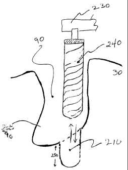
As shown in Fig. 4, the dental surgeon preferably thereafter, in accordance
with methods known in the art, utilizes a dental handpiece 230 and a bone
drill 240 to drill a
hole 210 3 to 6 mm (depth indicated by line axis 250) apically to the root
extraction socket
90. The hole 210 promotes marrow bleeding in the root extraction socket 90 and
serves as an
extension of the root extraction socket 90 into which a dental implant is
secured as explained
below.
Following drilling of the hole 210 as in Fig. 4, the dental surgeon hydrates
any
one of many bone graft materials at the area surrounding the root extraction
socket 90.
Although many bone graft materials, such as Bioglass , Osteograf , Oestrogen ,
etc. may
be utilized, Bioplant , Inc.'s Hard Tissue Replacement or HTR , which is a
synthetic bone
alloplast, is preferably used. As will be explained, using HTR to fill the
area surrounding
the implant promotes bone growth in the socket area whether used with or
without a barrier
membrane (e.g., a resorbable or non-resorbable membrane) thereby maintaining
the height
and width of the alveolar ridge and preventing the natural process of atrophy
which normally
follows root extraction, i.e, "Ridge Preservation". The dental surgeon may
additionally mix
the graft material with the patient's own bone (e.g., from the hip bone, or
from other areas of
the jawbone, e.g., chin) in order to promote faster and more effective growth
of bone in the
alveolar ridge through the use of bone precursor cells.
Although the HTR can be wetted (hydrated) with liquid antibiotic, liquid
recombinant bone-inducer factors or sterile saline solution, blood from the
surgical area of the
patient's alveolar marrow is preferably used to wet the HTR or other graft
material utilized.
Accordingly, as shown in Fig. 5a, the dental surgeon uses a filter-tipped
straight or curved
syringe 300, such as HTR -24 Straight Syringe, Item #H216102 or HTR -24 Curved
7
CA 02387845 2002-04-17
WO 01/28448 PCT/USOO/41370
Syringe, Item #H216112 available from Bioplant , Inc. of 20 North Main Street,
Norwalk,
Connecticut 06854, filled with 750 micron diameter HTR to absorb blood 310
from the
bleeding root extraction socket 90 and the hole 210. The dental surgeon
thereafter allows the
blood wetted HTR 400 to congeal for 2 to 4 minutes at the conclusion of which
time he
removes the filter tip 320 from the syringe 300. U.S. Patent Application
Serial No.
08/831,941 describing the syringe 300, and special tip 320, and a method for
using the same
is hereby incorporated in its entirety by reference.
As illustrated in Fig. 5b, during hydration of the graft material, the dental
surgeon inserts an implant 200, preferably a threaded titanium screw, into the
hole 210 apical
to the root extraction socket 90. Alternately, a HA coated screw or cylinder
or non-HA
coated cylinder implant may be used. The installation of the implant 200 is
done in the
normal manner and preferably utilizes torque reduction rotary instruments at
500 r.p.m. using
copious irrigation with chilled sterile physiological saline solution. Lower
speeds may be
used without irrigation. Hand instruments may also be used for insertion. By
installing the
implant 200 into the alveolar bone 30 at 210, the implant 90 is firmly
anchored to the alveolar
bone 30 rendering the implant sufficiently immobile. If the implant is not
sufficiently
immobile (i.e., it is loose) following implant installation, the implant 200
is removed and
replaced with a larger implant. The dental surgeon thereafter places a healing
cap 410 (i.e.,
an abutment) onto the head of the implant 200 using a hand instrument with a
rachet with no
irrigation. The dental surgeon may utilize any of the screw implants of
appropriate
composition, length and width known in the art, depending upon the size and
dimension of
the extracted tooth's socket and the state of the alveolar ridge.
As illustrated in Fig. 6, the dental surgeon then expels the wetted HTR 400
into and around the root extraction socket 90. The dental surgeon expels an
amount of wetted
8
CA 02387845 2002-04-17
WO 01/28448 PCT/US00/41370
HTRO 400 sufficient to fill the root extraction socket 90 up to the height of
the alveolar ridge
surrounding the bone voids around the dental implant 200. The wetted HTRO 400
is of a
paste-like, moldable form which lends itself to being shaped. The dental
surgeon compacts
the wetted HTRO 400 up to and surrounding the implant neck (e.g.,
"backfilling"), but does
not cover the healing cap 410. Firm but minimal pressure is used in packing
the HTRO 400,
such that the implant 200 does not move as a result of the HTRO packing. If
the implant 200
is loose enough in the root extraction socket 90 to move at this point, it
should be removed
and replaced with a larger sized implant because graft material will not help
to tighten a loose
implant. The wetted HTRO 400 adheres immediately to the alveolar bone 30 and
the implant
200, causing the root extraction socket 90 bleeding to clot.
After the remaining void of the root extraction socket 90 is filled with
wetted
HTRO 400, the dental surgeon accomplishes primary closure using any of the
varying
methods known in the art. The dental surgeon may utilize two vertical relief
incisions and
undermining using silk sutures for soft tissue closure. Alternately, the
dental surgeon may
use a surgical foil (e.g., Biofoil(g) or a collagen dressing or any other
protective device to
hold the bone graft material in place and to protect the bone graft material
from the patient's
tongue or food displacement. When HTRO is used as the bone graft material, a
dense fibrous
barrier "membrane" is naturally formed by the HTRO under the gingiva flap and,
accordingly, it is possible that no further barrier membrane or other bone
graft holding
material may be necessary.
As is known in the art, when accomplishing primary closure the dental surgeon
may leave the healing cap 410 exposed (one-stage implant) or he may
alternately cover the
healing cap via soft tissue closure (two-stage implant). As will be explained
and as is known
in the art, the two-stage implant procedure requires that an additional
surgical procedure be
9
CA 02387845 2002-04-17
WO 01/28448 PCT/US00/41370
performed at a later point after implantation whereas use of the one-stage
implant procedure
does not require that an additional surgical procedure be performed. The
dental surgeon is
free to utilize either the one-stage or two-stage implant procedure
considering such factors as,
e.g., the possibility of infection and/or other post-operative considerations.
The dental surgeon may thereafter prescribe systemic antibiotics and
analgesics for seven to ten days as is known in the art. If resorbable sutures
were used to
promote healing, removal of the sutures is not thereafter necessary. The
dental surgeon then
carefully cleans the area. The patient should keep the area clean during this
time preferably
using a germ reducing (e.g., Peridex rinse).
With reference to Fig. 7, approximately 4 to 10 months after implant
installation (depending upon the patient), the dental surgeon exposes the
healing cap 410 of
the implant 200 using gum surgery or punch techniques where the two-stage
implant
procedure was utilized. As previously explained, if a one-stage implant
procedure was
utilized, gum surgery and/or punch techniques are not necessary to expose the
healing cap
410 because the healing cap 410 will already be exposed.
In either case, as shown in Fig. 7, the dental surgeon thereafter performs
prosthetic procedures which may include the mounting of a prosthetic crown 500
on the
implant 200 in the normal manner.
Over the course of the 4 to 10 month post-implant period, the HTR will have
promoted bone growth in the area of the root extraction socket 30 by
osteoconduction such
that the implant 200 will have been osseointegrated in an HTR -bone complex
510. By the
addition of bone growth factors to a graft material's surface (e.g. BMP, OP-1,
angiogenic
factors, plasma factors, synthetic peptides, etc...), HTR is made
osteoinductive.
Osteoinductivity reduces the bone healing rate, and subsequent bone
regeneration is
CA 02387845 2002-04-17
WO 01/28448 PCT/US00/41370
considerably faster (e.g. it may take weeks instead of months or years).
Furthermore by
having immediately backfilled the extraction socket 30 with HTR or other
graft bone
substitute materials months earlier, the normal resorption rate (40 to 60% in
2 to 4 years) of
the jaw bone associated with tooth extractions is avoided. Also, because the
implant was
inserted immediately after the extraction of the root, an additional surgical
procedure, namely
the implantation, is avoided. The patient is given an immediate implant
directly after losing a
tooth.
The methods described above may be modified to support other prosthetic
structures such as a superstructure. When multiple, contiguous, and damaged or
otherwise
unhealthy roots are extracted, the above procedure is used to install an
implant into each
socket and then backfill each root extraction socket with bone graft material
in accordance
with the methods described above.
Thereafter, in accordance with the above-described method, approximately 4
to 10 months after the multiple implants are installed, the dental surgeon
exposes the healing
caps of the implants using gum surgery or punch techniques where the two-stage
implant
procedure was utilized. If a one-stage procedure was utilized, a secondary
surgical technique
is not necessary. In either case, the dental surgeon thereafter performs
prosthetic procedures
which may include the mounting of superstructure on the implants in the normal
manner.
2. Delayed Post-Extraction Implant
In addition to the above-described method and apparatus for immediately
installing an implant in a root extraction socket, i.e, immediate post-
extraction installation of
an implant, an alternate method for post-extraction installation of an implant
(i.e., delayed
11
CA 02387845 2002-04-17
WO 01/28448 PCT/USOO/41370
post-extraction installation of an implant) includes: (a) filling a root
extraction socket 90 with
bone graft material and (b) delaying implantation of an implant in the root
extraction socket
until a later time, i.e., after the bone graft material has promoted
sufficient new bone growth
in the root extraction socket.
In accordance with the above-mentioned delayed post-extraction implant
installation method, the dental surgeon proceeds as previously described with
respect to the
inunediate post-extraction implant installation implant method as shown in
Fig. 2, i.e., the
dental surgeon extracts the damaged or decayed root 20 in the normal manner.
Rather than proceeding to the step of drilling the hole 210 apically to the
root
extraction socket 90 as illustrated in Fig. 4, however, the dental surgeon
instead proceeds to
the step of hydrating the bone graft material, e.g., the HTR 400, as shown in
Fig. 5a. The
dental surgeon utilizes blood caused by the root extraction as the hydrating
agent for the
HTR .
As with the immediate post-extraction implant installation procedure, the
dental surgeon allows the blood-wetted HTR to congeal for 2 to 4 minutes in
the syringe
300 and then expels the wetted HTR 400 into the root extraction socket 90 as
shown in Fig.
6 (without implant 200). As previously described, the HTR may be mixed with
the
patient's own bone.
The dental surgeon then compacts the wetted HTR up to the gum line 50 and
accomplishes primary closure using any of the methods known in the art.
Alternately the
dental surgeon may use a surgical foil or collagen dressing to hold the graft
material in place.
The dental surgeon may thereafter prescribe systemic antibiotics and
analgesics for seven to ten days as is known in the art. If resorbable sutures
were used to
promote healing, removal of the sutures is not thereafter necessary. The
dental surgeon then
12
CA 02387845 2002-04-17
WO 01/28448 PCT/USOO/41370
carefully cleans the area. The patient should keep the area clean during this
time preferably
using a germ reducing rinse, e.g., "Peridex" rinse.
Thereafter, depending upon the patient, the dental surgeon returns to the
extraction site 2 to 12 months after extraction and installs an implant in the
HTR generated
bone complex. From the time of the implantation, the HTR will have promoted
sufficient
bone growth in the root extraction socket 90 so as to allow the secure
installation of an
implant using known methods for installing an implant in a normal, non-
atrophied jaw bone.
Any of the implants and methods for installing an implant in a normal non-
atrophied jaw
bone that are known in the art may be used.
The amount of time the dental surgeon waits prior to proceeding with implant
installation is dependent upon the patient and, more particularly, upon the
bone growth rate of
the HTR -bone complex. The longer the HTR is permitted to remain in the root
extraction
socket prior to implant installation, the greater the amount of dense bone
that will have been
created by the HTR -bone complex and, accordingly, the greater will be the
density of the
bone created in the root extraction socket. Greater bone density provides for
a more secure
implant.
As previously stated, an implant may be installed in the root extraction
socket
as early as 2-6 months after the HTR is inserted into the root extraction
socket. The bone
that will have been formed at this point by the HTR -bone complex will be
immature bone,
i.e., osteoid. If the dental surgeon installs the implant in osteoid, the
dental surgeon waits
approximately 6 months before returning to the site to install a crown 500 on
the implant as in
Fig. 7 in the manner known in the art. However, if the dental surgeon waits a
longer period
of time before installing the implant, e.g., more than 6 months, the bone into
which the
implant is installed will be more mature and, therefore, denser. Accordingly,
the dental
13
CA 02387845 2002-04-17
WO 01/28448 PCT/USOO/41370
surgeon may wait a shorter period of time, e.g., 3 months, before placing a
crown 500 on the
implant in the manner known in the art.
The above described delayed post-extraction implant installation method may
be modified to support other prosthetic structures such as a superstructure.
When multiple,
contiguous, and damaged or otherwise unhealthy roots are extracted, the
delayed post-
extraction implant method is used to backfill each of the multiple root
extraction sockets with
bone graft material and thereafter, e.g., 2 to 12 months later, an implant is
installed in each of
the root extraction sockets having HTR -bone generated complex therein in
accordance with
the above- described delayed post-extraction implant installation method for a
single root
extraction socket.
While the present invention has been particularly shown and described with
reference to the preferred embodiment thereof, it will be understood by those
skilled in the art
that various changes in form and details may be made therein without departing
from the
spirit and scope of the invention.
14