Note: Descriptions are shown in the official language in which they were submitted.
CA 02388033 2002-04-05
WO 01/24845 PCT/US00/27654
DEVICE AND METHOD FOR NONINVASIVE CONTINUOUS
DETERMINATION OF PHYSIOLOGIC CHARACTERISTICS
FIELD OF THE INVENTION
The present invention relates generally to noninvasive methods of
quantitatively
determining various physiologic parameters relating to cardiovascular and
respiratory
function. More particularly, the invention relates to a method and apparatus
for
continuous, noninvasive determination of: arterial blood pressure, venous
pressure, arterial
1o oxygen saturation, venous oxygen saturation, arterial pulse wave velocity,
aortic pulse
wave velocity, aortic pulse flow velocity, cardiac stroke volume, cardiac
output, heart rate,
and respiratory rate.
BACKGROUND OF THE INVENTION
15 Critically ill and seriously injured patients require constant care and
attention.
Doctors, nurses, and hospital technicians need a continuous flow of
information about the
many patients under their care. Heart rate and blood pressure measurements are
two
primary vital signs that indicate the health of patients under their care.
When these two
common indices of wellness fall below normal readings, a patient is usually in
distress and
20 requires immediate attention.
Dangerous conditions brought about by a cardio-vascular or pulmonary disease,
severe trauma, or drug abuse may bring about a failure of the lungs and heart
to supply the
bloodstream with life-giving oxygen. Such a fatal deficiency can be detected
by
continually gauging the amount of hemoglobin in the bloodstream that is
carrying oxygen.
25 This third vital sign, which manifests oxygen saturation of the blood, is
especially critical
because a rapid decline in oxygen in the bloodstream is associated with
increased risk of
patient mortality.
It is well known that blood pressure can be directly measured by placing a
fluid-
filled catheter directly into the vessel and coupling this to an electro-
mechanical
3o transducer. This is the most accurate means, but has all the disadvantages
of invasive
measurement, including pain on insertion, risk of infection or disease
transmission, risk of
bleeding or thrombosis, and great expense. A further disadvantage is the
creation of toxic
medical waste (needle, gloves, skin dressing, etc).
CA 02388033 2002-04-05
WO 01/24845 PCT/US00/27654
Blood pressure measurement can also be measured indirectly using an occlusive
cuff (with either auscultation or oscillometry to make the determination).
This is the most
common means of blood pressure measurement. Illustrative are Pat. Nos.
5,582,179,
5,048,533, 5,152,296 and 4,793,360.
A further occlusive cuff apparatus is disclosed in U.S. Pat. No.5,766,130.
According to the invention, the apparatus includes multiple "pressurized
pneumatic cuffs"
that are used to "plot blood pressure and/or volumetric blood flow wave forms
from a
plurality of separate digits and/or extremities of a patient so that
circulatory parameters
may be measured rapidly and recorded from a great number of the patient's
digits or
limbs".
Although commonly employed, the occlusive cuff also has numerous
disadvantages, which include discomfort, intermittent readings, and poor
reliability.
An additional means of determining blood pressure is through an assessment of
"pulse wave velocity". Several prior art references disclose methods and/or
apparatus
employing such means. Illustrative is U.S. Pat. No.5,649,543.
There are also several prior art references that disclose methods and/or
apparatus
for determining blood pressure through a "pulse wave amplitude" assessment
Illustrative
are U.S. Pat. Nos. 4,735,213, 4,872,461, 4,793,360, and 5,385,149.
Although most of the noted noninvasive blood pressure methods and apparatus,
2o particularly the occlusive cuff, have been employed for many years by
health care
personnel, the conventional methods and apparatus have one major, common
drawback -
the need for separate calibration.
Accordingly, there is a need for noninvasive methods and devices for
determining
various physiological characteristics, such as central venous pressure and
cardiac output,
without separate calibration. There is also a similar need for noninvasive
methods and
devices for determining various blood parameters including pulse amplitude,
pulse delay,
pulse velocity, pulse contour, flow velocity and flow delay.
As will be appreciated by one having ordinary skill in the art, the present
invention
satisfies these and other needs.
CA 02388033 2002-04-05
WO 01/24845 PCT/US00/27654
SUMMARY OF THE INVENTION
The present invention includes a device for the noninvasive monitoring of a
physiologic characteristic of a patient's blood. In one embodiment, the device
comprises a
tissue probe having a radiation emitter and a radiation detector configured to
receive the
radiation after absorbance through the patient's blood; a position sensor for
determining
the relative height of the probe compared to a level corresponding to the
patient's heart;
and a controller for computing the physiologic characteristic of the patient's
blood based
on the absorbance of the first wavelength of radiation and the relative height
of the probe.
The radiation emitters of the invention can utilize a single wavelength or a
plurality of
discrete wavelengths and may include visible light, infrared light, and
ultraviolet light.
The probes are adapted for use with hands, fingers, feet, toes, ears,
earlobes, nares, lips,
tongue and the like. Additional radiation emitters and detectors may also be
used.
Preferably, the probe further comprises ECG leads.
An alternative embodiment of the device of the invention comprises a tissue
probe
and controller in conjunction with a movement generator for inducing a
position change of
the probe with respect to a level corresponding to the patient's heart.
Preferably, the
movement generator induces a known position change of the probe and moves the
probe to
positions above and below a level corresponding to the patient's heart.
The invention also comprises method for determining a physiological
characteristic
of a patient's blood noninvasively. In one embodiment, absorbance
characteristics of the
blood are measured at varying positions relatively to the level of the
patient's heart. By
comparing blood parameters such as pulse amplitude, pulse velocity, pulse
delay, pulse
contour, flow velocity and flow delay to hydrostatic pressure differences
induced by the
position changes, characteristics such as arterial and central venous blood
pressure and
cardiac output can be determined. Alternatively, two probes are used to
compute pulse
delays between coupled tissues or opposing tissues.
The subject invention relates novel methods for noninvasive determination of
physiologic characteristics. The first new and unique method and device
utilizes changes
in hydrostatic pressure induced by positional changes to facilitate
measurements. A
3o second new and unique method and device for noninvasive determination of
cardiac
output by measuring delays in pulse arrival times in coupled organs or members
on
CA 02388033 2002-04-05
WO 01/24845 PCT/US00/27654
opposite sides of the body is also described. The two methods are such that
they can
advantageously be used together.
By varying the hydrostatic pressure in an extremity, one can not only perform
self
calibration for a blood pressure determination, but also change the pulse wave
velocity and
s pulse propagation delay with respect to the opposite extremity. With this
information,
pulse wave velocity, and consequently flow wave velocity at the aortic root
can be
determined.
Similar techniques of varying hydrostatic pressure can be used to assess
venous
pressure and saturation. The technique of repetitious determinations made
while altering
1o position or other variables allows a multitude of additional analyses to be
made. The
determinations can be made intermittently or continuously.
Further objects of the invention are exemplified by the following potential
applications:
(al). A patient is anesthetized for a surgical procedure. Probes are attached
to
15 the index forgers of each hand, and a movement generator is placed on one
arm. A
complete set of vital signs and physiologic characteristics is generated
continuously,
including: arterial blood pressure, venous pressure, arterial oxygen
saturation, venous
oxygen saturation, arterial pulse wave velocity, aortic pulse wave velocity,
aortic pulse
flow velocity, cardiac stroke volume, cardiac output, heart rate, and
respiratory rate. Other
2o characteristics can be calculated if desired.
(a2). A patient is anesthetized for a cardiac surgical procedure. As access to
the
arms is difficult, probes are attached to the patient's temples. A complete
set of vital signs
and physiologic characteristics is continuously generated.
(a3). A patient is anesthetized for a cardiac surgical procedure; this time
the
25 procedure includes valvular repair or replacement. Since the cardiac output
and other
characteristics can be continuously computed, the adequacy of the surgical
repair can be
judged immediately.
(a4). As the number of endoscopic or minimally invasive cardiac surgical
procedures is expected to increase, the demand for less invasive monitoring
will also
3o increase. The device described herein provides noninvasive, continuous
monitoring of
essentially all cardiovascular characteristics.
4
CA 02388033 2002-04-05
WO 01/24845 PCT/US00/27654
(a5). Cardiac catheterization procedures are often done on critically ill
patients.
As the procedures are usually relatively brief and accomplished without
general
anesthesia, invasive monitoring methods are often not desired despite the
illness of the
patients. The device described herein will provide the necessary monitoring
that is
typically provided by much more invasive, expensive, and time consuming
monitors
(a6). A patient is hospitalized in the intensive care unit of a hospital after
a heart
attack. Probes are attached to the index fingers of each hand, and a movement
generator is
placed on an arm or a leg. A complete set of vital signs and physiologic
characteristics
can be continuously generated. In addition, arrhythmias can be detected and
diagnosed.
(a7). The patient noted above is now moved to a "step-down" or telemetry unit
from the intensive care unit. Because the device described herein eliminates
the need for
invasive monitoring lines, a complete set of vital signs and physiologic
characteristics can
still be continuously generated. As the patient has mobility of arms and legs,
a movement
generator is no longer needed, as the patient's spontaneous motion, even
during sleep, will
generate hydrostatic pressures in the limbs, allowing all computations to be
made. In
addition, the probes may be made wireless, and connected to a central nursing
station by
means of infrared or radio frequency communication.
(a8). The patient noted in applications 6 and 7 above is now moved to a
regular
hospital bed, and does not require continuous monitoring. However, vital signs
can still be
2o recorded by a technician moving the device from bedside to bedside on a
cart. The device
does not require highly trained nursing personnel to operate.
(a9). The patient noted in applications 6, 7, and 8 above has now been
discharged
from the hospital, and now presents to his physician's office for follow-up.
The same
device can be used in physician's offices, as it provides better care at lower
cost.
(a10). Ambulances, emergency vehicles, and military vehicles can also employ
this device as it is very simple to operate, and provides data that currently
is impossible for
them to obtain. In addition, the information can be transmitted to central
stations where
medical personnel are available for help and advice.
(al 1). The device and methods of the invention will provide means of
monitoring
3o patients or checking vital signs for extended care facilities, nursing
homes, and other
health-related facilities
CA 02388033 2002-04-05
WO 01/24845 PCT/US00/27654
(a12). Blood pressure screening clinics and drugstores will have a greatly
improved means of determining patient's blood pressures and other vital signs.
Airports
and airplanes are able to purchase medical equipment, but often do not have
personnel
trained to operate the equipment. The device is simple and quick to operate.
(a13). The patient noted in applications 6 through 9 above can also monitor
his
heart disease and health care at home. The operation of the device is
straightforward
enough to be used by the layman with minimal instruction, and inexpensive
enough for
personal home use. The patient can measure his cardiovascular characteristics
daily, or as
frequently as he and his physician desire. A communication means, such as a
modem, can
t o easily be incorporated into the device. This, with appropriate software
and support, would
allow essentially instantaneous communication with a physician's office,
clinic, or
hospital. In addition, a permanent record can be made and stored
electronically. If
desired, the device could automatically "sign on" to the Internet or other
network, and link
to the appropriate website or other address. The ability to participate more
fully in their
own health care will improve the welfare of individuals.
(a14). The patient of above presents to the emergency room of a hospital with
chest pain. The ER physician can access, via the Internet or other means, the
patient's
vital sign history, including ECG. This allows the physician to determine if
abnormalities
are new or chronic. Changes, such as dysrhythmias, can be identified as to
when they first
occurred, perhaps to within a time frame of hours or less.
(a15). People without diagnosed cardiovascular disease can use the device to
allow themselves to participate in their own health care. This will allow
virtually
immediate diagnosis of any problems, allowing early intervention. In addition,
a
permanent record can be created if desired.
(a16). The device will impact fitness and physical training for everyone from
lay
people to military personnel to professional athletes.
(a17). The device can be employed in the diagnosis and management of
peripheral
vascular disease. Measurement of pulse wave velocity in the extremities, and
particular
differential pulse wave velocities in the lower extremities, can be used to
diagnose
3o peripheral vascular disease. Since measurements are real time and
continuous, they can
also be used in management. For example, if balloon angioplasty of an artery
is
6
CA 02388033 2002-04-05
WO 01/24845 PCT/US00/27654
performed, the clinician can tell immediately if flow has improved. In the
case of
angioplasty of coronary arteries, the clinician can follow cardiac
characteristics on a beat-
by-beat basis.
(a18). In addition to peripheral vascular disease, other diseases, such as
abdominal
aortic aneurysm, can be diagnosed and managed. Changes in pulse wave velocity
and
waveform can be followed for years if desired.
(a19). Some of the most important potential uses of the device relate to the
health
care of neonates and young children. For these patients, the measurement of
common
characteristics such as blood pressure can be difficult even for highly
trained personnel in
well-equipped facilities. The simple placement of probes on fingers will
alleviate this.
The device will also allow noninvasive diagnosis of congenital cardiac defects
and
anomalies. Analysis of differential pulse wave velocity and blood pressure
will allow
rapid, accurate, and specific diagnosis of many disorders, including Tetralogy
of Fallot and
transposition of the great vessels. The ability to distinguish both arterial
and venous
saturations and pressures will allow diagnosis of patent ductus arteriosus,
truncus
arteriosus, atrial septal defect, and ventricular septal defect. Differential
arm and leg pulse
wave velocities and pressures will confirm diagnosis of coarctation of the
aorta. Because
of its continuous measurements, the device can be used for only for diagnosis
but
confirmation of adequacy of repair, including intraoperatively. As the device
is
2o inexpensive and easy to operate, it may become a screening tool for
newborns and infants.
(a20). The device can be used in conjunction with intra-aortic balloon pump
(IABP) counterpulsation. Beat-by-beat analysis of effectiveness and ability to
wean from
counterpulsation can be made.
(a21). The device can be used in conjunction with placement of cardiac
pacemakers, to set proper rate and timing intervals. In addition, efficacy of
pacemakers
can be checked as frequently as desired, and scheduling of reprogramming or
replacement
made automatically.
(a22). It is straightforward to incorporate other devices, such as the
electroencephalogram (EEG) or electromyogram (EMG), into probes of the
invention. As
3o a general-purpose monitor, the device will invite the addition of
specialized add-ons.
CA 02388033 2002-04-05
WO 01/24845 PCT/US00/27654
(a23). Many enhancements are included in the invention. For example, addition
of chest (horizontal) leads allows full diagnostic ECGs to be performed.
(a24). Under some circumstances, such as severe hypotension, the pulse cannot
be
identified in the periphery. In such cases, many of the determinations claimed
herein
cannot be made. However, the ability of the device to identify venous blood
can still give
important information.
(a25). Forces other than gravity can be used. In a microgravity environment
such
as a space station orbiting the Earth, a device such as the one described
could be
constructed to perform all indicated determinations using acceleration caused
by
t o movement in place of gravitational acceleration.
(a26). As mentioned in the examples above, an anticipated use is in the field
of
home health care, with the possibility of automatic sign-on and direction to a
website. As
the user is already participating in his or her health care, the extension of
providing access
to related health or other information via the Internet~ is a natural one.
(a27). A verification means, such as fingerprint scanning, can be incorporated
into
a personal-use device, to ensure that any medical information gathered
belonged to the
individual using the device.
(a28). The device will be used in conjunction with the Penaz technique or
other
methods, such as calibration with a cuff or other means, as desired.
BRIEF DESCRIPTION OF THE FIGURES
Further features and advantages will become apparent from the following and
more
particular description of the preferred embodiments of the invention, as
illustrated in the
accompanying drawings, and in which like referenced characters generally refer
to the
same parts or elements throughout the views, and in which:
FIGURE 1 is a diagram of the central cardiovascular system, showing the
asymmetry of origins of the vessels off the aortic arch;
FIGURE 2 shows a representative probe of the invention with a single emitter-
detector pair;
3o FIGURE 3 shows an alternative embodiment of a probe of the invention with a
single emitter-detector pair;
s
CA 02388033 2002-04-05
WO 01/24845 PCT/US00/27654
FIGURE 4 shows a probe of the invention with two emitter-detector pairs spaced
a
known distance apart. This can be used to measure the velocity of the pulse
wave within
the probe itself;
FIGURE 5 shows a probe with a single emitter-detector pair and a single
electrocardiogram (ECG) electrode;
FIGURE 6 shows a probe with a single emitter-detector pair and two ECG
electrodes;
FIGURE 7 shows a probe with a two emitter-detector pairs and two ECG
electrodes;
1o FIGURE 8 shows a probe of the invention further comprising a position
sensor;
FIGURE 9 shows an embodiment of the invention with probes placed on opposite
digits of a subject;
FIGURE 10 shows an embodiment of the invention with probes placed on opposite
temples of a subject;
15 FIGURE 11 shows a circuit schematic of the invention comprising a
photoplethysmogram;
FIGURE 12 shows a circuit schematic of the invention comprising a
photoplethysmogram with an ECG amplifier;
FIGURE 13 shows a circuit schematic of the invention comprising a
2o photoplethysmogram with an ECG amplifier and a level signal;
FIGURE 14 shows a circuit schematic of the invention comprising a
photoplethysmogram with two independent channels;
FIGURE 15 shows a circuit schematic of the invention comprising a
photoplethysmogram with two independent channels and an ECG amplifier;
25 FIGURE 16 shows an embodiment of the invention with probes placed on the
digit
and on the arm near the brachial artery;
FIGURE 17 shows an embodiment of the invention with probes placed on a finger
and on a toe;
FIGURE 18 shows an embodiment of the invention with probes placed on opposite
30 fingers and on a toe;
9
CA 02388033 2002-04-05
WO 01/24845 PCT/US00/27654
FIGURES 19 and 20 show embodiments of the invention with probes placed on
opposite digits of a subject positioned at differential heights relative to
the patient's heart;
FIGURE 21 shows an embodiment of the invention with probes placed on
opposite fingers positioned at differential heights and on a toe; and
FIGURES 22-25 are graphical representations of an oscilloscope screen showing
recordings using methods of the invention.
DETAILED DESCRIPTION OF THE INVENTION
Functionally the heart is divided into two sides or sections. The right or
pulmonary
1 o circulation section that receives blood from the veins of the body and
pumps it through the
lungs and the left or systemic circulation section that receives the blood
from the lungs and
pumps it to the body. Th blood is then collected in the veins to be returned
to the right
side of the heart. This anatomy is generally shown in Figure 1. The arterial
system begins
at the aorta l, to which the left ventricle of the heart pumps. The first
three branches of the
15 aorta are the brachiocephalic or innominate artery 2, the left (common)
carotid artery 3,
and the left subclavian artery 4. The brachiocephalic artery branches into the
right
subclavian 5 and right (common) 6 carotid arteries. These arteries provide the
blood
supply for the head and upper extremities. The aorta then passes down (caudad)
through
the body, continuing to provide arterial branches to organs, terminating as a
bifurcation
2o creating the iliac arteries. The brachiocephalic or innominate artery is
the first branch of
the aorta. It in turn branches into the right subclavian and right carotid
arteries. In
contrast, the left subclavian and left carotid arteries originate directly off
the aortic arch.
Thus, the subclavian and carotid arteries and any of their branches have
different paths
from their counterparts on the opposite side of the body.
25 Because of the different origins from the aorta and different branching
pattern of
the arterial tree, it can be appreciated that blood ejected from the left
ventricle will not
follow symmetrical pathways to opposite arms or opposite sides of the head.
Similarly,
the pressure pulse wave associated with left ventricular ejection will follow
different
pathways, and can be expected to arrive at different times for paired organs
or members of
3o the upper body.
CA 02388033 2002-04-05
WO 01/24845 PCT/US00/27654
Measurements performed by the inventor have shown this delay can range from
less than one millisecond to several milliseconds, depending on the subject
and
circumstances. In addition, the inventor has found that this delay can be
altered by several
methods disclosed herein. This propagation delay, its alterations, and other
factors make
possible several determinations heretofore not possible by noninvasive means.
Blood pressure is the pressure exerted by the blood within a vessel upon the
wall of
the vessel. It is measured in units of force per unit area. Central venous
pressure is the
pressure within the large veins in the chest and the right atrium, which is
the common
emptying point for the venous system. Cardiac output is the amount of blood
pumped by
the heart, expressed in units of volume per time.
Central venous pressure (CVP) is defined as the distending pressure present in
the
veins in the chest (proximate to the heart), and is considered equal to the
pressure in the
right atrium (which is the emptying point for the venous system). Pressure
should be the
same throughout the venous system, but there are valves to ensure that the
blood does flow
back toward the heart (for example, when standing the venous blood must flow
uphill, and
there is no pump as on the arterial side).
As discussed in detail below, the present invention generally includes a
radiation
emitter having at least one wavelength being applied through a patient's
tissue to the
patient's blood; a radiation detector which detects reception of the at least
one wavelength
2o after absorbance through the blood, a movement generator for inducing
position changes
in the tissue; and a controller for computing the various characteristics
based on the
absorbance of the at least one wavelength of radiation at various position
levels. In a
preferred embodiment, the radiation emitter and detector are inserted in a
probe which can
be placed about the tissue/blood to be measured. A number of suitable
configurations for
probes are shown in Figures 2-8.
For example, Figure 2 shows a representative probe 10 with a single emitter-
detector pair 12. The emitter and detector are placed such that transmittance
through a
body member, such as a finger 13, is measured. Generally, any part of the body
that can be
successfully transilluminated with the radiant energy used can be utilized.
Thus, toes,
3o ears, etc. could also be used. In addition, pulse oximetry can be
accomplished with this
and all of the following embodiments. Figure 3 shows a representative probe 14
with a
11
CA 02388033 2002-04-05
WO 01/24845 PCT/US00/27654
single emitter-detector pair 16 placed such that reflectance of a body member,
such as a
finger, is measured. Further, Figure 4 shows a probe 18 with two emitter-
detector pairs 20
and 22 spaced a known distance apart. This can be used to measure the velocity
of the
pulse wave within the probe itself.
In certain embodiments of the invention, the probe comprises one or more
electrocardiogram (ECG) electrodes in conjunction with the emitter-detector
pairs. For
example, Figure 5 shows a probe 24 with a single emitter-detector pair 26 and
a single
electrocardiogram (ECG) electrode 28. Similarly, Figure 6 shows a probe 30
with a single
emitter-detector pair 32 and two ECG electrodes 34 and 36 and Figure 7shows a
probe 38
1o with a two emitter-detector pairs 40 and 42 and two ECG electrodes 44 and
46. Such
probes, if placed on opposite extremities of a patient, can be used to measure
central and
peripheral pulse wave velocity as well as ECG. Other configurations, such as
double
emitter-detector pairs and single ECG electrode, can be envisioned.
In yet other embodiments of the invention, the probe further comprises a
position
15 sensing or measuring device together with the emitter-detector pairs and/or
ECG
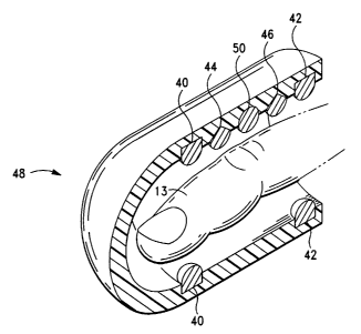
electrodes. Figure 8 shows a probe 48 similar to that shown in Figure 7 with
the addition
of a position sensor 50. This position sensor could be used in conjunction
with a position
sensor placed at heart level in order to determine the hydrostatic pressure
difference
between the two position sensors.
2o As discussed in detail herein, the invention employs hydrostatic pressure
to enable
precise self callibration of the devices in a completely non-invasive manner.
Hydrostatic
pressure affects all liquids. Gravity or other acceleration will affect both
the arterial and
venous sides of the circulation. It affects all aspects of the blood pressure
equally - mean,
systolic, diastolic. For example, an increase in height which causes a change
of 10 torr
25 will change every pressure measurement during the cardiac cycle by this
amount.
For example, if the "true" blood pressure (taken level with the heart) is
120/80,
when the arm is raised an amount needed to decrease the measured pressure by
10 torr, the
measured pressure in the arm will be I 10/70 . The pulse pressure will be the
same, but the
transmural pressure will be 10 torn lower at all times. In addition, the
vessel will be
3o smaller at all points.
12
CA 02388033 2002-04-05
WO 01/24845 PCT/US00/27654
The heart is taken to be the center of the circulatory system, and all values
are in
reference to it. This is not necessary for the practice of the invention, but
serves as
reference points for values in the current medical literature.
The electromagnetic radiation in this description will refer to light in the
visible
and infrared range although, as noted in the attached claims, it is
conceivable that other
forms could be used.
Similarly, while the present invention primarily describes the use of
transillumination, it will be appreciated that reflectance spectrophotometry
may
alternatively be employed.
O~eratin P~ciples
It is well known that Incident radiation passing through a body part is
attenuated
(absorbed) in the tissue. The theoretical basis for spectrophotometic
techniques is Beer's
law (the Beer-Lambert-Bouguer law) which expresses the incident intensity in
terms of
transmitted intensity and extinction coefficients of the tissue compartments
through which
the radiation has passed. The equation can be written as:
Eq.l 1n(1 / lo) = E*C*L
Where:
Io = the incident intensity of the source radiation;
I = the transmitted intensity of the source through the sample;
E = the extinction coefficient of the component of interest;
C = the concentration of the component in the tissue itself;
L = the optical path length (distance) through the absorber; and
E*C*L = absorbance.
Beer's law and the practice of spectrophotometry and oximetry have been
exhaustively reviewed in the literature. Generally, pulse oximetry in effect
filters out
3o signals other that pulsating (AC). In the body, it can be assumed that the
pulsatile
13
CA 02388033 2002-04-05
WO 01/24845 PCT/US00/27654
component of the signal is arterial blood, while all other tissue absorbers
should be non-
pulsatile (DC).
An additional feature of this invention, not found in any previous disclosure,
is the
use of hydrostatic pressure changes to vary the amount of venous blood within
a body
member such as a finger. Thus, hydrostatic changes can be used in a similar
manner to the
pulse to perform measurements on both arterial and venous blood. If a finger
is contained
within a probe, raising the probe will lower the hydrostatic pressure of all
vessels in the
finger, both arterial and venous. Both arteries and veins (and arterioles and
venules) will
be smaller due to lower pressure distending their walls. Most change will
occur on the
1o venous side of the circulation due to lower pressure. Total absorbance of
the finger will
decrease. As the arterial oxygen saturation can be measured by pulse oximetry,
the venous
oxygen saturation can be calculated in a similar manner.
A light signal of a known intensity and wavelength is produced by means of
light-
emitting diodes (LEDs) as in currently used oximeters or, as in one possible
embodiment,
a broad-band light source whereby wavelengths are isolated by a rotating
filter or diffusion
grating. In the latter case, the emitted light is distilled through a filter
which allows a
known wavelength and intensity of light to penetrate. Use of tunable lasers or
other
equipment is also possible. If the light source is proximate to the point of
use, no further
mode of
2o transmission will be needed. If it is not, the light will be transported to
the desired point
by means such as a fiber optic cable, preserving the wavelength and intensity.
Several means of motion induction are possible. Various means of position
measurement are also possible. For example, a liquid filled tube with an end
open to the
atmosphere can be employed. Other position sensors are known to those having
skill in
the art, and include electromagnetic, spectroscopic, and chemical means.
A broad-band photo detector (in the case of visible or infrared light) or
other means will be
utilized to measure the quantity of transmitted light.
To generate a single data point, the movement induction means is used
to bring the forger (or other space of interest) to a known position relative
to the heart.
3o Light of known wavelength and intensity is emitted (and transmitted if
necessary) on the
surface of interest. Detection of the light signal at a distinct point
(normally opposing
14
CA 02388033 2002-04-05
WO 01/24845 PCT/US00/27654
surface) is made and the relative absorbance and extinction of the signal is
calculated.
Signal processing is used to determine the pulsatile portion of the signal.
The arrival time
of the pulse is recorded, as is the amplitude and waveform. This measurement
may be
repeated one or more times to ensure the accuracy of the measurement; this can
be done
within a very short time frame (less than a millisecond).
To generate multiple data points, the process outlined in the previous step
will be
repeated at the next chosen wavelength, while still at the same predetermined
position. The
range and number of wavelengths can be selected, and changed for different
applications.
Once the desired number of wavelengths has been examined, the movement
1 o induction means would bring the finger or other volume to a predetermined
second
position, and the data collection of steps would be repeated. At the
completion of
measurements and determinations for this second position, the movement
induction means
will bring the space to a third predetermined position, and the measurements
and
determinations repeated. This process would be continued until the desired
range of
positions has been scrutinized.
In order to make computations of pulse propagation delay, identical
measurements
would be made simultaneously with a probe on the same member on the opposite
side of
the body. For example, if one probe were placed on the index finger of the
right hand, the
other probe would be placed on the index finger of the left hand.
2o Because the arterial path to the arm is essentially identical after the
second part of
the subclavian artery, any differences in pulse wave velocity and pulse wave
propagation
time must occur prior to this point; that is, very close to the root of the
aorta. In any case,
pulse wave velocity increases rapidly as the pulse wave propagates down the
aorta and into
the periphery (Fung). Thus, any timing differences in the periphery will be
greatly
reduced by the high wave velocity, leaving central effects as the most
prominent.
The apparatus of the invention can be operated intermittently or continuously.
In
the intermittent mode, a single set of calculations can be used for analysis
to produce the
determinations claimed. However, the device can also be easily operated in
continuous
mode, with the process outlined above repeated as often as wished (constantly
if desired).
3o In addition, a rapid ("stat") mode can be offered with the minimum number
of
CA 02388033 2002-04-05
WO 01/24845 PCT/US00/27654
measurements made that will provide an accurate estimation of correct values.
Such a
rapid mode would be useful in emergency situations.
While this methodology should give precise values, further adjustment may be
desired to compensate for any discrepancies between theoretical and in vivo
s measurements. Contemporary oximeters in fact use a calibration curve when
determining
oxygen saturation, with the curve being generated with data from normal
volunteers.
Calculations and Analysis
The following algorithms are further examples of the use of the present
invention.
1o Some variables have degrees of co-dependence. In these cases, values are
calculated by
iterative computational techniques.
Generally, measurement of pulse wave amplitude and timing is made using probes
such as that shown in Figure 2, using methods similar to standard oximetry
described in
the prior art. As shown in Figure 9, a first probe 52 is placed on a finger
and set at a known
15 position relative to the heart. Another, simultaneous measurement of pulse
wave
amplitude and timing is made by a second probe 54 placed on a finger on the
hand
opposite that of the first probe. The pulse delay occurring between the two
measurements
is made. Alternatively, as shown in Figure 10, probes 52 and 54 can be placed
on opposite
temples of the patient to measure pulse wave values and delay. The probes can
also be
2o placed on the patient's ears.
From this information alone, an estimate of pulse wave velocity at the aortic
root
could be made, by utilizing a table of normal values for the distance of the
central
anatomical difference.
If a measurement of blood pressure is then made, one can perform the following
2s calculation:
Eq.2 p=c*u*p
Where:
3o c = pulse wave velocity;
a = flow wave velocity; and
16
CA 02388033 2002-04-05
WO 01/24845 PCT/US00/27654
p = the density of the blood (approximately 1.055 grams/cm3)
According to the invention, p and c have been measured, p is known. This
allows one to solve for a , which is the flow wave velocity at the aortic
root. This by itself
is a measure of cardiac output. If one makes an estimate of aortic root
diameter, one can
then compute cardiac stroke volume.
Techniques described by O'Rourke and others describe reconstruction techniques
that can be used to convert or "transform" peripheral blood pressures and
waveforms to the
corresponding pressure and waveform at the aortic root. Ideally, the blood
pressure at the
1o aortic root should be used as the pressure term in Fung's equation.
One can improve on the above determination in several ways. The first way is
by
additionally measuring the peripheral pulse wave velocity. To do this,
measurement of
pulse wave amplitude and timing is made by a first probe such as that shown in
Figure 5.
The probe is at a set known position relative to the heart. Another,
simultaneous
measurement of pulse wave amplitude and timing is made by a second probe
placed on a
finger on the hand opposite that of the first probe. The pulse delay occurring
between the
two measurements is made. The respective peripheral pulse wave velocities are
also
computed. If the peripheral pulse wave velocities are different, it can be
assumed that this
is because of the different central anatomies from which the respective pulses
traveled.
2o This information alone may be enough to compute central pulse wave velocity
from a
table of normals. However, when combined with the pulse wave delay
information, this
data enables one to construct a function of pulse wave speed from the
periphery back to
the aortic root, thus giving another measure of central pulse wave velocity.
Another method of the invention is to vary the position of the probes relative
to the
2s heart. If the first probe is at heart level and the second probe is raised
above (with respect
to the earth) heart level, the hydrostatic pressure of the blood vessels
within the second
probe will be lower than those within the first probe. In turn, in accordance
with Fung's
equation stated above, this means that the pulse wave velocity of the arterial
vessels within
the second probe will be lower than that in the arterial vessels within the
first probe. This
3o will change both the measured pulse delay between the two probes, and the
measured
17
CA 02388033 2002-04-05
WO 01/24845 PCT/US00/27654
peripheral pulse wave velocities. This creates additional measurements by
which to
compute central pulse wave velocity.
According to the invention, changes in hydrostatic pressure are controlled by
the
following equation:
Eq.3 p=p*g*h
Where:
p = blood density;
1 o g = gravitational acceleration; and
h = height above a reference point (with respect to the earth).
The difference in hydrostatic pressure between the vessels in two probes is
thus
governed completely by their difference in heights relative to the heart
(referenced to the
t 5 surface of the earth). Therefore, a known change in position produces a
known change in
hydrostatic pressure.
According to the invention, the above measurements can be employed to derive a
number of physiological properties. Preferably, the probes of the invention
are connected
to a controller to aid the data collection and analysis used to make the
desired
2o determination. The controller includes a computing device or standard
personal computer
(PC) with a monitor. Included within the controller are algorithms for the
calculation of
variables not measured directly.
For example, Figure 11 shows a circuit schematic for a one or two wavelength
photo-plethysmograph. Emitters 56 and 58 and detector 60 are positioned
adjacent the
25 tissue being measured, such as a finger 61. Emitters 56 and 58 are driven
by drive
circuitry 62, which is in turn governed by control signal circuitry 64.
Detector 60 is
connected to amplifier 66. The signal from amplifier 66 is sent to demodulator
68, which
is also synched to control signal circuitry 62. The signal from the
demodulator 68 is sent
to analog-digital converter 70. The desired computations are performed on the
output
30 from the converter 70 by signal processor 72 and the results sent to
display 74. Emitters
56 and 58 operate specific wavelengths, such as 805 nm, and may comprise light
emitting
18
CA 02388033 2002-04-05
WO 01/24845 PCT/US00/27654
diodes (LEDs) or laser diodes. Detector 60 preferably comprises a silicon
photodiode.
Such emitter-detector pairs are shown in Figures 2 and 3.
Figure 12 shows a schematic of an alternate embodiment of suitable circuitry.
As
with Figure 10, emitters 76 and 78 are connected via LED drive circuitry 79
and control
signal circuitry 80 to demodulator 82. Signal from detector 84 is amplified at
circuit block
86 and sent to demodulator 82. Output from demodulator 82 is sent to A/D
converter 88.
In addition, ECG leads 90 are connected to differential amplifier 92 and the
signal is sent
to converter 88. Output from converter 88 is processed at block 94 and the
results sent to
display 96. A probe such as those shown in Figures 5 and 6 may be used with
the
1o circuitry. The ECG leads are preferably silver/silver chloride or stainless
steel.
Yet another embodiment of the invention is shown in Figure 13. Emitters 98 and
100 are connected via LED drive circuitry 101 and control signal circuitry 102
to
demodulator 104. Signal from detector 106 is amplified at circuit block 108
and sent to
demodulator 104. Output from demodulator 104 is sent to A/D converter 109. ECG
leads
t 5 110 are connected to differential amplifier 112 and the signal is sent to
converter 109.
Digit level sensor 114 and heart level sensor 116 are connected to amplifier
118 and the
signal is sent to converter 109. Output from converter 109 is processed at
block 120 and
the results sent to display 122.
Figure 14 shows a circuit schematic suitable for use with a probe having two
2o physically independent channels, such as the one shown in Figure 4. A first
emitter-
detector pair comprising emitters 124 and 126 and detector 128 are positioned
adjacent the
tissue being measured, such as a finger. A second pair comprising emitters 132
and 134
and detector 136 are positioned a selected distance from the first pair.
Emitters 124, 126,
132 and 134 are driven by drive circuitry 138, which is in turn governed by
control signal
25 circuitry 140. Signal from detector 128 is amplified by block 142 and sent
to demodulator
144. Independently, signal from detector 136 is amplified and demodulated at
blocks 146
and 148, respectively. Output from demodulators 144 and 148 is sent to analog-
digital
converter 150. The desired computations are performed on the output from the
converter
150 by signal processor 152 and the results sent to display 154.
3o An alternative embodiment configured for use with a probe having two
physically
independent channels and an ECG lead, such as the one shown in Figure 7, is
19
CA 02388033 2002-04-05
WO 01/24845 PCT/US00/27654
schematically shown in Figure 15. A first emitter-detector pair comprising
emitters 156
and 158 and detector 160 are positioned adjacent the tissue being measured,
such as a
finger. A second pair comprising emitters 164 and 166 and detector 168 are
positioned a
selected distance from the first pair. Emitters 156, 158, 164 and 166 are
driven by drive
circuitry 170 which is in turn governed by control signal circuitry 172.
Signal from
detector 160 is amplified by block 174 and sent to demodulator 176.
Independently, signal
from detector 168 is amplified and demodulated at blocks 178 and 180,
respectively.
Output from demodulators 176 and 180 is sent to analog-digital converter 182.
ECG leads
184 are connected to differential amplifier 186 and the signal is also sent to
converter 182
1o The desired computations are performed on the output from the converter 182
by signal
processor 188 and the results sent to display 190.
As one of ordinary skill in the art will appreciate, the placement of the
various
probes discussed above will effect the types of measurements that can be
taken. As
discussed above, Figures 9 and 10 show probes placed on opposite extremities
to enable
measurement of pulse wave delay. Figure 16 shows an embodiment of the
invention with
probe 52, such as in Figure 1, placed on the digit, and a probe 54, such as in
Figure 2,
placed on the arm near the brachial artery. This could measure the pulse wave
velocity in
the arm (as well as pulse oximetry). A similar embodiment could measure pulse
wave
velocity in the leg. Figure 17 shows probes 52 and 54 placed on a finger and
on a toe to
2o measure the pulse wave delay. Figure 18 shows probes 52 and 54 placed on
opposite
digits and probe 55 placed on a toe. This allows measurement of the
differential pulse
wave delay between the fingers and toe, and allows calibration of the toe
probe to be used
in place of a finger probe (if only one finger probe could be used, such as in
hand surgery).
The use of appropriate probes also allows a diagnostic-quality ECG. Figures 19
and 20
2s show probes 52 and 54 placed on opposite digits. One arm of the subject is
placed at the
level of the heart, while one arm is moved to different positions, both above
and below the
level of the heart. By generating different hydrostatic pressures in the
vessels, the pulse
velocity and hence pulse wave delay changes. In addition, the amplitude of the
pulse
wave, and amplitude of venous absorbance changes. This allows the additional
3o computations of arterial blood pressure and venous pressure. Figure 21
shows probes 52
and 54 placed on opposite digits and probe 55 placed on a toe. The
differential hydrostatic
CA 02388033 2002-04-05
WO 01/24845 PCT/US00/27654
pressures in the vessels allow measurements of pulse wave velocity and pulse
wave delay,
as well as arterial blood pressure and venous pressure. Use of probes with
suitable ECG
leads will also allow the invention to perform a diagnostic-quality ECG. In
addition, heart
rate and respiratory rate can be calculated, and cardiac output and several
other
cardiovascular characteristics computed.
As discussed above, the controllers of the invention preferably output the
results of
the measurements and computations to a display. Figure 22 shows an
oscilloscope screen.
The two tracings are from pulse oximeter probes, such as those shown in Figure
1, placed
on the index fingers of both hands. The pulse wave delay is visable as the
slight phase
t0 difference between the two tracings. As the probes are at the same level,
the pulse
amplitudes are essentially identical. Figure 23 shows the oscilloscope screen
after the
hand with the probe displayed as the top tracing has been placed at a level
higher than the
heart and the hand with the probe displayed as the bottom tracing has been
placed at a
level lower than the heart. The induction of a pressure differential between
the two probes
~ 5 effects a change in the pulse delay. The change in pressure also
correspondingly alters the
pulse amplitudes. Figure 24 shows the oscilloscope screen after the hand with
the probe
displayed as the top tracing has been placed at a level lower than the heart
and the hand
with the probe displayed as the bottom tracing has been placed at a level
higher than the
heart. Here, the pulse delay has substantially reversed as have the pulse
amplitudes.
20 Figure 25 shows an oscilloscope screen displaying an electrocardiogram in
conjunction
with a pulse waveform.
The algorithms outlined below serve as examples, but modifications are
possible to
arrive at the indicated results, and are meant to be included within the
spirit of this
application. Various additional components of the device will be discussed in
more detail
25 below with reference to the following examplary determinations.
(dl). Determination of Arterial Blood Pressure
A probe such as that shown in Figure 1 is placed on an extremity, and that
extremity is moved in relation to the heart. As mentioned above, the
hydrostatic pressure
within the arteries and arterioles changes as a function of height with
respect to the heart.
3o Because of this, both the pulse wave velocity and pulse wave amplitude
change as a
function of probe height. These two parameters can be mapped against known
distance
21
CA 02388033 2002-04-05
WO 01/24845 PCT/US00/27654
above or below the heart. In this way, function curves of pressure vs. pulse
wave
amplitude and pressure vs. pulse wave velocity can be drawn. For example, a
full
excursion of the arm in a standing adult produces hydrostatic changes of
greater than 50
cm of water in both directions. Using an arm and a leg, a gradient of well
over 200 cm of
water can be generated. This is a significant portion of the normal blood
pressure range,
and certainly enough to produce the function curves mentioned above.
There is a huge amount of medical literature describing arterial behavior, so
the
curves can be extrapolated if necessary. These curves serve as calibration.
It can thus be determined if "recalibration" is necessary -- if either pulse
amplitude
or pulse wave velocity changes, and the other parameter does not change
correspondingly.
In other words, a shift on one curve should matched by a corresponding shift
on the other
curve. If this shift does not occur as predicted, recalibration is required.
Of course, the
process of recalibration is the simple procedure outlined above.
In a preferred embodiment, a first probe having a position sensor is placed
level
with the patient's heart. A second probe, such as one shown in Figure 8,
having a position
sensor and a pulse detector is placed on the patient's finger. The patient's
arm is held out
level with the heart so there is zero displacement between probes. Pulse
amplitude is
recorded from probe. The patient's arm is slowly raised, while pulse amplitude
and
relative displacement of probe are recorded. The hydrostatic pressure
difference between
2o probes is also computed. By comparing the recorded pulse amplitude to the
hydrostatic
pressure difference, a mathematical function relating pressure to pulse
amplitude can be
derived. Preferably, circuitry similar to that shown in Figure 13 is used to
aid the process.
This process is repeated while lowering the arm back to heart level, then
lowering the arm
to below heart level and, finally, raising the arm back to heart level.
Similar steps can be
applied to measure pulse delay, pulse velocity and pulse contour.
(d2). Determination of Cardiac Output
Cardiac output can be determined by measuring delays in pulse arrival times in
coupled organs or members on opposite sides of the body. In a preferred
embodiment of
the invention, probes such as those shown in Figure 1, having sensors for
detecting a
3o patient's pulse are placed on opposite fingers of the patient. The patient
positions both
arms straight out from the side. The blood pressure of the patient can be
determined either
22
CA 02388033 2002-04-05
WO 01/24845 PCT/US00/27654
through conventional means or by the methods of the invention. The pulse delay
between
the two probes can be measured utilizing circuitry such as that shown in
Figures 14 or 15,
for example. The dicrotic notch of the pulse may be determined by standard
methods, and
used to calculate the ejection time based on the timing. The size of the
aortic root can be
estimated by standard means and the consequently the pulse distance
differential at the
aortic root. This allows the calculation of the pulse velocity c at the aortic
route by the
following equation:
Eq. 4 c=(pulse distance)/(pulse delay).
The value of c can then be used to solve for flow wave velocity based on the
following equation:
Eq.S p=c*u*p
~ 5 Where:
c = pulse wave velocity;
a = flow wave velocity; and
p = density of the blood (approximately 1.055 grams/cm3).
2o According to the invention, cardiac stroke volume can be determined by
multiplying the aortic root area by the flow wave velocity and by the cardiac
ejection time.
Cardiac minute output can be calculated by multiplying the cardiac stroke
volume by the
pulse rate. These steps may be augmented by raising and lowering the patient's
arms with
respect to each other to vary the pressure and the pulse wave velocity.
25 Alternatively, cardiac output can be determined by placing probes such as
those
shown in Figure 5 on a patient's finger and toe. The probes measure oxygen
saturation at
each pulse. The oxygen saturation for each pulse at the first probe is
compared to the
oxygen saturation of that pulse and subsequent pulses at the second probe.
With
continuous monitoring, this allows the determination matching oxygen
saturation, within
3o given tolerance limits, of the pulses from the probes. The patient's blood
volume and the
physical separation of the probes can be determined by standard methods. This
allows the
23
CA 02388033 2002-04-05
WO 01/24845 PCT/US00/27654
computation of caridac stroke volume by dividing the blood volume displaced by
the
number of pulses. Then, the cardiac minute output can be calculated by
multiplying the
cardiac stroke volume by pulse rate. Circuitry such as that shown in Figures
11 or 12 is
suitable for use with this embodiment.
(d3). Determination of Venous Saturation and Pressure
Determination of arterial oxygen saturation can be determined by pulse
oximetry
and techniques well delineated in both the patent and medical literature.
Hydrostatic
changes as described in this application allow the determination of venous
saturation and
pressure as well.
Place a probe such as that shown in Figure 1 on a finger. Make measurements of
both total absorbance and pulsatile absorbance. Raise the probe a known
distance. Again
measure both total absorbance and pulsatile absorbance. Both will be
decreased. This is
because the pulse amplitude is less because the arterial blood pressure within
the probe is
less (due to decrease in hydrostatic pressure). However, the total absorbance
will also
15 decrease, as the distending pressure in the venous system is less, and
hence the veins and
venules are smaller. All changes in absorbance can be assumed to be due to
changes in
blood volume. Saturation is calculated using the ratios of absorbance of
distinct
wavelengths.
In one embodiment, the central venous pressure (CVP) can be estimated. A probe
2o containing a position sensor is place level with a patient's heart. A
second probe, such as
the one shown in Figure 8, also comprising a position sensor is placed on the
patient's
finger. The patient positions the arm so that the second probe is initially
lower than the
first probe. The total absorbance measured at the second probe is continuously
monitored.
The patient's arm is slowly raised, and the rate of change of absorbance of
the second
25 probe is computed with respect to the relative displacement to the first
probe. When the
rate of change changes by a predetermined amount representing an abrupt
decrease, the
arm position corresponding to the point of central venous drainage has been
reached. The
CVP can then be calculated by computing the hydrostatic pressure difference
between the
first probe and the second probe at that arm position. The circuitry shown in
Figure 13 is
3o suitable for use with this embodiment.
24
CA 02388033 2002-04-05
WO 01/24845 PCT/US00/27654
(d4). Determination of Heart Rate
According to the invention, heart rate can be determined by counting the
pulsatile
arterial signal for a known length of time, or by the ECG impulse.
(d5). Determination of Respiratory Rate
The impedance changes of the chest due to filling and emptying can be measured
from the electrocardiogram tracing. During normal breathing, negative pressure
is created
within the chest by lowering of the diaphragm and expansion of the rib cage.
This
negative pressure causes blood to empty more rapidly from the peripheral into
the central
veins. This is also the case when respiration is assisted by a negative-
pressure device such
1 o as the "iron lung".
During modern mechanically-assisted ventilation (with "ventilators"), positive
pressure is created within the chest by forcing air into the lungs. For both
positive- and
negative-pressure ventilation, expiration is passive. This respiratory
variation by itself can
be used as an estimate of cardiac filling, giving left heart pressures. This
determination
~ 5 can be assisted by the use of the hydrostatic techniques described above.
(d6). Diagnosis of Congenital Heart Disease and Anatomic Anomalies
Diagnosis of many disorders with anatomic anomalies can be made by the
detection of unexpected propagation times, and abnormal propagation delays
between
right- and left-sided organs.
2o The ability to measure both arterial and venous saturation, as well as
arterial and
venous pressures, can aid further in investigations.
(d7). Diagnosis of Dysrhythmias
By measuring blood pressure and the electrocardiogram simultaneously, the
diagnosis of dysrhythmias can be aided greatly. Both arterial and venous
pressure are
25 recorded with the ECG, allowing differentiation of atrial vs. ventricular
arrhythmias.
(d8). Determination of Additional Cardiovascular Characteristics
By measuring blood pressure and the electrocardiogram simultaneously, many
additional characteristics, such as systolic and diastolic pressure time
indices, can be
determined.
3o An enormous amount of information can be gleaned from the use of probes on
opposite sides of the body combined with hydrostatic perturbations. It is
important to
CA 02388033 2002-04-05
WO 01/24845 PCT/US00/27654
realize that the time of arrival of a pulse to paired members is different,
but the velocity of
the pulse is also different. Examination of pulse propagation time, pulse
propagation
phase or delay, pulse velocity, and pulse amplitude yields four parameters
that may change
in different ways for each perturbation. Particularly, raising and lowering an
arm by the
same amount may give different changes. Raising and lowering the other arm by
the same
amount may give still different changes. Further, raising an arm by a given
amount, then
raising again by the same amount, may give different changes. Raising the
other arm by
the given amount, then raising again by the same amount, may give still
different changes.
Similar effects can be obtained by lowering the extremity.
Without departing from the spirit and scope of this invention, one of ordinary
skill
can make various changes and modifications to the invention to adapt it to
various usages
and conditions. As such, these changes and modifications are properly,
equitably, and
intended to be, within the full range of equivalence of the following claims.
26