Note: Descriptions are shown in the official language in which they were submitted.
CA 02388448 2008-04-24
METHOD AND APPARATUS FOR OPHTHALMIC REFRACTIVE CORRECTION
SPECIFICATION
TECHNICAL FIELD
The invention relates to refractive laser ablation systems, and, more
particularly,
an excimer laser refractive ablation system employing an aperture card that
passes a
unique ablation profile.
BACKGROUND ART
Systems for reprofiling the eye for refractive correction have become
extremely
popular. Such systems typically employ a 193-nanometer (nm) argon-fluoride
excimer
laser, passing the light to the corneal tissue, where a very precise amount of
tissue is
"ablated" from the eye with a laser shot. A variety of delivery mechanisms are
commercially used, including systems in which a fixed spot size is moved over
the
surface of the eye, in which the spot size is varied, and in which erodible
masks are
placed in the path of the excimer laser beam. In all of these systems, the
ultimate goal is
to change the profile of the comeal surface by volumetrically altering the
amount of
tissue within the cornea. Further, these techniques have been employed on the
surface of
the cornea underneath the epithelia using a technique for photorefractive
keratectomy
(PRK) as well as the intra-cornea technique known as LASIK, or laser in situ
keratomileusis.
U.S. Patent 5,376,086, issued to Khoobehi et al., discloses a laser surgical
method
of sculpting a patient's cornea that uses a mask system with multiple openings
in which
laser power transmission is controlled through the use of diffi=action and
absoiption.
Each hole in the mask acts like an individual light source, distributing laser
power as a
function of the hole's size, shape, and overlaid coatings. By summing the
power output
of each hole pattern over a given area, an average power distribution is
generated. The
I
CA 02388448 2002-03-27
WO 01/28478 PCT/EP00/10379
technique, however, is limited because the power transmission of the mask is
tailored
only to a particular comeal surface by using topographical information of that
surface.
The corneal surface topographic data is the controlling mechanism for
constructing the
pattern of the mask itself. The surgeon can observe the topographic
information and then
pattern the mask according to that topographic information.
The present invention is directed to improving laser ablation of eye tissue
that
avoids or reduces shortcomings of previous methods.
SUMMARY OF THE INVENTION
According to certain features of the invention, a laser refractive ablation
system
for the eye, such as a 193 nm excimer laser system, is implemented to pass a
unique
"truncated" intensity ablation profile, for example, a non-Gaussian profile or
a truncated-
Gaussian profile referred to herein as a "soft spot" profile. The "top" of the
soft spot
profile is substantially flattened whereas the sides of the profile slope
until an ablation
intensity threshold is reached, at which point the edge or sides become nearly
vertical.
The profile is provided using an aperture card prepared based on diffractive
effects. The
aperture card preferably includes 1 and 2 millimeter (mm) apertures surrounded
by a
plurality of extremely small holes referred to herein as "soft spot" apertures
that allow
the diffractive effect of the laser light to accumulate to form the desired
profile. Further,
the aperture card includes a "square-sided" profile aperture (referred to
herein as a "hard
spot" aperture) for testing the fluence of the excimer laser system.
The aperture card is intended for a single surgical use because it can exhibit
changes in characteristics over time, although in some embodiments, this is
not
necessarily true. The card is preferably loaded for single use into the system
from an
aperture card holder, and transported into place using a horizontal and
vertical movement
robot mechanism. Then, a laser system determines whether the aperture card is
properly
positioned, inhibiting lasing action if the card is not positioned within
tolerance.
Alternatively, the laser system can determine the position and adjust the
computed
ablation profile or otherwise adjust the optical system to adapt for the
misalignment of
the aperture within the aperture card.
2
CA 02388448 2002-03-27
WO 01/28478 PCT/EP00/10379
BRIEF DESCRIPTION OF THE DRAWINGS
Figure 1 illustrates a laser refractive ablation system for the eye in
accordance
with an embodiment of the invention;
Figure 2 illustrates the laser system of Figure 1 that includes an aperture
card in
accordance with an embodiment of the invention;
Figure 3 illustrates a mechanism for holding an aperture card in place in
accordance with an embodiment of the invention;
Figure 4 is another view of the mechanism in Figure 3;
Figure 5 illustrates a view of a portion of the mechanism of Figure 3,
including
the aperture card of Figure 2 in accordance with an embodiment of the
invention;
Figure 6 illustrates another view of the portion of Figure 5 with the aperture
card
of Figure 2 removed;
Figure 7 illustrates a mechanism for holding an aperture card in place
according
to another embodiment of the invention;
Figure 8 illustrates an alternative position and alignment mechanism for the
aperture card corresponding to Figure 7;
Figure 9 is a mechanical drawing illustrating an aperture mask that forms a
portion of an aperture card in accordance with an embodiment of the invention;
Figure 10 illustrates a square-sided spatial intensity profile of an aperture
having
a square edge that forms part of the aperture mask of Figure 7 in accordance
with an
embodiment of the invention;
Figure 11 illustrates a "soft" spot aperture in accordance with an embodiment
of
the invention;
Figure 12 illustrates a useful ablation profile passed by a soft spot aperture
in
accordance with an embodiment of the invention;
Figure 13 illustrates a comparison between a square profile and a soft spot
profile
in accordance with an embodiment of the invention; and
3
CA 02388448 2002-03-27
WO 01/28478 PCT/EP00/10379
Figure 14 illustrates a comparison between a square profile and a soft spot
profile
with their resulting tissue ablation profiles.
MODE(S) FOR CARRYING OUT THE INVENTION
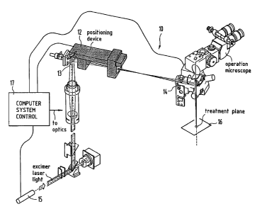
Turning to Figure 1, an exemplary excimer laser system 10 is illustrated in
which
a unique profile and aperture card handling or conveying system 12 (also
referred to as a
linear translation module) is shown in accordance with an embodiment of the
invention.
The system 12 takes the place of an iris diaphragm for beam dimensioning. It
is
mounted in a frame between a bending mirror 13 and a scanner block 14 (see
Figure 1).
Specifically, the excimer laser system is a typical 193 nm excimer laser
system. It
includes an excimer laser 15 and operates as a scanning laser system employing
mirrors
(e.g., galvanometer driven high precision mirrors for 193 nm) to scan the
laser beam to
appropriate points on the cornea in a treatment plane 16. Preferably, the
laser system 10
employs an eye tracking system with a tracking speed of at least 100 Hz. The
laser
system 10 is controlled by a control system 17, for example, a computer. The
control
system either can compute locally a shot pattem to achieve a desired ablation
profile, or
can receive an ablation profile remotely, such as according to U.S. Patent
5,891,132
entitled "Distributed Excimer Laser Surgery System," and issued to Hohla. Such
systems will be understood by those skilled in the art. Further, other lasers
than excimer
lasers may be used.
In any case, according to one aspect in the invention, the system is
implemented
to receive an aperture card or application card 100 as shown in Figure 2. The
card 100 is
precisely positioned within the laser path of the excimer laser system to pass
light
through apertures forming part of the aperture card. In one embodiment, the
aperture
card is a mask holder similar to a card-based system for chip card designs
that includes a
set of several high precision drillings used as reference points to the
geometrical
assembly of the card. The accuracy of these devices is typically down to less
than 30
microns on both axes, and manufacturing processes can be automated and checked
by
microscopic measuring tools. In an alternative embodiment the aperture card is
positioned and aligned via pressure points and fixation points in conjunction
with
4
CA 02388448 2002-03-27
WO 01/28478 PCT/EP00/10379
precision machining and manufacturing. This produces single use card
positioning with
a repeatable accuracy in the order of 5 m or better.
Referring to Figure 3, an aperture card system 12 is illustrated according to
an
embodiment of the invention. Figure 4 shows another view of the aperture card
system
12. After starting the laser system 10 and entering patient data in the
computer 17,
software of the system 10 requires the aperture card 100 to be inserted into
the system
12. Arrow 101 shows the direction in which the card 100 is inserted into the
system 12,
preferably from a sleeve or holder (not shown) that protects the aperture card
100 and
keeps it clean. The aperture card 100 is fed in a first orientation, for
example, laterally,
to card catchment or receiving machinery 103 (e.g., a lateral loading
mechanism) by
hand through a slot 102, although, in other embodiments, this can be
automated. The
card catchment machinery 103 pulls the aperture card 100 inside the laser
system 10. A
pickup-and positioning sled 104 (e.g., a vertical loading mechanism) moves
forward to
the card catchment machinery 103, as generally indicated by arrow 101' in
Figure 3,
which transfers the aperture card 100 over to the pickup-and positioning sled
104. The
pickup-and positioning sled 104 moves backwards, generally indicated by arrow
101" in
Figure 3, to a desired diaphragm position, and loads the aperture card 100 in
a second
orientation, for example, vertically, for accurate positioning and locking
(e.g., in a
vertical position) in the optical path of the excimer laser 15 by pins, as
described below.
Then, the pickup-and positioning sled 104 extracts itself to a place away from
the
secured card 100 and out of the way of the laser shots. The laser treatment
procedure of
the eye then can be started.
Also shown in Figures 3 and 4 are three positioning holes 105, which are used
to
position the card 100 on pins within the laser system 10, as will be described
below.
Near the middle of the card 100 is an aperture mounting slot 106 to hold an
aperture
mask 108. The mask 108 is mounted into the slot 106 of the aperture card 100,
preferably being glued in place.
Referring to Figure 5, according to an embodiment of the invention, the
aperture
card 100 is illustrated positioned, after transfer, in an aperture card holder
200 of the
laser system 10. As can be seen, three pins 202 pass through the pin holes 105
(see also
CA 02388448 2002-03-27
WO 01/28478 PCT/EP00/10379
Figure 3) for precise positioning of the aperture card 100 (one pin 202 is
obscured from
view in Figure 3 and all are obscured in Figure 4). Further, in some
embodiments, an
alignment hole 204, shown in Figures 3-5, is provided for reference by lasers
alignment
within the excimer laser system 10 to align the aperture card 100 within the
system
before the aperture card 100 is employed. The laser system determines whether
the
aperture card 100 is properly positioned, inhibiting lasing action if the card
is not
positioned within tolerance. Alternatively, the laser system 10 can determine
the
position and adjust the computed ablation profile or otherwise adjust the
optical system
to adapt for the misalignment of the aperture within the aperture card. Figure
6
illustrates the pins 202 in more detail along with clips 208 used to hold the
aperture card
100 (not shown in Figure 5) in place in the aperture card holder 200. It will
be apparent
to those skilled in the art that variations on this embodiment could be used
to mount the
aperture card 100 in position, including, for example, a different number of
pins like the
pins 202 and a different number of holes in the card 100 like the holes 104.
In an alternative aspect of this embodiment, illustrated with reference to
Figures 6
and 7, a pressure based mechanism is used in place of alignment holes and
positioning
pins to position, align and secure the aperture card 100. Figure 7 shows a
preferred
aperture card system 220 having a common structure, in part, with the aperture
card
system 12 in Figures 3 and 4. The primary distinction resides in the
replacement of the
alignment pins 202 (Figure 5) and corresponding alignment holes 105 in the
aperture
card 100 with fixation points 222 and pressure points 224, 224v as shown in
Figure 8.
In an aspect of this embodiment, the fixation points 222 comprise three
hardened
cylinder pins that are press fit into the card holder 26 with high accuracy.
The card 100
is pushed into the holder 226 from right to left (as viewed in Figure 8) until
the left edge
227 of the card touches fixation point 222Y and the bottom edge 229 of the
card touches
fixation points 222v1 and 222,2. The card is fixated against the fixation
points by
pressure points 224x, 224,, which, preferably, are spi'ings. By manufacturing
the card 100
with high precision such that the exact location of the apertures are known,
and the
fixation points engage the card edges at the same locations, repeated
positioning of the
cards has shown a measured accuracy of 5 m or better.
6
CA 02388448 2002-03-27
WO 01/28478 PCT/EP00/10379
The mask 108 is preferably constructed of an opaque coating (e.g., chromium
deposited or otherwise coated or layered) on quartz. More preferably, the
coating is a
multilayer coating including a layer of titanium overlaying the chromium
layer, and a
layer of gold overlaying the titanium layer. Most preferably, the multilayer
coating
consists of a chromium layer approximately 80 nm thick adjacent the substrate,
a
titanium layer approximately 40 nm thick adjacent the chromium layer, and a
gold layer
approximately 80 nm. thick adjacent the titanium layer. The aforementioned
layered
coating advantageously reduces unwanted reflection of laser light. The mask
includes a
2 mm effective diameter (or substantially 2 mm effective diameter) "soft spot"
(defined
in more detail below) aperture, a similar 1 mm (or substantially 1 mm
effective diameter)
soft spot aperture, and a center, 2 mm effective diameter (or substantially 2
mm) "hard"
aperture having a square edge. Unlike the mask in the aforementioned U.S.
Patent
5,376,086, patterning of the mask 108 is independent of eye topography data
and can be
used for any eye topographical surface, including any corneal surface.
Depending upon
the optical system employed by the excimer laser system 10, the actual overall
diameter
of the apertures referred to above may be larger or smaller than the
corresponding image
or irradiance pattern projected onto an eye. For example, typical
illustratively useful 2
mm diameter and 1 mm diameter spots on the eye can correspond to 3 mm and 1.5
mm
overall diameter aperture patterns, respectively.
The "hard" square edge aperture is used for fluence testing with a standard
fluence plate, such as a polyethylene foil coated on both sides with aluminum.
It is
preferable to perform the fluence test with a square edge aperture for system
calibration
because it is easier to see how many shots are required to ablate through
material from
one layer to the next or to penetrate to a particular depth using a square
edge profile
beam than it is with the rounded profile ablation of a non-square edge
aperture.
Referring to Figure 9, a mechanical drawing is illustrated of an aperture mask
300, which is exemplary of the aperture mask 108 that can fit into the
aperture slot 106,
and preferably glued into place. As shown in Figure 9, a "hard"-edged, center
aperture
302 has an overall diameter of 3 mm or substantially 3 mm that simply passes a
standard
square-sided (i.e., square-profiled) laser shot, such as that shown in Figure
10. A 3 mm
overall diameter soft spot aperture 304 includes a center aperture area 305
surrounded by
7
CA 02388448 2002-03-27
WO 01/28478 PCT/EP00/10379
a pattern of microscopically small holes 306 that, through direct diffractive
effects, create
the appropriate ablation profile. Finally, a third soft spot aperture 307 is
1.5 mm or
substantially 1.5 mm in overall diameter, which also includes a center
aperture area 308
and small microscopic holes 309 similar to those of the aperture 304 to create
the
appropriate ablation profile, an exemplary embodiment of which is discussed
below in
connection with Figure 11. Although Figure 9 illustrates the aperture mask 300
having a
single 3 mm diameter hard edged aperture, a 3 mm diameter soft spot aperture,
and a 1.5
mm diameter soft spot aperture, it is contemplated that more or less of these
numbers and
types, and possibly different diameters, of these apertures could be included
in the
aperture mask 300, all of which are included within the scope and spirit of
the present
invention. As discussed above, these apertures typically are projected onto
the eye either
reduced or enlarged; here, the 3 mm diameter and 1.5 mm diameter apertures
preferably
create 2 mm diameter and 1 mm diameter spots on the eye.
Prior to performing an ablation, the card holder 200 moves (e.g., laterally),
according to the desired excimer laser system, to employ a fluence test (in
which case the
3 mm center square profile aperture 302 is placed into position) and then for
the laser
ablations of the eye, either the soft spot aperture 304 (e.g., 2 mm imaged
spot or 307
(e.g., 1 mm imaged spot is placed into position. The soft spot apertures are
employed in
laser ablating by laterally moving the aperture card 100 (e.g., left and
right) within the
aperture card holder 200.
Again, the soft spot apertures 304 and 307 of Figure 9 are focused by the
system
10, preferably down to 2 mm and 1 mm or substantially those values,
respectively.
Again, the apertures 302, 304, and 307 are preferably formed on a quartz plate
308, using
a suitably deposited mask as described herein. The mask can then be
appropriately
etched using a laser etching system, as will be appreciated by those skilled
in the art.
Alternatively, photolithography, silicon wafer technology, chip card
technology, or other
techniques can be employed to create the mask 300.
Figure 11 is a more detailed illustration of an exemplary soft spot aperture
350
that can be used for the soft spot apertures 304 or 307. The soft spot
aperture 350 is
shown having a central open aperture 352 like the apertures 305 and 308 of
Figure 9,
surrounded by microscopic holes 354 like the holes 306 and 309. Once the
spatial
8
CA 02388448 2002-03-27
WO 01/28478 PCT/EP00/10379
intensity profile is specified, a variety of known techniques can be employed
to design
and position the holes, and knowledgeable artisans would be able to create
such an
aperture. By providing a spatial intensity profile, a soft spot aperture that
would produce
such a profile (e.g., like that of Figure 11) can be obtained from Fraunhofer
Institut
Siliziumtechnologie, Faunhoferstra(3e 1, D-25524 Itzehoe, Germany, and from
others.
Referring to Figure 12, a useful ablation profile (or spatial intensity
distribution)
400 passed by the soft spot aperture 304 is shown in accordance with an
embodiment of
the invention. In Figure 12, the profile is normalized and only one-half the
profile 400 is
illustrated, solely for simplicity of the drawing, it being understood that
the full profile
400 would be as if mirrored about the ordinate axis of Figure 12. The aperture
307
would pass a similar, but narrower, profile. As can be seen, a center portion
401 of the
aperture profile 400 is flat or substantially flat, whereas an edge 402 of the
profile 400,
continuous with the portion 401, is rounded. The portion 401 is preferably
symmetric
about the radius of the profile and extends across about 60-80%, and, more
preferably,
across about 65-70% of the profile 400. At a certain point, such as an
intensity threshold
point 404 at which the eye tissue ablation intensity threshold is no longer
reached, the
profile 400 preferably quickly drops off or diminishes as a substantially
square, vertical,
or truncated edge 406. The ablation threshold and any variations in it are
known in the
art. The amount of energy falling below the threshold for ablation is
preferably about
5% or less of the total energy encompassed by the profile 400. The profile 400
is non-
Gaussian, for example, between square and Gaussian-shaped, or a truncated
Gaussian.
Thus, an automatic system is employed to position the aperture card 100 at the
position in which the aperture mask 300 passes an ablation profile with a
substantially flat
or flattened top, rounded edges, and substantially truncated sides for
ablation of corneal
tissue. Further, the card 100 can be laterally moved into positions for laser
shots at a larger
"soft edged" spot size, a smaller "soft edged" spot size, or at a center
portion suitable for
adjusting fluence levels.
One advantage of the profile 400 of Figure 12 is that, by having a "flat" top,
a
relatively unifonn ablation is produced, which assists in steepening the sides
of an
ablation, although the resulting profile in tissue is more round than for a
square profile.
This, like for a Gaussian profile, is also advantageous in avoiding a "haze"
that can result
9
CA 02388448 2002-03-27
WO 01/28478 PCT/EP00/10379
from square profile ablations. The goal in the design and use of the profile
400 is to
maximize the ablation per shot while avoiding the haze encountered with square
profile
ablations. The width of the flat portion and the amount of energy below
threshold, as
discussed above, is driven by this goal. Also, the rounded and vertical edges
reduce the
"stair-step" effect of typical ablations with square-sided ablation profiles,
which could
affect healing. Further, by employing the profile 400, by appropriate mask 300
design,
nearly all of the eye tissue can be removed that normally would be removed
with a
square-sided ablation profile passed, for example, by the aperture 302, but
with rounded
and vertical edges. Thus, the soft spot profile combines both the advantages
of the
Gaussian profile and the square profile.
In Figure 12, although there is a small portion 408 of the ablation profile
400 that
might not reach the necessary ablation threshold, this portion is designed to
be a very
small fraction of the overall ablation profile 400. By contrast, typically for
a Gaussian
profile, a much larger amount of energy falls below the ablation threshold.
When the
ablation threshold is not reached, typically the tissue merely is heated,
rather than
ablated. Without ablation, the resulting heating diminishes one of the
advantages of a
193-nm laser system. It is therefore desirable to reduce this effect because
heating may
effect a later ablation of the heated tissue or other nearby tissue, or may
have other
effects, such as producing scarring or opacities. The total energy below
threshold of the
profile 400 is preferably limited to reduce heating while maximizing ablation.
By
designing the portion 408 to be small, possible detrimental thermal heating
below
threshold is reduced or minimized. Figure 13 illustrates a comparison of a
square profile
(e.g., if the aperture 302 or a similar aperture were used to ablate) with the
soft aperture
profile 400. Little of the total energy of the profile 400 appears outside the
square
profile, as indicated by the outside hatched area.
Compared to standard ablation profiles, the profile 400 of Figure 12 has many
advantages, including that it uniformly removes nearly all of the eye tissue
normaliy
removed by a square-sided ablation profile (e.g., typically within 90%,
although other
designs, such as 80%, are possible), but its edges are still "rounded,"
thereby rounding
the tissue removed on each laser shot. Figure 14 illustrates a comparison
between a
square profile 500 with a resulting laser shot ablation profile 504 in eye
tissue and a
CA 02388448 2002-03-27
WO 01/28478 PCT/EP00/10379
profile 5021ike the profile 400, according to the invention, with its rounded
laser shot
ablation profile 506 in eye tissue. By rounding the edges of the tissue
removed in each
shot, advantages also include allowing iris tracking systems to more
continuously and
accurately track the location of the pupil. This is because using square-sided
shot
profiles, the "haze" discussed above can appear on the eye before a LASIK flap
is
returned into position that can interfere with the ability of the eye tracker
to track the
pupil position on the eye. Further, there are fewer healing effects and the
eye can more
quickly return to a natural sight condition when the edges are rounded.
It is possible that the aperture mask 300 of Figure 9 (or the aperture mask
108, or
even the aperture card 100, of Figure 4) may wear out more quickly than a
typical
diaphragm card using an excimer laser system. This is for a variety of
reasons, including
the possibility that the quartz employed for the aperture mask 300 will become
slightly
more opaque over time. Because chromium-on-quartz is the preferred
manufacturing
technique, it is desirable thus to replace the aperture card with each excimer
treatment for
a new patient in the interest of surgical precautionary considerations and to
achieve the
highest quality ablations, in addition to reproducibility. The card can, for
example, be
treated with a laser blast or provided with an electronic signature that
disables the card
from further use.
Further, the aperture card 100 can also incorporate electronic circuitry to
provide
the laser system 10 information and validation of procedures to be performed.
For
example, the aperture card 100 can include an SLE 4428 secure chip memory.
This
typically is encoded with a variety of data, such as the serial number of the
machine (or
machines, such as for a laser center) on which the card 100 is validated for
use, the
number of procedures available with the particular card 100, what types of
treatments are
permitted, and perhaps storage of a control number and patient name after the
treatment
is performed for tracking purposes. With sufficient storage, the actual
treatment profiles
or iris alignment and verification data could also be included. Preferably,
the electronics
prevent use of the excimer laser system without appropriate validation, such
as through a
PIN number, and prevent extra uses of the card 100.
A variety of techniques can be employed to create ablation profiles using
these
soft spot apertures for custom refractive correction in which the laser system
determines
11
CA 02388448 2002-03-27
WO 01/28478 PCT/EP00/10379
the shot pattern necessary to achieve the ablation profile. In a preferred
embodiment,
laser beams having the soft spot intensity profile of this invention are used
in a dual-
mode, laser surgery system for the eye. In the dual mode system, the eye is
first treated
("shaped") for primary corneal defects, such as myopia, hyperopia, and
astigmatism,
using a larger, fixed spot size. Then, a smaller fixed spot size is used to
remove
remaining irregularities ("polished"). The larger size spot provides faster
treatment. The
smaller size spot provides more precision in the treatment of irregular
topographies. The
size of the larger spot is desirably a relatively large fraction of the
typical area of the
cornea to be subject to ablation. The larger spot is typically between about 2
and 3.5
mm, preferably about 2 mm in diameter. The smaller spot is typically not
larger than
about 1 mm in diameter, and is preferably about 1 mm in diameter. For example,
in U.S.
Patent application Serial No. 09/591,313 and International Patent Publication
WO
98/48746 (PCT application No. PCT/EP98/02428), a dual spot size system is
described
that employs 1 mm and 2 mm square profile spot sizes that are scanned over a
corneal
surface to create a desired ablation profile. As a particular, non-limiting
example, the 2
mm soft spot aperture can be used to treat 80% of a desired ablation in a
lower resolution
first pass and the remaining 20% treated with the 1 mm soft spot aperture in a
higher
resolution second pass. Other relative percentages are possible.
Using the scanning ("Plano-scan") techniques described in U.S. Patent
(U.S. Patent application Serial No. 08/324,782; PCT application No.
PCT/EP95/04028, and in International Patent Publication WO 94/07447 (PCT
application PCT/EP93/02667), the soft aperture spots of the present invention,
in another
embodiment, can be employed to create any desired ablation profile, and
particularly,
customized ablation profiles for irregular ablations other than simple myopic,
hyperopic,
and astigmatic profiles. Moreover, the soft spot apertures of the present
invention, in
another embodiment, can be employed to create spiral shot patterns and
randomized shot
patte:-r,s using the techniques described in International Patent Publication
WO 94/11655
(PCT application PCT/EP95/04028). These techniques can include ablations made
with
a single, fixed spot size.
Eye topography systems, such as the ORBSCAN and ORBSCAN II by Bausch
& Lomb/Orbtek , Inc., Salt Lake City, Utah, are known in the art. Eye
topography data,
12
WLII;~7M'js:Z~.wir; Technoias GmbH Ophthalmologische Systeme SIECERTSTR. 4
f3
Our Ref.: D 2510 PCT 1675 h/l U NG HE N
CA 02388448 2002-03-27
Jan. 2002
preferably elevation-based eye topography data, including comeal topography
data, as well as wavefront sensor data, for example, as disclosed in U.S.
Patent 5,777,719,
issued to Williams et al., can be used by an eye surgeon or automated for
identifying
regions of the cornea requiring ablations for vision correction. Such
techniques are
known to those skilled in the art. These data can be transformed for use in
conjunction
with the soft spot apertures and the aperture card of the present invention
for making
ablations in performing customized refractive correction surgery. _
Although preferred embodiments of the present invention have been described in
detail herein above, it should be clearly understood that many variations
and/or
modifications of the basic inventive concepts taught herein, which may appear
to those
skilled in the art, will still fall within the spirit and scope of the present
invention as
defined in the appended claims and their equivalents.
13
1 AMENDED SHEET 14.-01-2002