Note: Descriptions are shown in the official language in which they were submitted.
CA 02388861 2009-12-31
-I-
S
SYSTEM AND METHOD OF TREATING ABNORMAL TISSUE IN THE
HUMAN ESOPHAGUS
FIELD OF THE INVENTION
A system and method for treating abnormal epithelium in an esophagus.
BACKGROUND OF THE INVENTION
Two of the major functions of the human esophagus are the transport of food
from intake to the stomach and the prevention of retrograde flow of
gastrointestinal
contents. The retrograde flow is, in part, prevented by two esophageal
sphincters
which normally remain closed and which are functional rather than distinct
entities.
In particular, a lower esophageal sphincter normally remains closed until
parasympathetic activation causes its relaxation, allowing food to pass into
the
stomach from the esophagus. Various types of food and other activity may cause
relaxation of the sphincter, such as fatty meals, smoking and beverages having
xanthine content. Certain drugs or pharmaceuticals also may cause relaxation
of this
lower esophageal sphincter, as well as localized trauma or other problems such
as
neuromuscular disorders.
Regardless, patients having such difficulties may present with clinical
indications including dysphagia, or difficulty in swallowing, as well as more
classic
symptoms of heartburn and other similar complaints. Recurrent problems of this
nature often lead to a disorder known as reflux esophagitis, consisting of
esophageal
mucosa damage due to the interaction of the gastric or intestinal contents
with
portions of the esophagus having tissue not designed to experience such
interaction.
As suggested above, the causative agent for such problems may vary.
The treatment for the underlying cause of such inflammatory mechanisms is
not the subject of this patent application, but rather the invention is
focused on
treatment of secondary damage to tissue in the effected region of the
esophagus.
CA 02388861 2009-12-31
2
SUMMARY OF THE INVENTION
An ablation catheter and method of use is provided to endoscopically
access portions of the human esophagus experiencing undesired growth of
columnar epithelium. The ablation catheter system and method includes
controlled depth of ablation features and use of either radio frequency
spectrum,
non-ionizing ultraviolet radiation, warm fluid or microwave radiation, which
may
also be accompanied by improved sensitizer agents.
In accordance with an aspect of the present invention, there is provided a
system for ablating abnormal tissue from a human esophagus, comprising:
a. energy distribution means for distributing radio frequency energy,
microwave energy, ultraviolet light energy or energy generated by a heated
fluid
medium, said energy distribution means associated with an expandable member
sized and conformed to the shape of the esophagus such that it is positioned
and
expanded in said esophagus;
b. power means for powering the energy distribution means at levels
appropriate to ablate human tissue within said human esophagus to a
predetermined depth of ablation; and
c. control means designed for accurate control and positioning of the
member.
In accordance with another aspect of the present invention, there is
provided a system for ablating abnormal tissue from a human esophagus,
comprising:
a. energy distribution means capable of distributing radio frequency
energy, microwave energy, ultraviolet light energy or energy generated by a
heated fluid medium; associated with an expandable member, sized and
conformed to the shape of the esophagus such that it is positioned and
expanded
in said esophagus wherein the energy distribution means comprises an
expandable balloon having an electroconductive member associated with its
outer surface and wherein the electroconductive member comprises a pattern
that is at least one of a plurality of bipolar rings spaced one from the
other, a
plurality of monopolar rectangles spaced one from the other, or a bipolar
axial
pattern of interlaced finger electrodes spaced apart one from the other;
b. power means for powering the energy distribution means at levels
appropriate to ablate human tissue within a human esophagus; and
c. control means designed for accurate control and positioning of the
expandable member.
CA 02388861 2009-12-31
2a
In accordance with another aspect of the present invention, there is
provided the endoscopic use of an expandable member in a proximate portion of
a human esophagus to ablate abnormal tissue wherein the expandable member
is connectable to a power source for generating radio frequency energy,
microwave energy, ultraviolet light energy or thermal energy transmitted from
a
heated fluid mechanism, the expandable member being expandable and
positionable so as to be able to provide properly focused energy to a site of
abnormal tissue for ablation of the tissue, and further wherein ablation
energy is
providable to a portion of the expandable member to effect tissue ablation.
In accordance with another aspect of the present invention, there is
provided a system for ablating abnormal tissue from a human esophagus,
comprising:
a. an expandable member shaped and configured for insertion into,
positioning and expansion in a human esophagus connected to an energy
distributing device capable of distributing radio frequency energy, microwave
energy, ultraviolet light energy or thermal energy transmitted from a heated
fluid
medium;
b. a power source for powering the energy of distributing device at
levels appropriate to ablate human tissue within a human esophagus; and
c. control apparatus designed for accurate control and positioning of
the expandable member within the esophagus.
In accordance with another aspect of the present invention, there is
provided the endoscopic use of an expandable member proximate a portion of a
human esophagus for accessing and ablating abnormal tissue that is Barrett's
epithelium, variants of Barrett's epithelium, dysplastic tissue or malignant
tissue, wherein the expandable member is connectable to a power source for
generating radio frequency energy, microwave energy, ultraviolet light energy
or
thermal energy transmitted from a heated fluid medium; and wherein the
expandable member being expandable and positionable so as to provide properly
focused energy to a site of abnormal tissue for ablation of the tissue and so
that
its outer surface may be firmly pressed into the abnormal tissue to be ablated
so
that blood flow to the tissue is reduced or prevented; and energy is
providable to
a portion of the expandable member to effect tissue ablation.
In accordance with another aspect of the present invention, there is
provided a device for ablating abnormal tissue from a human esophagus,
comprising:
CA 02388861 2009-12-31
2b
a. a balloon catheter comprising an inflatable balloon positioned on
the distal end of a catheter said balloon configured to expand to the shape of
the
esophagus;
b. energy distribution means comprising an electrode array positioned
on the outside surface of the balloon capable of distributing radio frequency
energy; and
c. power means configured to power the energy distribution means so
that energy is applied to the tissue at levels appropriate to ablate the
tissue to a
predetermined depth of ablation.
In accordance with another aspect of the present invention, there is
provided a device for ablating abnormal tissue from a human esophagus,
comprising:
a. a balloon catheter comprising a flexible shaft having a proximal and
distal end and an inflatable balloon positioned on the distal end of the shaft
said
balloon configured to expand to the shape of the esophagus;
b. radio frequency energy distribution means comprising an electrode
array position around the circumference of a predetermined length of the
balloon; and
c. power means connected to the energy distribution means configured for
powering the energy distribution means to apply energy to the tissue at levels
appropriate to ablate the tissue to a predetermined depth of ablation.
In accordance with another aspect of the present invention, there is
provided a device for ablating abnormal tissue from a human esophagus,
comprising:
a. a balloon catheter comprising a flexible shaft having a proximal and
distal end and an inflatable balloon positioned on the distal end of the
shaft; the
balloon configured to expand to a predetermined shape and diameter of said
esophagus, the diameter being selected so that when the balloon is positioned
in
the esophagus at the site of ablation and inflated to its full diameter, its
outer
surface will be firmly pressed into the esophageal tissue to be ablated so
that
blood flow to the tissue is reduced or prevented;
b. energy distribution means comprising an electrode array
positioned
on the outside surface of the balloon capable of distributing radio frequency
energy uniformly to the tissue of the circumference of the inner lumen of the
esophagus when the balloon is inflated; and
CA 02388861 2010-11-08
-2c-
c. power means configured to power the energy distribution means
so that
energy is applied to the tissue at levels appropriate to ablate the tissue to
a predetermined
depth of ablation.
Various embodiments of this invention provide a system for ablation of
abnormal
tissue from a human esophagus, comprising: an elongated member; an expandable
member
coupled to a distal portion of the elongated member and sized to be positioned
and
expanded in the esophagus; and a plurality of RF electrode pairs at least
partially extending
around a circumference of the expandable member, a width of each RF electrode
and a
spacing between adjacent RF electrodes selected to provide a selectable
ablation of an
esophagus mucosal layer, a spacing between adjacent RF electrodes being no
more than 2
mm, and the plurality of RF electrode pairs being arranged in a pattern.
Various embodiments of this invention provide a system for ablation of
abnormal
tissue from a human esophagus, comprising: an elongated member; an expandable
member
coupled to a distal portion of the elongated member and sized to be positioned
and
expanded in the esophagus; and a plurality of RF electrode pairs at least
partially extending
around a circumference of the expandable member, the plurality of RF
electrodes being
arranged in a pattern of bipolar axial interlaced finger electrodes, a width
of each RF
electrode and a spacing between adjacent RF electrodes selected to provide a
selectable
ablation of an esophagus mucosal layer.
Various embodiments of this invention provide a system for ablation of
abnormal
tissue from a human esophagus, comprising: an elongated member; an expandable
member
coupled to a distal portion of the elongated member and sized to be positioned
and
expanded in the esophagus; and a plurality of RF electrode pairs at least
partially extending
around a circumference of the expandable member, the plurality of RF
electrodes being
arranged in a pattern of RF monopolar electrodes, a width of each RF electrode
and a
spacing between adjacent RF electrodes selected to provide a selectable
ablation of an
esophagus mucosal layer.
Various embodiments of this invention provide a system for ablation of
abnormal
tissue from a human esophagus, comprising: an elongated member; an expandable
member
coupled to a distal portion of the elongated member and sized to be positioned
and
expanded in the esophagus; and a plurality of bipolar RF electrode pairs at
least partially
extending around a circumference of the expandable member, a width of each RF
electrode
and a spacing between adjacent RF electrodes in a pair selected to provide a
selectable
CA 02388861 2012-03-22
-2d-
ablation of an esophagus mucosal layer, the plurality of RF electrodes being
arranged in a pattern of
bipolar axial interlaced finger electrodes, wherein a spacing of electrodes in
an electrode pair is 2
mm or less.
Various embodiments of this invention provide a system for ablation of
abnormal tissue
from a human esophagus, comprising: an elongated member; an expandable member
coupled to a
distal portion of the elongated member and sized to be positioned and expanded
in the esophagus;
and a plurality of RF electrodes at least partially extending around a
circumference of the
expandable member, a width of each RF electrode and a spacing between adjacent
RF electrodes
selected to provide a selectable ablation of an esophagus mucosal layer, the
RF electrodes being
arranged as a contiguous sequence of arrays and a spacing between adjacent RF
electrodes being no
more than 2 mm.
Various embodiments of this invention provide a system for ablation of
abnormal tissue
from a human esophagus, comprising: an elongated member; an expandable member
coupled to a
distal portion of the elongated member and sized to be positioned and expanded
in the esophagus;
and a plurality of bipolar RF electrode pairs at least partially extending
around a circumference of
the expandable member, a width of each RF electrode and a spacing between
adjacent RF
electrodes is selected to provide a selectable ablation of an esophagus
mucosal layer, a spacing
between adjacent RF electrodes being no more than 2 mm, the RF electrodes
being arranged as a
contiguous sequence of arrays with a single common RF electrode along an
entire length of the
arrays, each electrode pair having an electrode that is divided into a
sequence of selectable lengths.
Various embodiments of this invention provide an energy delivery device for
use in
ablating mucosal tissue in an esophagus, wherein at least a portion of the
energy delivery device is
for positioning at a mucosal tissue surface of the esophagus to deliver
sufficient energy from the
energy delivery device to the mucosal tissue surface to create a lesion in the
mucosal tissue, while
controlling a depth of the lesion.
Various embodiments of this invention provide an energy delivery device
comprising a
dilation and ablation catheter, a balloon member for expansion in an esophagus
and a plurality of
RF electrodes positioned on an outside surface of the balloon member, for use
in treating a tissue
site in the esophagus, wherein at least a portion of the energy delivery
device is configured for
positioning at a mucosal surface of the tissue site in the esophagus and to
deliver sufficient energy
to create a lesion with a controlled depth in the mucosal tissue.
CA 02388861 2012-03-22
-2e-
Various embodiments of this invention provide an energy delivery device that
includes a
plurality of RF electrodes for use in treating a tissue site in an esophagus,
wherein a width of
each RF electrode and a spacing between adjacent RF electrodes is selectable
for control of a
depth of ablation in a mucosal layer of the esophagus and at least a portion
of the energy
delivery device is configured for positioning at the tissue site in the
esophagus to deliver
sufficient energy to create a desired ablation depth in the mucosal layer.
BRIEF DESCRIPTION OF THE DRAWINGS
Figure 1 is a schematic view of portions of an upper digestive tract in a
human.
Figure 2 is a schematic view of a device of the invention, in an expanded
mode, within an
esophagus.
Figure 3 is a schematic view of a device of the invention.
Figure 4 is a photograph of the device of Figure 3.
Figure 5 is a view of a device of the invention.
Figure 6 shows the electrode patterns of the device of Figure 3.
Figure 7 shows electrode patterns of that may be used with a device of the
invention.
DETAILED DESCRIPTION OF THE INVENTION
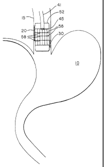
Various inflammatory disorders result in human patients who experience
retrograde flow of
gastric or intestinal contents from the stomach 10, as shown in Figure 1, into
the esophagus 15.
This flow is shown by arrows A and B in Figure 1. Although the causation of
these problems are
varied, this retrograde flow may result in secondary disorders which require
treatment independent
of and quite different from treatments appropriate for the primary disorder ¨
such as disorders of
the lower esophageal sphincter 18. One type of inflammatory disorder is known
as Barrett's
esophagus, in which the stomach acids, bile acids and enzymes regurgitated
from the stomach and
duodenum enter into the lower esophagus causing damage to the esophageal
mucosa. Indeed, when
this type of retrograde flow occurs frequently enough, damage may occur to
esophageal epithelial
cells 20. When normal replacement of damaged cells is overcome by the rate of
damage, then the
result may be symptomatic destruction of the healthy squamous epithelium. When
this occurs, the
squamous cells can be replaced by columnar epithelium 30 of the lower
esophageal passageway. It
is well established that although some of the columnar
CA 02388861 2002-05-15
WO 01/35846 PCT/US00/31561
-3-
cells may be benign, others may result in adenocarcinoma. Accordingly,
attention has
been focused on identifying and removing this columnar epithelium in order to
mitigate more severe implications for the patient. Examples of efforts to
properly
identify these growths, referred to as Barrett's epithelium or more generally
as
Barrett's esophagus, have included conventional visualization techniques known
to
practitioners in the field. Although certain techniques have been developed to
characterize and distinguish such epithelium cells, such as disclosed in
United States
Patent No. 5,524,622 and United States Patent No. 5,888,743, there has yet to
be
shown efficacious means of accurately removing undesired growths of this
nature
from portions of the esophagus to mitigate risk of malignant transformation.
Means for accomplishing this procedure according to this invention includes
use of the radio frequency spectrum at conventional levels to accomplish
ablation of
mucosal or submucosal level tissue. Such ablation is designed to remove the
columnar growths 30 from the portions of the esophagus 15 so effected. In one
embodiment, as shown in Figure 2, an elongated flexible shaft 41 is provided
for
insertion into the body in any of various ways selected by the medical
provider. The
shaft may be preferably placed endoscopically, e.g. through the esophagus, or
it may
be placed surgically, or by other means. Radiant energy distribution means is
provided at a distal end 45 of the flexible shaft to provide appropriate
energy for
ablation as desired. It is recognized that radiant energy of a preferred type
includes
radio frequency energy, microwave energy, or ultraviolet light, the latter
possibly in
combination with improved sensitizing agents. It is also recognized that
another
embodiment of this invention may utilize heatable fluid as an ablation energy
medium.
In one embodiment the flexible shaft comprises a coaxial cable surrounded by
an electrical insulation layer and comprises a radiant energy distribution
means
located at its distal end. In one form of the invention, a positioning and
distending
device around the distal end of the instrument is of sufficient size to
contact and
expand the walls of the body cavity in which it is placed (e.g. the esophagus)
both in
the front of the distribution means as well as on the sides of the
distribution means.
For example, the distal head of the instrument can be supported at a
controlled
distance from the wall of the esophagus by an expandable balloon member 52 so
as to
CA 02388861 2009-12-31
-4-
regulate and control the amount of energy transferred to the tissue comprising
the
esophageal wall. The balloon is preferably bonded to a portion of the flexible
shaft at
a point spaced from the distal head means.
Another embodiment comprises using the distending or expandable balloon
member as the vehicle to deliver the ablation energy. A critical feature of
this
embodiment includes means by which the energy is transferred from the distal
head
portion of the invention to the membrane comprising the balloon member. For
example, one type of energy distribution that may be appropriate
is shown in United States Patent No. 5,713,942, in which an
expandable balloon is connected to a power source which provides radio
frequency
power having the desired characteristics to selectively heat the target tissue
to a
desired temperature. The balloon 52 of the current invention may be
constructed of
an electroconductive elastomer such as a mixture of polymer, elastomer, and
electroconductive particles, or it may comprise a nonextensable bladder having
a
shape and a size in its fully expanded form which will extend in an
appropriate way to
the tissue to be contacted. In another embodiment, an electroconductive member
may
be formed from an electroconductive elastomer wherein an electroconductive
material
such as copper is deposited onto a surface and an electrode pattern is etched
into the
material and then the electroconductive member is attached to the outer
surface of the
balloon member. In one embodiment, the electroconductive member, e.g. the
balloon
member 52, has a configuration expandable in the shape to conform to the
dimensions
of the expanded (not collapsed) inner lumen of the human lower esophageal
tract. In
addition, such electroconductive member may consist of a plurality of
electrode area
segments 58 having therrnistor means or the like associated with each
electrode
segment by which the temperature from each of a plurality of segments is
monitored
and controlled by feedback arrangement. In another embodiment, it is possible
that
the electroconductive member may have means for permitting transmission of
microwave energy to the ablation site. In yet another embodiment, the
distending or
expandable balloon member may have means for carrying or transmitting a
heatable
fluid within one or more portions of the member so that the thermal energy of
the
heatable fluid may be used as the ablation energy source.
CA 02388861 2002-05-15
WO 01/35846 PCT/US00/31561
-5-
A preferred device, such as that shown in Figure 2, includes steerable and
directional control means, a probe sensor for accurately sensing depth of
cautery, and
appropriate alternate embodiments so that in the event of a desire not to
place the
electroconductive elements within the membrane forming the expandable balloon
member it is still possible to utilize the balloon member for placement and
location
control while maintaining the energy discharge means at a location within the
volume
of the expanded balloon member, such as at a distal energy distribution head
of
conventional design.
In one embodiment, the system disclosed herein may be utilized as a
procedural method of treating Barrett's esophagus. This method includes the
detection and diagnosis of undesired columnar epithelium within the esophagus.
After determining that the portion or portions of the esophagus having this
undesired
tissue should be partially ablated, then the patient is prepared as
appropriate according
to the embodiment of the device to be utilized. Then, the practitioner
prepares the
patient as appropriate and inserts, in one embodiment, via endoscopic access
and
control, the ablation device shown and discussed herein through the mouth of
the
patient. Further positioning of portions of the device occur until proper
location and
visualization identifies the ablation site in the esophagus. Selection and
activation of
the appropriate quadrant(s) or portion(s)/segment(s) on the ablation catheter
member
is performed by the physician, including appropriate power settings according
to the
depth of cautery desired. Additional settings may be necessary as further
ablation is
required at different locations and/or at different depths within the
patient's
esophagus. Following the ablation, appropriate follow-up procedures as are
known in
the field are accomplished with the patient during and after removal of the
device
from the esophagus. The ablation treatment with ultraviolet light may also be
accompanied by improved sensitizer agents, such as hematoporphyrin derivatives
such as Photofrmn (porfimer sodium, registered trademark of Johnson & Johnson
Corporation, New Brunswick, NJ).
In yet another embodiment of the method of the invention, the system
disclosed herein may be utilized as a procedural method of treating dysplasia
or
cancerous tissue in the esophagus. After determining that the portion or
portions of
the esophagus having undesired tissue which should be partially ablated, then
the
CA 02388861 2002-05-15
WO 01/35846 PCT/US00/31561
-6-
patient is prepared as appropriate according to the embodiment of the device
to be
utilized and treatment is provided as described above.
In yet another method of the invention, the practitioner may first determine
the
length of the portion of the esophagus requiring ablation and then may choose
an
ablation catheter from a plurality of ablation catheters of the invention,
each catheter
having a different length of the electrode member associated with the balloon
member. For example, if the practitioner determined that 1 centimeter of the
esophageal surface required ablation, an ablation catheter having 1 centimeter
of the
electrode member could be chosen for use in the ablation. The length of the
electrode
member associated with the balloon member can vary in length from 1 to 10 cm.
In yet another embodiment, a plurality of ablation catheters wherein the
radiant energy distribution means are associated with the balloon member can
be
provided wherein the diameter of the balloon member when expanded varies from
12mm to 25 mm. In this method, the practitioner will choose an ablation
catheter
having a diameter when expanded which will cause the esophagus to stretch and
the
mucosal layer to thin out, thus, reducing blood flow at the site of the
ablation. The
esophagus normally is 5 to 6 mm thick, with the method of the invention the
esophagus is stretched and thinned so that the blood flow through the
esophageal
vasculature is occluded. It is believed that by reducing the blood flow in the
area of
ablation, the heat generated by the radiant energy is less easily dispersed to
other
areas of the esophagus thus focusing the energy to the ablation site.
One means a practitioner may use to determine the appropriate diameter
ablation catheter to use with a particular patient would be to use in a first
step a highly
compliant balloon connected to pressure sensing means. The balloon would be
inserted into the esophagus and positioned at the desired site of the ablation
and
inflated until an appropriate pressure reading was obtained. The diameter of
the
inflated balloon would be determined and an ablation device of the invention
having a
balloon member capable of expanding to that diameter would be chosen for use
in the
treatment. It is well known that the esophagus may be expanded to a pressure
of 60-
120 lbs./square inch. In the method of this invention, it is desirable to
expand the
expandable electroconductive member such as a balloon sufficiently to occlude
the
vasculature of the submucosa, including the arterial, capillary or venular
vessels. The
CA 02388861 2002-05-15
WO 01/35846 PCT/US00/31561
-7-
pressure to be exerted to do so should therefore be greater than the pressure
exerted
by such vessels.
Operation and use of a device of the invention are described as follows. The
device used is shown schematically in Figures 3 and 5 and a photograph of the
device
is shown in Figure 4. As shown in Figure 5, the elongated flexible shaft 41 is
connected to a multi-pin electrical connector 94 which is connected to the
power
source and includes a male luer connector 96 for attachment to a fluid source
useful in
expanding the expandable member. The elongated flexible shaft has an electrode
98
wrapped around the circumference. The expandable member of the device shown in
Figures 3 and 4 further includes three different electrode patterns, the
patterns of
which are represented in greater detail in Figure 6. Normally, only one
electrode
pattern would be used in a device of this invention. In this device, the
elongated
flexible shaft 41 comprises six bipolar rings 62 with 2mm separation at one
end of the
shaft (one electrode pattern), adjacent to the bipolar rings is a section of
six
monopolar bands or rectangles 65 with lmm separation (a second electrode
pattern),
and another pattern of bipolar axial interlaced finger electrodes 68 is
positioned at the
other end of the shaft (a third electrode pattern). In this device, a null
space 70 was
positioned between the last of the monopolar bands and the bipolar axial
electrodes.
The catheter used in the study was prepared using a polyimide flat sheet of
about 1
mil (0.001") thickness coated with copper. The desired electrode patterns were
then
etched into the copper.
The electrode patterns of the invention may vary, other possible electrode
patterns are shown in Figure 7 as 80, 84, 88, and 92, respectively. Pattern 80
is a
pattern of bipolar axial interlaced finger electrodes with .3mm separation.
Pattern 84
includes monopolar bands with .3mm separation. Pattern 88 includes bipolar
rings
with .3mm separation. Pattern 92 is electrodes in a pattern of undulating
electrodes
with .2548mm separation.
In this case the electrodes were attached to the outside surface of an
esophageal dilation balloon 72 having a diameter of 18 mm. The device was
adapted
to use radio frequency by attaching wires 74 as shown in Figure 4 to the
electrodes to
connect them to the power source.
CA 02388861 2002-05-15
WO 01/35846 PCT/US00/31561
-8-
The balloon was deflated and the catheter inserted into the esophagus as
described below. In addition to the series of three different electrode
patterns a
number of different energy factors were applied to the esophagus of a normal
immature swine (about 25 kgs). First, an endoscope was passed into the stomach
of
the subject. The device of the invention was placed into the distal esophagus
using
endoscopic guidance. The balloon member was inflated to press the electrodes
against the esophageal mucosa. There was no indication that balloon dilation
resulted
in untoward effects on the esophagus.
Once the balloon member and electrodes were in place the first set of radio
frequency ("RF") applications were made. Following endoscopic evaluation of
the
treated areas, the device was withdrawn proximally. The placement of the
device was
evaluated endoscopically to assure a gap of normal tissue between the area of
the first
application and the second application, which gap will assure identification
of the two
treatment areas during post procedure evaluations. The procedure was repeated
a
third time using a similar procedure to that of the second application. During
the
treatment the tissue impedance was monitored as an indicator of the progress
of the
treatment, high impedance being an indication of desiccation. Accordingly, the
practitioner can determine through monitoring the tissue impedance when
sufficient
ablation has occurred.
The treatment parameters and observations from the first set of RF
applications are shown in Table 1. The effect of the treatment was evaluated
endoscopically. The areas of the esophagus treated (the "treatment patterns")
were
clearly visible as white bands. Untreated areas had the normal red/pink color.
CA 02388861 2002-05-15
WO 01/35846 PCT/US00/31561
-9-
TABLE 1
Treatment Set 1: Parameters and Observations
Observed Impedance
Device Location & Treatment Protocol Initial Terminal
Configuration (Ohms)1 (Ohms)
Distal// Bipolar 25 watts @30 secs + 33 258
40 watts @ 30 secs
Monopolar Band 1 25 watts @ 30 secs 125 Shut off at 29
secs`
Band 2 25 watts @ 30 secs 107 Shut off at 20
secs
Band 3 25 watts @ 30 secs 125 Shut off at 25
secs
Band 4 25 watts @ 30 secs 105 Shut off at 22
secs
Band 5 25 watts @ 30 secs 125 Full3 at 30 secs
Band 6 25 watts @ 30 secs 90 Shut off at 19
secs
Proximal // Bipolar 15 watts @ 30 secs + No data No change from
40 watts @ 30 secs baseline
Transformer tap = 50
Shut off usually occurs at 300 ohms.
"Full" indicates treatment progressed for the entire scheduled interval
without an
automatic termination event.
As can be seen from the table, once the observed impedance at the ablation
site reached 300 ohms the radio frequency generator shut off the signal.
The treatment parameters and observations from the second set of RF
applications
made mid level in the esophagus are shown in Table 2. As before the effect of
the
treatment was evaluated endoscopically. The treatment patterns were clearly
visible.
CA 02388861 2002-05-15
WO 01/35846
PCT/US00/31561
-10-
TABLE 2
Treatment Set 2: Parameters and Observations
Observed Impedance
Device Location & Treatment Protocol Initial Terminal
Configuration (Ohms)4 (Ohms)
Distal// Bipolar 25 watts @ 60 secs 30 121
(jump at 30 secs)
Monopolar Band 1 20 watts @ 60 secs 112
103
Full at 60 secs5
Band 2 20 watts @ 60 secs 108
300
Shut off at 25 secs
Band 3 20 watts @ 60 secs 109
301
Shut off at 31 secs
Band 4 20 watts @ 60 secs 108
300
Shut off at 27 secs
Band 5 20 watts @ 60 secs 115
301
Shut off at 42 secs
Band 6 20 watts @ 60 secs 109
301
Shut off at 24 secs
Proximal// Bipolar 40 watts @ 60 secs 32 37
Transformer tap = 50
"Full" indicates treatment progressed for the entire scheduled interval
without an
automatic termination event.
The treatment parameters and observations from the third set of RF
applications are depicted in Table 3. The effect of the treatment was
evaluated
endoscopically. The treatment patterns were clearly visible as white bands as
compared to the normal red/pink color.
CA 02388861 2002-05-15
WO 01/35846 PCTXS00/31561
-11-
TABLE 3
Treatment Set 3: Parameters and Observations
Observed Impedance
Device Location Treatment Protocol Initial Terminal
& Configuration (Ohms)6 (Ohms)
Distal// Bipolar 25 watts @ 120 secs 67 168
Dec at 106 secs
7Monopolar Band 15 watts @ 90 secs 104 283
1 Full at 90 secs8
Band 2 15 watts @ 90 secs 110 301
Shut off at 37 secs
Band 3 15 watts @ 90 secs 115 300
Shut off at 43 secs
Band 4 15 watts @ 90 secs 105 287
Full at 90 secs
Band 5 15 watts @ 90 secs 104 281
Full at 90 secs
Band 6 15 watts @ 90 secs 105 289
(inc at 38 secs)
Proximal // 40 watts @ 120 secs 87 105
Bipolar
Bipolar transformer tap = 35; Monopolar = 50
Monopolar treatment usually resulted in a dramatic decreased in "watts" read
out
within the middle and the end of the treatment interval. The decrease was from
15
watts (initial setting) to 3 or 4 watts at the end of the treatment cycle.
"Full" indicates treatment progressed for the entire scheduled interval
without an
automatic termination event.
The treatment transformer tap was changed for the bipolar treatments from 50
to 35. Of note is the observation that towards the end of the monopolar
treatments,
the watts output as reported on the generator decreased from a setting of 15
watts to a
reading of 3 to 4 watts. The increase in impedance observed in the study may
be
useful as an endpoint for controlling the RF energy at the ablation site.
The RF energy can be applied to the electroconductive members in a variety
of ways. In one embodiment, it is applied in the bipolar mode to the bipolar
rings
through simultaneous activation of alternating rings. In another embodiment,
it is
applied to the bipolar rings through sequential activation of pairs of rings.
In another
embodiment, the RF energy can be applied in monopolar mode through sequential
CA 02388861 2002-05-15
WO 01/35846 PCT/US00/31561
-12-
activation of individual monopolar bands or simultaneous activation of the
monopolar
bands.
After the treatment of the swine esophagus as described above using radio
frequency, the esophagus was extirpated and fixed in 10 percent normal
buffered
formalin (NBF). Three distinct lesion areas were observed corresponding to the
three
treatment sites and the esophagus was divided into three sections that
approximated
the three treatment zones. Each segment was cut into 4 to 5 mm thick serial
cross
sections. Selected sections from each treatment segment were photographed and
the
photographs of representative treatment segments were assembled side by side
to
compare similar catheter electrode patterns among the three treatment
regimens. The
following observations were made. Almost all the treated segments demonstrated
necrosis of the mucosa. Changes with the submucosal, muscularis and
adventitial
layers were observed, typically demonstrated by tissue discoloration
suggestive of
hemorrhage within the tissue. Finally in comparing the tissue to the normal
esophageal morphology, most treated segments were dilated with thinned walls.
Thus, all the electrode patterns and treatment parameters resulted in ablation
of the
mucosal layer of the esophagus.
The treated esophagus was sectioned into 44 sections with each section
labeled as either a treatment region or a region adjacent to a treatment
region. Each
section was processed for histological examination and stained with H&E and
reviewed twice. The following parameters were estimated and noted.
a. Percent Epithelial Slough:
Slough was defined as a separation of one or more layers of the
epithelium as visualized at 100-x magnification.
b. Epith: Percent cell death:
The basal layers of the epithelium were reviewed at 400-x
magnification. Determination of "cell death" was based upon the
following criteria:
Condensation of the nuclear material.
Loss of well-defined nuclear outline.
Loss of well-defined cellular detail.
c. Lamina propria// Muscularis mucosa// Submucosa:
CA 02388861 2002-05-15
WO 01/35846 PCT/US00/31561
-13-
Percent death:
Cell death was based primarily on the condensation of nuclear
material.
d. Muscularis/Adventitia:
Same as above.
The following table summarizes the percent slough, percent death in the
mucosa and submucosa and percent death in the muscularis as determined during
the
above-described study.
TABLE 4
Section Section Percent Percent death
// Percent death //
Number Location Slough Mucosa & submucosa Muscularis
1 Distal spacer 0 0 0
2 Distal // Bipolar Ring 0 0 0
3 Distal // Bipolar Ring 33 100 75
4 Distal // Bipolar Ring 100 100 50
5 Distal // Monopolar Band 100 100 75
6 Distal // Monopolar Band 100 100 75
7 Distal // Null 100 100 50
band
8 Distal // Null 100 100 75
band
9 Distal // Bipolar axial 50 95 50
10 Distal // Bipolar axial 75 90 25
11 Distal // Bipolar axial 50 75 25
12 Distal // Bipolar axial 50 75 25
13 Distal // Bipolar axial 50 100 25
14 Distal <> Mid spacer 0 0 0
Distal <> Mid spacer 0 0 0
16 Distal <> Mid spacer 0 0 0
17 Distal <> Mid spacer 0 0 0
18 Distal <> Mid spacer 5 5 5
19 Mid tmt // Bipolar ring 75 100 25
Mid tmt // Bipolar ring 60 100 25
21 Mid tmt // Bipolar ring 90 100 25
22 Mid tmt // Monopolar 60 75 25
band
23 Mid tmt // Null band 65 95 10
24 Mid tmt // Null band 75 100 10
Mid tmt // Bipolar axial 65 95 10
26 Mid tmt // Bipolar axial 35 25 25
27 Mid tmt // Bipolar axial 25 25 10
CA 02388861 2002-05-15
WO 01/35846 PCT/US00/31561
-14-
Section Section Percent Percent death //
Percent death //
Number Location Slough Mucosa & submucosa Muscularis
28 Mid tmt // Bipolar axial 30 50 25
29 Mid tmt <> proximal 65 25 50
spacer
30 Proximal // Bipolar ring 50 75 50
31 Proximal // Bipolar ring 25 75 25
32 Proximal // Bipolar ring 50 80 25
33 Proximal // Bipolar ring 75 75 50
34 Proximal // Monopolar 90 50 50
band
35 Proximal // Monopolar 100 99 75
band
36 Proximal // Monopolar 100 100 75
band
37 Proximal // Null band 90 95 75
38 Proximal // Bipolar axial 50 25 50
39 Proximal // Bipolar axial 90 50 50
40 Proximal // Bipolar axial 100 75 75
41 Proximal // Bipolar axial 90 90 50
42 Proximal spacer_ 0 0 0
43 Proximal spacer 0 0 0
44 Proximal spacer 0 0 0
Various modifications to the above-mentioned treatment parameters can be
made to optimize the ablation of the abnormal tissue. To obtain shallower
lesions
than the ones obtained in the above-mentioned study the RF energy applied may
be
increased while decreasing the treatment time. Also, the electrode patterns
may be
modified such as shown in Figure 7 to improve the evenness and shallowness of
the
resulting lesions. The system and method of the invention may also be modified
to
incorporate temperature feedback, resistance feedback and/or multiplexing
electrode
channels.
While a preferred embodiment of the present invention has been described, it
should be understood that various changes, adaptations and modifications may
be
made therein without departing from the spirit of the invention and the scope
of the
appended claims.