Note: Descriptions are shown in the official language in which they were submitted.
CA 02389134 2002-04-26
WO 01/15770 PCT/IB00/01480
ELECTROMAGNETIC METHOD OF TREATMENT OF
LESIONS ASSOCIATED WITH INADEQUATE BLOOD
PERFUSION, PARTIAL DENERVATION, TISSUE LOSS,
PAIN, EDEMA, INFLAMMATION & INFECTION
$ BACKGROUND OF THE INVENTION
Field of the Invention
The present invention relates generally to a method for treatment of skin
lesions and internal
wounds such as viable but underperfused myocardium. More specifically, this
invention is related to
a method for treatment of a subject afflicted with inadequate blood perfusion,
tissue loss, partial
denervation, wounds, bone fractures, burns and/or ulcers, by administration of
time varying
electromagnetic fields alone or simultaneously with static magnetic fields to
the subject of specific
characteristics.
Description of the Background
Locally applied electromagnetic fields (EMFs) have been shown to increase skin
and nerve
regeneration, collagen maturation and tensile strength during healing of skin
wounds in the rat. In
vitro, EMFs have been shown also to induce the proliferation and collagen-
mediated production of
fibroblasts and foster angiogenesis. In addition, locally applied EMFs have
long been known to
promote soft tissue repair. Chronic venous leg ulcers and pressure ulcers have
been treated by
application of various therapies, including the local application of growth
factors, hyperbaric oxygen,
local infrared irradiation, EMFs, ultra violet and low energy lasers as well
as ultrasound, directly to
the ulcerated site. The Agency for Health Care Policy and Research of the U.
S. Department of
Health and Human Services, however, recommended locally applied EMFs as the
sole adjunctive
therapy with sufficient supporting evidence for the treatment of pressure
ulcers. Although the local
application of EMFs has been used to treat chronic venous leg ulcers, nerve
regeneration, bone
nonunionis and to protect animal models from the sequela of provoked ischemia,
all attempts to attain
healing by administration of EMFs at a point distant from the afflicted site
have failed up to the
present time.
The ability of treating wounds and other ailments without administering the
EMFs at the
wound site while attaining an improvement, and even full healing of the wound,
would be of great
help, particularly in cases where the wound site is somewhat inaccessible or
simply to avoid
inconveniencing the patient. Such treatment could be administered on an out-
patient basis, would not
require highly skilled personnel and would reduce the cost of treatment while
freeing medically
trained personnel for the diagnosis and prescription of therapies and for
following up the results
attained in any particular round of applications.
Accordingly, there is a clear need for an improved method for accelerating and
fostering of
healing of a subject's wounds, particularly those wounds that prove to be
resistant to other more
conventional treatments, which is simple, does not inconvenience the subject,
may be performed on
an out-patient's basis by non-highly skilled personnel, and that does not
inconvenience the patient.
Such treatment would be highly therapeutic and cost effective.
SUMMARY OF THE INVENTION
This invention relates to a method of treating lesions, burns, skin ulcers,
and viable but
underperfused myocardium, which comprises applying externally and non-
invasive extremely iow
frequency electromagnetic fields (EMFs) of specific characteristics to a
subject afflicted with a ;.esion
burn and ulcer, at a site removed from the site of the lesion under conditions
effective to accelerate
CA 02389134 2002-04-26
WO 01/15770 PCT/IB00/01480
2
lesion, bum, bone or ulcer healing, to enhance angiogenesis and vasculogenesis
and to improve the
effect of other subsequently applied therapies. Suitable EMFs comprise a time-
varying
electromagnetic field component generated at frequencies of a few Hertz (more
than one) to less than
about 300 Hz and static magnetic field components having an intensity of about
few micro Tesla,
e.g., about 2-5 wT, about 20 wT to about 100 ~T, about 0.3 mT, about 0.8 mT.
These EMFs may
be used alone or in combination with a homogeneous static magnetic field of
about 40, about 50 to
about 70, about 80 mT (or about 400, about 500 to about 700, about 800 Gauss).
Although in some
cases, the measured frequencies inside the coils are about 0.3 to about 0.8
p,T, in those areas of the
skin close but not inside the coils, the intensities of the EMF are
progressively reduced down to the
environmental magnetic fields.
The method of this invention is particularly suited for the therapeutic
treatment of subjects
afflicted with skin lesions such as burns, internal wounds such as bone
fractures, partial denervation,
and ulcers, particularly chronic wounds, such as venous and arterial leg
ulcers, pressure ulcers,
unhealing wounds, infected wounds, painful inflamed tissue, areas of
inadequate blood perfusion,
such as viable but underperfused myocardium and the like, which are present
either by themselves or
in association with other causes, e.g. atherosclerosis, varicose veins,
diabetes, hypertension,
rheumatoid arthritis, trauma, and the like. Some lesions are resistant or non-
responsive to treatment
with surgical or conservative conventional methods, but the administration of
the present therapy
makes them highly responsive to previously ineffective treatments. The present
method may be
administered for a short period of time as well as through periodically
exposing an already treated
subject to repeated treatments over a period of time effective for reducing
the intensity of pain,
edema, inflammation and infection, increasing angiogenesis, vasculogenesis,
nerve regeneration,
bone union and reestablishing the subject's own immune-mediated anti-
inflammatory response and
wound healing processes.
The present therapeutic method greatly reduces the healing time in subjects
who had proved
previously highly resistant to other conventional medical treatments.
The present invention will be further illustrated by reference to the various
drawings and the
several examples hereinafter described.
BRIEF DESCRIPTION OF THE DRAWINGS
There are two preferred ways to expose a body part to the magnetic fields of
the present
invention. The first way utilizes a design to combine time-varying magnetic
fields with static
magnetic fields. Two different embodiments of the apparatus of this invention
related to the first
design are provided for this purpose.
The first embodiment of this design of the apparatus is described in Figures 1
to 7.
Figure 1 depicts a simplified partial perspective view of the apparatus of the
present
invention as viewed from the outside.
Figure 2 depicts a partial perspective and sectional view of the apparatus
shown in Figure 1.
Figure 3 shows a schematic representation of the magnetic flux generated by
the combined
variable and static magnetic fields generated in accordance with this
invention.
Figure 4 shows a graphic representation of the waveform generated by the
electric current
supplied to the coils by the electrical circuit in all the embodiments of the
apparatus of this invention.
Figure 5 shows a graphic representation of the frequency distribution of the
electromagnetic
signals generated by all the embodiments of the apparatus of this invention.
CA 02389134 2002-04-26
WO 01/15770 PCT/IB00/01480
3
Figure 6 is a simplified schematic top view of an arm extended inside the
solenoid and the
permanent magnets such as those shown in Figure 2.
Figure 7 is a schematic diagram of the electrical power supply circuitry used
to energize the
solenoid of the apparatuses of this invention.
$ The second embodiment of the first design of the apparatus of the invention
is shown in
Figures 8 to 10. This embodiment is an enlarged version of the first
embodiment shown in Figures 1
and 2.
Figure 8 depicts a perspective view of the apparatus of the present invention
exposing a
human body to EMFs.
Figure 9 depicts a side perspective view of the apparatus shown in Figure 8.
A second design of the apparatus of the invention exposes the body part only
to the time-
varying magnetic fields. Five different embodiments of the apparatus of this
invention are
exemplified for this purpose.
The first of the five embodiments is shown in Figures 10 to 13. In this
embodiment, the
permanent magnets of the apparatus described above are eliminated while the
other components
remain unchanged.
Figure 10 depicts a simplified partly perspective view of the apparatus of the
present
invention as viewed from the outside.
- Figure 11 depicts a partial perspective and sectional view of the apparatus
shown in Figure
10.
Figure 12 shows a schematic representation of the electromagnetic flux
generated by the
time-varying magnetic fields in accordance with this invention.
Figure 13 is a simplified schematic top view of an arm extended between the
magnetic coils
such as the coils shown in Figure 11.
The second of the five embodiments is shown in Figures 14 to 16. This is a
simplified
version of the embodiment shown in Figures 10 to 13.
Figure 14 depicts a simplified perspective view of the apparatus of the
present invention as
viewed from the outside.
Figure 15 depicts a partial perspective and sectional view of the apparatus
shown in Figure
14.
Figure 16 is a simplified schematic top view of an arm extended between the
magnetic coils
such as the coils shown in Figure 15.
The third of the five embodiments is shown in Figures 17 and 18, and is an
enlarged version
of the embodiment shown in Figures 10 to 13.
Figure 17 depicts a side perspective view of the apparatus of the present
invention exposing
a body to EMFs.
Figure 18 depicts a perspective view of the apparatus shown in Figure 17.
In the fourth of the five embodiments of the apparatus of this invention, the
time- varying
magnetic fields of the present invention are generated by two Hemholtz coils.
Figures 19 to 23 describe this embodiment.
CA 02389134 2002-04-26
WO 01/15770 PCT/IB00/01480
4
Figure 19 depicts a simplified perspective view of the apparatus of the
present invention as
viewed from the outside.
Figure 20 depicts a perspective and sectional view of the apparatus shown in
Figure 19.
Figure 21 shows a schematic representation of the electromagnetic flux
generated by the
S time-varying magnetic fields in accordance with this invention.
Figure 22 shows a simplified perspective view of a body extended between the
magnetic
coils of the apparatus shown in Figure 19.
Figure 23 depicts a simplified perspective view of a body extended between the
magnetic
coils of the apparatus shown in Figure 22 wherein the apparatus is placed
upside down and is fined to
the bed.
Figure 24 is a schematic diagram of the electrical power supply circuitry used
to energize
the magnetic coils of the apparatus of this invention.
The fifth embodiment of the apparatus is shown in Figures 25 to 28. In this
embodiment,
the time-varying magnetic fields of the present invention are generated by
coils in a saddle
disposition.
Figure 25 depicts a simplified perspective view of the apparatus of the
present invention as
viewed from the outside.
Figure 26 depicts a partly perspective and sectional view of the apparatus
shown in Figure
25. -____
Figure 27 shows a schematic representation of the electromagnetic flux
generated by the
time-varying magnetic fields in accordance with this invention.
Figure 28 is a simplified perspective view of a limb of an animal extended
between the
magnetic coils such as the coils of the apparatus as shown in Figure 26.
Figure 29 shows the effect of the magnetic field on the proliferation of
peripheral blood
mononuclear cells (PBMCs) obtained from normal subjects. The stimulation of
PMBCs proliferation
by phytohaemaglutinin alone without EMF effect is taken as 100% . The effect
of combined static and
time-varying magnetic fields produced an 11.1 % increase in PBMCs
proliferation (p < 0.001 by
Student "T" test). The static magnetic field alone had no statistically
significant effect on the
proliferation of PBMCs.
The foregoing and other objects and features of the present invention will be
more fully
appreciated from the following detailed description of the invention and the
accompanying drawings.
DETAILED DESCRIPTION OF THE PREFERRED EMBODIMENTS
This invention arose from a desire by the inventor to improve on prior art
technology and
various somewhat ineffective therapeutic treatments which, in addition,
require direct administration
to the site of the lesion. This makes them cumbersome, difficult to carry out,
and definitely
inconvenient to the patient.
The present inventor surprisingly discovered that when a subject with chronic
skin lesions)
and or internal wounds such as bone fracture, partial denervation or viable
but underperfused
myocardium, is treated by applying an electromagnetic field (EMFs) to a
portion of the subject's
body in accordance with this invention, the lesions show activated
inflammatory reaction and wound
repair, and as a result the lesions are drastically and readily reduced, and
in many cases readily
healed and/or completely eliminated. This is the case even for lesions that
are resistant to other
CA 02389134 2002-04-26
WO 01/15770 PCT/IB00/01480
generally effective therapies. In viable but underperfused myocardium, the
application of EMFs to a
portion of the subject's body in accordance with this invention, produce
increased collateral
circulation and myocardial contraction which are expressed as better clinical
improvement. The
beneficial results obtained by the inventor are realized by subjecting a site
of the subject's body such
5 as a limb, e.g. arm, hand, leg or foot, or even the whole body to the action
of specific
electromagnetic fields (EMFs) having specific constant and variable
components. It is a matter of
clinical experience that some subjects with internal and/or external lesions
develop resistance to the
action of the generally effective wound healing drngs and other therapies upon
their administration
over a period of time. In addition, other lesions, internal wounds,
underperfused tissue, burns and
ulcers remain unresponsive to these treatments throughout.
The present inventor has surprisingly found that when such subjects are
exposed to external,
non-invasive electromagnetic fields (EMFs) in accordance with this invention,
they become
responsive to previously ineffective treatments, e.g., with known wound
healing drugs and other
therapies. Many subjects that may benefit from the present treatment have
histories of acute
myocardial infarction, venous or arterial leg ulcers, bums, pressure ulcers,
surgically infected
wounds, bone fractures, tissue loss, partial denervation, inadequate blood
perfusion and painful
inflamed soft tissue, among others, which are resistant or non-responsive to
treatment with surgical,
pharmacological, or more conventional methods available.
This invention utilizes especially configured electromagnetic fields (EMFs)
which may be
advantageously applied at a site removed from the wound. The present EMFs
stimulate a subject's
immune system, including and preferably the subject's activated and memory
peripheral blood
mononuclear cells (PBMCs); although EMF's action on other EMF's exposed body
tissues is not
excluded. An activation of the subject's immune system elicits a natural
response providing the
lesion site and other areas of immune neuroendocrine regulation with the
necessary cells, cytokines,
angiogenic, osteogenic and neural growth factors, hormones and other
immunomodulators which
bring about the therapeutic healing of lesions such as burns, venous and
arterial leg ulcers, pressure
ulcers, chronic infected wounds internal or external areas with inadequate
blood perfusion such as
viable but underperfused myocardium and the like, muscle loss, bone fractures,
partial denervation,
painful inflamed soft tissue, and other skin lesions. The electromagnetic
fields (EMFs) of this
invention comprise time-varying magnetic fields, i.e. alternating currents
that oscillate at specified
frequencies. In one embodiment of the present invention, the alternating
currents are generated in a
solenoid with frequencies that range from a few Hertz (more than about 1 cycle
per second), about
10 Hertz, about 50 Hertz to about 100 Hertz, about 200 Hertz, about 300 Hertz,
with maximum
intensities around 120 Hertz and its harmonics. See Figure 5 of this patent.
These time varying
magnetic fields have small static magnetic fields associated to them from a
few microTeslas, e.g.
about 2-5 ~T, about 10 ~T, to about 100 ~T, about 0.3, about 0.8 mT. Strong
static magnetic fields
with an intensity of about 40, about 50 to about 60 ~T, about 70 ~T, about 80
mT (400 to 800
gauss), are generated by the motion of electrons around atoms and of electrons
in crystal lattices
within the permanent magnets of this invention placed around the solenoid.
The present method attains healing of a wounded tissue either directly or
through immune
cells or other exposed tissues or through cell's products, and affords a
reduction of pain, edema and
infection while restoring or stimulating the subject's immune- mediated
signaling network which is
necessary to increase angiogenesis, vasculogenesis and wound repair. When a
body part such as the
arm or the whole body is exposed to the ENTs of this invention, PBMCs as well
as other tissues such
as the skin, muscle, blood and lymphatic vessels, erythrocytes, bone, nerve
and extra cellular matrix,
are under the EMFs effects of this invention. Under these conditions the
tissues simultaneously
exposed, could be stimulated to secrete products that act at a distance in the
wound, in she
underperfused tissue, in the partially denervated area, in bone fracture
healing or in the imr.:une
neuroendocrine centers contributing to the regulation of wound repair.
CA 02389134 2002-04-26
WO 01/15770 PCT/IB00/01480
6
The present invention, thus, relates to a method of treating a body lesion
associated with
pain, edema, inflammation, bone fractures, partial denervation and/or
infection, impaired wound
repair and/or underperfused tissue such as viable but underperfused myocardium
and the like,
inadequate blood perfusion at the site of the lesion, and/or who exhibit an
impaired immune-mediated
inflammatory response and wound repair process which comprises applying to a
subject afflicted with
a lesion, externally and non-invasively and at a site removed from the lesion,
analgesic, angiogenic,
vasculogenic, nerve growth, anti-edema and wound repair effective
electromagnetic fields (EMFs)
comprising frequencies of a few Hertz, preferably about 0.1 Hz, about 1 Hz,
about 5 Hz, about 10
Hz, about 20 Hz, about 60 Hz, about 80 Hz, about 100 Hz, about 120 Hz to less
than about 180 Hz,
about 240 Hz, about 300 Hz, and static magnetic field components having a
minimum intensity of a
few microTesla, e.g. about 2-4 ~,T, about 10 ~.T , about 50 ~T, about 100 ~,T
to about 0.3, about
0.4, about 0.5, about 0.6, about 0.7, about 0.8 mT. These EMFs may be used
alone or in
combination with a static magnetic field of about 40, about 50 to about 70,
about 80 mT (or about
400, about 450, about 500, about 550 Gauss to about 600, about 650, about 700,
about 800 Gauss).
1 S In the method of invention, the EMFs may be applied to a limb of the
subject, that is to an
arm or a leg, and in some instances to a hand or a foot, to other parts of the
subject's body, or to the
whole body. The present method may be conducted for various periods of time,
including about 5
minutes to about 7 hours per administration. In one preferred embodiment, the
electromagnetic
fields (EMFs) administration is conducted over a period of time effective to
expose the subject's
entire blood volume at least once to the EMFs. This procedure ensures that the
entire blood volume
is exposed to the EMFs and the inductive effect imparted to all cells present
in the blood stream,
particularly activated and memory peripheral blood mononuclear cells, and
their progenitor cells.
Although in many cases, one application of the electromagnetic fields (EMFs)
may suffice, the
application may be repeated as many times as necessary. Preferably once a
week, twice a week, 3,
4, 5, 6, and 7 times a week and up to about 1, 2, 3, 4, 5, 6, 7 and 8 weeks,
and even several months
and years. In addition, the method also includes the periodic reapplication of
EMFs.
The method of this invention may be applied to the treatment of diseases and
conditions
associated with and/or caused by, and/or resulting in, a body lesion(s). In
all cases, a sufficient
number of applications of the electromagnetic fields (EMFs) maybe delivered
until the lesion is
alleviated and/or fully healed. Among the lesions that may be treated with the
present technology are
chronic wounds, partial denervation, loss of muscular tissue, bone fractures,
bums, lesions caused by
inadequate blood perfusion, such as viable but underperfused myocardium and
the like, consequences
of unsuccessful surgery, and different types of venous, arterial and vascular
problems, including
arterial and venous ulcers. Examples of various lesions and their descriptions
are provided below as
well as the various other treatments presently available and their degree or
lack of success in
overcoming the diseases and conditions.
~ARONIC VENOUS AND ARTERIAL LEG ULCERS
I. chronic Venous Leg Ulcers
Initially, chronic venous leg ulcers are generally caused by venous
insufficiency and valve
incompetence that lead to increased ambulatory pressure. This effect extends,
in most cases to small
veins, venules and capillaries. The pressure results in augmented
interendothelial spaces, which, in
turn, enhance extravasion of small solutes, macromolecules and blood cells.
This is seen as a
leakage of small and large molecules as well as blood cells out of the
endothelial tissues and into the
interstitial space. Concurrent with this, lymphoangiopathy and reduced
lymphatic drainage are seen,
which result in fall clinical edema. Slow blood flow develops and increased
adherence of white
blood cells and platelets to endothelium leads to the formation of
microthrombi which reduce the
CA 02389134 2002-04-26
WO 01/15770 PCT/IB00/01480
7
number of capillaries present. These changes ultimately lead to necrosis,
which is substituted by
fibrous tissue, which predispose the skin of the gaiter area to ulceration.
L1 Venous Leg Ulcers Treatment
Most macrovascular alterations may be conservatively treated by leg-elevation,
external
S graduated compression and/or surgical procedures. Microvascular changes are
targeted by means of
systemic pharmacological treatment with, for example, venotonic drugs,
platelet aggregation
inhibitors, white blood cells activation inhibitors and fibrinolytic and
thrombolytic agents, which are
among the most commonly prescribed medicines. However at present no convincing
evidence exists
to recommend these agents in venous leg ulcer treatment.
II. Chronic Arterial Leg Ulcers
In most cases, atherosclerosis of the lower limbs is the first cause of
arterial leg ulcers and it
leads to a progressive narrowing of the arterial lumen and a gradual reduction
of blood flow to the
distal circulation. As the disease progresses, the atherosclerotic lesions
generally extend. When the
blood flow becomes sufficiently reduced and the perfusion pressure of
nutritional capillaries uneven,
insufficient nutritional support of peripheral microcirculation ensues. In
addition, an increase in
leukocyte adhesion, platelet aggregation, rheological alterations and
arteriolar microthrori~bi are
observed. These contribute to further reducing the number of perused
capillaries. The described
arterial alterations are expressed clinically as rest pain, throphic skin
changes and ulceration. In
addition, these lesions often get infected.
IL1 Chronic Arterial Leg Ulcer Treatment
Present invasive treatments include angioplasty, vascular reconstruction and
amputation.
Most pharmacological treatments are focused to reduce pain, and to reduce
infection. At present the
European Consensus for Critical Leg Ischemia does not recommend any additional
pharmacological
treatment for arterial leg ulcers.
III. pressure Ulcers lDecubitus Ulcersl
Most pressure ulcers are commonly produced as a consequence and a complication
of
immobility. In many instances, they are resistant to medical treatment. This
resistance is greatly
increased by malnutrition, infections and old age. These ulcers generally
increase the risk of death of
patients that remain for extended periods in acute care hospitals and nursing
homes. In spinal cord
injured patients, for example, this problem accounts for 7-8% of deaths.
Although pressure, friction,
shearing forces and moisture have been implicated in the pathogenesis of skin
breakdown, the process
may be also aggravated by secondary infection.
IIL1 Pressure Ulcer (Decubitus Ulcers) Treatment
A most common treatment starts with the repositioning of the patient to avoid
prolonged
pressure in any specific area of the body. In addition to this, the basic
treatment procedures
commonly applied to these patients are general wound care and treatments aimed
to control the basic
disease. The patients are also advised on adequate nutritional habits and
recommended to take
supplements of ascorbic acid, zinc, and selenium.
IV. Chronic Wounds
In most cases of chronic wounds, inadequate nutritious blood supply, edema,
tissue anoxia,
cell death and infection, reinforced by aging and sustained stress, reduce
peptide signaling among
cells involving growth factors, hormones and the like as well as their effects
in the immune-
neuroendocrine communication network. Healing is impaired due to the absence
of a normal
immune-mediated inflammatory response and/or repair process. The presently
available treatments
are only partially effective and the long-term prognosis is poor for these
patients. The present status
CA 02389134 2002-04-26
WO 01/15770 PCT/IB00/01480
8
emphasizes the need of new methods to treat chronic wounds such as activating
the cell's and
cytokine's signaling language necessary for wound repair, nerve growth,
vasculogenesis and
angiogenesis.
V. MyocardialInfarction
Atherosclerosis is a generalized disease responsible for occlusive arterial
disease such as
lower-limb arterial disease (chronic arterial leg ulcers), myocardial
infarction and stroke. The
etiologic factors (arterial hypertension, high serum cholesterol, smoking,
stress, diabetes mellitus)
are similar in these diseases. Most deaths in patients with myocardial
infarction are caused by
arrythmias, infarct expansion and post-infarction remodeling.
V.1 Myocardial Infarction Treatment
Preventive measurements are focused to eliminate or control the etiological
factors.
Reduction of early mortality of myocardial infarction can be achieved by the
use of B-blockers,
thrombolytic therapy followed by catheter based revascularization, angioplasty
with or without
stenting, by-pass surgery, angiotensin converting enzyme inhibitors and
aspirin. To those patients
1 S with viable but underperfused myocardium who are not candidates for
coronary revascularization,
angiogenic growth factors are a novel approach still under clinical
investigation.
VI. Bone Fractures
In bone fractures, three overlapping phases of fracture healing have been
described: a) the
inflammatory phase which is similar to the inflammatory stage of an skin
wound. b) the reparative
phase in which osteoblast proliferation and endosteal tissues are central for
callus _formation. a~.d c)
the remodeling phase, where the fracture callus and bone are remodeled to
restore normal shape and
strength. In the three phases of fracture healing PBMCs and cytokines play a
central role. Current
treatment includes surgery, fixation and locally applied electromagnetic
fields to induce osteogenesis.
VII. Spinal Muscular Athro~hies
The spinal muscular athrophies of infancy and childhood are diseases in which
there is a
progressive degeneration of the motor neurons of the spinal cord, with early
age of onset and genetic
determination. Although clinically resembling muscular dystrophies, in these
patients the muscular
atrophy is secondary to nerve degeneration. Some infants develop normally for
several months
before weakness become apparent. The trunk, pelvic and shoulder girdle muscle
groups are
disproportionally affected. The disease runs a steadily downhill course, and
most patients have a
short period of life. A few may survive for several years, and even are able
to work with support.
At the present time there is no cure for these diseases. Treatment is
restricted to rehabilitation
therapy and orthopaedic support. New approaches to nerve regeneration are
neurothophic factors,
Schwann cells manipulation, and locally applied electric currents or
electromagnetic fields to induce
electric currents, although these treatments are not used to treat spinal
muscular atrophies.
Types of diseases and conditions which are normally associated with body
lesions are, for
example, partial denervation, tissue loss, bums, bone fractures, ischemic and
traumatic lesions,
chronic wounds and ulcers. By means of more specific examples, the following
diseases and
conditions have been observed to fall in the above described categories:
diseases and conditions
accompanied by inadequate blood perfusion such as viable but underperfused
myocardium and the
like, tissue necrosis, pain, edema and infection, such as internal and
external chronic wounds,
produced by atherosclerosis, arterial hypertension, diabetes, vasculitis,
venous insufficiency, varicose
veins, deep vein thrombosis, rheumatoid arthritis, prolonged immobility
obesity, vascular necrosis,
chronic wounds and aggravated bums, lesions produced by trauma, burns and
surgery. The present
treatment is particularly suitable for application to conditions where
nutritious blood supply and tissue
regeneration is needed such as skin, muscle, nerve and connective tissue
including, but not lii-nited to
CA 02389134 2002-04-26
WO 01/15770 PCT/IB00/01480
9
viable but underperfused myocardium, ulcers such as venous ulcers, arterial
ulcers, decubitus ulcers,
deep pressure ulcers, medial malleolus ulcers, factitial ulcers, ulcers
produced by trauma and surgery
and lower limb ulcers, spinal muscle atrophy, among others. The lower limb
ulcers are individually
or collectively located on different areas of the subject's leg or foot.
Wounds that are treatable with
$ the present method are of surgical or non-surgical origin. Common non-
surgical wounds are viable
but underperfused myocardium, chronic venous and arterial leg ulcers, pressure
ulcers and the like
and common surgical wounds are wounds associated with or caused by surgery,
silicon implants or
by a surgical drainage. Typical diseases and conditions associated with or
causing inadequate blood
perfusion, are for example: atherosclerosis, venous insufficiency, deep vein
thrombosis venous and
arterial hypertension, vasculitis, diabetes, rheumatoid arthritis, obesity,
and diseases associated with
long periods of inactivity, such as those occurring when a subject sits or
lies down for an extended
period of time.
The present method is also suitable for increasing the effect of a treatment
applied to a body
lesion associated with pain, edema and/or inflammation. In many cases, a
patient on whom a
treatment was previously ineffective is made to respond to the same treatment
upon a series of
electromagnetic fields (EMFs) applications. The present method does, in this
manner, increase the
subject responsiveness to the treatment, which, in turn, results in
alleviation of the body lesion and
the pain, edema and/or inflammation accompanying it.
The present method is of particular value where a subject had become resistant
to the agent
for treating the disorder or condition, and the application of the
electromagnetic fields (EMFs)
restores the ability of pharmaceutical or surgical treatments to improve the
subject's health and
mobility. Examples of such treatments are angioplasty, surgery, saphenectomy,
skin grafts, wound
closure, stitching, bandaging, anti-hypertensive therapy, pharmaceutical
composition administration,
nursing care and body lesion clean up, among others. Examples of
pharmaceutical compositions
which are typically administered to patients afflicted with the sort of
lesions described above are pain
killers, anti-inflammatory agents, antibiotics, flavinoids, tissue plasminogen
activator, aspirin,
prostacyclin analogs, prostanoids, venotonic drugs, fibrinolytic and
thrombolytic agents, diets and the
like. These agents may also be administered in conjunction with or subsequent
to the administration
of EMFs in accordance with this invention.
The preferred embodiments of the invention will now be described with
reference to the
drawings in so far as this may facilitate the understanding of this invention.
The present treatment
may be applied with the apparatus of the invention exemplarily illustrated in
Figures 1-2, 8-9, 10-11,
14-15, 17-18, 19-20, 22-23 and 25-26. The designs of the various embodiments
of the present
apparatus are derived from observations made in patients with similar problems
that were exposed to
time-varying magnetic fields of this invention, either alone, or in
combination, with static magnetic
fields. Figures 1-9 illustrate the use of time-varying magnetic fields in
combination with static
magnetic fields. Figures 8 and 9 illustrate an enlarged embodiment of this
invention which has a
large enough internal chamber to expose a large part of part of a human body
such as the torso.
In particularly preferred embodiments of the apparatus used many times with
success, the
permanent magnets are eliminated but the time-varying electromagnetic fields
are maintained. See,
Figures 10 to 16. Another embodiment provides an apparatus similar to the one
previously described
but equipped with a large enough internal chamber to expose a part of the
human body, e.g. from
the upper limit of the aorta to the lower limit of the liver and spleen,
inside the chamber to the
magnetic field. See, Figures 17 and 18. In this manner time-varying magnetic
fields of the same
characteristics can be applied to the patient's large blood and lymph vessels:
aorta, vana cava and
thoracic duct, as well as to the large blood and lymph reservoirs: heart,
lung, liver, spleen, and
paravertebral ganglia. This treatment simultaneously exposes large numbers of
peripheral blood
mononuclear cells to the electromagnetic fields, and reduces treatment time.
These embodiments of
CA 02389134 2002-04-26
WO 01/15770 PCT/IB00/01480
the apparatus were designed to provide a comfortable position for the patient
during exposure to the
magnetic fields.
In still another embodiment of the apparatus of the invention, the
characteristics of the time
varying magnetic fields are the same as in the above-described apparatus but
are generated by, for
5 example, two parallel coils, i.e. Hemholtz coils. The coils produce
homogeneous electromagnetic
fields in the center between them. This embodiment can be used to expose part
of the human body
lying on a bed to the time varying magnetic fields. See, Figures 19-22.
Yet another embodiment provides a slight variation of the apparatus described
above, but
where similar equipment is placed upside down and fixed-to the sides of a
narrow bed made of non
10 magnetic material. The magnetic flux produced by the coils now runs
perpendicular to the body axis.
The controls may be easily located at bedside, as shown. See, Figure 23.
Another embodiment of the present apparatus is provided with coils arranged in
a saddle
disposition so the magnetic fields run perpendicular to the limb. This
equipment was built for
veterinary applications and has been successfully used to cure an inflamed
joint and skin wound of a
horse. However, it may also be used in humans. See, Figures 25 to 28.
In still two further embodiments of the invention, the electromagnetic fields
of this invention
may be generated in two different manners. One manner is where the
electromagnetic fields are
generated by modulating continuous about 27.12 Mhz waves that transport
electrical and magnetic
energy through space (transporting wave) at the same or similar frequencies of
the present invention
(i.e.: an antenna to focus the electromagnetic fields of the present invention
in any direction). The
other way is by modulating electromagnetic fields of high intensity and
frequency (transporting
waves) such as those electromagnetic fields produced by a Tesla coil, to the
same or similar
frequencies as employed in the present invention. In the latter case, the
electromagnetic fields
traveling through space may reach a patient lying in bed or seated without
being attached to the
apparatus.
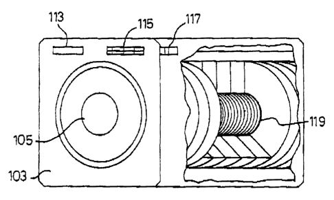
With reference to Figures 1 and 2, an embodiment of the apparatus of the
present invention
may be in the general form of a rectangular or square box 101, which may be
fabricated from wood,
plastic or other non-magnetic material to prevent interference with the
application of an
electromagnetic field to a patient's body part. The front or face panel 103 of
the apparatus may have
generally a circular opening 104 which extends as a elongated annular
passageway 105 in the box
101. The passageway 105 generally extends horizontally inside the box 101, and
when not in use, it
may be closed and has an end 109 at the rear wall or panel 111. Preferably
installed on the face
panel 103 are a timer 113 and a control mechanism 115, both of which are
connected to an electronic
panel 117, for varying and controlling the intensity and duration of the
magnetic field(s).
As shown in Figure 6, disposed on each interior longitudinal side of the
passageway 105 is a
solenoid coil 119 for supplying a time-varying magnetic field. In addition,
Figure 6 shows the
permanent magnets 121 for supplying the static magnetic field. Referring to
Figure 7, an electric
current is supplied from an AC power source (not shown) through power lines
123, 125, and into a
transformer 127 and a rectifier bridge 129 to the magnetic coil 119. The
manner and details for
supplying electric current to the solenoid are well known in the art, and are
not per se critical in this
invention.
With reference to Figures 8 and 9, another embodiment provides an apparatus
similar to the
one described in the previous paragraph. This embodiment is an enlarged
version of the embodiment
shown in Figures 1 to 7. This second embodiment allows exposure to a part or
the whole body to the
EMFs as shown in Figures 8 and 9. In this design, the major blood vessels
(aorta, cava veins)
thoracic duct, paravertebral lymph nodes and major blood reservoirs (liver,
spleen, lung and heart)
CA 02389134 2002-04-26
WO 01/15770 PCT/IB00/01480
11
can be exposed simultaneously to the combination of time-varying magnetic
fields and static magnetic
fields (not shown), reducing the patient exposure time.
This apparatus has a circular shaped housing 130 containing the coil 119 and
strong
permanent magnets 121. The circular shaped housing 130 has an internal chamber
132 large enough
to expose part of the body of a patient 134 to the magnetic field. The housing
130 may be fixed to a
narrow bed 136 made of non-magnetic material. The timer 113 and control
mechanism 115
connected to an electronic panel, as in the previous embodiment, may be
located at the side of the
bed. Preferably, the housing is made from a non-magnetic material such as wood
or plastic.
In another embodiment shown in Figures 10 and 11, the identical apparatus of
the
embodiment of Figures 1 and 2 is used without the permanent magnets 121. As
shown in Figure 13,
this embodiment also includes a coil support 250 for supporting the coil 119
and a tube wall 252.
In another embodiment, shown in Figures 14 to 16, which is a simplified
version of the
previous embodiment shown in Figures 10 to 13, a solenoid, such as a magnetic
coil 157, is mounted
within a cylindrically shaped housing 158. The cylindrically shaped housing
158 includes an
elongated annular passage 159 formed therethrough. The magnetic coil 157 is
disposed within the
cylindrically shaped housing 158 and around the annular passage 159. The
housing and the annular
passage are sized to comfortably accommodate the limb of a patient, such as
the forearm 160 or
upper arm 162 of a patient, allowing more mobility to the patient while being
exposed to the EMFs.
Therefore, this design permits comfortable and mobile limb exposure to the
ENVs of the
invention. Preferably, the cylindrically shaped housing is made of a light non-
magnetic material.
Such non-magnetic materials include plastic or wood. Also, the transformer 127
and rectifier bridge
129 (shown in Figure 7), along with a pair of contacts 152 which are
connectable to a power source,
are contained within a small adapter box 150, to readily obtain electric
current from an AC power
source (not shown). The adapter box 150 with the contacts, the transformer,
and the rectifier bridge
contained therein, is connected to the coil 157 by two long cables 154 which
provide current to the
coil 157.
With reference to Figures 17 and 18, another embodiment provides an apparatus
similar to
the embodiment shown in Figures 10 to 13. This embodiment is an enlarged
version of the
embodiment shown in Figures 10 to 13. This embodiment allows a part or the
whole body to be
exposed to the EMFs as shown in Figures 17 and 18. In this design the major
blood vessels (aorta
and cava veins) thoracic duct, paravertebral lymph nodes and major blood
reservoirs (liver, spleen,
lung and heart) can be exposed simultaneously to EMFs (not shown), reducing
the patient exposure
time. This apparatus has a circular shaped housing 260 containing the coil 119
and includes an
internal chamber 262 large enough to expose part of the body of a patient 204
to the magnetic field.
The housing 260 may be fixed to a narrow bed 245 made of non-magnetic
material. The timer 113
and the control mechanism 115 connected to an electronic panel, may be located
at the side of the
bed. Preferably, the housing is made from a non-magnetic material such as wood
or plastic.
A magnetic field may also be applied with another embodiment illustrated in
Figures 19 and
20. This apparatus may be in the general form of a U- shaped housing 200.
Referring to Figure 22,
part of the body of a patient 204 may be exposed to a homogeneous
electromagnetic field in
accordance with this invention between the lateral walls 208 of the housing
200 of the apparatus.
The apparatus may be fabricated from wood, plastic, or other non-magnetic
material, to prevent
interference with the application of an electromagnetic field to a patient's
body part. With reference
again to Figure 19, installed on the face panel 212 are a timer 214 and a
control mechanism 216,
both of which are connected to an electronic panel 218 shown in Figure 20, for
varying and
controlling the intensity and duration of the electromagnetic field(s).
CA 02389134 2002-04-26
WO 01/15770 PCT/IB00/01480
12
As shown in Figure 20 disposed on each interior of the lateral walls 208 is a
coil, 224 for
supplying time-varying magnetic fields. The lateral walls serve as supports to
maintain the coils in a
Helmholtz disposition. Figure 21 shows a schematic representation of the
electromagnetic flux
generated by the opposed Hemholtz coils 224. Referring to Figure 24, an
electric current is supplied
S from an AC power source (not shown) through power lines 230, 232 and into a
transformer 234 and
a rectifier bridge 236. To reduce heating of the coils by the current, a
capacitor 238, and resistors
240 are used to reduce the effective current to the magnetic coils 224. A fan
is used to cool the
resistors. The manner and details for supplying electric current to the coils
are well known in the
art, and are not per se critical in this invention.
A slight variation of this embodiment is shown in Figure 23. In this
embodiment, similar
equipment is used, but the housing 200 is turned upside down and fixed to the
sides of a narrow bed
245 made of non-magnetic material. The magnetic flux produced by the coils now
runs
perpendicular to the axis of the body 204. The timer and control mechanism 214
and 216 may be
easily located at the side of the bed.
Another embodiment of the apparatus of this invention is illusuated in Figures
25 and 26.
The apparatus may be in the general form of a saddle 280. Referring to Figure
28, the saddle may
be placed on a limb or a body part 282 to expose a patient to the homogeneous
electromagnetic field
in accordance with this invention. The apparatus may be fabricated from wood,
plastic or other non-
magnetic material to prevent interference with the application of an
electromagnetic field to a
patient's body part. As shown in Figures 25 and 26, the saddle is connected
through electric cables
288 to the control unit 290 which contains a timer 292 and a control mechanism
294, both of which
are connected to an electronic panel (not shown) for varying and controlling
the intensity and
duration of the electromagnetic field(s).
As shown in Figure 26 disposed on the. interior of the saddle walls are the
coils 298 for
supplying the time-varying magnetic field. Referring to Figure 24, the same
circuit as used in the
previous embodiment, Figures 19 to 23, can be used to deliver current to the
coils 298 of the current
embodiment illustrated in Figures 25 to 28. An electric current is supplied
from an AC power source
(not shown) through power lines 230, 232 and into the transformer 234 and the
rectifier bridge 236.
To prevent heating of the coils by the current, a capacitor 238, and resistors
240 are used to reduce
the effective current to the magnetic coils 298 and a fan is used to cool the
resistors. The manner
and details for supplying electric current to the coils are well known in the
art, and are not per se
critical in this invention.
There are two additional ways in which the electromagnetic fields of this
invention may be
generated. One is by modulating a continuous 27.12 MHz waves that transport
electrical and
magnetic energy through space (transporting waves) at the same frequencies of
the present invention
(i.e. an antenna to focus the electromagnetic fields of the present invention
in any direction), and the
other by modulating to the same frequencies of the present invention the
electromagnetic fields of
high intensity and frequency produced by a Tesla coil, (transporting waves).
The electromagnetic
fields of this last design travel through space so a patient lying in bed or
seated to a nearby source
may be treated without seeing the apparatus.
In order to treat a patient afflicted with skin lesions or internal wounds in
accordance with
the method of this invention, the patient's limb (e.g. a hand or part of an
arm) may be simply
inserted through the opening 104, and partly extended through the passageway
105. The power
source is then turned on to supply an electric current to the solenoid coil
119. The lines of flux
produced by the magnetic fields generated within the space 107 are shown in
Figure 3, and are seen
to comprise variable as well as static magnetic flux lines. It has been found
that the treatment is most
effective when the electromagnetic field is applied at extremely low
frequencies of more thin nne
Hertz to less than about 300 Hz. The frequencies spectral content of the time-
varying magnetic field
CA 02389134 2002-04-26
WO 01/15770 PCT/IB00/01480
13
are higher around approximately 120 Hz and its harmonics. See, for example
Figure 5. These
frequencies have a static magnetic field component with an intensity of about
0.3 to about 0.8 mT
root mean square (rms) and these EMFs can be used alone or in combination with
a homogeneous
static magnetic field of about 40 to about 80 mT (about 400 to about 800
Gauss). Thus, there are
S two static magnetic fields: one is generated jointly with the time-varying
magnetic field and
preferably has a low magnetic flux density of a few microteslas to a maximum
of about 0.3 to about
0.8 mT (rms) and the second has a higher density and is provided by the
permanent magnets with a
magnetic flux density of about 40 to about 80 mT, which is equivalent to about
400 to about 800
Gauss. The relationship between the instantaneous current supplied by the
electric circuit to the
magnetic coils as a function of time are shown as a series of waves in the
forms shown in Figure 4.
As previously indicated, the application of time-varying magnetic fields with
frequencies of a few
Hertz (more than one) to less than about 300 Hz and static magnetic field
components having an
intensity of about 0.3 to about 0.8 mT used alone or with the simultaneous
application of an
homogeneous static magnetic field of about 40 to 80 mT or about 400 to about
800 Gauss, constitutes
1 S an essential (critical) feature of the method of this invention.
The wound healing effects of the present method and apparatus which were
observed in
patients are obtained by applying the time-varying electromagnetic fields
described in this patent,
either alone or in combination with static electromagnetic fields. Generally,
however, each
component separately and their combination evidence different degrees of
effect on the proliferation
of peripheral blood mononuclear cells (PBMCs). This may be seen clearly in
Figure 29
accompanying this patent, showing the different degree of effects on cell
proliferation evidenced by
different types of magnetic fields when applied to PBMCs obtained from normal
subjects (the 100%
cell proliferation control are PBMCs stimulated to proliferate with
phytohaemaglutinin without the
presence of any of the magnetic fields (EMFs) of this invention). The
application of a combination
of static and time-varying fields produced 11.1 % increase of PBMCs
proliferation (P < 0.001
Student "T" test), the application of a time-varying magnetic field alone
produced 67 % reduction of
PBMCs proliferation (P < 0.001 Student "T" test), and the application of a
static magnetic field
alone had not significant effect on PBMCs proliferation.
For maximum benefit from the treatment, the magnetic fields are preferably
applied to the
patient's limb for about several minutes to about eight hours. The preferred
exposure time was
calculated so that a blood volume equivalent to the patient's entire blood
volume is exposed to the
magnetic field at least once. The exposure frequency may be varied from a
maximum of about 30
minutes to about seven hours a day, to a minimum of about 30 minutes a week,
depending on the
observed lesion repair and wound healing observed.
The effect of EMFs in accordance with the present invention applied on
isolated peripheral
blood mononuclear cells (PBMCs) to modulate their proliferation patterns,
their systemic effects in
vivo and the application of external time-varying magnetic fields alone or in
combination with static
variable magnetic field for treatment of patients with chronic skin ulcers,
chronic wounds, viable but
underperfused myocardium, bone fractures and partial denervation is
illustrated in the following
examples.
The following examples illustrate the effectiveness of the method of this
invention. These
examples, however, are merely illustrative of the invention and are not to be
construed so as to limit
the scope thereof.
EXAMPLES
IN VITRO EFFECT
CA 02389134 2002-04-26
WO 01/15770 PCT/IB00/01480
14
The following experiments show the effect of electromagnetic fields (EMFs),
applied in
accordance with the present invention, on the peripheral blood mononuclear
cells (PBMCs).
Example 1: In Vitro Effects of Different Electromagnetic
Fields on PBMC Proliferation
Peripheral blood mononuclear cells (PBMCs) were obtained by venous puncture
and
fractionated on a ficoll gradient. The fraction containing PBMCs was washed 3
times and
resuspended in phosphate buffered saline to a final concentration of 5 x 10⁶
cells/ml. PBMCs
aliquots 0.2 ml aliquots of 5 x 10⁶ cells/ml, plus 1~ m/ml of
phytohemaglutinin were placed in
Eppendorf tubes. Four subsets were formed (three tubes per subset). The first
subset was shielded
from the magnetic fields. Subset 2 was exposed to the combination of time-
varying electromagnetic
fields and static magnetic fields in accordance with this invention Subset 3
was exposed only to the
time-varying electromagnetic fields in accordance with this invention, and
Subset 4 was exposed only
to the static magnetic field in accordance with this invention. Four subsets
were prepared without
phytohemaglutinin (controls) and placed under the same experimental
conditions.
All tubes were incubated at 37 °C in a COZ humidifier incubator (Forma
Scientific Mod 3325
Dual -Chamber C02-water jacketed incubator). After an incubation period of 58
hours 1 ~.Ci of3H-
thymidine (5-6 ~.Ci/mM final concentration) was added to all tubes and
incubated for an additional 14
hours. All tubes were harvested at 72 hours and washed through glass fiber
filter paper with a cell
harvester, (Skraton, Tranby, Norway). 'H-thymidine incorporation was
determined using a liquid
scintillator counter (Beckmann Mod LS 6000 SE). The mean number of counts per
minute was
obtained by triplicate in each sample. Cell viability was always greater than
90% determined on the
basis of trypan blue exclusion. Figure 27 accompanying this patent shows the
magnetic fields effects
on PBMCs obtained from normal subjects.
The combined static and time-varying electromagnetic fields produced an 11 %
increase in
PBMCs proliferation (P < 0.001 Student "T" test). Time-varying electromagnetic
fields alone
produced about 67% reduction in cell proliferation (P < 0.001 Student "T"
test). Static magnetic
fields have no effect on PBMCs proliferation. Therefore, the in vitro
experiments demonstrate an
effect of the magnetic fields on isolated PBMCs, which is different for each
type of magnetic field or
its combination.
IN VIVO EFFECT & SAFETY STUDY
Example 2: Effect of EMFs on Body Wounds
(Safety and Tolerance Study)
Since electromagnetic fields cross biological tissues, they also produce in
vivo effects on
circulating PBMCs. The in vivo effects of the time varying electromagnetic
fields of this invention,
3 S alone or combined with the static magnetic fields, are observed in chronic
skin wounds even after
only about thirty minutes to one hour of exposing a body part situated far
from the lesion site to the
EMFs of this invention. In this period of time, it is common to observe the
drying of a lesion and a
reduction of its diameter.
The magnetic fields generated by the apparatus described in this patent are
below the
exposure limits recommended as safe by the World Health Organization (WHO).
However, due the
fact that this is a pioneering procedure, the following study was performed to
illustrate the safety of
the external application of magnetic fields of this invention to humans. After
submitting a letter of
voluntary consent to participate in the magnetic field protocol, each patient
was subjected to a
complete clinical history, physical examination, blood chemistry, cell blood
count, and urine analysis
before magnetic field exposure was applied. Before, during and after exposure
to an electromagnetic
field, an electrocardiogram was performed and the blood pressure measured.
Three groups of
volunteers were formed. The first group was exposed to the electromagnetic
forces (EMFs) for 30
CA 02389134 2002-04-26
WO 01/15770 PCT/IB00/01480
minutes a day, the second group for 30 minutes twice a week, and the third
group for 30 minutes
once a week. These three cohorts were followed for one year without presenting
negative side
effects.
In the last 5 years, more than 100 patients have been exposed one to seven
hours a day one
5 to seven times a week to external magnetic fields in accordance with the
method of this invention.
During the course of these treatments, no adverse or negative side effects
were observed in any of
these patients during or after the period of exposure to EMFs. The application
of EMFs by the
method and apparatus of this invention showed no effects on their
electrocardiograms even in patients
who had heart disease. Nor did such treatment have any adverse effects on the
patient's motor and
10 sensory nerve function or conduction.
CLINICAL TRIALS
The following are clinical trials evidencing the efficacy of the treatment of
the invention as
applied to the limb of patients with histories of chronic wounds, which were
previously unresponsive
to other therapeutic treatments.
15 A Phase I protocol following the Good Clinical Practice Guidelines as
defined by the U.S.
Food and Drug Administration (Phase I Study) was presented to the Juarez
Hospital Instir.~tional
Review Board/Ethics Committee and was approved September 24, 1995. Standard
operating
procedures were followed for monitoring the clinical trial. A total of 42
patients were studied.
The following clinical trials demonstrate the efficacy of the treatment of the
invention as
applied to the limb of patients with histories of chronic wounds, which were
previously unresponsive
to other therapeutic treatments and to patients with bone fractures.
Electromagnetic stimulation as defined below was applied in the arm two to
three hours a
day, several times a week, or a period of two to eight months. All exposed
tissues of the arm were
considered under the combined effect of the static andlor time variable
magnetic fields. The intensity
of the induced variable electric fields in each segment of the arm varied with
the radius according to
Faraday's law.
Twenty five patients were admitted for treatment of chronic leg ulcers. The
complete case
record for each patient contains a signed voluntary consent form. Their
clinical histories include
definition of venous and arterial diseases, presence of other underlying
diseases, ulcer history and
treatments used. Only lesions that remained unhealed at the time of
examination were followed up.
Each ulcer was described and documented by photographs, its size and
approximate area calculated
(length x width). When more than one ulcer existed, an approximate total ulcer
area was determined.
Laboratory tests include blood chemistry, hematological examinations,
electrocardiogram and other
diagnostic tests. The peripheral circulation was evaluated by doppler-color
ultrasonography,
arteriography, venography and pletismography. The follow-up records include a
periodic
photographic registration of chronic leg ulcer history, a weekly clinical
commentary and a report of
physical examination.
I. CHRONIC VENOUS LEG ULCERS
This group included patients whose underlying afflictions were, in addition to
the leg ulcers,
primary varicose veins, deep vein thrombosis, diabetes, hypertension,
rheumatoid arthritis, obesity
and cardiovascular disease among others. Pain, edema infection and limited
mobility, which
accompanied the leg ulcers.
Example 3: Old Female with Primary Varicose Veins
Patient No. 1 is a 75 year old woman afflicted with primary varicose veins and
had a 5 cm2
leg ulcer for over 9 months. The ulcer was accompanied by pain, edema and
limited mooility.
CA 02389134 2002-04-26
WO 01/15770 PCT/IB00/01480
16
Electromagnetic stimulation in accordance with the invention was then applied
to one of her arms for
about 2 hours/day 5, days/week over a period of 5 months. No other treatment
was administered
during this time.
The pain and edema disappeared after the first two weeks of treatment, at
which point the
patient became fully mobile. The ulcer was covered with granulation tissue and
appeared completely
healed 5 months after the initiation of the EMFs treatment. As treatment
continued, the patient
became more alert and optimistic about her life and her memory improved. The
patient was kept
under observation for a further period of eight months after the ulcer was
healed and during all this
time, the patient remained asymptomatic.
Ex m 4: Young Male with Primary Varicose Veins
Patient No. 2 is a 33 year old man afflicted with primary varicose veins, who
had a 4 cm2
leg ulcer for over 9 months, which was accompanied by pain and edema.
Electromagnetic
stimulation in accordance with this invention was applied to his arm for about
2 hours/day 5
days/week for a period of 1 week in the absence of additional treatments.
One week after initiation of the treatment, the pain and edema disappeared and
the ulcer
appeared completely healed, 6 weeks later, however, when the patient returned
to the hospital, he
had a new leg ulcer in all appearances produced by trauma, not by
reoccurrence.
A second series of EMFs treatment was then applied to one of his arms 2
hours/day for four
days. As the first time, the second treatment healed- this ulcer in one week.
xam 1 Middle Aged Female with Primary Varicose Veins
Patient No. 3 is a 66 year old woman that was afflicted with primary varicose
veins and had
a 6 cm2 leg ulcer for over a year. The ulcer was accompanied by pain, edema,
and limited mobility.
The only treatment she received was electromagnetic stimulation, applied to
one of her arms
2 hours/day, 3 days/week for a period of 3 months.
After the first 2 weeks of treatment, the pain and edema disappeared and the
patient became
fully mobile. The treatment enhanced the growth of granulation tissue and
reduced the ulcerated area
on her leg by about 48 % , 3 months after treatment began.
Exam In a 6: Young Female with Deep Vein Thrombosis
Patient No. 4 is a 38 year old woman, afflicted with deep vein thrombosis for
over 8 years
and had a 6 cm2 leg ulcer for over 7 years. The ulcer was accompanied by pain,
edema and limited
mobility. During this time, the patient was given medical and surgical
treatment, including
saphenectomy and skin graft procedures, as well as the application of an
ambulatory pressure
bandage to the ulcer, but they were ineffective in improving her condition.
Electromagnetic stimulation in accordance with this invention was then applied
to one of her
arms 2 hours/day, 5 days/week for a period of over 5 months. During this time
the patient continued
to use her loose ambulatory bandage on the leg ulcer she used before the
electromagnetic therapy.
After the first two weeks of treatment the pain and edema disappeared, and
after 6 weeks
she became fully mobile. After 5 months of treatment the ulcer base was
superficial and its size
reduced by about 80% .
Example 7: Middle Aged Female with Deep Vein Thrombosis
and Superficial Venous Insufficiency
Patient No. 5 is an obese 41 year old woman, afflicted with deep vein
thrombosis and
superficial venous insufficiency, with chronic leg ulcers for over 7 years in
both lower limbs. The
ulcerated areas were accompanied by pain, edema, itching and limited mobility.
During this time,
CA 02389134 2002-04-26
WO 01/15770 PCT/IB00/01480
17
she received local and systemic medical treatment, as well as phlebotomy with
Litton technique in
both legs which were ineffective in improving her condition.
Electromagnetic stimulation was then applied to one of her arms for 2
hours/day, 3
days/week. After the first two weeks of treatment pain and edema disappeared
and in two months
the ulcers were completely healed and EMFs exposure was suspended. Four months
later, the ulcers
remained healed and the patient was asymptomatic.
Example 8: Middle Aged Female with a Deep and Superficial
Venous Insufficiency and Arterial Hypertension
Patient No. 6 is an obese 59 year old woman, afflicted with deep and
superficial venous
insufficiency and arterial hypertension, with a chronic leg ulcer for over one
and a half years. The
ulcer was accompanied by pain, edema, a big area of erythema and limited
mobility. During this
time, arterial hypertension was kept under control and the ulcer was treated
with conventional
medical and surgical treatments which were ineffective in improving her
condition.
The electromagnetic stimulation was then applied to one of her arms for 2
hours/day, 3
days/week for about 2 months while she continued to take the same anti-
hypertensive treatment as
before. After the first two weeks of electromagnetic therapy pain and edema
disappear and in three
weeks the ulcer was covered by scab. The area of erythema was reduced in four
weeks and the scab
began to fall being substituted by scarred tissue.
Example 9: Female with Lymphatic and Superficial Venous Insufficiency
Patient No. 7 is a 42-year-old female afflicted with lymphatic and superficial
venous
insufficiency caused by a bum on her left leg, which occurred when she was 2
years old, In
addition, for a period of over 5 years she had an extensive ulcerated area on
her leg totaling 395 cm2,
which was accompanied, by pain, edema and limited mobility. During that time,
she had received
compressive ambulatory bandage, medical and surgical treatments, including
skin grafts, which were
ineffective in improving her condition.
Electromagnetic stimulation was then applied to one of her arms for 2
hours/day, 5
days/week for a period of over 6 months in accordance with the invention.
During the course of this
therapy, she continued to use the loose, compressive ambulatory bandage, which
she used before
electromagnetic therapy. No other additional treatment was administered to
her.
After 2 weeks of electromagnetic treatment, the pain and edema disappeared,
and after 4 to
8 weeks she became fully mobile. After 4 to 7 weeks of electromagnetic
treatment, angiogenesis was
evident by the presence of abundant granulation tissue on the ulcerated area.
At week 8 she became
fully mobile. The lesion was completely healed after 7 months of treatment.
Example 10: Female with Superficial Venous Insufficiency
Patient No. 8 is a 52 year old woman with superficial venous insufficiency who
had a 7 cm2
leg ulcer for over 7 years. The ulcer was accompanied by pain, edema,
infection and limited
mobility. The electromagnetic stimulation was applied to one of her arms for
about 2 hours/day, 3
days/week over a period of two months. No other treatment was administered
during this time.
The pain and edema disappeared after the first week of treatment at which
point the patient
become fully mobile. The ulcer size was reduced by more than 95 % after 1
month of treatment.
Example 11: Old Female with Superficial Venous Insufficiency
Patient No. 9 is a 62 year old woman afflicted with superficial venous
insufficiency and a
chronic leg ulcer which opened and closed for over 15 years. A week before
admission to the ~tz:dy
the ulcer scar became very painful producing a 0.5 cmz ulcerated area with
serum exudatc. The
electromagnetic stimulation was then applied to one of her arms for about 2
hours/day, 3 days/week
CA 02389134 2002-04-26
WO 01/15770 PCT/IB00/01480
18
for two weeks. The ulcer healed in three days and the patient remained
asymptomatic during the
following month.
Example 12: Middle Aged Female with Superficial Venous
Insufficiency Factitial Ulcer and Mental Depression
S Patient No. 10 is a S1 year old, mentally depressed woman who had two leg
ulcers of
approximately 2S cm2 that were artificially maintained open by the patient for
over 21 years. The
lesions were accompanied by pain, edema and limited mobility. During that time
the patient had
been given medical treatment but it was ineffective in treating her condition.
Electromagnetic
stimulation in accordance with this invention was then applied to the
patient's arm 2 hours/day, 3
days/week for a period of two months. No other treatment was administered to
her during this time.
After 4 weeks of the treatment the pain and edema disappeared, and after 2
months of the
treatment the ulcer had been reduced by about 32 % .
L1 PATHOLOGIES ASSOCIATED WITH CHRONIC VENOUS LEG ULCERS AND/OR
1 S VENOUS DISEASE THAT LIIVVIIT BENEFICIAL EFFECT
This group includes patients whose underlying afflictions are, in addition to
the chronic leg
ulcers and venous disease, non pitting edema, obesity with very high body mass
index (more than 40
kg/MZ) and dioderma gangrenosum among others. Pain, edema and infection
accompanied the
chronic leg ulcers.
- Exam In a 13: Superficial and/or Deep Venous Insufficiency, Non-pitting
Edema, Obesity &
Chronic Venous Leg Ulcers
This example describes treatment to a group of four patients. The patients
(Patients Nos.
11-14) are one woman and three men S6 to 71 years old afflicted with
superficial and/or deep venous
insufficiency, brawny edema, obesity and chronic venous leg ulcers for a
period of 9 to 17 years.
2S All patients had been with preventively ueated with present recommended
medical treatments, and
surgery intended to alleviate venous hypertension and/or to cover the
ulcerated areas with skin grafts.
All these treatments were ineffective to improve their condition.
Electromagnetic stimulation in accordance with this invention was then applied
to each of
patients 11 to 14 on either arm 2 hours/day, 3 days/week. Two weeks after the
initiation of EMFs
treatment, pain was reduced and the ulcers look dried and deep. These changes
improved patient's
condition for a while, however the size of the ulcers remained the same and in
some cases increased
their size during the EMFs treatment period. These patients were eliminated
from the protocol.
Example 14: Female with Severe Superficial Venous Insufficiency, Obesity with
High Body
Mass Index & Chronic Venous Leg Ulcer
3S Patient No. 1S is a 46 year old woman afflicted with superficial venous
insufficiency since
she was 13 years old, obesity with very high Body Mass Index (more than 40 k
g/M2 ) and a painful
large chronic venous leg ulcer in her left leg for a period of over 4 years.
During that time, she
received preventive measurements and recommended medical treatments in
specialized centers
without improving her condition.
Electromagnetic stimulation in accordance with this invention was then applied
to one of her
arms 2 hours/day, 3 days/week, for a period of four months. Two weeks after
the initiation of
EMFs treatment, pain was reduced, exudate stop and the ulcer borders look
inflamed. However the
size of the ulcer remained the same during the EMFs treatment period. This
patient was eliminated
from the protocol.
CA 02389134 2002-04-26
WO 01/15770 PCT/IB00/01480
19
Example 15: Middle Aged Female with Deep Vein
Thrombosis & Pioderma Gangrenosum
Patient No. 16 is a 46 year old woman afflicted with deep vein thrombosis and
two atypical
ulcers located in the posterior part of her right leg for a period over 4
years. During that time, she
received preventive measurements and recommended medical treatments in
specialized centers
without improving her condition.
Electromagnetic stimulation in accordance with this invention was then applied
to one of her
arms 2 hours/day, 3 days/week, for a period of four months. Two weeks after
the initiation of
EMFs treatment, pain was reduced and one of the ulcers became superficial.
After that point the
ulcers began to grow and the patient was eliminated from the protocol. A skin
biopsy revealed
changes compatible with Piodernia Gangrenosum the patient was medically
treated.
II. ARTERIAL LEG ULCERS
This group consists of patients whose underlying afflictions, in addition to
the chronic
arterial leg ulcers, were generalized atherosclerosis, diabetes, arterial
and/or venous hypertension,
1 S rheumatoid arthritis, obesity, cardiovascular disease and vascular
necrosis, among others. Rest pain,
edema infection and limited mobility accompanied the leg ulcers.
Example 16: Old Woman with Critical Leg Ischemia & Diabetes
Patient No. 17 is a 73 year old woman, afflicted with 3 arterial ulcers and
diabetes, who
had a 22 cmz ulcerated area on her left leg for over 4 months accompanied by
intense pain that
prevented the patient from sleeping at night. During that time she received
medical treatment, but it
was ineffective in improving her condition.
Electromagnetic stimulation in accordance with this invention was then applied
to one of her
arms for 3 hours/day, 4 days/week for a period of over 7 months. No other
treatment was
administered during this time.
After 4 weeks of treatment, the skin temperature increased and the superficial
veins became
more noticeable reflecting increased vasculogenesis. The ulcer bottom display
abundant granulation
tissue reflecting increased angiogenesis. Pain was greatly reduced allowing
the patient to sleep at
night. After 7 months of electromagnetic treatment the three ulcers were
completely healed. During
this time the mental alertness and memory of the patient improved
significantly. This patient was
followed for an additional year. During this time the ulcers remained healed.
Example 17: Old Woman with Rheumatoid Arthritis & Atherosclerosis
Patient No. 18, is an 80 year old woman afflicted with two arterial ulcers
associated with
rheumatoid arthritis and atherosclerosis. The ulcerated area was approximately
35 cm~ produced by
elastic stockings after knee surgery. These lesions remained open for 19
months and were
accompanied by intense pain that kept the patient awake at night. Skin grafts
and pharmacological
treatments were ineffective in improving her condition. Electromagnetic
stimulation in accordance
with this invention was applied to one of her arms for three hours/day, 3
days/week, for a period of
4 1/2 months. No other treatment was administered during this time.
Four weeks after the initiation of the electromagnetic treatment pain was
reduced, allowing
the patient to sleep at night. The skin temperature increased and the
superficial veins became more
noticeable reflecting increased vasculogenesis. The ulcer bottom display
abundant granulation tissue
reflecting increased angiogenesis. In two months the patient had no pain and
the ulcerated area was
completely healed in four and a half months. During this period the patient
became more alert,
enthusiastic and her memory improved. The patient was followed for an
additional eight months.
During this time the ulcers remained healed.
CA 02389134 2002-04-26
WO 01/15770 PCT/IB00/01480
Example 18: Old Man with Generalized Atherosclerosis
Patient No 19 is a 70 year old man afflicted with a two year old chronic
arterial ulcer 65
cmz, caused by generalized atherosclerosis on the anterior part of his leg.
The femoral artery lumen
was 85% reduced and the anterior tibial artery lumen had 90% stenosis. The
ulcer was accompanied
S by intense pain that kept the patient awake at night. Skin grafts and
pharmacological treatments were
ineffective in improving his condition.
Electromagnetic stimulation in accordance with this invention was applied to
one of his arms
for three hours/day, 3 days/week, for a period of 12 months. No other
treatment was administered
during this time.
10 After two weeks of treatment the ulcer bottom was red and the tendon was
epithelialized,
pain was greatly reduced allowing the patient to sleep at night and mobility
was improved. Two an a
half months after the skin temperature increased and the superficial veins
became more noticeable
reflecting increased vasculogenesis. The ulcer bottom display abundant
granulation tissue reflecting
increased angiogenesis and the patient was without pain. During this time the
patent became more
15 optimistic, alert and his mood improved significantly. The overall
reduction of the ulcerated surface
area on his leg brought about by 12 months of treatment was about 80 % .
Example 19: Female with Generalized Atherosclerosis, Superficial & Deep Venous
Insufficiency & Rheumatoid Arthritis
Patient No. 20 is a 57-year-old woman afflicted with generalized
atherosclerosis, superficial
20 and deep venous insufficiency and rheumatoid arthritis. Additionally, she
had two 9 month old
ulcers. One ulcerated area 6.5 cm2 on the front part of her leg, and the other
a 1 cm2 medial
malleolus ulcer. Electromagnetic stimulation was then applied on one of the
patient's arms, 3
hours/day, 4 days/week, for about 7 months. No other treatment was
administered at this time.
After 3 months of electromagnetic treatment, the medial malleolus ulcer healed
and the ulcer
in the front part of her leg healed in seven months. During this time her
depressive mood improved.
Example 20: Middle Aged Female with Vascular Necrosis
Patient No. 21 is a 45 year old woman afflicted with vascular necrosis of the
right leg
resulting from receiving a silicon implant. Reconstructive surgery saved her
leg from amputation.
Two years after, the use of a tight prosthesis lead to arterial occlusion in
the posterior-medial area
other leg, resulting in a very painful 23 cm2 ulcer that lasted for over 5
months. Pain and edema
accompanied the ulcer, and she had limited mobility. During that time she
received medical
treatment, but it was ineffective in relieving her condition. Before
electromagnetic therapy was given
to the patient, she required local anesthesia while her ulcer was cleaned. The
electromagnetic
stimulation was applied to her arm 1 hour/day, 5 days/week for a period of
over 2 months, during
which time her ulcer was cleaned daily. No other treatment was administered at
this time.
After 4 weeks of treatment the pain and edema disappeared, anesthesia was no
longer
needed to clean the ulcer, and the patient became fully mobile. Abundant
granulation tissue appeared
in the ulcer bottom and the ulcer was completely healed after 2 months of the
treatment.
Example 21: Middle Aged Male with Uncontrolled Arterial
Hypertension & Deep Vein Thrombosis
Patient No. 22 is a 54 year old man, afflicted with uncontrolled arterial
hypertension and
deep vein thrombosis which had chronic leg ulcers totaling 20 cmz for over 7
years. The ulcerated
area was accompanied by pain, edema, and limited mobility. During this time,
he received medical
treatment, including anti-hypertensive therapy, but it was ineffective in
improving his condition.
CA 02389134 2002-04-26
WO 01/15770 PCT/IB00/01480
21
Electromagnetic stimulation was then applied to one of his arms for 2
hours/day, 3
days/week for about 4 months while he continued to take the same anti
hypertensive treatment as
before.
After the first 3 weeks of treatment, the pain, edema, and the necrotic tissue
of the ulcer
disappeared. He became fully mobile after 6 weeks of treatment and the ulcers
completely healed in
less than 3 months of treatment.
example 22: Female with Uncontrolled Arterial Hypertension, Superficial Venous
Insufficiency, Controlled Type H Diabetes Mellitus & Obesity
Patient No. 23 is a 60 year old woman who had 2 leg ulcers totaling 11 cmz,
one of which
she had for over a year and the other for over 3 months. During that time she
received medical
treatment including anti-hypertensive therapy, but it was ineffective in
improving her condition.
Electromagnetic stimulation in accordance with the present method was then
applied on one
of her arms for 3 hours/day, 3 days/week for a period of over 4 1/2 months,
during which time she
received a continuation of her anti-hypertensive treatment. During the course
of electromagnetic
therapy the patient developed a third lesion which was produced by trauma.
After one week of treatment, the pain, edema, and the three-month-old leg
ulcer
disappeared. The lesion induced by subsequent trauma healed after 6 weeks of
electromagnetic
treatment. The patient was followed for an additional year and the ulcers
remained healed.
Example 23: Old Female with Generalized Atherosclerosis
Patient No. 24 is an 85 year old woman afflicted with generalized
atherosclerosis and a
chronic leg ulcer 1.2 cm2 for over one year. During that time she received the
recommended
medical treatment but it was ineffective in relieving her condition.
Electromagnetic stimulation in accordance with the present method was applied
to one of her
arms 2 hours/day, 5 days/week for a period of over 2 months. After two weeks
of treatment the pain
and edema disappeared. These changes were followed by a reactivation of the
inflammatory reaction
around the ulcer with an exuberant increase of granulation tissue that cover
the ulcer bottom
reflecting enhanced angiogenesis. In three months the ulcerated area was
reduced by more than
70 % .
Example 24: Young Male with Uncontrolled Type I
Diabetes Mellitus & Hypercholesterolemia
Patient No. 25 is a 24 year old man afflicted with uncontrolled Type I
diabetes mellitus and
hypercholesterolemia, afflicted with a three month old plantar ulcer on his
left foot. The patient
received local surgical and medical treatments that were ineffective in
relieving his condition. The
electromagnetic stimulation was applied to one of his arms 2 hours/day, 5
days/week for a period of
over 2 months. After three weeks of treatment the deep ulcer base was at the
level of the dermis.
The ulcer healed completely in four weeks. The patient was followed for an
additional four months
remaining asymptomatic.
IH. PRESSURE ULCERS (DECUBITUS ULCERS)
This group includes patients whose underlying afflictions were, in addition to
the pressure
ulcers, paraplegia, deep vein thrombosis and obesity, among others. Pain,
edema and infection
accompanied the pressure ulcers.
Example 25: Middle Aged Incontinent, Paraplegic Female with Leg Wounds
Patient No. 26 is a 64 year old, paraplegic and incontinent woman who had 2
wounds. The
24 cmz wound located on her left thigh had been caused by an attempt to
surgically repair an electric
blanket burn and was 6 months old. The second wound was a 4-month-old, stage 5
Yarkony-Kirk
CA 02389134 2002-04-26
WO 01/15770 PCT/IB00/01480
22
classification decubitus sacrum ulcer. During that time, she received systemic
antibiotic treatment
and three attempts were made to surgically repair her wounds. Those treatments
were ineffective in
improving her condition. Electromagnetic stimulation was then applied on one
of her arms for 6
hours/day, 7 days/weeks for a period of over 8 1/2 months. The patient
received nurse care for 24
hours/day and continued to receive systemic antibiotic treatment. No other
additional wound
treatment was administered at this time.
After 6 weeks of electromagnetic treatment the thigh wound healed, the
decubitus ulcer
infection was controlled, and wound retraction and epithelialization continued
to heal the wound.
After 13 weeks of electromagnetic treatment the abundant granulation and
epithelial tissues were
removed and the wound was surgically closed after leaving a surgical drainage.
After 3 further weeks of electromagnetic treatment, the surgical wound was
healed and an
ulcerated area was formed at the surgical drainage. An autologous skin graft
placed on the ulcer site
was eventually lost. The decubitus ulcer healed completely after three
additional months of
electromagnetic therapy.
Example 26: Old Female with Pressure Ulcer & Deep Vein Thrombosis
Patient No. 27 is an obese 90 year old woman who had an 18 month old, 60 cm2
painful,
edematous, bleeding, friable, purple, deep pressure ulcer on her left ischial
tuberosity caused by her
sitting in the same position for 8 to 12 hours/day while being afflicted with
deep vein thrombosis.
Pain and edema accompanied the ulcer. During that time, the patient received
medical treatment
including local wound cleaning, but the treatment was ineffective in improving
her condition.
Electromagnetic stimulation was then applied to one of her arms for 2
hours/day, 7
days/week for a period of over 5 months. She continued to receive local wound
cleaning, but no
additional treatment was given to her at this time.
After 2 weeks of electromagnetic treatment the pain and edema disappeared,
except for a 1 x
10 mm open skin area which did not heal because of the constant pressure from
sitting. During the
course of treatment, her skin resistance increased. The color of the skin was
pink in the morning and
bright red after 8 to 12 hours of sitting. After 4 months of treatment, the
ulcer was completely
healed and the skin returned to normal.
IV. CHRONIC WOUNDS
This group included patients whose underlying afflictions were, in addition to
the chronic
wounds, peripheral arterial disease, intense stress and peritonitis, among
others. Pain, edema and
infection accompanied the chronic wounds.
Example 27: Old Male with Infected Surgical Wound
Patient No. 28 is a 78 year old man who had a 126 cm2 infected surgical wound
after a long
3 S and complicated abdominal surgery.
Electromagnetic stimulation was applied to one of his arms 1 hours/day for 3
weeks. After
2 weeks of treatment the infection disappeared, and the wound was surgically
closed because of
granulation tissue and wound healing were under optimal conditions for this
procedure.
Example 28: Old Man with Unhealed Surgical Wounds
Patient No. 29 is a 73 year old man with lower limb arterial disease
associated with
smoking, diabetes and generalized atherosclerosis. Chronic lower-limb ischemia
had been present
for years. Critical leg ischemia was evident in the last year and the patient
was subjected to surgical
re-vascularization. However, 3-1/2 months after the operation, the surgical
wounds located in his
left thigh and leg were not healed and the knee surgical wound remained an
open 30 cm2 wour.4.
CA 02389134 2002-04-26
WO 01/15770 PCT/IB00/01480
23
Electromagnetic stimulation in accordance with this invention was then applied
to one of his
arms 2 hours/day 3 days/week for a period of five months. No additional
treatment was
administered to him.
After 4 weeks of electromagnetic treatment the thigh and leg wounds healed and
wound
retraction and epithelialization continued to heal the knee wound, reducing
the wound area to about
97% in 5 months. The wound was completely healed in one additional month.
Example 29: Middle Aged Female with an Unhealed Ulcer
Patient No. 30 is a 65 year old woman with lower limb arterial disease
associated with
smoking. As a consequence of a trauma, a small bruise developed into a 2.6 cmz
infected, very
painful ulcer, in two weeks causing reduced mobility. This wound was resistant
to antibiotic
treatment.
Electromagnetic stimulation in accordance with this invention was then applied
to one of her
arms 2 hours/day, 5 days/week in addition to a new antibiotic treatment. After
two weeks the
infection was controlled, edema and pain were drastically reduced and mobility
became almost
normal. The previously unhealed wound displayed abundant granulation tissue on
the ulcer bottom.
A week later the patient was asymptomatic and ulcer healed in less than one
month since the
beginning of EMFs treatment.
Example 30: Middle Age Male with an Infected Shoulder Wound
Patient No. 31 is a 57 year old man under intense stress who by scratching an
old scar
developed a 3 cm3 very painful abscess in his left shoulder in two weeks. The
lesion was controlled
with antibiotics but two weeks later pain, edema, immobility and a 3 cmz open
wound were still
present.
Electromagnetic stimulation in accordance with this invention was then applied
to one of his
arms 2 hours/day, 5 days/week. Three weeks later, pain, edema and immobility
disappeared and the
wound was reduced by more than 70% . The ulcer healed completely two weeks
later.
V. DILATED ISCHEMIC CARDIOMYOPATHY
This group includes patients with viable but underperfused myocardium, who
were not
candidates for coronary revascularization.
Atherosclerosis is a generalized disease responsible for occlusive arterial
disease such as
lower-limb arterial disease (chronic arterial leg ulcers), myocardial
infarction and stroke. The
etiological factors (arterial hypertension, high serum cholesterol, smoking,
stress, diabetes mellitus)
are similar, as are preventive measurements (eliminate or control the
etiological factors), and some
treatments (angioplasty or by-pass when indicated). The enhancement of the
body's natural process
of collateral blood vessel growth is a novel approach. Therapeutic
angiogenesis has been attempted
using angiogenic growth factors. Acid and basic fibroblast growth factors have
been successfully
tested in animal models but positive clinical results have not been reported
in clinical trials. Vascular
endothelial growth factor (VEGF) recombinant protein has been successfully
tested in patients with
heart infarction that were not ideal candidates for coronary revascularization
and intramuscular
injection of naked plasmid DNA encoding the 165 isoform of VEGF has been
successfully tested in
patients with critical limb ischemia.
Example 31: Young Male with Sequels of Anteroseptal Acute Myocardial
Infarction
Patient No. 32 is a 40 year old man with sequels of anteroseptal acute
myocardial infarction.
The patient was evaluated by clinical examination, blood tests, chest X-Ray,
electrocardiogram,
stress test, echocardiogram, coronary angiography, and cardiac
ventriculography. The observed
lesions include anteroseptal myocardial infarction with extensive anterior and
lateral sub-epicardic
CA 02389134 2002-04-26
WO 01/15770 PCT/IB00/01480
24
ischemia and left bundle anterior hemi- branch block. 90% stenosis in middle
third of left anterior
descending coronary artery with a severe stenosis of first diagonal artery at
its origin, slow TIMI 2
flow, anterior, apical, inferior and septal akinesis. Ejection fraction 20 % .
Patient was classified as
Functional Class III of the New York Heart Association (NYHA) being not
candidate for coronary
revascularization. Patient remained on isorbide 10 mg 3/day, aspirin 150
mg/day and rest during
four additional months without showing any improvement. Electromagnetic force
(EMFs) was then
applied to one of his arms 2 hours/day, 3 days/week for a period of one month
while continuing with
the same medical treatment as before.
After one month of EMFs treatment, patient improvement was very fast even in
the
presence of bad evolution of the left descending coronary artery lesion that
changed from 90%
stenosis to occlusion. Clinical evaluation and stress test classified the
patient in Functional Class I of
the New York Heart Association (NYHA), instead of the Class III he had before.
Anterior, apical,
inferior and septal akinesis diagnosed by ventriculograin and echocardiogram
improved to
hypokinesis. Collateral circulation and contractility of hybernating
myocardium improved.
1 S These changes may not be explained by the medical treatment he received
during four
months after AMI and previously to be exposed to EMFs. During all this period
the patient remained
in Functional Class III (NYHA). The most plausible explanation for the
patient's better clinical
condition in only one month after EMFs exposure, Functional Class I (NYHA), is
enhanced
angiogenesis and vasculogenesis in viable but underperfused myocardium induced
by the EMFs
activated peripheral blood mononuclear cells.
example 32: Obese Woman with Arterial Hypertension & Sequels of Three Month
Old
Acute Myocardial Infarction
Patient 33 is a 54 year old obese woman afflicted with arterial hypertension
and sequels of a
three month old Acute Myocardial Infarction (AMI). Patient evaluation included
clinical
examination, blood tests, chest X-Ray, electrocardiogram, stress test,
echocardiogram, coronary
angiography, and cardiac ventriculography. Lesions included Arterial
Hypertension, elevated blood
values of Glucose, Uric Acid, Triglycerides and Cholesterol. Anteroseptal and
lateral subendocardic
necrosis. Extensive anterior sub-epicardic ischemia. Right atrium enlargement.
Coronary
angiogram. reported occlusion of left descending coronary artery at its origin
and 80% stenosis of
circumflex. Patient was treated with Metoprolol 50 mg./12 Hs., Isorbide 10
mg./12 Hs., Pravacol
20 mg./24 Hs. Norvas 2.5 mg./24 Hs. and ASA 100 mg./24 Hs. Three months later
arterial
hypertension and blood tests were within normal limits but the cardiovascular
symptomatology
deteriorated, from Functional Class II New York Heart Association (NYHA) to
Functional Class III
(NYHA). An stress test was attempted but had to be suspended at the beginning
of the test by
fatigue, hypertension and pain. Functional Class III (NYHA).
Electromagnetic stimulation was then applied to one of her arms 2 hours/day, 4
days/week
for a period of five months while continuing with her previous treatment.
After one month of EMFs treatment, patient evaluation changed from Functional
Class III
(NYHA) to Functional Class I (NYHA) and collateral circulation and
contractility of hibernating
myocardium improved. These changes occur in the presence of more severe
lesions than the ones
observed in her previous coronary angiogram including additional occlusion of
the right coronary
artery at its middle third, 90% stenosis of the first diagonal artery and left
ventricle diastolic final
pressure of 15 mm Hg.
These changes in only one month after EMFs may not be explained by the medical
treatment
she received during nine months after AMI and previously to be exposed to
EMFs. During all this
period the patient evolved from Functional Class II (NYHA) to functional class
III (NYHA~. The
most plausible explanation for the patient's better clinical condition,
Functional Class I (N YHA), are
CA 02389134 2002-04-26
WO 01/15770 PCT/IB00/01480
enhanced angiogenesis and vasculogenesis in viable but underperfused
myocardium induced by the
EMFs activated peripheral blood mononuclear cells.
VI. ANKLE FRACTURES
This group included patients with no other underlying affliction. This
orthopaedic problem
S was selected to evaluate the benefits of the EMFs of this invention in
reducing pain and edema in an
acute problem as well as to observe if it could accelerate the healing process
of a different type of
internal wound, i.e. a bone fracture.
Example 33: Eight Patients with Ankle Fractures Type Weber A or B
This example presents information on a group of eight patients (patients 34 to
41). The
10 patients are two women and six men 19 to 65 years old, afflicted with uni
or bi-malleolar ankle
fractures type Weber A or B. All fractures were accompanied with intense pain,
and edema. All
fractures were treated with internal fixation. Compression of fracture
surfaces was obtained with lag
screws with or without semitubular compression plates. Surgical repair of the
deltoid ligament was
done when indicated. Sterile dressings were applied to the wounds and then a
plaster splint to hold
15 the foot in neutral position.
Electromagnetic stimulation was then applied to each patient on either arm 2
hours/day, 2-3
days/week for 10 sessions beginning the next day after surgery. Leg was
maintained elevated and
pain was controlled with Dipirone 500 mg 3/day.
In all patients, pain was reduced from 10 to 3-4 days. Edema from 10 to 5-6
days and bone
20 -union-was observed in-four weeks instead of six, at that point patients
were allowed full-weight
bearing.
VII. SPINAL MUSCULAR ATROPIES
This patient was selected as an example of nerve regeneration in a disease
with no present
treatment. In partial denervation, the axon and the nerve terminal innervating
a muscle fiber
25 degenerates. Healthy denervated tissue responds by releasing cytokines that
induce the terminal
Schwann cells on the denervated muscle fiber to grow processes that reach the
nerve terminal of a
neighboring innervated muscle fiber. Then the Schwann cell process induce a
nerve sprout in the
normal nerve terminal that grows along the Schwann cell process towards the
denervated endplate
until reinnervation of the denervated endplate is completed. At early stages
of certain types of spinal
muscular atrophies when a motor neuron of the spinal cord dies, cytokine
signaling language
stimulated by EMF's activated PBMCs could trigger these events in neighboring
healthy axons not
affected by the disease producing reinervation of the denervated muscle
fibers.
Exam In a 34: Young Boy with Spinal Muscular Atrophy
Patient 42 is a 4 year old boy with Spinal Muscular Atrophy. Being the second
boy of a
normal twin pregnancy and delivery, their parents had the benefit to compare
one twin to the other.
Their recollection suggests that early development was delayed. He started
walking at 15 months but
his gait appeared abnormal and since age 2 he has been unable to run or hop,
has difficulty walking
up one or two steps, tends to fall frequently and needs help getting back on
his feet. At physical
examination weakness of all muscle groups was noticeable but more so in the
shoulder and pelvic
girdles. These findings together with the subtle fasciculations of the tongue,
electromyography,
magnetic resonance imaging and left quadriceps biopsy support the diagnosis of
Spinal Muscular
Atrophy. During the last year, diets, rehabilitation therapy and the use of
stabilizing artroses have
been ineffective in improving his condition.
After signing a voluntary consent form, electromagnetic stimulation was then
applied to nne
of his arms, 2 hours/day, 2 days/week for one month, however this was
difficult to achieve because
CA 02389134 2002-04-26
WO 01/15770 PCT/IB00/01480
26
of his age. Therefore a new design with Helmholtz coils was used to expose
part of the body while
the child was sleeping. This modality of electromagnetic stimulation was
applied 2 hours/day, 5
days/week for a period of two years.
Periodic examinations by the inventor, the pediatrician, the neurologist, the
rehabilitation
specialist, notes of the mother and teacher, and videotapes, were used to
follow his evolution. After
the first month his equilibrium was better, more confident to play with his
brother, sister and peers at
school. He started gaining weight, and at 4 months he was able to walk without
falling and began to
run and hop. At six months, an increase in muscular strength and mass of the
shoulder and pelvic
girdles was noticeable. One year after the initiation of treatment the child
was able to rise from the
floor without help. Two years after, his movements and development were almost
normal.
CONCLUSIONS
The above examples show that the application of external, non-invasive
electromagnetic
fields (EMFs) in accordance with the method of this invention, either alone or
combined with other
medical and surgical treatments healed body lesions in patients who were
previously unresponsive to
conventional medical and surgical treatments alone, or improve the effects of
these treatments.
Thus, from the foregoing detailed description of the invention, and the
illustrative examples
provided herein, it may be appreciated that the treatment offers a remarkably
effective clinical means
for improving a patient's health and well being without producing adverse or
negative side effects.
This has been exemplified on patients with various diseases and conditions,
such as a history of
partial denervation, internal or external lesions such as wounds, areas with
inadequate blood
perfusion such as viable but underperfused myocardium and the like, burns and
ulcers alone or where
the patient is suffering from other disorders as well.