Note: Descriptions are shown in the official language in which they were submitted.
CA 02389569 2002-05-15
WO 01/37194 PCT/US00/31602
CRYSTALLIZABLE COMPOSITIONS COMPRISING
A CASPASE-7
TECHNICAL FIELD OF INVENTION
The present invention relates to a data
storage medium encoded with the structural coordinates
of crystallized molecules and molecular complexes which
comprise the active site binding pockets of caspase-7.
Such data storage material is capable of displaying
such molecules and molecular complexes, or their
structural homologues, as a graphical three-dimensional
representation on a computer screen. This invention
also related to methods of using the structure
coordinates to solve the structure of similar or
homologous proteins or protein complexes. In addition,
this invention relates to methods of using the
structure coordinates to screen and design compounds,
including inhibitory compounds, that bind to caspase-7
or homologues thereof. This invention also relates to
molecules or molecular complexes which comprise the
active site binding pockets of caspase-7 or close
structural homologues of the active site binding
pockets. The present invention also relates to
compositions and crystals of a caspase-7 in complex
with a caspase inhibitor.
BACKGROUND OF THE INVENTION
Apoptosis, or programmed cell death, is a
principal mechanism by which organisms eliminate
unwanted cells. The deregulation of apoptosis, either
excessive apoptosis or the failure to undergo it, has
been implicated in a number of diseases such as cancer,
CA 02389569 2002-05-15
WO 01/37194 PCT/US00/31602
- 2 -
acute inflammatory and autoimmune disorders, ischemic
diseases and certain neurodegenerative disorders [see
generally Science, 281, pp. 1283-1312 (1998); Ellis et
al., Ann. Rev. Cell. Biol., 7, p. 663 (1991)].
Caspases are a family of cysteine protease
enzymes that 'are key mediators in the signaling
pathways for apoptosis and cell disassembly [N. A.
Thornberry, Chem. Biol., 5, pp. R97-8103 (1998)].
These signaling pathways vary depending on cell type
and stimulus, but all apoptosis pathways appear to
converge at a common effector pathway leading to
proteolysis of key proteins. Caspases are involved in
both the effector phase of the signaling pathway and
further upstream at its initiation. The upstream
caspases involved in initiation events become activated
and in turn activate other caspases that are involved
in the later phases of apoptosis.
The caspases have been classified into three
groups depending on their predominant functional roles
and their substrate specificities [N. A. Thornberry,
Chem. Biol., 5, pp. R97-8103 (1998); N.A. Thornberry &
Y. Lazebnik, Science, 281, pp. 1312-1316 (1998); M.
Garcia-Calvo et al., J. Biol. Chem., 273, pp. 32608-
32613 (1998) ] .
The first subfamily consists of caspases-1
(ICE), 4, and 5.. These caspases have been shown to be
involved in pro-inflammatory cytokine processing and
therefore play an important role in inflammation.
Caspase-1, the most studied enzyme of this class,
activates the IL-1(3 precursor by proteolytic cleavage.
This enzyme therefore plays a key role in the
inflammatory response. Caspase-1 is also involved in
the processing of interferon gamma inducing factor
CA 02389569 2002-05-15
WO 01/37194 PCT/US00/31602
- 3 -
(IGIF or IL-18) which stimulates the production of
interferon gamma, a key immunoregulator that modulates
antigen presentation, T-cell activation and cell
adhesion.
The remaining caspases make up the second and
third subfamilies. These enzymes are of central
importance in the intracellular signaling pathways
leading to apoptosis. One subfamily consists of the
enzymes involved in initiating events in the apoptotic
pathway, including transduction of signals from the
plasma membrane. Members of this subfamily include
caspases-2, 8, 9, and 10. The other subfamily,
consisting of the effector caspases 3, 6, and 7, are
involved in the final downstream cleavage events that
result in the systematic breakdown and death of the
cell by apoptosis. Caspases involved in the upstream
signal transduction activate the downstream caspases,
which then disable DNA repair mechanisms, fragment DNA,
dismantle the cell cytoskeleton and finally fragment
the cell.
The utility of caspase inhibitors to treat a
variety of mammalian disease states associated with an
increase in cellular apoptosis has been demonstrated
using peptidic caspase inhibitors. For example, in
rodent models, caspase inhibitors have been shown to
reduce infarct size and inhibit cardiomyocyte apoptosis
after myocardial infarction, to reduce lesion volume
and neurological deficit resulting from stroke, to
reduce post-traumatic apoptosis and neurological
deficit in traumatic brain injury, to be effective in
treating fulminant liver destruction, and to improve
survival after endotoxic shock [H. Yaoita et al.,
Circulation, 97, pp. 276-281 (1998); M. Endres et al.,
CA 02389569 2002-05-15
WO 01/37194 PCT/US00/31602
- 4 -
J. Cerebral Blood Flow and Metabolism, 18, pp. 238-247,
(1998); Y. Cheng et al., J. Clin. Invest., 101, pp.
1992-1999 (1998); A.G. Yakovlev et al." J. Neurosci.,
17, pp. 7415-7424 (1997); I. Rodriquez et al., J. Exp.
Med., 184, pp. 2067-2072 (1996); Grobmyer et al., Mol.
Med., 5, p. 585 (1999)].
Caspase-7 is considered a potential target
for therapeutic agents. The current understanding of
caspase-7 has not however led to satisfactory
treatments for caspase-7 mediated disease. Thus, there
is a need for more effective caspase-7 inhibitors.
There is also a need for inhibitors that either inhibit
caspase-7 selectively or inhibit caspase-7 as well as
other caspases.
Drug discovery efforts directed towards
caspase-7 have been hampered by the lack of structural
information about caspase-7. Such structural
information would be valuable in the discovery of
selective caspase-7 inhibitors and pan-caspase
inhibitors. However, efforts to determine the
structure of caspase-7 have been hampered by
difficulties in crystallizing caspase-7. There have
been no crystals reported of a caspase-7 protein.
Thus, x-ray crystallographic analysis of such proteins
has not been possible.
SUMMARY OF THE INVENTION
Applicants have solved this problem by
providing, for the first time, the crystallization of a
caspase-7 in complex with a caspase-7 inhibitor and the
structure coordinates of that complex. Solving the
three-dimensional crystal structure of that complex has
allowed applicants to determine key structural features
CA 02389569 2002-05-15
WO 01/37194 PCT/US00/31602
- 5 -
of caspase-7, particularly the shape of its active site
binding pockets.
Thus, the present invention provides
molecules or molecular complexes that comprise all or
parts of these binding pockets, or homologues of these
binding pockets that have similar three-dimensional
shapes.
The present invention relates to a data
storage medium that comprises the structural
coordinates of crystallized molecules and molecular
complexes which comprise caspase-7, including all or
any parts of the caspase-7 active site binding pockets.
The data storage medium is capable of displaying the
molecules and molecular complexes, or their structural
homologues, as a graphical three-dimensional
representation on a computer screen.
The invention also provides a method for
determining at least a portion of the three-dimensional
structure of molecules or molecular complexes which
contain at least some structurally similar features to
a caspase-7. This is achieved by using at least some
of the structure coordinates obtained for caspase-7.
In addition, this invention relates to
methods of using the structure coordinates to screen
and design compounds, including inhibitory compounds,
that bind to caspase-7 or homologues thereof.
The invention also provides crystallizable
compositions and crystals of a caspase-7/inhibitor
complex and methods for making such crystals.
CA 02389569 2002-05-15
WO 01/37194 PCT/US00/31602
- 6 -
BRIEF DESCRIPTION OF THE FIGURES
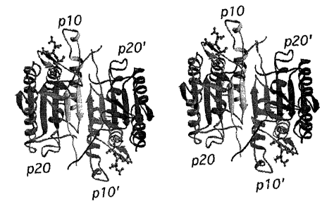
FIGS. 1A and 1B depict a stereoview of a
caspase-7 tetrameric assembly made using RIBBONS [M.
Carson, J. Appl. Cryst., 24, pp. 958-961 (1991)]. The
p20, p10 and their symmetry related equivalents, p20'
and p10', are shown moving from left to right in each
of FIG. 1A and FIG. 1B. The ball-and-stick model, near
the top left of each FIG., represents the Ac-DEVD-CHO
inhibitor.
FIGS. 2A, 2B, and 2C depict secondary
structural elements of caspase-1, caspase-3, caspase-7,
and caspase-8.
FIG. 2A depicts the conserved fold for
superimposed Caspase-1, Caspase-3, Caspase-7, Caspase-8
with covalently bound tetrapeptide inhibitor, Ac-DEVD-
CHO.
FIG. 2B depicts superimposed S4 loops for
caspase-1 (378-386), caspase-3 (244-262), caspase-7
(270-288), and caspase-8 (451-463).
FIG. 2C depicts prime side helix-turn-helix
insertion in caspase-8 ranging from residues 245-253
proximal to superimposed insertion loop of caspase-1
(residues 249-254). FIG. 2B and FIG. 2C have been
rotated slightly relative to FIG. 2A.
FIG. 3 depicts the sequence alignment of
caspase-1, caspase-3, caspase-7, and caspase-8. The
alignment was heavily biased upon the superposition of
conserved secondary structural elements and active site
residues of caspase-1, caspase-3, caspase-7, and
caspase-8. The alignment was performed with the MVP
program and then adjusted manually [M. H. Lambert,
Pract. Appl. Comput.-Aided Drug Des., pp. 243-303, P.S.
Charifson, ed., Dekker, New York. (1997)]. Boxed
CA 02389569 2002-05-15
WO 01/37194 PCT/US00/31602
-
residues denote direct or water-mediated interactions
with Ac-DEVD-CHO as shown in FIG. 4.
FIG. 4 depicts hydrogen bonding and van der
Waals interactions of covalently bound Ac-DEVD-
thiohemiacetal with binding site residues of: a)
caspase-1; b) caspase-3; c) caspase-7; and d)
caspase-8. "Wat" indicates a water molecule.
FIG. 5A-5D depicts electrostatic potentials
mapped onto molecular surface for binding site residues
of caspase-1, caspase-3, caspase-7, and caspase-8.
Areas of positive electrostatic potential, negative
electrostatic potential, and neutral electrostatic
potential are depicted. The molecular surface and
electrostatic potentials calculated with GRASP [A.
Nicholls et al., Proteins: Structure Function,
Genetics, 11, pp. 281-296 (1991)].
FIG. 6 depicts a stereoview diagram showing
the superposition of caspase-3 with caspase-7 generated
by RIBBONS. The N-acetyl group of the tetrapeptide
inhibitor, Ac-DEVD-CHO, bound to caspase-7 is
translated approximately 2.5 A relative to that in
caspase-3 due to the substitution of proline (235) for
serine at the same position of the P4-binding site.
FIG. 7 lists the atomic structure coordinates
for caspase-7 in complex with a synthetic tetrapeptide
inhibitor as derived by X-ray diffraction from a
crystal of that complex. The preparation of the
complex is described in Examples 1 and 2. The
following abbreviations are used in FIG. 7:
"Atom type" refers to the element whose
coordinates have been determined. Elements are defined
by the first letter in the column.
CA 02389569 2002-05-15
WO 01/37194 PCT/US00/31602
_ g -
"X, Y, Z" crystallographically define the
atomic position determined for each atom.
"Occ" is an occupancy factor that refers to
the fraction of the molecules in which each atom
occupies the position specified by the coordinates.. A
value of "1" indicates that each atom has the same
conformation, i.e., the same position, in all molecules
of the crystal.
"B" is a thermal factor that measures
movement of the atom around its atomic center.
FIG. 8 shows a diagram of a system used to
carry out the instructions encoded by the storage
medium of FIGS. 9 and 10.
FIG. 9 shows a cross section of a magnetic
storage medium.
FIG. 10 shows a cross section of an
optically-readable data storage medium.
DETAILED DESCRIPTION OF THE INVENTION
The following abbreviat ions used
are
throughout
the application:
A = Ala = Alanine T = Thr Threonine
=
V = Val = Valine C = Cys Cysteine
=
L = Leu = Leucine Y = Tyr Tyrosine
=
I - Ile = Isoleucine N = Asn Asparagine
=
P = Pro = Proline Q = Gln Glutamine
=
F = Phe = Phenylalanine D = Asp Aspartic Acid
=
W = Trp = Tryptophan E = Glu Glutamic Acid
=
M = Met = Methionine K = Lys Lysine
=
G = Gly = Glycine R = Arg Arginine
=
S = Ser = Serine H = His Histidine
=
Additional definitions are set forth in the
specification
where
necessary.
CA 02389569 2002-05-15
WO 01/37194 PCT/US00/31602
- 9 -
In order that the invention described herein
may be more fully understood, the following detailed
description is set forth.
Applicants have solved the above problems by
providing, for the first time, crystallizable
compositions comprising a caspase-7 in complex with a
caspase-7 inhibitor.
Thus, in one embodiment of this invention is
provided a crystallizable composition comprising a
caspase-7 in complex with a tetrapeptide aldehyde
inhibitor, acetyl-Asp-Glu-Val-Asp-CHO (Ac-DEVD-CHO).
Preferably, the caspase-7 has amino acids 1-303
according to Y. Gu et al., J. Biol. Chem., 271, pp.
10816-10820 (1996).
The caspase-7 polypeptide portion of the
complex is any polypeptide which has the cysteine
protease activity of the naturally occurring caspase-7.
Preferably, the caspase-7 polypeptide in the
compositions of this invention is the recombinantly
produced caspase-7 protein that is prepared as
described herein.
The caspase-7 polypeptide and Ac-DEVD-CHO may
be produced by any well-known method, including
synthetic methods, such as solid phase, liquid phase
and combination solid phase/liquid phase syntheses;
recombinant DNA methods, including cDNA cloning,
optionally combined with site directed mutagenesis;
and/or purification of the natural products, optionally
combined with enzymatic cleavage methods to produce
fragments of naturally occurring caspase-7 polypeptide.
The inhibitor Ac-DEVD-CHO is commercially available
(Peptides International) or may be produced by~any
well-known method, including synthetic methods, such as
CA 02389569 2002-05-15
WO 01/37194 PCT/US00/31602
- 10 -
solid phase, liquid phase and combination solid
phase/liquid phase syntheses.
In another embodiment of this invention is
provided a crystal comprising a caspase-7 in complex
with a tetrapeptide aldehyde inhibitor, acetyl-Asp-Glu-
Val-Asp-CHO (Ac-DEVD-CHO). Preferably, the caspase-7
has amino acids 1-303 according to Y. Gu et al., J.
Biol. Chem., 271, pp. 10816-10820 (1996). Preferably,
0
the crystal has unit cell dimensions of 88.2 A, 88.2 A,
0
186.2 A, a=90.0°, (3=90.0°, y=120.0° and belongs to space
group P3221. More preferably, the crystallized enzyme
' is a tetramer.
Importantly, applicants' invention has
provided, for the first time, information about the
shape and structure of the caspase-7 active site
binding pockets.
Binding pockets are of significant utility in
fields such as drug discovery. The association of
natural ligands or substrates with the binding pockets
of their corresponding receptors or enzymes is the
basis of many biological mechanisms of action.
Similarly, many drugs exert their biological effects
through association with the binding pockets of
receptors and enzymes. Such associations may occur
with all or any parts of the binding pocket. An
understanding of such associations will help lead to
the design of drugs having more favorable associations
with their target receptor or enzyme, and thus,
improved biological effects. Therefore, this
information is valuable in designing potential
inhibitors of caspase-7-like binding pockets.
CA 02389569 2002-05-15
WO 01/37194 PCT/US00/31602
- 11 -
The term "binding pocket", as used herein,
refers to a region of a molecule or molecular complex,
that, as a result of its shape, favorably associates
with another. chemical entity or compound.
The term "caspase-7-like binding pocket" refers
to a portion of a molecule or molecular complex whose
shape is sufficiently similar to all or any parts of
the caspse-7 active site binding pockets as to bind
common ligands. This commonality of shape is defined
by a root mean square deviation from the structure
coordinates of the backbone atoms of the amino acids
that make up the binding pockets in caspse-7 (as set
forth in Figure 1) of not more than 1.5 A. How this
calculation is obtained is described below.
The "active site binding pockets" or "active
site" of caspse-7 refers to the area on the caspse-7
enzyme surface where cleavage of a substrate occurs,
and where Ac-DEVD-CHO exerts its inhibitory effect.
The terms "P binding pocket", "S pocket" "S
region" and the like, refer to binding subsites, or
portions of the substrate binding site on the caspase
molecule. The amino acid residues of the substrate are
given designations according to their position relative
to the scissile bond, i.e., the bond that is broken by
the protease. The residues are designated P1, P2,
etc., for those extending toward the N-terminus of the
substrate and P1', P2', etc., for those extending
toward the C-terminus of the substrate. The portions
of an inhibitor that correspond to the P or P' residues
of the substrate are also labeled P1, P1', etc., by
analogy with the substrate. The binding subsites of
the caspase molecule that receive the residues labeled
P1, P1', etc., are designated S1, S1', etc., or may
CA 02389569 2002-05-15
WO 01/37194 PCT/US00/31602
- 12 -
alternately be designated "the P1 binding pocket", "the
P1' binding pocket", etc. [I. Schechter & A. Berger,
Biochem. Biophys. Res. Communi., 27, pp. 157-162
(1967) ] .
In resolving the crystal structure of caspase-
7, applicants have determined that caspase-7 amino
acids 234, 235, 237, 276, 278, 281, and 284 are
responsible for the S4-binding region of caspase-7
being more hydrophilic than the S4-binding region of
caspase-3. Applicants have also determined that amino
acids 85, 86, 87, 88, 144, 145, 184, 186, 191, 223,
230, 231, 232, 233, 234, 235, 237, 240, 276, 278, 281,
282, and 284 are situated close enough to the Ac-DEVD-
CHO inhibitor to interact with this ligand.
Applicants have also determined that amino acid
residues 87, 184, and 233 are important in the Pl
binding pocket of caspase-7; that amino acid residues
191, 230, 232, and 282 are important in the P2 binding
pocket of caspase-7; that amino acid residues 86, 88,
233 are important in the P3 binding pocket of caspase-
7; and that amino acid residues 234, 235, 237, 240, and
276 are important in the P3 binding pocket of caspase-
7.
It will be readily apparent to those of skill
in the art that the numbering of amino acids in other
isoforms of caspase-7 may be different than that
described herein.
Advantageously, the crystal provided by this
invention is amenable to x-ray crystallography. Thus,
this invention also provides the three-dimensional
structure of a caspse-7 complex, specifically a
caspase-7/Ac-DEVD-CHO complex, at 2.35 A resolution.
CA 02389569 2002-05-15
WO 01/37194 PCT/US00/31602
- 13 -
Importantly, this has provided for the first time,
information about the shape and structure of caspase-7.
The three-dimensional structure of the
caspase-7/inhibitor complex of this invention is
defined by a set of structure coordinates as set forth
in FIG. 7. The term "structure coordinates" refers to
Cartesian coordinates derived from mathematical
equations related to the patterns obtained on
diffraction of a monochromatic beam of X-rays by the
atoms (scattering centers) of a caspase-7/inhibitor
complex in crystal form. The diffraction data are used
to calculate an electron density map of the repeating
unit of the crystal. The electron density maps are
then used to establish the positions of the individual
atoms of the caspase-7 or caspase-7/inhibitor complex.
Those of skill in the art will understand
that a set of structure coordinates for an enzyme or an
enzyme-complex or a portion thereof, is a relative set
of points that define a shape in three dimensions.
Thus, it is possible that an entirely different set of
coordinates could define a similar or identical shape.
Moreover, slight variations in the individual
coordinates will have little effect on overall shape.
The variations in coordinates discussed above
may be generated because of mathematical manipulations
of the structure coordinates. For example, the
structure coordinates set forth in FIG. 7 could be
manipulated by crystallographic permutations of the
structure coordinates, fractionalization of the
structure coordinates, integer additions or
subtractions to sets of the structure coordinates,
inversion of the structure coordinates or any
combination of the above.
CA 02389569 2002-05-15
WO 01/37194 PCT/US00/31602
- 14 -
Alternatively, modifications in the crystal
structure due to mutations, additions, substitutions,
and/or deletions of amino acids, or other changes in
any of the components that make up the crystal could
S also account for variations in structure coordinates.
If such variations are within an acceptable standard
error as compared to the original coordinates, the
resulting three-dimensional shape is considered to be
the same.
Various computational analyses are therefore
necessary to determine whether a molecule or molecular
complex or a portion thereof is sufficiently similar to
all or parts of the caspase-7/inhbitor structure
described above as to be considered the same. Such
analyses may be carried out in current software
applications, such as the Molecular Similarity
application of QUANTA (Molecular Simulations Inc., San
Diego, CA) version 4.1, and as described in the
accompanying User's Guide.
The Molecular Similarity application permits
comparisons between different structures, different
conformations of the same structure, and different
parts of the same structure. The procedure used in
Molecular Similarity to compare structures is divided
into four steps: 1) load the structures to be
compared; 2) define the atom equivalencies in these
structures; 3) perform a fitting operation; and 4)
analyze the results.
Each structure is identified by a name. One
structure is identified as the target (i.e., the fixed
structure); all remaining structures are working
structures (i.e., moving structures). Since atom
equivalency within QUANTA is defined by user input, for
CA 02389569 2002-05-15
WO 01/37194 PCT/US00/31602
- 15 -
the purpose of this invention we will define equivalent
atoms as protein backbone atoms (N, Ca, C and O) for
all conserved residues between the two structures being
compared. We will also consider only rigid fitting
operations.
When a rigid fitting method is used, the
working structure is translated and rotated to obtain
an optimum fit with the target structure. The fitting
operation uses an algorithm that computes the optimum
translation and rotation to be applied to the moving
structure, such that the root mean square difference of
the fit over the specified pairs of equivalent atom is
an absolute minimum. This number, given in angstroms,
is reported by QUANTA.
For the purpose of this invention, any
molecule or molecular complex that has a root mean
square deviation of conserved residue backbone atoms
(N, Ca, C, O) of less than 1.5 A when superimposed on
the relevant backbone atoms described by structure
coordinates listed in FIG. 7 are considered identical.
More preferably, the root mean square deviation is less
than 1.0 A.
The term "root mean square deviation" means
the square root of the arithmetic mean of the squares
of the deviations from the mean. It is a way to
express the deviation or variation from a trend or
object. For purposes of this invention, the "root mean
square deviation" defines the variation in the backbone
of a protein or protein complex from the relevant
portion of the backbone. of the caspase-7 polypeptide
portion of the complex as defined by the structure
coordinates described herein.
CA 02389569 2002-05-15
WO 01/37194 PCT/US00/31602
- 16 -
Once the structure coordinates of a protein
crystal have been determined they are useful variety of
purposes, such as drug discovery and x-ray
crystallographic determinations of related proteins.
Thus, in accordance with the present
invention, the structure coordinates of a caspase-7
polypeptide/inhibitor complex, and portions thereof is
provided. Such data may be used for the purposes of,
for example, drug design and molecular replacement.
Accordingly, one embodiment of this invention
provides a molecule or molecular complex comprising all
or any part of a binding pocket defined by structure
coordinates of caspase-7 amino acids 234, 235, 237,
276, 278, 281, and 284 according to FIG. 7, or a
homologue of said molecule or molecular complex,
wherein said homologue comprises a binding pocket that
has a root mean square deviation from the backbone
atoms of said amino acids of not more than 1.5 A, and
wherein said molecule or molecular complex has a S4
binding region that is more hydrophilic than the S4
binding region of caspase-3.
Another embodiment of this invention provides
a molecule or molecular complex comprising all or any
part of a binding pocket defined by structure
coordinates of caspase-7 amino acids 85, 86, 87, 88,
144, 145, 184, 186, 191, 223, 230, 231, 232, 233, 234,
235, 237, 240, 276, 278, 281, 282, and 284 or a
homologue of said molecule or molecular complex,
wherein said homologue comprises a binding pocket that
has a root mean square deviation from the backbone
atoms of said amino acids of not more than 1.5 A, and
wherein said molecule or molecular complex has a S4
CA 02389569 2002-05-15
WO 01/37194 PCT/US00/31602
- 17 -
binding region that is more hydrophilic than the S4
binding region of caspase-3.
Yet another embodiment of this~invention
provides a molecule or molecular complex defined by all
or part of the structure coordinates according to
FIG. 7, or a homologue thereof, wherein said homologue
has a root mean square deviation from the conserved
backbone atoms of said amino acids of not more than
a
1.5 A, and wherein said molecule or molecular complex
has a S4 binding region that is more hydrophilic than
the S4 binding region of caspase-3. Preferably, a
molecule or molecular complex is defined by all of the
structure coordinates according to FIG. 7 or a
homologue thereof.
Yet another embodiment of this invention
provides a molecule or molecular complex defined by all
or part of the structure coordinates of caspase-7 amino
acids 58-302 according to FIG. 7, or a homologue
thereof, wherein said homologue has a root mean square
deviation from the conserved backbone atoms of said
0
amino acids of not more than 1.5 A, and wherein said
molecule or molecular complex has a S4 binding region
that is more hydrophilic than the S4 binding region of
caspase-3. Preferably, the molecule or molecular
. complex is defined by all of the structure coordinates
of caspase-7 amino acids 58-302 according to FIG. 7 or
a homologue thereof.
Preferably, in any of these embodiments, the
homologue has a root mean square deviation from the
conserved backbone atoms of said amino acids of not
0
more than 1.0 A.
In a more preferred embodiment, the molecule
or molecular complex is defined by the structure
CA 02389569 2002-05-15
WO 01/37194 PCT/US00/31602
- 18 -
coordinates of caspase-7 amino acids 58-302 according
to FIG. 7.
This invention also provides a machine-
readable data storage medium, comprising a data storage
material encoded with machine readable data, wherein
said data is defined by the all or a portion of the
structure coordinates of a caspase-7 complex according
to FIG. 7, or a homologue of said complex, wherein said
homologue comprises backbone atoms that have a root
mean square deviation from the backbone atoms of the
complex of not more than 1.5 A.
Preferably, the data is defined by the
structure coordinates of caspase-7 amino acids 234,
235, 237, 276, 278, 281, and 284 according to FIG. 7,
or a homologue of said complex, wherein said homologue
comprises backbone atoms that have a root mean square
deviation from the backbone atoms of the complex of not
more than 1.5 A.
More preferably, the data is defined by the
structure coordinates of caspase-7 amino acids 85, 86,
87, 88, 144, 145, 184, 186, 191, 223, 230, 231, 232,
233, 234, 235, 237, 240, 276, 278, 281, 282,~and 284
according to FIG. 7, or a homologue of said complex,
wherein said homologue comprises backbone atoms that
have a root mean square deviation from the backbone
atoms of the complex of not more than 1.5 A.
Even more preferably, the data is defined by
the structure coordinates for caspase-7 amino acids 58-
302 according to FIG. 7, or a homologue of said
molecule or molecular complex, said homologue having a
root mean square deviation from the backbone atoms of
said amino acids of not more than 1.5 A.
CA 02389569 2002-05-15
WO 01/37194 PCT/US00/31602
- 19 -
Preferably, in any of these embodiments, the
homologue has a root mean square deviation from the
conserved backbone atoms of said amino acids of not
a
more than 1.0 A.
More preferably, the data is defined by the
structure coordinates for caspase-7 amino acids 58-302
according to FIG. 7.
According to this invention, these caspase-7
complexes and homologues thereof have a S4 binding
region that is more hydrophilic than the S4 binding
region of caspase-3.
To use the structure coordinates generated
for the caspase-7 complex or one of its binding pockets
or homologues thereof, it is sometimes necessary to
convert them into a three-dimensional shape. This is
achieved through the use of commercially available
software that is capable of a generating a three-
dimensional representation of molecules or portions
thereof from a set of structure coordinates. The
three-dimensional representations may be displayed as a
graphical representation.
Therefore, according to another embodiment of
this invention is provided a machine-readable data
storage medium, comprising a data storage material
encoded with machine readable data, when using a
machine programmed with instructions for using said
data, is capable of producing a three-dimensional
representation of any of the molecule or molecular
complexes, or homologues thereof, that are described
herein.
This invention also provides a computer for
producing a three-dimensional representation of:
CA 02389569 2002-05-15
WO 01/37194 PCT/US00/31602
- 20 -
a. a molecule or molecular complex
comprising a binding pocket defined by structure
coordinates of caspase-7 amino acids 234, 235, 237,
276, 278, 281, and 284 according to FIG. 7; or
b. a homologue of said molecule or
molecular complex, wherein said homologue comprises a
binding pocket that has a root mean square deviation
from the backbone atoms of said amino acids of not more
than 1.5 A, wherein said computer comprises:
i. a machine-readable data storage
medium comprising a data storage material encoded with
machine-readable data, wherein said data comprises the
structure coordinates of caspase-7 amino acids 234,
235, 237, 276, 278, 281, and 284 according to FIG. 7;
ii. instructions for processing said
machine-readable data into said three-dimensional
representation.
Preferably, the computer is for producing a
three-dimensional representation of a molecule or
molecular complex defined by the set of structure
coordinates for caspase-7 amino acids 85, 86, 87, 88,
144, 145, 184, 186, 191, 223, 230, 231, 232, 233, 234,
235, 237, 240, 276, 278, 281, 282, and 284 according to
FIG. 7, or a three-dimensional representation is of a
homologue of said molecule or molecular complex, said
homologue having a root mean square deviation from the
backbone atoms of said amino acids of not more than
1.5 A.
More preferably, the computer is for
producing a three-dimensional representation of a
molecule or molecular complex defined by all or a
portion of the set of structure coordinates according
to FIG. 7, or a homologue of said molecule or molecular
CA 02389569 2002-05-15
WO 01/37194 PCT/US00/31602
- 21 -
complex, said homologue having a root mean square
deviation from the backbone atoms of said amino acids
of not more than 1.5 A.
Most preferably, the computer is for
producing a three-dimensional defined by the set of
structure coordinates of caspase-7 amino acids 58-302
according to FIG. 7, or a homologue of said molecule or
molecular complex, or a homologue thereof having a root
mean square deviation from the backbone atoms of said
amino acids of not more than 1.5 A.
Preferably, in any of these embodiments, the
homologue has a root mean square deviation from the
conserved backbone atoms of said amino acids of not
more than 1.0 A.
Most preferably, the computer is for
producing a three-dimensional defined by the set of
structure coordinates of caspase-7 amino acids 58-302
according to FIG. 7.
In a preferred embodiment, a computer
according to this invention also comprises a working
memory for storing instructions for processing the
machine-readable data, a central-processing unit
coupled to the working memory and to said machine-
readable data storage medium for processing said
machine-readable data into the three-dimensional
representation, or a display for displaying the three-
dimensional representation. More preferably, a
computer according to this invention comprises a
display. Most preferably, a computer according to this
invention comprises the above working memory, central-
processing unit, and display.
FIG. 8 demonstrates one version of these
embodiments. System 10 includes a computer 11
CA 02389569 2002-05-15
WO 01/37194 PCT/US00/31602
- 22 -
comprising a central processing unit ("CPU") 20, a
working memory 22 which may be, e.g., RAM
(random-access memory) or "core" memory, mass storage
memory 24 (such as one or more disk drives or CD-ROM
drives), one or more cathode-ray tube ("CRT") display
terminals 26, one or more keyboards 28, one or more
input lines 30, and one or more output lines 40, all of
which are interconnected by a conventional
bidirectional system bus 50.
Input hardware 36, coupled to computer 11 by
input lines 30, may be implemented in a variety of
ways. Machine-readable data of this invention may be
inputted via the use of a modem or modems 32 connected
by a telephone line or dedicated data line 34.
Alternatively or additionally, the input hardware 36
may comprise CD-ROM drives or disk drives 24. In
conjunction with display terminal 26, keyboard 28 may
also be used as an input device.
Output hardware 46, coupled to computer 11 by
output lines 40, may similarly be implemented by
conventional devices. By way of example, output
hardware 46 may include CRT display terminal 26 for
displaying a graphical representation of a binding
pocket of this invention using a program such as QUANTA
as described herein. Output hardware might also'
include a printer 42, so that hard copy output may be
produced, or a disk drive 24, to store system output
for later use.
In operation, CPU 20 coordinates the use of
the various input and output devices 36, 46,
coordinates data accesses from mass storage 24 and
accesses to and from working memory 22, and determines
the sequence of data processing steps. A number of
CA 02389569 2002-05-15
WO 01/37194 PCT/US00/31602
- 23 -
programs may be used to process the machine-readable
data of this invention. Such programs are discussed in
reference to the computational methods of drug
discovery as described herein. Specific references to
components of the hardware system 10 are included as
appropriate throughout the following description of the
data storage medium.
FIG. 9 shows a cross section of a magnetic
data storage medium 100 which can be encoded with a
machine-readable data that can be carried out by a
system such as system 10 of FIG. 8. Medium 100 can be
a conventional floppy diskette or hard disk, having a
suitable substrate 101, which may be conventional, and
a suitable coating 102, which may be conventional, on
one or both sides, containing magnetic domains (not
visible) whose polarity or orientation can be altered
magnetically. Medium 100 may also have an opening (not
shown) for receiving the spindle of a disk drive or
other data storage device 24.
The magnetic domains of coating 102 of medium
100 are polarized or oriented so as to encode in manner
which may be conventional, machine readable data such
as that described herein, for execution by a system
such as system 10 of FIG. 8.
FIG. 10 shows a cross section of an
optically-readable data storage medium 110 which also
can be encoded with such a machine-readable data, or
set of instructions, which can be carried out by a
system such as system 10 of FIG. 8. Medium 110 can be
a conventional compact disk read only memory (CD-ROM)
or a rewritable medium such as a magneto-optical disk
which is optically readable and magneto-optically
writable. Medium 100 preferably has a suitable
CA 02389569 2002-05-15
WO 01/37194 PCT/US00/31602
- 24 -
substrate 111, which may be conventional, and a
suitable coating 112, which may be conventional,
usually of one side of substrate 111.
In the case of CD-ROM, as is well known,
coating 112 is reflective and is impressed with a
plurality of pits 113 to encode the machine-readable
data. The arrangement of pits is read by reflecting
laser light off the surface of coating 112. A
protective coating 114, which preferably is
substantially transparent, is provided on top of
coating 112.
In the case of a magneto-optical disk, as is
well known, coating 112 has no pits 113, but has a
plurality of magnetic domains whose polarity or
orientation can be changed magnetically when heated
above a certain temperature, as by a laser (not shown).
The orientation of the domains can be read by measuring
the polarization of laser light reflected from coating
112. The arrangement of the domains encodes the data
as described above.
For the first time, the present invention
permits the use of drug discovery techniques, including
structure-based, rational drug design, or database
screening techniques, to design, select, and/or
synthesize chemical entities, including inhibitory
compounds that are capable of binding to caspase-7, or
any portion thereof.
For example, the structure encoded by the
data may be computationally evaluated for its ability
to associate with chemical entities. Chemical entities
that associate with caspase-7 may inhibit caspase-7,
and are potential drug candidates. Alternatively, the
structure encoded by the data may be displayed in a
CA 02389569 2002-05-15
WO 01/37194 PCT/US00/31602
- 25 -
graphical three-dimensional representation on a
computer screen. This allows visual inspection of the
structure, as well as visual inspection of the
structure's association with chemical entities.
~ Thus, according to another embodiment, the
invention relates to a method for evaluating the
potential or ability of a chemical entity to associate
with any of the molecules or molecular complexes set
forth above. This method comprises the steps of: a)
employing computational means to perform a fitting
operation between the chemical entity and a binding
pocket of the molecule or molecular complex; and b)
analyzing the results of said fitting operation to
quantify the association between the chemical entity
and the binding pocket. The term "chemical entity", as
used herein, refers to chemical compounds, complexes of
at least two chemical compounds, and fragments of such
compounds or complexes.
For the first time, the present invention
permits the use of molecular design techniques to
identify, select and design chemical entities,
including inhibitory compounds, capable of binding to
caspase-7-like binding pockets.
Applicants' elucidation of the caspase-7
binding sites provides the necessary information for
designing new chemical entities and compounds that may
interact with the caspase-7 binding pockets, in whole
or in part. This elucidation also enables the
evaluation of structure-activity data for compounds
which bind to caspase-7 binding pockets. Applicants'
elucidation of the caspase-7 binding sites also
provides important structural information for comparing
the requirements for interacting with caspase-7 binding
CA 02389569 2002-05-15
WO 01/37194 PCT/US00/31602
- 26 -
pockets selectively or in addition to other caspase
binding pockets.
Thus, according to another embodiment, the
invention relates to a method for a method for
identifying a potential agonist or antagonist of any of
the molecules or molecular complexes set forth above.
This method comprises the steps of: a. generating a
three-dimensional structure of the molecule or
molecular complex; b. employing said three-dimensional
structure to design or select said potential agonist or
antagonist; c. providing said potential agonist or
antagonist; and d. contacting said potential agonist or
antagonist to interact with said molecule.
Throughout this section, discussions about
the ability of an entity to bind to, associate with or
inhibit a caspase-7 binding pocket refers to features
of the entity alone. Assays to determine whether a
compound binds to caspases are well known in the art
[see for example, the assays described in W097/22619].
The design of compounds that bind to or
inhibit caspase-7 according to this invention generally
involves consideration of two factors. First, the
entity must be capable of physically arid structurally
associating with parts or all of the caspase-7 binding
pockets. Non-covalent molecular interactions important
in this association include hydrogen bonding, van der
Waals interactions, hydrophobic interactions and
electrostatic interactions.
Second, the entity must be able to assume a
conformation that allows it to associate with the
caspase-7 binding pocket directly. Although certain
portions of the entity will not directly participate in
these associations, those portions of the entity may
CA 02389569 2002-05-15
WO 01/37194 PCT/US00/31602
- 27 -
still influence the overall conformation of the
molecule. This, in turn, may have a significant impact
on potency. Such conformational requirements include
the overall three-dimensional structure and orientation
of the chemical entity in relation to all or a portion
of the binding pocket, or the spacing between
functional groups of an entity comprising several
chemical entities that directly interact with the
caspase-7 binding pocket or homologues thereof.
The potential inhibitory or binding effect of
a chemical entity on a caspase-7 binding pocket may be
analyzed prior to its actual synthesis and testing by
the use of computer modeling techniques. If the
theoretical structure of the given entity suggests
insufficient interaction and association between it and
the caspase-7 binding pocket, testing of the entity is
obviated. However, if computer modeling indicates a
strong interaction, the molecule may then be provided
or synthesized and tested for its ability to bind to a
caspase-7 binding pocket. This may be achieved by
testing the ability of the molecule to inhibit caspase-
7 using assays that are known to those of ordinary
skill in the art. In this manner, the obtaining and
testing of inoperative compounds may be avoided.
A potential inhibitor of caspase-7 may be
computationally evaluated by means of a series of steps
in which chemical entities or fragments are screened
and selected for their ability to associate with the
caspase-7 binding pockets.
One skilled in the art may use one of several
methods to screen chemical entities or fragments for
their ability to associate with a caspase-7 binding
pocket. This process may begin by visual inspection
CA 02389569 2002-05-15
WO 01/37194 PCT/US00/31602
- 28 -
of, for example, a.caspase-7 binding pocket on the
computer screen based on the caspase-7 structure
coordinates in FIG. 7 or other coordinates which define
a similar shape generated from the machine-readable
storage medium. Selected fragments or chemical
entities may then be positioned in a variety of
orientations, or docked, within that binding pocket as
defined supra. Docking may be accomplished. using
software such as Quanta and Sybyl, followed by energy
minimization and molecular dynamics with standard
molecular mechanics force fields, such as CHARMM and
AMBER.
Specialized computer programs may also assist
in the process of selecting fragments or chemical
entities. These include:
1. GRID (P. J. Goodford, "A Computational Procedure
for Determining Energetically Favorable Binding Sites
on Biologically Important Macromolecules", J. Med.
Chem., 28, pp. 849-857 (1985)). GRID is available from
Oxford University, Oxford, UK.
2. MOSS (A. Miranker et al., "Functionality Maps of
Binding Sites: A Multiple Copy Simultaneous Search
Method." Proteins: Structure, Function and Genetics,
11, pp. 29-34 (1991)). MCSS is available from
Molecular Simulations, San Diego, CA.
3. AUTODOCK (D. S. Goodsell et al., "Automated
Docking of Substrates to Proteins by Simulated
Annealing", Proteins: Structure, Function, and
Genetics, 8, pp. 195-202 (1990)). AUTODOCK is
available from Scripps Research Institute, La Jolla,
CA.
4. DOCK (I. D. Kuntz et al., "A Geometric Approach to
Macromolecule-Ligand Interactions", J. Mol. Biol.,
CA 02389569 2002-05-15
WO 01/37194 PCT/US00/31602
- 29 -
161, pp. 269-288 (1982)). DOCK is available from
University of California, San Francisco, CA.
Once suitable chemical entities or fragments
have been selected, they can be assembled into a single
compound or complex. Assembly may be preceded by
visual inspection of the relationship of the fragments
to each other on the three-dimensional image displayed
on a computer screen in relation to the structure
coordinates of caspase-7. This would be followed by
manual model building using software such as Quanta or
Sybyl [Tripos Associates, St. Louis, MO].
Useful programs to aid one of skill in the
art in connecting the individual chemical entities or
fragments include:
1. CAVEAT (P. A. Bartlett et al., "CAVEAT: A Program
to Facilitate the Structure-Derived Design of
Biologically Active Molecules", in Molecular
Recognition in Chemical and Biological Problems",
Special Pub., Royal Chem. Soc., 78, pp. 182-196 (1989);
G. Lauri and P. A. Bartlett, "CAVEAT: a Program to
Facilitate the Design of Organic Molecules", J. Comput.
Aided Mol. Des. , 8, pp. 51-66 (1994)). CAVEAT is
available from the University of California, Berkeley,
CA.
2. 3D Database systems such as ISIS (MDL Information
Systems, San Leandro, CA). This area is reviewed in Y.
C. Martin, "3D Database Searching in Drug Design", J.
Med. Chem., 35, pp. 2145-2154 (1992).
3. HOOK (M. B. Eisen et al., "HOOK: A Program for
Finding Novel Molecular Architectures that Satisfy the
Chemical and Steric Requirements of a Macromolecule
Binding Site", Proteins: Struct., Funct., Genet., 19,
CA 02389569 2002-05-15
WO 01/37194 PCT/US00/31602
- 30 -
pp. 199-221 (1994). HOOK is available from Molecular
Simulations, San Diego, CA.
Instead of proceeding to build an inhibitor
of a caspase-7 binding pocket in a step-wise fashion
one fragment or chemical entity at a time as described
above, inhibitory or other caspase-7 binding compounds
may be designed as a whole or "de novo" using either an
empty binding site or optionally including some
portions) of a known inhibitor(s). There are many de
novo ligand design methods including:
1. LUDI (H.-J. Bohm, "The Computer Program LUDI: A
New Method for the De Novo Design of Enzyme
Inhibitors", J. Comp. Aid. Molec. Design, 6, pp. 61-78
((1992)). LUDI is available from Molecular Simulations
Incorporated, San Diego, CA.
2. LEGEND (Y. Nishibata et al., Tetrahedron, 47, p.
8985 (1991)). LEGEND is available from Molecular
Simulations Incorporated, San Diego, CA.
3. LeapFrog (available from Tripos Associates, St.
Louis, MO).
4. SPROUT (V. Gillet et al., "SPROUT: A Program for
Structure Generation)", J. Comput. Aided Mol. Design,
7, pp. 127-153 (1993)). SPROUT is available from the
University of Leeds, UK.
Other molecular modeling techniques~may also
be employed in accordance with this invention [see,
e.g., N. C. Cohen et al., "Molecular Modeling Software
and Methods for Medicinal Chemistry, J. Med. Chem., 33,
pp. 883-894 (1990); see also, M. A. Navia and M. A.
Murcko, "The Use of Structural Information in Drug
Design", Current Opinions in Structural Biology, 2, pp.
202-210 (1992); L. M. Balbes et al., "A Perspective of
Modern Methods in Computer-Aided Drug Design", in
CA 02389569 2002-05-15
WO 01/37194 PCT/US00/31602
- 31 -
Reviews in Computational Chemistry, Vol. 5, K. B.
Lipkowitz and D. B. Boyd, Eds., VCH, New York, pp. 337-
380 (1994); see also, W. C. Guida, "Software For
Structure-Based Drug Design", Curr. Opin. Struct.
Biology, 4, pp. 777-781 (1994)].
Once a compound has been designed or selected
by the above methods, the efficiency with which that
entity may bind to a caspase-7 binding pocket may be
tested and optimized by computational evaluation. 'For
example, an effective caspase-7 binding pocket
inhibitor must preferably demonstrate a relatively
small difference in energy between its bound and free
states (i.e., a small deformation energy of binding).
Thus, the most efficient caspase-7 binding pocket
inhibitors should preferably be designed with a
deformation energy of binding of not greater than about
10 kcal/mole, more preferably, not greater than 7
kcal/mole. Caspase-7 binding pocket inhibitors may
interact with the binding pocket in more than one
conformation that is similar in overall binding energy.
In those cases, the deformation energy of binding is
taken to be the difference between the energy of the
free entity and the average energy of the conformations
observed when the inhibitor binds to the protein.
An entity designed or selected as binding to
a caspase-7 binding pocket may be further
computationally optimized so that in its bound state it
would preferably lack repulsive electrostatic
interaction with the target enzyme and with the
surrounding water molecules. Such non-complementary
electrostatic interactions include repulsive charge-
charge, dipole-dipole, and charge-dipole interactions.
CA 02389569 2002-05-15
WO 01/37194 PCT/US00/31602
- 32 -
Specific computer software is available in
the art to evaluate compound deformation energy and
electrostatic interactions. Examples of programs
designed for such uses include: Gaussian 94, revision
C (M. J. Frisch, Gaussian, Inc., Pittsburgh, PA ~1995);
AMBER, version 4.1 (P. A. Kollman, University of
California at San Francisco, ~1995); QUANTA/CHARMM
(Molecular Simulations, Inc., San Diego, CA ~1995);
Insight II/Discover (Molecular Simulations,. Inc., San
Diego, CA ~1995); Delphi (Molecular Simulations, Inc.,
San Diego, CA ~1995); and AMSOL (Quantum Chemistry
Program Exchange, Indiana University). These programs
may be implemented, for instance, using a Silicon
Graphics workstation such as an Indigo2 with "IMPACT"
graphics. Other hardware systems and software packages
will be known to those skilled in the art.
Another approach enabled by this invention,
is the computational screening of small molecule
databases for chemical entities or compounds that can
bind in whole, or in part, to a caspase-7 binding
pocket. In this screening, the quality of fit of such
entities to the binding site may be judged either by
shape complementarity or by estimated interaction
energy [E. C. Meng et al., J. Comp. Chem., 13, pp. 505-
524 (1992)].
Thus, enabled by this invention are compounds
that inhibit caspase-7 by associating directly with the
caspase-7 active site. Preferably, such compounds have
a strain energy of 10 kcal/mol or less. More
preferably, these compounds contain fewer than three
secondary amide bonds. Even more preferably, these
compounds have a molecular weight of less than 1000.
CA 02389569 2002-05-15
WO 01/37194 PCT/US00/31602
- 33 -
Those of skill in the art will realize that
association of natural ligands or substrates with the
binding pockets of their corresponding receptors or
enzymes is the basis of many biological mechanisms of
action. Similarly, many drugs exert their biological
effects through association with the binding pockets of
receptors and enzymes. Such associations may occur
with all or any parts of the binding pockets. An
understanding of such associations will help lead to
the design of drugs having more favorable associations
with their target receptor or enzyme, and thus,
improved biological effects. Therefore, this
information is valuable in designing potential ligands
or inhibitors of receptors or enzymes, such as
inhibitors of caspase-7.
The term "associating with" refers to a
condition of proximity between chemical entities or
compounds, or portions thereof. The association may be
non-covalent -- wherein the juxtaposition is
energetically favored by hydrogen bonding or van der
Waals or electrostatic interactions -- or it may be
covalent.
Another particularly useful drug design
technique enabled by this invention is iterative drug
design. Iterative drug design is a method for
optimizing associations between a protein and a
compound by determining and evaluating the three-
dimensional structures of successive sets of
protein/compound complexes.
In iterative drug design, crystals of a
series of protein or protein complexes are obtained and
then the three-dimensional structures of each crystal
is solved. Such an approach provides insight into the
CA 02389569 2002-05-15
WO 01/37194 PCT/US00/31602
- 34 -
association between the proteins and compounds of each
complex. This is accomplished by selecting compounds
with inhibitory activity, obtaining crystals of this
new protein/compound complex, solving the three-
s dimensional structure of the complex, and comparing the
associations between the new protein/compound complex
and previously solved protein/compound complexes. By
observing how changes in the compound affected the
protein/compound associations, these associations may
be optimized.
In some cases, iterative drug design is
carried out by forming successive protein-compound
complexes and then crystallizing each new complex.
Alternatively, a pre-formed protein crystal is soaked
in the presence of an inhibitor, thereby forming a
protein/compound complex and obviating the need to
crystallize each individual protein/compound complex.
Advantageously, the caspase-7/inhibitor crystals
provided by this invention may be soaked in the
presence of a compound or compounds, to provide other
crystal complexes.
As used herein, the term "soaked" refers to a
process in which the crystal is transferred to a
solution containing the compound of interest.
In another embodiment of this invention is
provided a method for preparing a composition
comprising a caspase-7 comprising the steps described
in Examples 1 and 2. Preferably, the composition
comprises a caspase-7 in complex with Ac-DEVD-CHO.
Once the structure coordinates of a protein
crystal have been determined they are useful in solving
the structures of other crystals. Thus, the structure
coordinates set forth in FIG. 7 can also be used to aid
CA 02389569 2002-05-15
WO 01/37194 PCT/US00/31602
- 35 -
in obtaining structural information about another
crystallized molecule or molecular complex. This may
be achieved by any of a number of well-known
techniques, including molecular replacement.
A machine-readable data storage medium
comprising a data storage material encoded with a first
set of machine readable data which, when combined with
a second set of machine readable data using a machine
programmed with instructions~for using said first set
of data and said second set of data, can determine at
least a portion of the structure coordinates
corresponding to~the second set of machine readable
data, wherein: said first set of data and said second
set of data comprises a Fourier transform of at least a
portion of the structural coordinates for caspase-7
according to FIG. 7; said second set of data comprises
an X-ray diffraction pattern of a molecule or molecular
complex of unknown structure.
The structure coordinates set forth in FIG. 7
can also be used for determining at least a portion of
the three-dimensional structure of molecules or
molecular complexes which contain at least some
structurally similar features to caspase-7. In
particular, structural information about another
crystallized molecule or molecular complex may be
obtained. This may be achieved by any of a number of
well-known techniques, including molecular replacement.
Therefore, in another embodiment this
invention provides a method of utilizing molecular
replacement to obtain structural information about a
crystallized molecule or molecular complex whose
structure is unknown comprising the steps of:
CA 02389569 2002-05-15
WO 01/37194 PCT/US00/31602
- 36 -
a) generating an X-ray diffraction pattern
from said crystallized molecule or molecular complex;
and
b) applying at least a portion of the
structure coordinates set forth in FIG. 7 to the X-ray
diffraction pattern to generate a three-dimensional
electron density map of the molecule or molecular
complex whose structure is unknown.
Preferably, the crystallized molecule or
molecular complex comprises a caspase-7. More
preferably, the crystallized molecule or molecular
complex is obtained by soaking a crystal of this
invention in a solution.
By using molecular replacement, all or part
of the structure coordinates of the caspase-7 complex
provided by this invention (and set forth in FIG. 7)
can be used to determine the structure of a
crystallized molecule or molecular complex whose
structure is unknown more quickly and efficiently than
attempting to determine such information ab initio.
Molecular replacement provides an accurate
estimation of the phases for an unknown structure.
Phases are a factor in equations used to solve crystal
structures that can not be determined directly.
Obtaining accurate values for the phases, by methods
other than molecular replacement, is a time-consuming
process that involves iterative cycles of
approximations and refinements and greatly hinders the
solution of crystal structures. However, when the
crystal structure of a protein containing at least a
homologous portion has been solved, the phases from the
known structure provide a satisfactory estimate of the
phases for the unknown structure.
CA 02389569 2002-05-15
WO 01/37194 PCT/US00/31602
- 37 -
Thus, this method involves generating a
preliminary model of a molecule or molecular complex
whose structure coordinates are unknown, by orienting
and positioning the relevant portion of the caspse-7
complex according to FIG. 7 within the unit cell of. the
crystal of the unknown molecule or molecular complex so
as best to account for the observed X-ray diffraction
pattern of the crystal of the molecule or molecular
complex whose structure is unknown. Phases can then be
calculated from this model and combined with the
observed X-ray diffraction pattern amplitudes to
generate an electron density map of the structure whose
coordinates are unknown. This, in turn, can be
subjected to any well-known model building and
structure refinement techniques to provide a final,
accurate structure of the unknown crystallized molecule
or molecular complex [E. Lattman, in Meth. Enzymol.,
115, pp. 55-77 (1985); M. G. Rossmann, ed., Int. Sci.
Rev. Ser., No. 13, Gordon & Breach, New York (1972)].
The structure of any portion of any
crystallized molecule or molecular complex that is
sufficiently homologous to any portion of the caspase-7
can be solved by this method.
In a preferred embodiment, the method of
molecular replacement is utilized to obtain structural
information about a molecule or molecular complex,
wherein the complex comprises a caspase-7. Preferably
the caspase-7 is the caspase-7 described herein, in
complex with the inhibitor Ac-DEVD-CHO.
The structure coordinates of caspase-7 as
provided by this invention are particularly useful in
solving the structure of other crystal forms of
caspase-7, or complexes thereof.
CA 02389569 2002-05-15
WO 01/37194 PCT/US00/31602
- 38 -
The structure coordinates are also
particularly useful to solve the structure of crystals
of caspase-7 complexes, particularly caspase-7,
co-complexed with a variety of chemical entities. This
approach enables the determination of the optimal sites
for interaction between chemical entities, including
interaction of candidate caspase-7 inhibitors with
caspase-7. For example, high resolution X-ray
diffraction data collected from crystals exposed to
different types of solvent allows the determination of
where each type of solvent molecule resides. Small
molecules that bind tightly to those sites can then be
designed and synthesized and tested for their caspase-7
inhibition activity.
All of the complexes referred to above may be
studied using well-known X-ray diffraction techniques
and may be refined versus 1.5-3 A resolution X-ray data
to an R value of about 0.20 or less using computer
software, such as X-PLOR [Yale University, ~1992,
distributed by Molecular Simulations, Inc.; see, e.g.,
Blundell & Johnson, supra; Meth. Enzymol., vol. 114 &
115, H. W. Wyckoff et al., eds., Academic Press
(1985)]. This information may thus be used to optimize
known caspase inhibitors, and more importantly, to
design new caspase inhibitors.
In order that this invention be more fully
understood, the following examples are set forth.
These examples are for the illustrative purposes only
and are not to be construed as limiting the scope of
this invention in any way.
CA 02389569 2002-05-15
WO 01/37194 PCT/US00/31602
- 39 -
EXAMPLE 1
Expression and Purification of Caspase-1, Caspase-3,
Caspase-7, and Caspase-8 for Crystallization
Recombinant human Caspase-1 was expressed in
Escherichia coli as an insoluble p32 protein spanning
residues 120-404 of the p45 precursor [N. A. Thornberry
et al., Nature, 356, pp. 768-774 (1992)]. The
insoluble p32 protein was solubilized and purified
under chaotropic conditions, then refolded and
autoprocessed in vitro producing the p20 and p10 active
subunits. Details, including previous X-ray
crystallographic analyses have been described elsewhere
[K. P. Wilson et al., Nature, 370, pp. 270-275 (1994);
N. Margolin et al., J. Biol. Chem., 272, pp. 7223-7228
(1997)]. Caspase-3 (residues 29-277) and Caspase-7
(residues 1-303) containing an N-terminal (His)6
affinity tag and thrombin cleavable site were expressed
in Escherichia coli [Y. Gu et al., J. Biol. Chem., 271,
pp. 10816-10820 (1996); J.A. Lippke et al., J. Biol.
Chem. 271, pp. 1825-1828 (1996)]. Both caspases were
soluble and active as their p20/p10 subunits, yielding
0.05 and 1 mg/gm cells of caspase-3 and caspase-7,
respectively. For purification of either protein, cell
paste was resuspended in 10 volumes of 50 mM HEPES
buffer containing 10% (v/v) glycerol, 300 mM NaCl, 5 mM
(3-mercaptoethanol ((3-ME), 0.05% (w/v) (3-OG, 25 mM
imidazole, 0.1 mM PMSF, pH 8.0 at 4°C. Following
mechanical disruption of the cells, the soluble
fraction was harvested by centrifugation at 30,000 x g
for 30 min at 4°C. The supernatant was incubated
batchwise overnight with 1 ml Ni-affinity resin
(Qiagen) per 5-10 mg of expected caspase-3 or caspase-
7. The resin was washed with 50 column volumes of the
CA 02389569 2002-05-15
WO 01/37194 PCT/US00/31602
- 40 -
extraction buffer, followed by 50 column volumes of the
extraction buffer adjusted to pH 7Ø caspase-3 and
caspase-7 were eluted with 50 and 300 mM imidazole,
respectively, in 50 mM HEPES, pH 7.0 containing 10
(v/v) glycerol, 100 mM NaCl, 5 mM (3-ME.
Recombinant caspase-8 (residues 233-479) was
expressed as an N-terminal His-tagged fusion in High-
five insect cells using a baculovirus expression vector
system by conventional methods [W. Chen et al., Protein
Expression and Purification, 9, pp. 69-75 (1997)].
Crystallization of caspase-8/Ac-DEVD-CHO was enhanced
by three Lys -~ Arg point mutations (at residues 246,
250 and 253), and removal of Pro370 and Va1371, which
appeared absent from other caspases in sequence
alignments. Caspase-8 was processed intracellularly
and secreted into the media during expression. The
cell culture media was centrifuged at 1,600 x g to
remove cells, and the supernatant (~ pH 6.4) adjusted
to 500 (w/v) ammonium sulfate and stirred gently for 60
min on ice. After centrifugation at 54,000 x g for 45
min at 4°C, the supernatant was decanted, 0.2 ~m
filtered and the ammonium sulfate concentration
increased to 85°s (w/v) and stirred for another 60 min
on ice. Centrifugation at 54,000 x g for 45 min at 4°C
resulted in the precipitation of caspase-8. Pellets
were resuspended in 20 of the starting volume of the
media using 100 mM HEPES, pH 8.0, containing 100 mM
NaCl, loo glycerol (v/v) and 5 mM (3-ME. The solution
was incubated with 1 ml Talon affinity resin (Clontech)
per 5-10 mg expected caspase-8 and gently mixed
overnight at 4°C. The resin washed with 150 column
volumes of 20 mM HEPES, pH 7.0, containing 100 mM NaCl,
CA 02389569 2002-05-15
WO 01/37194 PCT/US00/31602
- 41 -
10% glycerol (v/v) and 5 mM (3-ME. A second wash of 50
column volumes was performed using the same buffer with
25 mM imidazole. Caspase-8 was eluted with wash buffer
adjusted to 350 mM imidazole.
Characterization of Triple Mutant Caspase-8
V~lild-type and mutant caspase-8 enzymes were
characterized using the fluorogenic substrate Ac-DEVD-
AMC (Alexis Biochemicals, San Diego, CA). The
concentrations of active enzyme were determined by
active site titration with Ac-DEVD-CHO (Peptides
International, Louisville, KY). All assays were
performed in 100 mM HEPES, pH 8, containing 100 mM
NaCl, 5 mM DTT and 0.1% (w/v) CHAPS at 37°C using a 96-
well Fmax plate reader (Molecular Devices, Sunnyvale,
CA). The Ac-DEVD-AMC substrate concentration was
varied between 2 and 100 ~M and the reaction was
initiated by addition of 2 nM enzyme. Enzyme kinetic
data were analyzed by nonlinear regression in the
program EZ-Fit (Perrella Scientific, Amhurst, NH).
T7YTMDT.~' 7
Crystallization of Caspase-1, Caspase-3, Caspase-7, and
Caspase-8 in Complex with Ac-DEVD-CHO
Details of protein purification and
crystallization of the caspase-1/Ac-DEVD-CHO complex
has been reported [N. Margolin et al., J. Biol. Chem.,
272, pp. 7223-7228 (1997)J. Metal affinity purified
caspase-3, caspase-7 or caspase-8 were inhibited by
addition of a two-fold molar excess of Ac-DEVD-CHO
(Peptides International). The N-terminal (His)s tag was
then removed from caspase-3, caspase-7 or caspase-8 by
thrombin cleavage (20 units of thrombin /mg caspase) at
37°C for 60 mina Thrombin was removed by a 5 min
CA 02389569 2002-05-15
WO 01/37194 PCT/US00/31602
- 42 -
incubation with 100 ~.1 of benzamidine sepharose. The
free (His)6 tag and aggregated caspase were removed by
size-exclusion chromatography using a column (60 x 1.5
cm) packed with Superdex-75 resin (Pharmacia). The
column was equilibrated at 4°C in 20 mM HEPES, pH 7.0,
containing 10% glycerol (v/v), 100 mM NaCl, 5 mM ~i-ME
at a flow rate of 1 ml/min. Light-scattering (PD-2000,
Precision Detectors, Franklin, MA) analyses during
size-exclusion chromatography identified aggregated
protein, which was excluded from the pooled fractions.
Caspase-1/Ac-DEVD-CHO was purified on the same size-
exclusion column except 50 mM citrate, pH 6.5,
containing 2 mM DTT buffer was used. After size-
exclusion, equimolar Ac-DEVD-CHO was added prior to
concentration of the caspase/Ac-DEVD-CHO co-complexes
for crystallization. All protein samples were stored
at -70 °C.
Crystals of inhibited caspase-3 were grown by
vapor diffusion and macro-seeding. Thousands of micro-
crystals were initially obtained in 12 h when protein
(4.6 mg/ml in 20 mM Na HEPES, 2.0 mM DTT, 0.1 M NaCl,
10% glycerol, pH 7.0) was mixed with reservoir (0.2 M
ammonium acetate, 0.1 M Na citrate, 30% w/v PEG 4000)
at a 3~1:2~1 protein solution to reservoir ratio and
allowed to stand at room temperature. A
crystallization droplet setup in the same way but with
2% (v/v) methyl-pyrrolidinone (MeP) added to the
reservoir produced no crystals. A micro-crystal was
then transferred to this second drop and this seed
crystal grew over 10 days to a size of 0.4 mm x 0.3 mm
x 0.25 mm.
The autoproteolyzed active form caspase-7 was
inhibited by titrating Ac-DEVD-CHO into the protein
CA 02389569 2002-05-15
WO 01/37194 PCT/US00/31602
- 43 -
sample. The complex was further purified by size-
exclusion chromatography. Pooled fractions were
concentrated to about 5mg/ml for crystallization. Slow
vapor diffusion was used to obtain X-ray quality
crystals of the caspase-7/Ac-DEVD-CHO complex over a
few weeks at 4°C.
Caspase-8 was titrated with Ac-DEVD-CHO (12.0
mg/ml, 20 mM HEPES pH 7.0, 0.1 M NaCl, 5.0 mM (3-ME) and
was subsequently added to a reservoir solution (0.1 M
potassium phosphate, pH 6.0, 5% t-butanol, 40% (w/v)
ammonium sulfate) at a 2~1:2~1 ratio and suspended over
1.0 ml of reservoir at room temperature. A single
crystal was harvested within two weeks and has
dimensions of 0.40 X 0.20 X 0.10 mm. The same crystal
was flash cooled to 170°K in N2 gas stream prior to data
collection.
rwTrrtnr n -~
Crystal Structure Determination
Crystals of caspase-1-Ac-DEVD-CHO and
caspase-3-Ac-DEVD-CHO were mounted in glass capillaries
for X-ray data collection at -7°C and -4°C
respectively. X-ray data of both caspase-1/Ac-DEVD-CHO
and caspase-3 complexes were collected on a Raxis IIC
image plate equipped with Rigaku rotating anode
generator and processed using software provided by the
manufacture (Molecular Structures Corp., Woodlands,
Texas). R-merge for the data was 6.1% at 2.2 A
resolution. Analysis of the unit cell dimensions
suggested that each asymmetric unit contained two
caspse-3 heterodimers. A polyalanine model of a single
caspase-1. heterodimer was used to obtain a successful
rotation and translation function solution for a
CA 02389569 2002-05-15
WO 01/37194 PCT/US00/31602
- 44 -
caspase-3 heterodimer using the program AMoRe [J.
Navaza, Acta Crystallography, A50, pp. 157-163 (1994)].
The first solution was then held fixed while a second
polyalanine model was tried in the rotation and
translation function. Combining the two solutions
produced a polyalanine model for the caspase-3 dimer of
heterodimers with an R-factor of 45.10 for all observed
reflections between 8 and 2.8 A resolution, and an R-
free of 47.5% for l00 of the reflections set aside at
the start of the refinement. The resolution of the
maps and model was gradually increased to 2.2 A
resolution by cycles of model building, positional
refinement and thermal factor refinement, interspersed
with torsional dynamics runs. All model refinement was
carried out using the XPLOR suite of programs [A.
Brunger, "X-PLOR, A system for X-ray crystallography
and NMR" New Haven, Yale University Press (1996)].
Crystals of the caspase-7-Ac-DEVD-CHO complex
were transferred to cryoprotectant and flash-cooled to
100K in NZ gas stream prior to data collection. The
diffraction images were recorded on CCD 2X2K detector
at Brookhaven National Laboratories (BNL), Brookhaven,
NY. The data were processed using DENZO and SCALPACK
software [Z. Otwinowski & W.~Minor, Methods In
Enzymology (Macromolecular Crystallography, Part A),
276, pp. 307-326 (1997)]. The crystals have unit cell
dimensions of 88.2 A, 88.2 A, 186.2 A, a=90.0°,
(3=90.0°, Y=120.0° and belong to space group P3221.
Assuming that there is one tetramer in the asymmetric
unit, the calculated Matthew's specific volume was 2.6
A3/d. The structure was solved by molecular
replacement methods using a truncated caspase-3
CA 02389569 2002-05-15
WO 01/37194 PCT/US00/31602
- 45 -
tetramer molecule as the searching template. The
initial R-factor and correlation coefficient factors
are 44o and 650, respectively. A polyalanine model
from the solution was first refined against data
between 8.0 A to 3.0 A. The side chains of individual
amino acids were modeled into the electron density map
according to the protein sequence. The model was
refined using XPLOR and manually corrected using QUANTA
(QUANTA97, Molecular Simulations, Inc.). The final
model has a full tetramer assembly of 654 residues, two
sulfate anions and 374 water molecules.
A single crystal of caspase-8-Ac-DEVD-CHO
complex was flash cooled in Nz cold gas stream prior to
the synchrotron data collection at BNL. Data was
recorded on a 2X2K CCD image plate mounted on X25 beam
line. The space group of the crystal was determined as
C2221 with unit cell of 62.12 A, 344.33 A, 190.99 A,
a=90.0, ~3=90.0, Y=90Ø The Matthew's specific volume
calculation suggested that there are three independent
tetrameric molecules in the asymmetric unit giving a
calculated solvent content of 54% [B. W. Matthews,
Journal of Molecular Biology, 33, pp. 491-497 (1968)).
The crystal diffracts extremely anisotropically along
the a*, b*, and c* axes to 2.35 A, 2.80 A and 2.65 A
respectively. The data set was processed using DENZO
and SCALPACK software to a resolution of 2.65 A. The
structure solution was obtained by using AMoRe and by
using a truncated caspase-3 tetramer molecule as
searching template. Rigid-body and positional
refinement of the polyalanine model was initially done
using XPLOR. The side chains, insertions and deletions
of the molecule were modeled into the electron density
map manually using QUANTA programs. The final model
CA 02389569 2002-05-15
WO 01/37194 PCT/US00/31602
- 46 -
contains 1454 amino acids, 316 solvent molecules and
six Ac-DEVD-CHO compounds covalently attached to the
active site Cysteine residue.
The coordinates of the caspase-7-Ac-DEVD-CHO
structure have been deposited with the Protein Data
Bank under the accession code 1F1J.
~'Y21MDT.~' d
Crystallography
The model quality of all four structures was
assessed using PROCHECK and the crystallographic
statistics are given in Table 1 [R. A. Laskowski,
Journal of Applied Crystallography, 26, pp. 283-291
(1993)).
Table l.Crystallographic data and refinement statistics
Cspl Csp3 Csp7 Csp8
X-ray experiments
Temperature (K) 298 270 100 100
X-ray radiation
ro tating anode rotating anode BNSL BNSL
Wavelength (A) 1.5418 1.5418 1.10 1.09
Space group P43212 P212121 P3221 C2221
a(A) 62.45 89.46 88.18 62.12
b (P.) 62 . 45 97 . 33 88 . 344 . 33
18
c(A) 162.36 70.09 186.23 190.99
a() 90.0 90.0 90.0 90.0
) 90.0 90.0 90.0 90.0
y() 90.0 90.0 120.0 90.0
No of p20/p10 in asym. Unit
1 2 2 6
Recording apparatus Raxis IIC Raxis IIC CCD 2X2K CCD 2X2K
Resolution (A) 2.5 2.2 2.35 2.65
Total observation 48936 63425 139781 189632
Unique reflection 13468 12532 34359 53183
Completeness (%) 93 92 96.6 88.2
Rs5""* ( % ) 6 . 5 7 . 8 8 . 3 5 . 5
CA 02389569 2002-05-15
WO 01/37194 PCT/US00/31602
- 47 -
Structure solution
Methods ref 16. M.R. M.R. M.R.
Search model
ref 16. polyala of Cspl polyala Csp3 polyala of Csp3
of
Resolution range (A )
ref 16. 8.0-2.8 16.0- 4.0 16.0-4.0
Software ref 16. Amore Amore Amore
R-factor (%) ref 16. 45.1 41.0 34.7
Correlation coefficient Factor
ref 16. 54.6 61.0 72.5
Refinement
Resolution range (A) 7.5-2.4 8.0- 2.2 7.5 2.35 20.0- 2.6
-
Sigma cut off (6) 2.5 3.0 2.5 2.5
Programs XPLOR XPLOR XPLOR XPLOR
No of reflections 14658 24890 33303 48348
R-factor# (a) 23.1 18.9 18.5 21.9
Free R-factor.#(%) 28.2 24.7 26.3 28.6
Stereochemical parameters
R.m.s. bond distance (A)
0.011 0.009 0.009 0.006
R.m.s. bond angle ()
2.3 1.9 1.8 1.7
R.m.s. dihedral angle ()
24.1 23.6 23.4 23.4
Non-glycine residues
in the regions of
Most favored 90.0 89.3 89.0 86.3
Additional allowed 9.5 9.0 10.5 13.7
Generously allowed 0.5 0.7 0.5 0.0
Disallowed 0 0 0 0.0
*Rsym E~Ii-<I>~/EIi Where Ii and <I> are the intensities for the it
observation and mean of the reflection, respectively
#R-factor=EIF°bs-FcalI~~~Fobsl Where F°bs and F°al are
observed
and calculated model structure factors.
Comparison Of Caspase-7/Ac-DEVD-CHO Complex To
Caspase-3/Ac-DEVD-CHO Complex
The caspase-7-Ac-DEVD-CHO complex is similar
to the caspase-3-Ac-DEVD-CHO complex. The refined
model of caspase-7 contains a complete catalytic unit
comprising two p20-p10 heterodimers. The p20 and p10
CA 02389569 2002-05-15
WO 01/37194 PCT/US00/31602
- 48 -
polypeptide chains are composed of residues 57-196 and
212-302, respectively. The two interdigitating
heterodimers associate to form a tetramer of globular
shape. A ribbon stereo diagram of the caspase-7
complex is illustrated in FIG. 1. As expected, the
overall fold of caspase-7 is very similar to that of
caspase-1, caspase-3, and caspase-8. All four subunits
contribute to a total of 12 strands comprising the
central (3 sheet. This forms the care of the enzyme,
which is flanked by ten a helixes approximately
parallel to the [3 strands. In particular, the [i-sheet
structure of the p20 domain, which contains the
catalytic His285 and Cys186 residues, has a crossing-
over -1X, +2X, +1X linking topology according to
Richardson's definition [J. S. Richardson, Adv. Protein
Chem., 34, pp. 167-338 (1981)]. This motif has been
widely observed in serine hydrolases, most notably the
a/[i hydrolase superfamily [D. L. Ollis et al., Protein
Eng., 5, pp. 599-611 (1992)].
Among the three subfamilies of caspases,
caspase-7 belongs to the second group that includes
caspase-2 and caspase-3. Aside from the differences
that exist in the propeptide and linker regions,
caspase-7 is structurally and functionally very similar
to caspase-3. In fact, caspase-7 and caspase-3 can be
superimposed without any amino acid deletions or
insertions along the polypeptide chains (FIG. 2a) with
a root mean square (RMS) deviation of 0.37P. for
backbone Ca atoms. Sequence alignment and Ca atom
superpositioning of caspase-1, caspase-3, caspase-7 and
caspase-8 spanning all three caspase subfamilies
(FIGS. 2a and 3) demonstrated that the main differences
between their folds are in three regions. These
CA 02389569 2002-05-15
WO 01/37194 PCT/US00/31602
- 49 -
differences occur in the two loops on the prime side
and a third loop proximal to the S4 binding site
(FIG. 2a).
Similar to caspase-3, caspase-7 also has a
single-residue deletion within the strand C290-M294
(FIG. 3), which pairs with its symmetry-related
equivalent forming the tetrameric assembly of the
enzyme. In caspase-1 and other caspases of group 1,
there is an arginine residue (R391 in caspase-1) which
causes a bulge in the (3 strand and consequently induces
a rotation of one heterodimer relative to the other.
In caspase-3 [P.R.E. Mittl et al., J. Biol. Chem., 272,
pp. 6539-6547 (1997)] and caspase-7, the two dimeric
subunits are significantly less twisted. The
combination of~this single deletion and greater
hydrophobic nature of this strand in caspase-3 and
caspase-7 has not only changed the quaternary
structures of these enzymes but also reshaped the
central cavity formed at the interface of the two
heterodimers. The central cavity of the caspase-7
tetramer was occupied by 24 water molecules. As this
central cavity is adjacent to the prime side of the
substrate binding site, it may not effect directly
substrate binding to caspase-3 and caspase-7. However,
the variation in the central cavity among caspases
might be an important determinant of macromolecular
substrate recognition in the apoptosis cascade.
Conserved Fold And Topology
Like caspase-1 and caspase-3, the mature
forms of caspase-7 and caspase-8 are tetramers
comprised of two p20/p10 heterodimers. The
heterodimers are both structurally and functionally
associated to appear as a single domain. For all four
CA 02389569 2002-05-15
WO 01/37194 PCT/US00/31602
- 50 -
caspases, the protein core is a six-stranded ((3-sheet
surrounded by five or six a-helices which lie roughly
parallel to the (3-strands. Weak electron density for
the N-terminal helix was observed for Cspl [K. P. Wilson
& D.J. Livingston, Nature, 370, pp. 270-275 (1994)),
but little or no density could be observed for this
helix in caspase-3, caspase-7, and caspase-8. Both p10
and p20 subunits contribute to the ligand binding site
for all four caspase structures. The main differences
between the folds for caspases 1, 3, 7, and 8 occur in
two loop regions: one on the prime side and the other
proximal to the S4 binding site (FIG. 2A).
On the prime side, caspase-8 differs from the
others by the presence of a helix-turn-helix insertion
ranging from residues 245-253 (FIG. 2C). This agrees
with the recently reported caspase-8 structures
expressed in Escherichia coli that also possess some
helical content to this insertion loop [H. Blanchard et
al., Structure (London), 7, pp. 1125-1133 (1999); W.
Watt et al., Structure (London), 7, pp. 1135-1143
(1999)]. An analysis of a sequence alignment derived
from superposition of the four caspase structures
(FIG. 3), suggests this insertion to be unique to
caspase-8. Three of the lysines in this region
(Lys246, Lys250, Lys253) were mutated to arginine in
order to obtain diffraction quality crystals. A
comparison of wild-type and the mutant form of caspase-
8 used to determine the crystal structure showed that
both enzymes appear to be indistinguishable, yielding a
kcat/Km of 1 x 10⁵ Ml-sl-1 for Ac-DEVD-
aminoemethylcoumarin (Ac-DEVD-AMC) hydrolysis. Two of
these mutated arginines are stabilized by a salt bridge
with nearby glutamic residues (Arg250 with G1u249,
CA 02389569 2002-05-15
WO 01/37194 PCT/US00/31602
- 51 -
Arg253 with G1u330, Arg246 with a glutamic acid from a
symmetry-related neighbor. None of these mutated
residues is oriented in the prime side region nor are
they in close proximity to the tetrapeptide inhibitor.
Leu254, from this helix-turn-helix insertion, appears
to point inward towards the prime side region. The
spatial position of the helix-turn-helix insertion for
caspase-8 most closely resembles a short insertion loop
of caspase-1 (FIG. 2C: residues 249-254) which is
conserved among the members of the caspase-1 family.
The differences in size of the S4 loop among
the four caspases in this study are striking. These
loops involve the following residues: caspase-1 (378-
386), caspase-3 (244-262), caspase-7 (270-288),
caspase-8 (451-463). FIG. 2B illustrates that this
loop is largest for caspase-3 and caspase-7. Despite
the fact that this loop is identical in length for
these two closely related caspases, they are only 63%
identical in composition. The same loop is shorter for
caspase-8 and shorter still for caspase-1. In each
case there is an interaction between the P4 aspartic
acid of the Ac-DEVD-CHO inhibitor and a residue in this
loop.
Binding Of Ac-DEVD-CHO To Caspases From All Three
families
The four caspases included in this
application span three different functional and
phylogenetic groups of caspases and each exhibit subtle
differences in the way they bind Ac-DEVD-CHO. Although
there are differences in some of the protein amino
acids from each caspase contributing to binding Ac-
DEVD-CHO, the overall conformation of the inhibitor is
highly conserved. The peptide inhibitor has adopted an
CA 02389569 2002-05-15
WO 01/37194 PCT/US00/31602
- 52 -
extended conformation in all four complexes. Besides
the fact that the catalytic dyad and two residues
anchoring the aspartate residue in the P1 position are
from the p20 domain, most of the intimate contacts are
between the tetrapeptide and the groups from the p10
subunit. Although there are differences in some of the
amino acid residues from each caspase that contribute
to binding Ac-DEVD-CHO, the overall conformation of the
inhibitor is highly conserved. A schematic comparing
Ac-DEVD-CHO binding to caspase-1, caspase-3, caspase-7,
and caspase-8 is shown in FIG. 4, whereas FIG. 5 shows
the surface features of the respective binding sites.
Comparison Of The Binding Of Caspase-1, Caspase-2,
Caspase-3 and Caspase-4 To The Tetrapeptide Pl-P4
Residues
A schematic comparing Ac-DEVD-CHO binding to
caspase-1, caspase-3, caspase-7, and caspase-8 is shown
in FIG. 4. FIG. 5 shows the surface features of the
respective binding sites.
At the P1 site, all four of the complexes
show that the inhibitor is covalently bound to the
nucleophilic cysteine in the active site (FIG. 4)
although binding to the classic oxyanion hole was not
observed. In each case, the thiohemiacetal oxygen
resulting from nucleophilic thiol attack on the
aldehyde carbonyl forms a hydrogen bond with the
adjacent histidine that comprises the enzymes's
cysteine-histidine dyad. In addition, the
thiohemiacetal oxygen in all four complexes makes
hydrogen bonds of varying strength to the nucleophilic
cysteine backbone nitrogen and conserved glycine
backbone nitrogen. Previous work has shown that some
irreversible inhibitors exhibit classic oxyanion hole
CA 02389569 2002-05-15
WO 01/37194 PCT/US00/31602
- 53 -
binding to caspase-1 and caspase-3 [N.P.C. Walker et
al., Cell, 78, pp. 343-352 (1994); P.R.E. Mittl et al.,
J. Biol. Chem., 272, pp. 6539-6547 (1997)]. The other
interactions at P1 are also strikingly conserved among
the four structures. In every case, there are charge-
charge interactions between the P1 aspartate sidechain
and two arginine residues, as well as a hydrogen bond
with a conserved glutamine. FIG. 5 shows the conserved
nature of this overwhelmingly electropositive site and
the buried nature of the P1 aspartate. Additionally,
each complex retains a hydrogen bond between the P1
backbone nitrogen and the backbone carbonyl of a
conserved serine.
For the P2 valine of Ac-DEVD-CHO the overall
nature of the hydrophobic interaction is conserved, but
there are subtle differences in the x1 value for the
valine sidechain depending on the residues comprising
the S2 pocket. Interestingly, caspase-3 and caspase-7
possess an extra residue (phenylalanine) which
contributes to the S2 site and is also part of the S4
loop. Caspase-8 also possesses an aromatic residue
(Tyr365) at roughly the same spatial position as Phe256
of caspase-3 and Phe282 of caspase-7, but this tyrosine
residue is part of an extended strand near the C-
terminus of the p20 subunit. Residues corresponding to
Tyr365 of caspase-8 are Pro290 (caspase-1), Leul68
(caspase-3, and Leu191 caspase-7). None of these
contribute directly to the S2 site in these complexes
with Ac-DEVD-CHO.
The P3 glutamic acid of the tetrapeptide
inhibitor, makes at least one charge-charge interaction
with a conserved arginine in all four caspase complexes
(FIG. 4). Additionally, the P3 backbone nitrogen and
CA 02389569 2002-05-15
WO 01/37194 PCT/US00/31602
- 54 -
carbonyl oxygen of the P3 glutamic acid make strong
hydrogen bonds with the respective backbone carbonyl
oxygen and nitrogen of the same arginine involved in
the surface charge interaction. Caspase-8 also has an
additional charge-charge interaction from a second
arginine (Arg258) which follows the helix-turn-helix
insertion loop. This is in agreement with recent
findings in which a similar peptide inhibitor is
complexed with caspase-8 [H. Blanchard et al.,
Structure (London), 7, pp. 1125-1133 (1999)]. The
sequence alignment presented in FIG. 3 suggests that
caspase-6, caspase-9, caspase-10 and caspase-14 would
also be capable of providing an extra basic amino acid
at this position. Analysis of FIG. 5 shows that the P3
glutamic acid of the Ac-DEVD-CHO inhibitor is oriented
towards this second (i.e. Arg258) arginine on the
protein surface of caspase-8. A crystallographic water
is observed binding to the conserved arginine for
caspase-3, caspase-7, and caspase-8.
The major differences for the binding of Ac-
DEVD-CHO to caspase-1, caspase-3, caspase-7, and
caspase-8 occur at the P4 aspartic acid and the
acetylated amino terminus of the tetrapeptide. All of
the caspases included in this study involve either
direct or indirect binding by the S4 loop. FIG. 4
shows that caspase-1 interacts with the P4 aspartic
acid sidechain directly through an interaction with
Arg383. Caspase-3 interacts via the backbone nitrogen
of Phe250 and the sidechain of Asn208. In this case,
there is also an interaction of the P4 aspartic acid
with Trp214 through a water mediated hydrogen bond.
Additionally, the P4 backbone nitrogen and carbonyl
oxygen forms hydrogen bonds to two different water
CA 02389569 2002-05-15
WO 01/37194 PCT/US00/31602
- 55 _
molecules both of which form hydrogen bonds to the
carbonyl of Phe250. Caspase-7 binds the P4 aspartic
acid through both backbone and sidechain involvement of
G1n276. The P4 backbone nitrogen forms an additional
hydrogen bond with the backbone carbonyl of G1n276
while the P4 backbone carbonyl oxygen forms a hydrogen
bond to the same crystallographic water molecule
involved in another hydrogen bond with the P2 backbone
nitrogen. It is interesting to note that although the
caspase-3 and caspase-7 sequences share 57% identity
and 67o similarity (FIG. 3) there are significant
differences in and around the S4 binding region. The
replacement of caspase-3 residues (Asn208, Ser209,
Asp211, Phe250, Phe252, Thr255, and A1a258 by their
respective caspase-.7 residues (Ser234, Pro235, Arg237,
G1n276, Asp278, His281, and G1u284) has changed the
chemical content (FIG. 5) in the S4 binding region to
be more hydrophilic.
Caspase-8 interacts directly with the P4
aspartate residue via the sidechain nitrogens of Asn414
and Trp420 that are not part of the S4 loop. However,
the P4 aspartate carbonyl oxygen interacts with D455
via a water mediated hydrogen bond.
The N-acetyl P4 capping group also exhibits
differences in hydrogen bonding and hydrophobic
interactions. Caspase-l, caspase-7, and caspase-8 all
make a hydrophobic interaction between the N-acetyl
terminal methyl and a proline (FIG. 4). Caspase-3
lacks this hydrophobic interaction, but makes two
hydrogen bonds with 5209, one involving the 5209
backbone nitrogen, the other with the S209 sidechain.
Caspase-7 forms a water mediated hydrogen bond with
both the backbone nitrogen and sidechain of Asp278.
CA 02389569 2002-05-15
WO 01/37194 PCT/US00/31602
- 56 -
For caspase-8, the N-acetyl carbonyl oxygen forms a
hydrogen bond to the sidechain of Asn414. No hydrogen
bond is observed between caspase-1 and the N-acetyl
group. One final observation regarding differences in
binding the N-acetyl group occurs between caspase-3 and
the other three caspases included in this study. As
mentioned above, there are two strong hydrogen bonds
between the N-acetyl group and S209 for caspase-3. In
caspase-1, caspase-7, and caspase-8, (and caspase-9)
however, the residue corresponding to Ser209 is a
proline; any hydrogen bonding interactions of the N-
acetyl group involve different residues. Due to the
inability of the proline ring to form hydrogen bonds,
the P4 N-acetyl group is translated approximately
2.5 A. FIG. 6 illustrates this shift between caspase-3
and caspase-7. These are members of the same caspase
subfamily.
The differences in hydrogen bonding of the P4
and N-acetyl groups with the associated caspase
residues results in a variable width of the S4 site
with caspase-8 having the widest S4 site followed by
caspase-1 (FIG. 5); caspase-3 and caspase-7 have a
narrower S4 site. For caspase-3 and caspase-7 the
reduced width in the S4 site can be directly linked to
the greater extent of the hydrogen bond network between
this portion of the tetrapeptide and the S4 site which
serves to "pull" the walls of the S4 pocket towards the
P4 group. On this point, the electrostatic potential
surfaces illustrated in FIG. 5 are informative. The
surface potential of caspase-7 is unique because it is
the only caspase of the four studied which has an
unpaired basic residue (Arg237) near the P4 Asp.
Caspase-1 and caspase-8 also have basic residues that
CA 02389569 2002-05-15
WO 01/37194 PCT/US00/31602
- 57 -
contribute to the electropositive portion of the
surface potential in this region, but they also have
counterbalancing negative charges near by. Caspase-3
has a neutral S4 region. These observations suggest
that in terms of structure and associated surface
potential, there should be no absolute requirement for
a negatively charged group at P4.
Structural Insights And Binding Specificity
Based on positional scanning of a
combinatorial substrate library, Thornberry et al. were
able to determine the optimal.tetrapeptide substrate
sequences for ten human caspases [N.A. Thornberry, J.
Biol. Chem., 272, pp. 17907-17911 (1997)]. In this
study, a cleavable Asp-AMC was held constant at P1 and
the amino acids at P2 through P4 were varied. They
found that group I caspases (caspase-1, caspase-4 and
caspase-5) loosely preferred the sequence WEHD, while
group II caspases (caspase-2, caspase-3, and caspase-7)
strongly preferred the motif DEXD. Group III caspases
(caspase-6, caspase-8, caspase-9, and caspase-10)
preferred the motif (L/V)EXD. This information was
subsequently used to design and investigate
tetrapeptide aldehyde inhibitors [M. Garcia-Calvo et
al., J. Biol. Chem., 273, pp. 32608-32613 (1998)].
Some traditional reversible inhibitors and irreversible
inhibitors such as Z-VAD-FMK were also included in the
study.
Reported Ki values (nM) from reference
sequences for Ac-DEVD-CHO against caspase-1, caspase-3,
caspase-7 and caspase-8, respectively, are as follows:
l8nM, 0.23nM, l.6nM, 0.92nM [M. Garcia-Calvo et al., J.
Biol. Chem., 273, pp. 32608-32613 (1998)]. Although
this tetrapeptide inhibitor is most potent against
CA 02389569 2002-05-15
WO 01/37194 PCT/US00/31602
- 58 _
group II and group III caspases, it is the only
reported tetrapeptide aldehyde that significantly
inhibits representatives from all three groups of
caspases. The fact that all caspases considered here
can accommodate a valine at P2 attests to the general
tolerability for branched amino acids at this position.
However, branched amino acids at P2 tend to produce
less optimal substrates against caspase-1, caspase-4
and caspase-5 and are typically not present in the best
inhibitors for group I caspases [N.A. Thornberry, J.
Biol. Chem., 272, pp. 17907-17911 (1997)]. In general,
large groups are tolerated as both substrates and
inhibitors for all three caspase subfamilies and even
though tetrapeptide substrates containing tryptophan,
phenylalanine, and tyrosine at P2 are suboptimal,
histidine-containing tetrapeptide substrates were
generally good against group I and group III caspases
[N. A. Thornberry, J. Biol. Chem., 272, pp. 17907-17911
(1997)]. Additionally, in a previous crystallographic
study, a non-peptidic pyridone aldehyde inhibitor
containing a large 6-benzyl substituent on the pyridone
ring was found to place the phenyl ring of the benzyl
group over the S2 site in caspase-1 [J.M.C. Golec,
Bioorg. Med. Chem. Lett., 7, pp. 2181-2186 (1997)].
The optimal residue identified by the
combinatorial tetrapeptide substrate library at the P3
position for all three groups of caspases is glutamic
acid [N.A. Thornberry et al., J. Biol. Chem., 272, pp.
17907-17911 (1997)]. All four caspases in the present
study make at least one charge-charge interaction with
the P3 glutamic acid. The nature of this interaction
involves an absolutely conserved Arg, however, this
salt bridge is on the surface of the protein and is
CA 02389569 2002-05-15
WO 01/37194 PCT/US00/31602
- 59 -
solvent exposed. In terms of substrate specificity,
such an interaction might be optimal in terms of
recognition and proper positioning of the peptide
backbone, but would be expected to contribute minimally
to the overall binding energetics [S. Dao-Pin, Ciba
Found. Symp. (Protein Conform.) 161, pp. 52-62 (1991)].
This is illustrated by the inhibitor Ac-YVAD-CHO which
shows good inhibitory potency against caspase-1 and
modest inhibitory potency against group III caspases.
Although the surface charge-charge interactions) would
be lost, the P3 valine would be expected to still make
the required backbone hydrogen bonds and a potential
hydrophobic interaction with a either a proline
(caspase-1, caspase-7, caspase-8) (3-carbon of serine
(caspase-3). The branched P3 valine would also serve
to maintain the rigidity of the extended tetrapeptide
inhibitor and could potentially improve cell potency
due to one less formal charge on the molecule. A
recent report of peptidomimetic caspase inhibitors
shows reasonably potent inhibition of multiple caspases
in a compound class that would be incapable of making a
charge-charge interaction with the conserved S3
arginine [J. C. VJu & L.C. Fritz, Methods (Orlando,
Fla.), 17, pp. 320-328 (1999)].
In terms of both substrate specificity and
inhibitor selectivity, the P4 position offers the most
variability. In general, both hydrophobic groups and
the anionic aspartate are tolerated at the P4 position
for both substrates and inhibitors. For group II and
group III caspases, a tryptophan rests at the bottom of
the S4 pocket. For group I caspases, this same residue
is a smaller hydrophobic amino acid (e. g. Val or Ile,
including Va1348 in caspase-1). The binding of Ac-
CA 02389569 2002-05-15
WO 01/37194 PCT/US00/31602
- 60 -
DEVD-CHO to Cspl produces fewer total hydrogen bonds
with S4 residues relative to the other caspases studied
and this allows for a wider S4 pocket. The combination
of fewer hydrogen bonds along with a neutral
electrostatic potential in this portion of the binding
site suggests that group I caspases could accommodate
larger hydrophobic groups at S4, thereby, providing an
avenue to gain selectivity in the design of inhibitors.
The combinatorial tetrapeptide substrate study showed
that WEHD was the optimal substrate for caspase-1 and
was also a good substrate for the other group I
caspases, caspase-4, and caspase-5 [N.A. Thornberry, J.
Biol. Chem., 272, pp. 17907-17911 (1997)]. However,
leucine was roughly equal to tryptophan at P4 as a
substrate for these two caspases. Group II caspases
were shown to have a strong preference for substrates
with aspartic acid at P4. As alluded to above, the
replacement of hydrophobic residues in caspase-3 by
charged or hydrophilic residues in caspase-7 might
allow these two seemingly redundant caspases to act on
different substrates in different cell types or
cellular compartments (J. M. Chandler et al., J. Biol.
Chem., 273, 10815-10818 (1998); T. Machleidt,
Federation of European Biochemical Studies, 436, pp.
51-54 (1998)] even though their in vitro substrate
preferences are identical. Group III caspases prefer
either valine or leucine at the P4 position.
Interestingly, in terms of inhibitors, AcWEHD-CHO
showed the most potent inhibition against caspase-1
suggesting that the valine at the bottom of the S4
pocket leaves enough room for the P4 tryptophan to be
accommodated [M. Garcia-Calvo et al., J. Biol. Chem.,
273, pp. 32608-32613 (1998)]. Based on the sequence
CA 02389569 2002-05-15
WO 01/37194 PCT/US00/31602
- 61 -
alignment in FIG. 3, caspase-4 and caspase-5 would be
expected to possess an isoleucine instead of valine at
the same position at the bottom of the S4 pocket. This
one extra methyl group still allows the P4 tryptophan
from AcWEHD-CHO to fit, but not quite as well (caspase-
1, Ki=0.056 nM; Caspase-4, Ki=97 nM; Caspase-5, Ki=43
nM). AcWEHD-CHO was a poor inhibitor of all other
group II and group III caspases except caspase-8 (Ki=21
nM). Although caspase-8 has a large tryptophan residue
at the bottom of the S4 pocket, this portion of the
binding site is fairly wide and can accommodate the P4
group of AcWEHD-CHO. Similar trends are observed for
inhibitors containing tyrosine or benzyloxycarbonyl
groups at P4. Additionally, the first selective Cspl
inhibitor to reach clinical trials, VX-740, possesses
an isoquinoline group at P4. The X-ray
crystallographic structure of this inhibitor clearly
shows the P4 isoquinoline fitting snugly in the S4
pocket (manuscript in preparation). Thus, although it
is possible to design selective Cspl inhibitors by
incorporation of large hydrophobic groups at P4, it is
not clear that one can take advantage of the P4
position to design selective inhibitors of other
caspases.
L~YTMT~T L' C
Use of Caspase-7 Coordinates for Inhibitor Design
The coordinates in FIG. 7 are used to design
compounds, including inhibitory compounds, that
associate with caspase-7 or homologues of caspase-7.
This process may be aided by using a machine-readable
data storage medium encoded with a set of machine-
readable data storage medium encoded with a set of
CA 02389569 2002-05-15
WO 01/37194 PCT/US00/31602
- 62 -
machine-executable instructions, wherein the recorded
instructions are capable of displaying a three-
dimensional representation of the caspase-7 complex or
a portion thereof. The graphical representation is
used according to the methods described herein to
design compounds, including inhibitory compounds, that
bind to caspase-7. Such compounds may associate with
caspase-7 at all or part of the active site.
While we have described a number of
embodiments of this invention, it is apparent that our
basic examples may be altered to provide other
embodiments which utilize the products and processes of
this invention. Therefore, it will be appreciated that
the scope of .this invention is to be defined by the
appended claims rather than by the specific embodiments
which have been represented by way of example.