Note: Descriptions are shown in the official language in which they were submitted.
CA 02390591 2002-05-08
WO 02/28473 PCT/AU01/01231
"Cochlear implant elecfrode array"
Field of the Invention
The present invention relates to an implantable device and, in
particular, to an implantable cochlear electrode incorporating a bioresorbable
stylet.
Background of the Invention
Hearing loss, which may be due to many different causes, is generally
of two types, conductive and sensorineural. Of these types, conductive
hearing loss occurs where the normal mechanical pathways for sound to
1o reach the hair cells in the cochlea are impeded, for example, by damage to
the
ossicles. Conductive hearing loss may often be helped by use of conventional
hearing aid systems, which amplify sound so that acoustic information does
reach the cochlea and the hair cells.
In many people who are profoundly deaf, however, the reason for
deafness is sensorineural hearing loss. This type of hearing loss is due to
the
absence of, or destruction of, the hair cells in the cochlea which transduce
acoustic signals into nerve impulses. These people are thus unable to derive
suitable benefit from conventional hearing aid systems, because there is
damage to or absence of the mechanism for nerve impulses to be generated
2o from sound in the normal manner.
It is for this purpose that cochlear implant systems have been
developed. Such systems bypass the hair cells in the cochlea and directly
deliver electrical stimulation to the auditory nerve fibres, thereby allowing
the brain to perceive a hearing sensation resembling the natural hearing
sensation normally delivered to the auditory nerve. US Patent 4532930, the
contents of which are incorporated herein by reference, provides a
description of one type of traditional cochlear implant system.
Cochlear implant systems have typically consisted of two key
components, namely an external component commonly referred to as a
3o processor unit, and an implanted internal component commonly referred to
as a stimulator/receiver unit. Traditionally, both of these components have
cooperated together to provide the sound sensation to an implantee.
The external component has traditionally consisted of a microphone
for detecting sounds, such as speech and environmental sounds, a speech
processor that converts the detected sounds and particularly speech into a
CA 02390591 2002-05-08
WO 02/28473 PCT/AU01/01231
2
coded signal, a power source such as a battery, and an external antenna
transmitter coil.
The coded signal output by the speech processor is transmitted
transcutaneously to the implanted stimulator/receiver unit situated within a
recess of the temporal bone of the implantee. This transcutaneous
transmission occurs through use of an inductive coupling provided between
the external antenna transmitter coil which is positioned to communicate
with an implanted antenna receiver coil provided with the stimulator/receiver
unit. This communication serves two essential purposes, firstly to
1o transcutaneously transmit the coded sound signal and secondly to provide
power to the implanted stimulator/receiver unit. Conventionally, this link
has been in the form of a radio frequency (RF) link, but other such links have
been proposed and implemented with varying degrees of success.
The implanted stimulator/receiver unit typically included the antenna
receiver coil that receives the coded signal and power from the external
processor component, and a stimulator that processes the coded signal and
outputs a stimulation signal to an intracochlea electrode assembly which
applies the electrical stimulation directly to the auditory nerve producing a
hearing sensation corresponding to the original detected sound.
The external componentry of the cochlear implant has been
traditionally carried on the body of the implantee, such as in a pocket of the
implantee's clothing, a belt pouch or in a harness, while the microphone has
been mounted on a clip mounted behind the ear or on a clothing lapel of the
implantee.
More recently, due in the main to improvements in technology, the
physical dimensions of the speech processor have been able to be reduced
allowing for the external componentry to be housed in a small unit capable of
being worn behind the ear of the implantee. This unit has allowed the
microphone, power unit and the speech processor to be housed in a single
3o unit capable of being discretely worn behind the ear, with the external
transmitter coil still positioned on the side of the user's head to allow for
the
transmission of the coded sound signal from the speech processor and power
to the implanted stimulator unit.
With continuing future technological advancements it will be possible
to provide a cochlear implant system which is totally implanted within the
head of the user and requires no external devices to operate. The microphone
CA 02390591 2002-05-08
WO 02/28473 PCT/AU01/01231
will be implanted within the user as well as a power source, so that there
will
be no need to require an external link for the device to operate, at least for
a
period of time.
Together with improvements in available technology much research
has been undertaken in the area of understanding the way sound is naturally
processed by the human auditory system. With such an increased
understanding of how the cochlea naturally processes sounds of varying
frequency and magnitude, there is a need to provide an improved cochlear
implant system that delivers electrical stimulation to the auditory nerve in a
way that takes into account the natural characteristics of the cochlea.
It is known in the art that the cochlea is tonotopically mapped. In
other words, the cochlea can be partitioned into regions, with each region
being responsive to signals in a particular frequency range. This property of
the cochlea is exploited by providing the electrode assembly with an array of
z5 electrodes, each electrode being arranged and constructed to deliver a
cochlea-stimulating signal within a preselected frequency range to the
appropriate cochlea region. The electrical currents and electric fields from
each electrode stimulate the cilia disposed on the modiola of the cochlea.
Several electrodes may be active simultaneously.
2o It has been found that in order for these electrodes to be effective, the
magnitude of the currents flowing from these electrodes and the intensity of
the corresponding electric fields, are a function of the distance between the
electrodes and the modiola. If this distance is relatively great, the
threshold
current magnitude must be larger than if the distance is relatively small.
25 Moreover, the current from each electrode may flow in all directions, and
the
electrical fields corresponding to adjacent electrodes may overlap, thereby
causing cross-electrode interference. In order to reduce the threshold
stimulation amplitude and to eliminate cross-electrode interference, it is
advisable to keep the distance between the electrode array and the modiola as
30 small as possible. This is best accomplished by providing the electrode
array
in the shape which generally follows the shape of the modiola. Also, this
way the delivery of the electrical stimulation to the auditory nerve is most
effective as the electrode contacts are as close to the auditory nerves that
are
particularly responsive to selected pitches of sound waves.
35 In order to achieve this electrode array position close to the inside wall
of the cochlea, the electrode needs to be designed in such a way that it
CA 02390591 2002-05-08
WO 02/28473 PCT/AU01/01231
4
assumes this position upon or immediately following insertion into the
cochlea. This is a challenge, as the array needs to be shaped such that it
assumes a curved shape to conform with the shape of the modiola and must
also be shaped such that the insertion process causes minimal trauma to the
sensitive structures of the cochlea. In this sense it has been found to be
desirable for the electrode array be generally straight during the insertion
procedure.
Several procedures have been adopted to provide an electrode
assembly that is relatively straightforward to insert while adopting a curved
configuration following insertion in the cochlea. In one case, a platinum wire
stylet is used to hold a pre-curved electrode array in a generally straight
configuration up until insertion. Following insertion, the platinum stylet is
withdrawn allowing the array to return to its pre-curved configuration.
In another development, a bimetallic filament (such as nickel/titanium)
or a shape memory alloy (such as an alloy of nickel and titanium) is
positioned in the electrode assembly and used to again hold a pre-curved
electrode array in a generally straight configuration while the array is at
about
room temperature. On insertion into the body and exposure to body
temperature, the alloy or filament bends into a pre-selected curved
configuration.
In a still further arrangement, a longitudinal element that is arranged
on one side of the array and constructed to change its dimension on insertion
can be utilised. For example, the longitudinal element could include a
hydrogel, such as polyacrylic acid (PAA) or polyvinyl alcohol (PVA), which
expands after insertion by absorbing water from the cochlear fluid.
In developing such electrode array designs, it is of great importance
that the design be constructed to minimise potential damage to sensitive
structures in the cochlear on insertion and placement. Each of the above
constructions suffer from a number of disadvantages in this regard.
Still further, it has been proposed to straighten pre-curved electrode
arrays using inserted longitudinal elements or surrounding sheaths formed
from bioresorbable materials that dissolve or soften on implantation. A
disadvantage with use of such bioresorbable materials is that, due to the
generally wet nature of the surgical environment, the polymer can dissolve or
soften before the electrode array is appropriately positioned, causing
difficulties in placement and insertion procedures.
CA 02390591 2002-05-08
WO 02/28473 PCT/AU01/01231
The present invention is directed to an electrode assembly adapted to
overcome some of the difficulties of prior art electrode assemblies.
Any discussion of documents, acts, materials, devices, articles or the
like which has been included in the present specification is solely for the
5 purpose of providing a context for the present invention. It is not to be
taken
as an admission that any or all of these matters form part of the prior art
base
or were common general knowledge in the field relevant to the present
invention as it existed before the priority date of each claim of this
application.
Summary of the Invention
Throughout this specification the word "comprise", or variations such
as "comprises" or "comprising", will be understood to imply the inclusion of a
stated element, integer or step, or group of elements, integers or steps, but
not
the exclusion of any other element, integer or step, or group of elements,
i5 integers or steps.
The present invention relates to an implantable tissue stimulating
device having a first configuration prior to and at least on commencement of
insertion into an implantee's body and adapted to adopt at least a second
configuration following insertion.
According to one aspect, the present invention is an implantable tissue-
stimulating device comprising:
an elongate member having a plurality of electrodes mounted thereon
and having a first configuration selected to allow said member to be inserted
into an implantee's body and at least a second configuration wherein said
elongate member is adapted to apply a preselected tissue stimulation with the
electrodes, said elongate member being made of a resiliently flexible first
material;
a bioresorbable stiffening element having a configuration selected for
biassing said elongate member into said first configuration, said stiffening
3o element being made of a second material relatively stiffer than said first
material and which dissolves or softens on exposure to a fluid to permit said
elongate member to at least approach or adopt said second configuration; and
an outer layer surrounding the stiffening element, the layer being made
of a material sufficiently resiliently flexible to allow said elongate member
to
at least approach or adopt said second configuration, the outer layer having a
first rate of fluid ingress therethrough and having at least one fluid ingress
CA 02390591 2002-05-08
WO 02/28473 PCT/AU01/01231
6
means formed therein, the rate of fluid ingress through the fluid ingress
means being greater than the first rate of fluid ingress through the outer
layer.
In a preferred embodiment, the second configuration of the elongate
member is curved. More preferably, the elongate member adopts a spiral
configuration when in the second configuration.
In a further embodiment, the fluid is a saline solution. In another
embodiment, the fluid is a body fluid of the implantee.
According to a second aspect, the present invention is a cochlear
implant electrode assembly comprising:
1o an elongate electrode carrier member having a plurality of electrodes
mounted thereon and having a first configuration selected to allow said
member to be inserted into an implantee's cochlea and at least a second ,
configuration wherein said elongate member is curved to match a surface of
said cochlea, said elongate member being made of a resiliently flexible first
material;
a bioresorbable stiffening element having a configuration selected for
biassing said elongate member into said first configuration, said stiffening
element being made of a second material relatively stiffer than said first
material and which dissolves or softens on exposure to cochlear fluids to
2o permit said elongate member to at least approach or adopt said second
configuration; and
an outer layer surrounding the stiffening element, the layer being made
of a material sufficiently resiliently flexible to allow said elongate member
to
at least approach or adopt said second configuration, the outer layer having a
first rate of cochlear fluid ingress therethrough and having at least one
fluid
ingress means formed therein, the rate of cochlear fluid ingress through the
fluid ingress means being greater than the first rate of cochlear fluid
ingress
through the outer layer.
The elongate member is preferably preformed from a plastics material
with memory and is preformed to the second configuration. The elongate
member preferably has a first end that is firstly inserted into the implantee.
In a preferred embodiment, the first configuration is preferably
substantially straight. More preferably, the first configuration is straight.
In a preferred embodiment, the elongate member is formed from a
suitable biocompatible material. In one embodiment, the material can be a
CA 02390591 2002-05-08
WO 02/28473 PCT/AU01/01231
silicone, such as Silastic MDX 4-4210. In another embodiment, the elongate
member can be formed from a polyurethane.
In a further embodiment, the elongate member can have a resiliently
flexible tip member extending forwardly from the first end of the body. The
tip member preferably has a distal end and a proximal end. The tip member
can have a stiffness that is relatively less stiff than said stiffening
element.
The tip member can further be formed of a material that is substantially the
same or the same stiffness as the body of the elongate member. Tn another
embodiment, the tip member can be formed of a material that is relatively
less stiff than at least a portion of the elongate member. In a further
embodiment, the tip member can be formed of a material that undergoes a
change in stiffness, preferably a decrease in stiffness, on insertion into the
body, such as the cochlea.
In a further embodiment, the stiffness of the tip member can vary along
at least a portion of its length from its distal end to its proximal end. In
one
embodiment, the stiffness of the tip member can vary over the entire length of
the tip member or only a portion thereof. The stiffness can increase from the
distal end to the proximal end. In one embodiment, the stiffness of the tip
member over said portion or its length can increase gradually from its distal
end towards to the proximal end. The increase in stiffness can be
substantially smooth or increase in a stepwise fashion.
In a further embodiment, the tip member can be formed of the same
material as the body of the elongate member. In another embodiment, the tip
member can be formed of a different material to that of the body of the
elongate member. The tip member can be comprised of an inner relatively
stiff core of material having a tapered end, with at least the tapered end
being
overlaid by a relatively flexible material that extends beyond the tapered end
of the core material so that the tip member undergoes a gradual decrease in
flexibility in the region of the tapered end of the core moving away from the
3o distal end.
The tip member can be formed separately to the body of the elongate
member and mounted thereto. For example, the tip member can be adhered
to the first end of the body of the elongate member. In another embodiment,
the tip member can be integrally formed with the body of the elongate
member. The tip member can be formed from a silicone material. In another
CA 02390591 2002-05-08
WO 02/28473 PCT/AU01/01231
8
embodiment, the tip member can be formed of an elastomeric material, such
as polyurethane.
In another embodiment, the tip member can have a plurality of metallic
particles dispersed therethrough. The metallic particles can be substantially
evenly dispersed through the tip member. Alternatively, the metallic
particles can be non-evenly dispersed throughout the tip member. In one
embodiment, the metallic particles can increase in density away from the
distal end towards the proximal end of the tip member. By varying the
density of the metallic particles, it is possible to vary the relative
stiffness of
1o the tip member.
The metallic particles preferably comprise a biocompatible material,
such as platinum. The particles can be substantially spherical or spherical.
It will be appreciated that the particles can have other suitable shapes. In
one
embodiment, the particles can have a diameter between about 50~m and
100~,m.
In addition to, or instead of, being used to potentially modify the
physical characteristics of the tip member, the provision of the metallic
particles also result in the tip member being detectable by fluoroscopy and X-
ray techniques. This provides another means for the surgeon to monitor the
2o placement and position of the tip member during or after insertion of the
electrode array in the body, such as in the cochlea.
When the elongate member is in the first configuration, the tip member
is preferably substantially straight and, more preferably, straight.
In a further embodiment, the tip member can be coated with a
lubricious material. The lubricious material can be a bioresorbable or non-
bioresorbable material.
The tip member can be formed from, or incorporate as a portion
thereof, a bioresorbable material. The presence of the bioresorbable material
preferably results in the flexibility of the tip member increasing on
insertion
3o of the tip member into the body, such as the cochlea. The bioresorbable
material in the tip member can be selected from the group consisting of
polyacrylic acid (PAA), polyvinyl alcohol (PVA), polylactic acid (PLA) and
polyglycolic acid (PGA).
In another embodiment, the tip member can be formed from, or
incorporate as a portion thereof, a polymeric coating which becomes softer,
CA 02390591 2002-05-08
WO 02/28473 PCT/AU01/01231
9
and so increases in resilient flexibility, in the presence of moisture or body
heat.
The tip member preferably has a length from its distal end to its
proximal end in the range of about 0.3 to 4mm, more preferably about 1.0 to
3mm. The diameter of the tip member can be substantially constant for a
majority of its length or can vary in diameter. The tip member can be
substantially cylindrical, cylindrical, or non-cylindrical for a majority of
its
length. At the distal end, the diameter preferably gradually decreases to form
a rounded end. The maximum diameter of the tip member is preferably
about 0.55mm.
In one embodiment, the tip member can be solid. In another
embodiment, the tip member can have an external wall defining a cavity. In
one embodiment, the cavity can have a diameter greater than that of the
receiving portion of the body of the elongate member. In a further
embodiment, the cavity can extend from the proximal end towards the distal
end of the tip member. The cavity can decrease in diameter away from the
proximal end. The cavity can be in communication with a distal end of the
receiving portion of the body of the elongate member. In a further
embodiment, the stiffening means can extend into the cavity when positioned
2o within the device or assembly according to the respective aspects of the
present invention. In a preferred embodiment, the tip member can move
relative to the stiffening means when it extends into the cavity of the tip
member.
In general, the tip could be made of a combination of materials
arranged in a variety of geometries depending on the specific design goal.
The outside shape and size of the tip can also be made in a variety of forms
depending on the design goal.
In a further embodiment, the bioresorbable material of the stiffening
element is selected from the group consisting of polyacrylic acid (PAA),
polyvinyl alcohol (PVA), polylactic acid (PLA) and polyglycolic acid (PGA).
Other materials could also be used which provide the characteristics required
for the particular application.
The outer layer can be in turn surrounded by another layer. In a
further embodiment, the outer layer can be formed from a biocompatible
material. In one embodiment, the outer layer can be formed from a material
that has a relatively higher degree of resilient flexibility than the elongate
CA 02390591 2002-05-08
WO 02/28473 PCT/AU01/01231
member. In another embodiment, the material of the outer layer can have a
resilient flexibility identical to that of the elongate member. In a preferred
embodiment, the outer layer is formed from the same material as the elongate
member.
5 The outer layer can be bonded to the elongate member. In another
embodiment, the outer layer can be integrally formed therewith.
In one embodiment, the stiffening element can comprise a longitudinal
quasi-stylet disposed in a lumen extending within the elongate member. In
one embodiment, the lumen can be cylindrical or any other suitable shape
10 and also can have an opening formed therein, providing the fluid ingress
means of the assembly.
The opening is preferably at an end of the lumen distal the first end of
the elongate member. In this embodiment, the opening can be closed by a
closure means adapted to seal the opening of the lumen.
The closure means can comprise a plug adapted to be inserted into the
lumen and to form a seal therewith. The plug can have a frusto-conical outer
wall adapted to seal with the wall of the lumen on insertion. The plug in this
embodiment can be formed from a resiliently flexible material such as
silicone or polyurethane. Alternatively, the plug could be of any suitable
2o shape and could also be formed from a stiff plastic such as
polytetrafluoroethylene (PTFE) or a metal such as platinum or stainless steel.
In another embodiment, the closure means can comprise a cap adapted
to seal the opening of the lumen. In one embodiment, the cap can have a top
adapted to seal the opening and a skirt depending therefrom. The skirt can
have an engagement means formed on an outer surface thereof adapted to
engage with the inner surface of the lumen on mounting of the cap to the
lumen. In this embodiment, the inner surface of the lumen can have an
engagement means complementary to that on the outer surface of the skirt of
the cap. The engagement means on the skirt can comprise a screw thread
3o adapted to engage with a corresponding screw thread on the inner
cylindrical
surface of the lumen.
In another embodiment, the stiffening element can comprise a sheath
that at least partially surrounds the elongate member. In this embodiment,
the sheath preferably fully envelops the elongate member. In this
embodiment, an annular channel can be formed in the elongate member to
receive the sheath. Such an annular channel can have an annular opening at
CA 02390591 2002-05-08
WO 02/28473 PCT/AU01/01231
11
an end distal the first end of the elongate member. The annular opening of
the channel can be closed by a suitably shaped plug or cap.
In another embodiment, the opening of the lumen or channel can be
closed by a sealing layer bonded to the elongate member. The sealing layer
can be formed from a layer of silicone material that is used to close the
opening following placement of the stiffening element within the elongate
member. In another embodiment, the elongate member can be fabricated
such that the closure means is provided by an extension of the outer layer
over the opening of the lumen or channel or a drop of silicone or other
sealing material over the opening.
In the latter case, the closure means is preferably removable to form an
opening by slicing the sealing layer or outer layer extension with a blade,
such as that provided by a pair of scissors, to allow ingress of fluid into
the
lumen or channel.
In a further embodiment, a plurality of openings, comprising the fluid
ingress means, can be formed in the outer layer. In one embodiment, the
openings can be disposed along the length of the outer layer. In one
embodiment, the openings can be equally spaced along the length of the outer
layer. In a preferred embodiment, the openings can comprise slits formed in
2o the outer layer. The slits preferably slow but do not prevent ingress of
fluid
through the outer layer to the stiffening element. In a further embodiment,
the slits can be formed in a lateral surface of the elongate member such that
on the commencement of curvature of the elongate member, the slits are
caused to at least partially open and allow fluid ingress into the elongate
member.
The slits can be formed to all allow substantially the same or the same
rate of ingress of fluid through the outer layer. In another embodiment, at
least one slit can allow a different rate of progress of fluid through the
outer
layer compared to the other slits. In a still further embodiment, each slit
can
allow a different rate of progress of fluid through the outer layer compared
to
the other slits formed therein.
In one embodiment, the slit most distal the first end of the elongate
member can allow a greater rate of fluid ingress through the outer layer than
its adjacent slit positioned closer to said first end or vice versa. As such,
the
bioresorbable material beneath this slit preferably begins to dissolve or
soften
before the remainder of the stiffening element so allowing the elongate
CA 02390591 2002-05-08
WO 02/28473 PCT/AU01/01231
12
member to begin to firstly move from its first configuration to its second
configuration at or adjacent the position of this most distal or most proximal
slit.
The rate of progress provided by each slit can follow this pattern along
the length of the device towards the first end, with the next closer slit to
the
first end providing a relatively lesser rate of ingress than its adjacent more
distal slit. This pattern results in the bioresorbable material dissolving or
softening from an end distal the first end towards an end closer to the first
end or vice versa. Where the first configuration is straight and the second
1o configuration is curved, the elongate member begins to curve distal the
first
end and then continues to further adopt the curved configuration as the
stiffening element dissolves or softens towards the first end or vice versa.
In one embodiment, the slits or other fluid ingress means can be
sealable with a bioresorbable material. The bioresorbable material preferably
softens and/or dissolves on exposure to a fluid, such as cochlear fluid, to
allow ingress of the fluid into the elongate member. In this embodiment, the
slits or other fluid ingress means can be sealed with the same or a different
quantity and/or the same or different thicknesses of bioresorbable material.
Variations in thickness and/or quantity of the bioresorbable material provide
a means of varying the rate of ultimate dissolution of the stiffening element
of
the device.
In another embodiment, the fluid ingress means can comprise regions
of silicone having a thickness less than that of the remainder of the outer
layer. Due to the reduced thickness of these regions, the fluid passes through
the fluid ingress means more quickly than the remainder of the outer layer.
The thickness of the fluid ingress regions can be varied to suit the desired
rate of dissolution/softening of the stiffening element required by the
application. Different regions can have different thickness as required. For
example, a region distal the first end of the elongate member can have a
3o thickness that is thinner than that of a region closer to the first end.
In a further embodiment, at least a portion of an outer surface of the
elongate member can have a coating of a lubricious material. In one
embodiment, a substantial portion or the entire outer surface of the elongate
member can have a coating of the lubricious material.
In this embodiment, the lubricious material can be selected from the
group comprising polyacrylic acid (PAA), polyvinyl alcohol (PVA), polylactic
CA 02390591 2002-05-08
WO 02/28473 PCT/AU01/01231
13
acid (PLA) and polyglycolic acid (PGA). It is envisaged that other similar
materials could also be used.
In a further aspect, the present invention comprises a method of
implanting a tissue-stimulating device or cochlear electrode assembly device
as defined herein in a body of an implantee.
In this aspect, the method can comprise a step of accessing the
implantation site and then a step of inserting the device. Prior to insertion,
the device is preferably substantially straight or straight. On insertion, the
device can adopt an intermediate configuration (as defined herein). Either
1o prior to full insertion or following full insertion, the device preferably
adopts
its second configuration.
Once implanted, the electrodes can receive stimulation signals from a
stimulator means. The stimulator means is preferably electrically connected
to the elongate member by way of an electrical lead. The lead can include the
one or more wires extending from each electrode of the array mounted on the
elongate member.
In one embodiment, the lead can extend from the elongate member to
the stimulator means or at least the housing thereof. In one embodiment, the
lead is continuous with no electrical connectors, at least external the
housing
of the stimulator means, required to connect the wires extending from the
electrodes to the stimulator means. One advantage of this arrangement is that
there is no requirement for the surgeon implanting the device to make the
necessary electrical connection between the wires extending from the
electrodes and the stimulator means.
The stimulator means is preferably positioned within a housing that is
implantable within the implantee. The housing for the stimulator means is
preferably implantable within the bony well in the bone behind the ear
posterior to the mastoid.
When implantable, the housing preferably contains, in addition to the
stimulator means, a receiver means. The receiver means is preferably
adapted to receive signals from a controller means. The controller means is,
in use, preferably mounted external to the body of the implantee such that
the signals are transmitted transcutaneously through the implantee.
Signals can preferably travel from the controller means to the receiver
means and vice versa. The receiver means can include a receiver coil
adapted to receive radio frequency (RF) signals from a corresponding
CA 02390591 2002-05-08
WO 02/28473 PCT/AU01/01231
14
transmitter coil worn externally of the body. The radio frequency signals can
comprise frequency modulated (FIVI) signals. While described as a receiver
coil, the receiver coil can preferably transmit signals to the transmitter
coil
which receives the signals.
The transmitter coil is preferably held in position adjacent the
implanted location of the receiver coil by way of respective attractive
magnets
mounted centrally in, or at some other position relative to, the coils.
The external controller can comprise a speech processor adapted to
receive signals output by a microphone. During use, the microphone is
preferably worn on the pinna of the implantee, however, other suitable
locations can be envisaged, such as a lapel of the implantee's clothing. The
speech processor encodes the sound detected by the microphone into a
sequence of electrical stimuli following given algorithms, such as algorithms
already developed for cochlear implant systems. The encoded sequence is
transferred to the implanted stimulator/receiver means using the transmitter
and receiver coils. The implanted stimulator/receiver means demodulates the
FM signals and allocates the electrical pulses to the appropriate attached
electrode by an algorithm which is consistent with the chosen speech coding
strategy.
2o The external controller further comprises a power supply. The power
supply can comprise one or more rechargeable batteries. The transmitter and
receiver coils are used to provide power via transcutaneous induction to the
implanted stimulator/receiver means and the electrode array.
While the implant system can rely on external componentry, in another
embodiment, the controller means, including the microphone, speech
processor and power supply can also be implantable. In this embodiment,
the controller means can be contained within a hermetically sealed housing
or the housing used for the stimulator means.
Brief Description of the Drawings
By way of example only, preferred embodiments of the invention are
now described with reference to the accompanying drawings, in which:
Fig. 1 is a simplified cross-sectional view of one embodiment of an
electrode assembly according to the present invention;
Fig. 2 is a simplified cross-sectional view of another embodiment of an
electrode assembly according to the present invention;
CA 02390591 2002-05-08
WO 02/28473 PCT/AU01/01231
Fig. 3 is a diagrammatic view of the assembly of Fig. 1 being inserted in
to the scala tympani;
Fig. 4 is a diagrammatic view of the assembly of Fig. 1 deployed in the
scala tympani;
5 Fig. 5 is a simplified cross-sectional view of another embodiment of an
electrode assembly according to the present invention; and
Figs. 6a-6d depict alternative tip structures for the electrode assembly
depicted in Fig. 5.
Preferred Mode of Carrs~ing Out the Invention
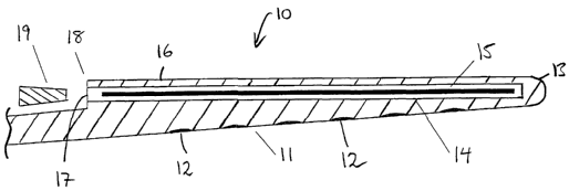
10 One embodiment of a cochlear implant electrode assembly is depicted
generally as 10 in Figs. 1, 3 and 4.
The depicted electrode assembly 10 preferably has an electrical lead
extending back to a stimulator/receiver housing. In considering this
invention, it is to be understood that each electrode may have one or more
15 wires (not depicted) electrically connected thereto and extending from each
respective electrode back through the lead to the stimulator/receiver. The use
of a stimulator/receiver as described herein is known in the art and the
present invention can be used with any such stimulator/receiver as known in
the art.
2o The assembly 10 comprises an elongate electrode carrier member 11
having a plurality of electrodes 12 mounted thereon. For the purposes of
clarity, the electrodes 12 depicted in Figs. 1, 2 and 5 are not necessarily
shown to scale. The depicted elongate member 11 is preformed from a
resiliently flexible silicone with memory and is preformed to a curved
configuration suitable for conforming with the inner wall of the scala tympani
of the cochlea as depicted in Fig. 4. The elongate member 11 has a first
end 13, distal the lead, that is firstly inserted into the implantee on
insertion
of the assembly 10.
As depicted in Fig. 5, the elongate member 11 can have a tip member
30 29 integrally formed with its first end 13. The tip 29 is formed from the
same
silicone used to fabricate the elongate member 11 and, in the depicted
embodiment, the material of tip member 29 has a resilient flexibility equal to
that of the material used for the carrier member 11.
Possible alternative constructions for the tip member 29 are provided
in Figs, 6a-6d. As depicted in Fig. 6a, the tip member 70 can be solid and
formed of an inner core 71 of relatively stiff material 71 and an outer layer
72
CA 02390591 2002-05-08
WO 02/28473 PCT/AU01/01231
16
of relatively flexible material. The core 71 can taper in diameter over region
73 towards the distal end 21. The taper 73 causes the overall stiffness of the
tip 70 to increase over the length of the taper 73 away from the distal end
21.
The outer layer 72 can be formed of the same material as the remainder of the
body of the elongate carrier member 11 or can be a different material.
As depicted in Fig. 6b, the tip member 40 can comprise a solid mass
integrally formed to the first end 13 of the elongate carrier 11.
Still further and as depicted in Fig. 6c, the tip member 50 can comprise
a solid mass 51 that is formed separately from the carrier member 11 and
subsequently adhered thereto.
As depicted in Fig. 6d, the tip member 60 can comprise an elastomeric
silicone material having a plurality of substantially spherical platinum
particles 61 dispersed therethrough. The particles 61 have a diameter
between about 50~,m and 100~,m. It will be appreciated that the particles 61
depicted in Fig. 6d are not drawn to scale.
In Fig. 6d, the particles 61 are depicted as substantially evenly
dispersed through the tip member 60. In another embodiment, the particles
could be non-evenly dispersed through the tip member. For example, the
particles could increase in density away from the distal end 21 towards the
proximal end of the tip member 60. By varying the density of the platinum
particles 61, it is possible to vary the relative stiffness of the tip member
60.
In addition to, or instead of, being used to potentially modify the
physical characteristics of the tip member, the provision of the metallic
particles 61 also result in the tip member 60 being detectable by fluoroscopy
and X-ray techniques. This provides another means for the surgeon to either
monitor the placement and position of the tip member 60 during or after
insertion of the electrode array 10 in an implantee's cochlea.
Disposed within a substantially cylindrical lumen 14 is a stylet-type
element 15. This stylet-type element 15 differs from a conventional stylet in
that it is formed from a bioresorbable polyacrylic acid (PAA) that is adapted
to dissolve or soften on exposure to fluids to permit the elongate member 11
to take its preformed curved configuration. It will be appreciated that the
stylet could be formed from other suitable bioresorbable materials. The
stylet-type element 15 has a straight configuration and has a stiffness
greater
than that of the silicone making up the elongate member 11. Accordingly, the
CA 02390591 2002-05-08
WO 02/28473 PCT/AU01/01231
17
stylet-type element 15, when in position biases the elongate member 11 into a
straight configuration as depicted in Figs. 1 and 3.
Overlaying the stylet-type element 15 is an integral outer layer 16 of
silicone material that surrounds and protects the stylet-type element 15. In
particular, the outer layer 16 serves to protect the stylet-type element 15,
at
least for some time, from dissolution or softening due to exposure of the
assembly to fluids, such as cochlear fluids, on insertion in the scala tympani
30.
As depicted in Fig. 1, the lumen 14 has an opening 17 at an end 18
distal the first end 13. In the embodiment depicted in Fig. 1, the opening 17
can be closed by a plug 19 that is adapted to seal the opening 17 of the lumen
14. While a frusto-conical plug is depicted in Fig. 1, other plug types can be
envisaged. For example, and as is depicted in Fig. 2, the opening 17 can be
sealed with a quantity 9 of silicone.
As an alternative to the plug 19 depicted in Fig. 1, the opening 17 of the
lumen 14 can be closed by a sealing layer bonded to the elongate member 11.
The sealing layer can be formed from a layer of silicone material that is used
to close the opening 17 following placement of the stylet-type element 15
within the lumen 14. In another alternative, the elongate member can be
fabricated such that the closure is provided by an extension of the outer
layer
16 over the opening 17 of the lumen 14.
In the latter case, the closure is preferably removed to form the opening
17 by slicing the sealing layer or outer layer extension with a blade, such as
that provided by a pair of scissors, to allow ingress of fluid into the lumen
14.
An alternative embodiment of the electrode array is depicted generally
as 20 in Fig. 2. In this embodiment, a plurality of transverse slits 21 are
formed in the outer layer 16. The slits preferably slow but do not prevent
ingress of fluid through the outer layer 16 to the stylet-type element 15.
In the depicted embodiment, each slit 21 is adapted to allow a
substantially equal rate of ingress of fluid into the lumen 14. It will be
appreciated that the slit design could be modified such that different slits
21
allowed different rates of progress of fluid through the outer layer 16. For
example, a slit 21 most distal the first end 13 could be adapted to allow a
greater rate of fluid ingress through the outer layer 16 than its adjacent
slit .
positioned closer to the first end 13. In this case, the bioresorbable
material
of the stylet-type element 15 beneath this slit would begin to dissolve or
CA 02390591 2002-05-08
WO 02/28473 PCT/AU01/01231
18
soften before the remainder of the stylet-type element 15 so allowing the
elongate member 11 to begin to move from the straight configuration to its
curved configuration at or adjacent the position of this most distal slit 21.
In
the depicted embodiment, each slit 21 can also be filled with a quantity of
bioresorbable material. In this case, each slit 21 can be filled with a
different
quantity or thickness of bioresorbable material so as to provide a means of
controlling the location of and rate of dissolution of the stylet-type element
15.
The rate of progress provided by each slit 21 can follow this pattern
1o along the length of the elongate member 11 towards the first end 13, with
the
next closer slit 21 to the first end 13 providing a relatively lesser rate of
ingress than its adjacent more distal slit 21. This pattern results in the
bioresorbable material of the stylet-type element 15 dissolving or softening
from an end distal the first end 13 towards an end closer to the first end 13.
As such, the straight elongate member 11 begins to curve distal the first end
13 and then continues to further adopt the curved configuration as the stylet-
type element 15 dissolves or softens towards the first end 13 or vice versa.
As an alternative to the slits 21, the outer layer 16 of the elongate
member 11 can be provided with one or more regions that more readily allow
ingress of bodily fluids, such as cochlear fluids. These regions can comprise
regions of the outer layer 16 that have a thickness less than that of the
remainder of the outer layer 16. Due to the reduced thickness of these
regions, the fluid passes through the regions more quickly than the remainder
of the outer layer 16. The thickness of the fluid ingress regions can be
varied
to suit the desired rate of dissolution of the stylet-type element 15 required
by
the application. Different regions can have different thickness as required.
For example, a region distal the first end 13 of the elongate member 11 can
have a thickness that is thinner than that of a region closer to the first end
13,
or vice versa. Such regions may also be formed by a matrix of pinholes or
other such structure to allow for a chosen rate of fluid ingress rather than
slits.
In use, the substantially straight assembly 10 or 20 will initially be
positioned at an entry to the scala tympani 30 as depicted in Fig. 3. At this
point, in those embodiments where present, the plug 19, or quantity 9, can be
removed or a covering over the slits 21 peeled away to allow bodily fluids,
such as cochlear fluids, to move into the lumen 14. Entry of the fluids into
CA 02390591 2002-05-08
WO 02/28473 PCT/AU01/01231
19
the lumen 14 commences dissolution or softening of the stylet-type element
15.
As dissolution or softening is occurring, the assembly 10 can be
carefully advanced into the scala tympani 30. Dissolution or softening of the
stylet-type element 15 causes the assembly 10 to begin to adopt a curved
configuration. As the assembly 10 continues to be advanced, it is preferably
positioned as depicted in Fig. 4, with the electrodes 12 facing the modiola
within the cochlea so that they are positioned as close as possible to the
spiral
ganglia thereof.
The control of the commencement of, and preferably the rate of, stylet
dissolution provides the surgeon with greater control of the implantation
procedure for the cochlear implant electrode assembly 10. The provision of
greater control minimises the potential for trauma to the sensitive tissues
inside the cochlea and also enhances the likelihood of successful placement
of the assembly 10 at the first attempt.
While the preferred embodiment of the invention has been described in
conjunction with a cochlear implant, it is to be understood that the present
invention has wider application to other implantable electrodes, such as
electrodes used with pacemakers.
2o It will be appreciated by persons skilled in the art that numerous
variations and/or modifications may be made to the invention as shown in the
specific embodiments without departing from the spirit or scope of the
invention as broadly described. The present embodiments are, therefore, to
be considered in all respects as illustrative and not restrictive.