Note: Descriptions are shown in the official language in which they were submitted.
CA 02391212 2002-05-08
WO 01/36977 PCT/US00/31492
METHODS AND COMPOSITIONS FOR IDENTIFYING
DISEASE MARKERS
Reference to Related Applications
This application claims priority to utility patent application identified by
Attorney Docket
No. MTP-026, entitled "Methods and Compositions for Identifying Disease
Markers," filed on
November 10, 2000, and the benefit of U.S. Serial No. 60/165,673, filed
November 16, 1999;
U.S. Serial No. 60/172,170, filed December 17, 1999; U.S. Serial No.
60/178,860, filed January
27, 2000; and U.S. Serial No. 60/201,721, filed May 3, 2000, the disclosures
of which are
incorporated by reference herein.
Field of the Invention
The present invention relates generally to methods and compositions for
identifying
1 o disease markers, for example, cancer markers, in a mammal. More
specifically, the present
invention relates to mass spectrometry-based methods and compositions for
identifying cancer
markers in a body fluid.
Background of the Invention
There is an ongoing need to identify new biological markers useful in the
detection and/or
treatment of various mammalian disorders, for example, cancer. Although a
variety of markers
have been identified for certain diseases, there is still the need to identify
markers for a disease
for which no markers presently are available, as well as new markers that are
more sensitive and
reliable than currently existing markers.
Biochemical markers can be identified by analyzing tissue or body samples from
a
2o mammal with the disease of interest and then comparing the results of the
analysis with those
obtained from a mammal without the disease. One successful approach using two-
dimensional
gel electrophoresis has led to the identification of a variety of marker
proteins that are present at
a higher concentration in tissue or body fluid samples of a diseased mammal
relative to a normal
mammal. See, for example, Partin et al. (1993) CANCER IZES. 53:744-746 which
describes the
identification of prostate cancer markers and Getzenberg et al. (1996) CANCER
IZES. 56:1690-
1694, which describes the identification of bladder cancer markers.
U.S. Patent No. 5,858,683 discloses a method for identifying cervical cancer
in an
individual. In the method, protein extracts from samples of normal cervical
tissue were
CA 02391212 2002-05-08
WO 01/36977 PCT/US00/31492
-2-
fractionated by two-dimensional gel electrophoresis. Similarly, a second
protein extract from
samples of cervical cancer biopsy tissue were also fractionated by two-
dimensional gel
electrophoresis. The resulting gels were compared and spots corresponding to
proteins present in
higher concentrations in the cancer sample versus the normal sample were
identified. Proteins
were eluted from the spots of interest on the two-dimensional gel and
subjected to conventional
protein microsequencing to identify the protein within the spot of interest.
This approach has
lead to the identification of at least two cervical cancer markers, referred
to in the art as TDP-43
and IEF-SSP-9502. Although this approach can be successful, there is still the
need to develop a
protocol for the more rapid identification of cancer markers and for
identifying markers which
otherwise may not be detectable using the gel electrophoresis approach.
More recently, an alternative non-electrophoretic-based method (i.e., does not
require an
electrophoresis step) for identifying cancer markers has been reported in
Chang et al. ( 1999)
RAPID COMMLTN. Mass SPECTRUM. 13, 1808-1812. Lysates from cultured cells
(either normal
breast cells or malignant breast cells) were fractionated by non-porous
reverse-phase high
performance liquid chromatography to give protein separation profiles. The
more abundant
proteins specifically present in the malignant cell lysates were harvested and
analyzed by matrix-
assisted laser desorption/ionization (MALDI) to determine the masses of the
abundant proteins.
In addition, a sample of each protein was trypsinized and the tryptic
fragments subjected to
MALDI to give masses of the fragments which were then compared to protein
databases to
2o identify the abundant proteins in the cancer cell based samples. Practice
of this method
permitted the identification of various proteins, for example, the
phosphoprotein p53, the proto-
oncogene tyrosine kinase SRC (C-SRC), the c-myc promoter protein and the
breast epithelial
antigen BA46, all of which were more abundant in the breast cancer lysates.
The usefulness of
this type of approach for analyzing samples more complex than cell lysates
still needs to be
evaluated.
There is, therefore, still a need in the art to develop new methods and
compositions that
can be used to rapidly identify disease markers present in actual tissue or
body fluid samples. It
is contemplated that such a new method can supplement the already existing
methods for
identifying disease markers so that additional disease markers can be
identified.
CA 02391212 2002-05-08
WO 01/36977 PCT/US00/31492
Summary of the Invention
The invention provides methods and compositions for the rapid detection and
characterization of disease markers, for example, cancer markers, in a mammal,
for example, a
human. Once identified the markers can be used as targets in assays for
detecting the disease, as
targets for treatment of the disease or both.
In one aspect, the invention provides a method for identifying a marker
molecule
indicative of a disease in a mammal. The method comprises the steps of: (a)
removing at least
one abundant protein from a sample harvested from a mammal with the disease;
(b) fractionating
the resulting sample depleted of abundant protein to produce a plurality of
fractions, each
1 o fraction comprising a plurality of molecules; (c) then, separating by mass
the molecules disposed
within a pre-selected fraction; (d) repeating steps (a) through (c) with a
sample harvested from a
mammal without the disease; and (e) comparing the molecules separated from the
sample from
the mammal with the disease with those separated from the sample from the
mammal without the
disease. As a result, it is possible to rapidly identify one or more marker
molecules present at a
15 higher concentration in the sample from the mammal with the disease
relative to the sample from
the mammal without the disease, wherein the presence of marker molecule is
indicative of the
disease.
In a preferred embodiment. the sample can be either a tissue or body fluid
sample.
Preferred body fluids include, for example, blood, serum, plasma, sweat,
tears, urine, peritoneal
2o fluid, lymph, vaginal secretion, semen, spinal fluid, ascitic fluid,
saliva, sputum, or breast
exudate. Serum, however, currently is most preferred.
It has been discovered that by removing one or more abundant proteins from the
sample,
it is easier to evaluate less abundant proteins as possible disease markers.
As used herein, an
abundant protein comprises greater than about 5% (w/w), more preferably
greater than about
25 20% (w/w) of total protein in the sample. When the sample is serum, the
abundant protein
typically is immunoglobulin or albumin. In a preferred embodiment, both
immunoglobulin and
albumin are removed from the serum to produce an immunoglobulin and albumin
depleted serum
suitable for further processing.
After depleting the samples of at least one abundant protein, the resulting
sample then is
3o fractionated to give a plurality of fractions, with each fraction
comprising a plurality of
molecules. In a preferred embodiment. the initial fractionation is by a non-
electrophoretic
CA 02391212 2002-05-08
WO 01/36977 PCT/US00/31492
-4-
method, for example, by chromatography, more specifically by affinity
chromatography. In a
more preferred embodiment, the affinity chromatography is ion exchange
chromatography, for
example, anion exchange chromatography. During ion exchange chromatography,
the sample of
interest is combined with an appropriate matrix, for example, an anionic or
cationic exchange
matrix, and molecules are allowed to bind to the matrix. After washing to
remove unbound
material, the bound molecules then are eluted selectively into different
elution buffers, each
buffer preferentially eluting a different population of molecules. In ion
exchange
chromatography, for example, the elution buffers can contain different salt
concentrations to
permit preferential elution of different types of molecules. It is
contemplated that by choosing
appropriate buffers it is possible to generate a plurality of fractions, each
comprising a plurality
of molecules. Alternatively, the affinity chromatography may be performed
using a solid support
having carbohydrate binding moieties, for example, lectin, disposed thereon.
As a result, it is
possible to separate carbohydrate containing molecules, for example,
glycosylated molecules
from non-glycosylated molecules.
I S One or more of the resulting fractions can then be analyzed by mass-
spectroscopy to give
the mass of the molecules disposed within a particular fraction. For example,
each fraction can
be analyzed by matrix assisted laser desorption/ionization-time of flight
(MALDI-TOF) mass
spectroscopy or, more preferably, by surface enhanced laser
desorption/ionization-time of flight
(SELDI-TOF) mass spectroscopy. During this protocol, the molecules are
separated by mass.
2o As a result, it is possible to produce a profile of masses within the
sample. By comparing the
molecules present at a higher concentration in a sample from a mammal with the
disease relative
to those present in a sample from a mammal without the disease, it is possible
to identify the
molecules that are found at elevated levels in the diseased mammal.
If necessary, it is possible to further identify the marker molecules. Further
analysis may
25 comprise isolating the molecule and, for example, if the molecule is a
protein, then the protein
can be further identified by conventional tryptic mapping and/or amino acid
sequencing
methodologies.
It is contemplated that the method of the invention is particularly effective
at identifying
markers when the disease is cancer. Accordingly, it is contemplated that the
method can be used
3o to identify markers for breast cancer, lung cancer, prostate cancer,
bladder cancer, cervical
CA 02391212 2002-05-08
WO 01/36977 PCT/US00/31492
-5-
cancer, ovarian cancer, colon cancer or colorectal cancer. The Examples
hereinbelow disclose
the identification of breast cancer markers.
In another aspect, marker proteins, once identified, can be used in an assay
for diagnosing
the disease in a mammal. In a preferred embodiment, the method comprises the
steps of: (a)
contacting a sample from the mammal with a binding moiety that binds
specifically to a disease-
associated protein to produce a binding moiety-disease-associated protein
complex, wherein the
binding moiety binds specifically to a marker protein identified by the method
of the invention;
and (b) detecting the presence of the complex, which if present is indicative
of the presence of
disease in the mammal.
1 o In a preferred embodiment, the binding moiety is an antibody, for example,
a monoclonal
antibody, a polyclonal antibody, or fragment thereof, for example, an Fv, Fab,
Fab', (Fab')2 or a
biosynthetic antibody binding site, for example, an sFv. The binding moiety
preferably is labeled
with a detectable moiety, for example, a radioactive label, a hapten label, a
fluorescent label, or
an enzymatic label.
15 The presence or amount of the marker protein can thus be indicative of the
presence of
the disease in the individual. For example, the amount of marker protein in
the sample may be
compared against a threshold value previously calibrated to indicate the
presence or absence of
the disease, wherein the amount of the complex in the sample relative to the
threshold value can
be indicative of the presence or absence of the disease in the individual.
Such methods can be
2o performed either on tissue, for example, breast tissue, or a body fluid,
for example, serum.
These and other numerous additional aspects and advantages of the invention
will
become apparent upon consideration of the following figures, detailed
description, and claims
which follow.
Description of the Drawings
25 The invention can be more completely understood with reference to the
following
drawings, in which:
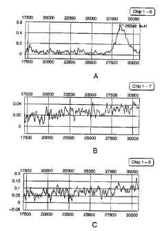
Figures 1 A-C are spectra resulting from the characterization via mass
spectrometry of 28
kD protein eluted from a polyacrylamide gel and applied to a nickel SELDI
chip. Figure 1A is a
spectrum of the heaviest 28 kD protein isolated from the gel, Figure 1B is a
spectrum of the
CA 02391212 2002-05-08
WO 01/36977 PCT/US00/31492
-6-
median 28 kD protein isolated from the gel, and Figure 1 C is a spectrum of
the lightest 28 kD
protein isolated from the gel.
Detailed Description of the Invention.
The present invention provides methods and compositions for the identification
of
disease markers useful as targets either in assays for the detection of the
disease or in treatment
of the disease. If the marker is, for example, a protein, it is contemplated
that the presence of the
disease in an individual can be detected using the marker protein and/or
binding moieties (e.g.
antibodies) that bind to the marker protein or to nucleic acid probes which
hybridize to nucleic
acid sequences encoding the marker protein. Furthermore, it is contemplated
that the skilled
artisan may produce novel therapeutics for treating the disease which include,
for example:
antibodies that can be administered to an individual and bind to and reduce or
eliminate the
biological activity of the target protein in vivo; nucleic acid or peptidyl
nucleic acid sequences
that hybridize with genes or gene transcripts encoding the target proteins
thereby to reduce
expression of the target proteins in vivo; or small molecules, for example,
organic molecules
which interact with the target proteins or other cellular moieties, for
example, receptors for the
target proteins, thereby to reduce or eliminate biological activity of the
target proteins.
Set forth below are methods for identifying disease markers and methods for
detecting the
disease by using the marker proteins as targets.
1. Methods for Identifying Disease Markers.
In general, the disease markers are identified by comparing the composition of
a sample
of tissue or body fluid of a mammal diagnosed with the disease against the
composition of a
sample similarly treated from an individual without the disease. Accordingly.
the resulting
markers can be used in assays to detect the presence or absence of a disease
in a mammal.
Furthermore, it is contemplated that the same method may be employed to
identify markers that
are present at higher concentrations in one disease state relative to another
disease state, for
example, an aggressive cancer versus a quiescent cancer.
As used herein, the term "marker" is understood to mean any biological marker,
for
example, a protein or nucleic acid, which is detectable at a higher level in a
tissue or body fluid
sample of an individual diagnosed with or diagnosable as having a disease
relative to a tissue or
3o body fluid sample of an individual free of the disease and includes species
and allelic variants
CA 02391212 2002-05-08
WO 01/36977 PCT/US00/31492
_7_
thereof and fragments thereof. The terms "marker" and "target" are used
interchangeably herein.
It is not necessary that the marker be unique to a disease state; rather the
marker should have a
signal to noise ratio high enough to discriminate between samples originating
from a diseased
individual and samples originating from an individual without the disease.
In one embodiment, the method of the invention comprises the steps of: (a)
removing at
least one abundant protein from a sample harvested from a mammal with the
disease; (b)
fractionating the resulting sample depleted of abundant protein to produce a
plurality of fractions,
each fraction comprising a plurality of molecules; (c) then, separating by
mass the molecules
disposed within a pre-selected fraction; (d) repeating steps (a) through (c)
with a sample
1 o harvested from a mammal without the disease; and (e) comparing the
molecules separated from
the sample from the mammal with the disease with those separated from the
sample from the
mammal without the disease. As a result, it is possible to rapidly identify
one or more marker
molecules present at a higher concentration in the sample from the mammal with
the disease
relative to the sample from the mammal without the disease. The resulting
markers, once
identified, can be used in an assay to detect the presence or status of a
disease, or as a target for
therapy.
It is contemplated that the method can be used to identify markers in tissue
or body fluid
samples. The method, however, is particularly useful in the identification of
disease markers in a
body fluid, for example, in blood, serum, plasma, sweat, tears, urine,
peritoneal fluid, lymph,
2o vaginal secretion, semen, spinal fluid, ascitic fluid, saliva, sputum, or
breast exudate. Serum,
however, is most preferred.
By removing one or more abundant proteins from the sample, it is easier to
evaluate less
abundant proteins as possible disease markers. As used herein, an abundant
protein comprises
greater than about 5% (w/w), more preferably greater than about 20% (w/w) of
total protein in
the sample. When the sample is serum, the abundant protein typically is
immunoglobulin or
albumin. It has been reported that in serum, albumin constitutes about 57-71%
of total serum
protein and that immunoglobulin constitutes 8-26% of total serum protein
(Lollo et al. ( 1999)
ELECTROPHORESIS 20:854-859). Accordingly, removal of these proteins alone
permits easier
evaluation of less abundant proteins as disease markers. Accordingly, it is
preferable to remove
both immunoglobulin and albumin from the serum to produce an immunoglobulin
and albumin
depleted serum suitable for further processing.
CA 02391212 2002-05-08
WO 01/36977 PCT/US00/31492
_g_
The immunoglobulin and/or albumin proteins can be extracted using conventional
methodologies, for example, affinity-based methodologies, known and used in
the art. For
example, immunoglobulin can be removed selectively from samples using binding
proteins, for
example, an antibody or a fragment thereof, Protein A, or Protein G,
immobilized on a solid
support. For example, a solution of interest can be passed through a
chromatography column
packed with such a solid support under conditions such that the immunoglobulin
molecules
preferentially bind to the matrix. The resulting column flow through,
therefore, is depleted of
immunoglobulin. A preferred matrix comprises Protein G coupled to agarose
particles, available
commercially from Pharmacia and Upjohn, Peapack, NJ under the trade name
Hitrap Protein G.
to Similarly, albumin can be removed selectively for samples of interest via
affinity
chromatography, using, for example, Sepharose coupled to Cibacron blue
available commercially
from Pharmacia and Upjohn, Peapack, NJ. Alternatively, both albumin and
immunoglobulin G
can be removed simultaneously from serum using ProtoClearTM (Lollo et al. (
1999)
ELECTROPHORESIS 20:854-859). The authors report that greater than 95% of human
serum
albumin and greater than 97% of human immunoglobulin can be removed using
ProtoClearTM.
After depleting the samples of at least one abundant protein, the resulting
sample then is
fractionated to give a plurality of fractions, with each fraction comprising a
plurality of
molecules. The initial fractionation preferably is by a non-electrophoretic
method, for example,
by chromatography, more specifically, affinity chromatography. In a more
preferred
2o embodiment, the affinity chromatography is ion exchange chromatography, for
example, anion or
cation exchange chromatography. With serum, this step preferably is performed
by anion
exchange chromatography. During ion exchange chromatography, the sample of
interest is
combined with an appropriate matrix, for example, an anionic exchange matrix,
and molecules
are allowed to bind to the matrix. After washing to remove unbound material,
the bound
molecules then are eluted selectively into different elution buffers, each
buffer preferentially
eluting a different population of molecules. It is contemplated that by
choosing appropriate
buffers it is possible to generate a plurality of fractions, each comprising a
plurality of molecules.
In a procedure described in detail in Example 1, serum substantially free of
immunoglobulin and
albumin was subdivided into twelve fractions containing approximately equal
amounts of protein
3o by anion exchange chromatography. "Substantially free" is understood to
mean at least 70%,
more preferably at least 80%, more preferably at least 90% and most preferably
at least 95% of a
particular molecule. Anion exchange chromatography produces different
populations of
CA 02391212 2002-05-08
WO 01/36977 PCT/US00/31492
-9-
samples, with each sample containing numerous molecules, but significantly
less in number than
the original starting material. These molecules can then be more easily
characterized as a
function of mass. In an exemplary protocol, serum is applied to a Mono Q
(Pharmacia and
Upjohn, Peapack, NJ) anion exchange column in phosphate buffer. The proteins
once bound can
be eluted by increasing the concentration of a salt, for example, sodium
chloride, in a series of
elution buffers. The choice of appropriate salt concentrations is considered
to be within the level
of skill in the art and will depend upon variables such as the type of
starting material, and the
types and numbers of proteins desired in each population.
Alternatively, the affinity chromatography may be performed using a solid
support having
1o carbohydrate binding moieties, for example, lectin, disposed thereon. As a
result, it is possible to
separate glycosylated from non-glycosylated molecules.
One or more of the resulting fractions can then be analyzed by mass, for
example, mass-
spectroscopy. For example, each fraction can be analyzed by matrix assisted
laser
desorption/ionization-time of flight (MALDI-TOF) mass spectroscopy or by
surface enhanced
15 laser desorption/ionization-time of flight (SELDI-TOF) mass spectroscopy.
See U.S. Patent No.
5,719,060.
Generally, analysis by mass spectrometry involves the vaporization and
ionization of a
sample of material using a high energy source, for example, a laser. Usually,
the material is
vaporized from the surface of a probe tip into the gas or vapor phase by a
laser beam, whereby
2o some of the individual molecules become ionized. The positively charged
molecules then are
accelerated using a high voltage field and allowed to fly into a high vacuum
chamber, at the end
of which is an detection surface. Because the time-of flight is a function of
mass of the ionized
molecule, the elapsed time between ionization and impact can be used to
determine molecule's
mass. As a result, using this type of mass spectrometry it is possible to
produce a profile of
25 masses within the sample. By comparing the molecules present at a higher
concentration in a
sample from a mammal with the disease relative to those present in a sample
from a mammal
without the disease, it is possible to identify the molecules (i.e., markers)
that are found at
elevated levels in the diseased mammal.
Using mass spectrometry, it is further possible to characterize the markers by
their
3o binding affinity to a particular surface. For example, in SELDI-TOF mass
spectroscopy, several
different surfaces are available commercially from Ciphergen Biosystems, Inc.,
Palo Alto, CA.
CA 02391212 2002-05-08
WO 01/36977 PCT/US00/31492
-10-
Each of the surfaces have different surface properties and thus bind different
populations of
markers. Available surfaces include copper-treated surfaces and nickel-treated
surfaces which
can be generated by adding a copper or nickel salt solution to a chip
comprising
ethylenediaminetriacetic acid. Other SELDI chip surfaces include: WCX-2 which
comprises
carboxylate moieties, and SAX-2 which comprises quarternary ammonium moieties.
The
markers therefore can be further characterized by their affinity to a
particular SELDI chip. For
example, as used herein, the term "affinity" to a particular SELDI chip is
understood to mean that
a marker binds preferentially to one type of SELDI chip (e.g., copper SELDI
chip) relative to one
or more of the other SELDI chips (e.g., the nickel, SAX-2 and WCX-2 chips)
disclosed herein.
As discussed in detail in Example l, comparison of the sera from diseased and
healthy
individuals revealed a number of proteins frequently present at detectable
levels in the sera of
diseased individuals, but infrequently present at comparable levels in the
sera of healthy
individuals.
Once the markers, for example, protein markers, have been identified by mass
spectrometry, the identified proteins can be isolated by standard protein
isolation methodologies
and sequenced using protein sequencing technologies known and used in the art.
For example,
each of the markers, once identified, can be purified to homogeneity using the
methodologies and
the information derived therefrom in the previous steps. For example, the
marker can be isolated
based on its mass as determined by mass spectrometry and its other physical
and chemical
features, for example, ability to bind to an affinity column, for example, an
ion exchange column.
The proteins can be further characterized by conventional amino acid
sequencing, for example,
by Edman degradation and/or mass spectrometry-based microsequencing of
proteolytic
fragments.
It is contemplated that the method of the invention is particularly effective
at identifying
markers when the disease is cancer. Accordingly, it is contemplated that the
method can be used
to identify markers for breast cancer, lung cancer, prostate cancer, bladder
cancer, cervical
cancer, ovarian cancer, colon cancer or colorectal cancer. The Examples
hereinbelow disclose
the identification of breast cancer markers.
CA 02391212 2002-05-08
WO 01/36977 PCT/US00/31492
-11-
2. Detection of Disease
Once a disease marker has been identified, the marker, for example, a protein
or a nucleic
acid encoding the protein, may be used to determine whether an individual has
the disease, and,
if so, suitable detection methods can be used to monitor the status of the
disease.
By using proteins or nucleic acids encoding the proteins as markers, the
skilled artisan
can produce a variety of detection methods for detecting a disease in a human.
The methods
typically comprise the steps of detecting, by some means, the presence of one
or more markers in
a tissue or body fluid sample of the human. The accuracy and/or reliability of
the method for
detecting markers in a human may be further enhanced by detecting the presence
of a plurality of
marker proteins or nucleic acids in a preselected tissue or body fluid sample.
The detection
assays may comprise one or more of the protocols described hereinbelow.
2.A. Protein-Based Assays
If the marker is a protein, the protein may be detected, for example, by
combining the
marker protein with a binding moiety capable of specifically binding the
marker protein. The
binding moiety may comprise, for example, a member of a ligand-receptor pair,
i.e., a pair of
molecules capable of having a specific binding interaction. The binding moiety
may comprise,
for example, a member of a specific binding pair, such as antibody-antigen,
enzyme-substrate,
nucleic acid-nucleic acid, protein-nucleic acid, protein-protein, or other
specific binding pair
known in the art. Binding proteins may be designed which have enhanced
affinity for a target
protein. Optionally, the binding moiety may be linked with a detectable label,
such as an
enzymatic, fluorescent, radioactive, phosphorescent or colored particle label.
The labeled
complex may be detected, e.g., visually or with the aid of a spectrophotometer
or other detector.
Marker proteins may also be detected using gel electrophoresis techniques
available in
the art. In two-dimensional gel electrophoresis, the proteins are separated
first in a pH gradient
gel according to their isoelectric point. The resulting gel then is placed on
a second
polyacrylamide gel, and the proteins separated according to molecular weight
(see, for example,
O'Farrell (1975) J. Biol. Chem. 250: 4007-4021).
One or more marker proteins may be detected by first isolating proteins from a
sample
obtained from an individual suspected of having a disease, and then separating
the proteins by
3o two-dimensional gel electrophoresis to produce a characteristic two-
dimensional gel
CA 02391212 2002-05-08
WO 01/36977 PCT/US00/31492
-12-
electrophoresis pattern. The pattern may then be compared with a standard gel
pattern produced
by separating, under the same or similar conditions, proteins isolated from
normal or known
cancer cells. The standard gel pattern may be stored in, and retrieved from an
electronic database
of electrophoresis patterns. The presence of a marker protein in the two-
dimensional gel
provides an indication that the sample being tested was taken from a person
with the disease. As
with the other detection assays described herein, the detection of two or more
proteins, for
example, in the two-dimensional gel electrophoresis pattern further enhances
the accuracy of the
assay. The presence of a plurality, e.g., two to five, marker proteins on the
two-dimensional gel
provides an even stronger indication of the presence of disease in the
individual. The assay thus
1 o permits the early detection and treatment of the disease.
A marker protein may also be detected using any one of a wide range of
immunoassay
techniques available in the art. For example, the skilled artisan may employ a
sandwich
immunoassay format to detect a disease marker in a body fluid sample.
Alternatively, the skilled
artisan may use conventional immuno-histochemical procedures for detecting the
presence of the
15 marker in a tissue sample using one or more labeled binding proteins.
In a sandwich immunoassay, two antibodies capable of binding the marker
protein
generally are used, e.g., one immobilized onto a solid support, and one free
in solution and
labeled with a detectable chemical compound. Examples of chemical labels that
may be used for
the second antibody include radioisotopes, fluorescent compounds, and enzymes
or other
2o molecules that generate colored or electrochemically active products when
exposed to a reactant
or enzyme substrate. When a sample containing the marker protein is placed in
this system, the
marker protein binds to both the immobilized antibody and the labeled
antibody, to form a
"sandwich" immune complex on the support's surface. The complexed protein then
is detected
by washing away non-bound sample components and excess labeled antibody, and
measuring the
25 amount of labeled antibody complexed to protein on the support's surface.
Alternatively, the
antibody free in solution, which can be labeled with a chemical moiety, for
example, a hapten,
may be detected by a third antibody labeled with a detectable moiety which
binds the free
antibody or, for example, the hapten coupled thereto.
Both the sandwich immunoassay and the tissue immunohistochemical procedure are
3o highly specific and very sensitive, provided that labels with good limits
of detection are used. A
detailed review of immunological assay design, theory and protocols can be
found in numerous
CA 02391212 2002-05-08
WO 01/36977 PCT/US00/31492
-13-
texts in the art, including "Practical Immunology", Butt, W.R., ed., (1984)
Marcel Dekker, New
York and "Antibodies, A Laboratory Approach", Harlow et al. eds. (1988) Cold
Spring Harbor
Laboratory.
In general, immunoassay design considerations include preparation of
antibodies (e.g.,
monoclonal or polyclonal antibodies) having sufficiently high binding
specificity for the target
protein to form a complex that can be distinguished reliably from products of
nonspecific
interactions. As used herein, the term "antibody" is understood to mean
binding proteins, for
example, antibodies or other proteins comprising an immunoglobulin variable
region-like
binding domain, having the appropriate binding affinities and specificities
for the target protein.
1 o The higher the antibody binding specificity, the lower the target protein
concentration that can be
detected. As used herein, the terms "specific binding" or "binding
specifically" are understood to
mean that the binding moiety, for example, a binding protein has a binding
affinity for the target
protein of greater than about 105 M-1, more preferably greater than about 10~
M-1.
Antibodies to an isolated marker or target protein which are useful in assays
for detecting
a breast cancer in an individual may be generated using standard immunological
procedures well
known and described in the art. See, for example, Practical Immunology, Butt,
N.R., ed., Marcel
Dekker, NY, 1984. Briefly, an isolated target protein is used to raise
antibodies in a xenogeneic
host, such as a mouse, goat or other suitable mammal. The marker protein is
combined with a
suitable adjuvant capable of enhancing antibody production in the host, and is
injected into the
host, for example, by intraperitoneal administration. Any adjuvant suitable
for stimulating the
host's immune response may be used. A commonly used adjuvant is Freund's
complete adjuvant
(an emulsion comprising killed and dried microbial cells and available from,
for example,
Calbiochem Corp., San Diego, or Gibco, Grand Island, NY). Where multiple
antigen injections
are desired, the subsequent injections may comprise the antigen in combination
with an
incomplete adjuvant (e.g., cell-free emulsion). Polyclonal antibodies may be
isolated from the
antibody-producing host by extracting serum containing antibodies to the
protein of interest.
Monoclonal antibodies may be produced by isolating host cells that produce the
desired antibody,
fusing these cells with myeloma cells using standard procedures known in the
immunology art,
and screening for hybrid cells (hybridomas) that react specifically with the
target protein and
3o have the desired binding affinity.
CA 02391212 2002-05-08
WO 01/36977 PCT/US00/31492
-14-
Antibody binding domains also may be produced biosynthetically and the amino
acid
sequence of the binding domain manipulated to enhance binding affinity with a
preferred epitope
on the target protein. Specific antibody methodologies are well understood and
described in the
literature. A more detailed description of their preparation can be found, for
example, in Butt
( 1984) "Practical Immunology " (supra).
In addition, genetically engineered biosynthetic antibody binding sites, also
known in the
art as BABS or sFv's, may be used in the practice of the instant invention.
Methods for making
and using BABS comprising (i) non-covalently associated or disulfide bonded
synthetic VH and
VL dimers, (ii) covalently linked VH-VL single chain binding sites, (iii)
individual VH or VL
domains, or (iv) single chain antibody binding sites are disclosed, for
example, in U.S. Patent
Nos.: 5,091,513; 5,132,405; 4,704,692; and 4,946,778. Furthermore, BABS having
requisite
specificity for the marker protein can be derived by phage antibody cloning
from combinatorial
gene libraries (see, for example, Clackson et al. (1991) Nature 352: 624-628).
Briefly, phage
each expressing on their coat surfaces, BABS having immunoglobulin variable
regions encoded
I S by variable region gene sequences derived from mice pre-immunized with
isolated marker
proteins, or fragments thereof are screened for binding activity against
immobilized breast
cancer-associated protein. Phage which bind to the immobilized marker proteins
are harvested
and the gene encoding the BABS sequenced. The resulting nucleic acid sequences
encoding the
BABS of interest may then be expressed in conventional expression systems to
produce the
BABS protein.
The isolated marker protein also may be used for the development of diagnostic
and other
tissue evaluating kits and assays to monitor the level of the proteins in a
tissue or fluid sample.
For example, the kit may include antibodies or other specific binding proteins
which bind
specifically to the marker proteins and which permit the presence and/or
concentration of the
marker proteins to be detected and/or quantitated in a tissue or fluid sample.
Suitable kits for detecting marker proteins are contemplated to include, e.g.,
a receptacle
or other means for capturing a sample to be evaluated, and means for detecting
the presence
and/or quantity in the sample of one or more of the marker proteins described
herein. As used
herein, "means for detecting" in one embodiment includes one or more
antibodies specific for
these proteins and means for detecting the binding of the antibodies to these
proteins by, e.g., a
standard sandwich immunoassay as described herein. Where the presence of a
protein within a
CA 02391212 2002-05-08
WO 01/36977 PCT/US00/31492
-15-
cell is to be detected, e.g., as from a tissue sample, the kit also may
comprise means for
disrupting the cell structure so as to expose intracellular proteins.
2.B. Nucleic Acidbased Assays
The presence of a disease in an individual may also be determined by
detecting, in a
tissue or body fluid sample, a nucleic acid molecule encoding the marker
protein. Using methods
well known to those of ordinary skill in the art, the marker proteins may be
sequenced, and then,
based on the determined sequence, oligonucleotide probes designed for
screening a cDNA library
(see, for example, Sambrook et al. (1989) supra).
A target nucleic acid molecule encoding a marker protein may be detected using
a labeled
to binding moiety capable of specifically binding the target nucleic acid. The
binding moiety may
comprise, for example, a protein, a nucleic acid or a peptidyl nucleic acid.
Additionally, a target
nucleic acid, such as an mRNA encoding a marker protein, may be detected by
conducting, for
example, a Northern blot analysis using labeled oligonucleotides, e.g.,
nucleic acid fragments
complementary to and capable of hybridizing specifically with at least a
portion of a target
~5 nucleic acid.
More specifically, gene probes comprising complementary RNA or, preferably,
DNA to
the disease-associated nucleotide sequences or mRNA sequences encoding the
marker proteins
may be produced using established recombinant techniques or oligonucleotide
synthesis. The
probes hybridize with complementary nucleic acid sequences presented in the
test specimen, and
2o can provide exquisite specificity. A short, well-defined probe, coding for
a single unique
sequence is most precise and preferred. Larger probes generally are less
specific. While an
oligonucleotide of any length may hybridize to an mRNA transcript,
oligonucleotides typically
within the range of 8-100 nucleotides, preferably within the range of 15-50
nucleotides, are
envisioned to be most useful in standard hybridization assays. Choices of
probe length and
25 sequence allow one to choose the degree of specificity desired.
Hybridization is carried out at
from 50° to 65°C in a high salt buffer solution, formamide or
other agents to set the degree of
complementarity required. The state of the art is such that probes can be
manufactured to
recognize essentially any DNA or RNA sequence. For further particulars, see,
for example,
Guide to Molecular Techniques, Berger et al., Methods of Enzymology, Vol. 152,
1987.
3o A wide variety of different labels coupled to the probes or antibodies may
be employed in
the assays. The labeled reagents may be provided in solution or coupled to an
insoluble support,
CA 02391212 2002-05-08
WO 01/36977 PCT/US00/31492
-16-
depending on the design of the assay. The various conjugates may be joined
covalently or
noncovalently, directly or indirectly. When bonded covalently, the particular
linkage group will
depend upon the nature of the two moieties to be bonded. A large number of
linking groups and
methods for linking are taught in the literature. Broadly, the labels may be
divided into the
following categories: chromogens; catalyzed reactions; chemiluminescence;
radioactive labels;
and colloidal-sized colored particles. The chromogens include compounds which
absorb light in
a distinctive range so that a color may be observed, or emit light when
irradiated with light of a
particular wavelength or wavelength range, e.g., fluorescers. Both enzymatic
and nonenzymatic
catalysts may be employed. In choosing an enzyme, there will be many
considerations including
the stability of the enzyme, whether it is normally present in samples of the
type for which the
assay is designed, the nature of the substrate, and the effect if any of
conjugation on the enzyme's
properties. Potentially useful enzyme labels include oxiodoreductases,
transferases, hydrolases,
lyases, isomerases, ligases, or synthetases. Interrelated enzyme systems may
also be used. A
chemiluminescent label involves a compound that becomes electronically excited
by a chemical
~ 5 reaction and may then emit light that serves as a detectable signal or
donates energy to a
fluorescent acceptor. Radioactive labels include various radioisotopes found
in common use
such as the unstable forms of hydrogen, iodine, phosphorus or the like.
Colloidal-sized colored
particles involve material such as colloidal gold that, in aggregate, form a
visually detectable
distinctive spot corresponding to the site of a substance to be detected.
Additional information
20 on labeling technology is disclosed, for example, in U.S. Patent No.
4,366,241.
A common method of in vitro labeling of nucleotide probes involves nick
translation
wherein the unlabeled DNA probe is nicked with an endonuclease to produce free
3'hydroxyl
termini within either strand of the double-stranded fragment. Simultaneously,
an exonuclease
removes the nucleotide residue from the 5'phosphoryl side of the nick. The
sequence of
25 replacement nucleotides is determined by the sequence of the opposite
strand of the duplex.
Thus, if labeled nucleotides are supplied, DNA polymerase will fill in the
nick with the labeled
nucleotides. Using this well-known technique, up to 50% of the molecule can be
labeled. For
smaller probes, known methods involving 3'end labeling may be used.
Furthermore, there are
currently commercially available methods of labeling DNA with fluorescent
molecules, catalysts,
3o enzymes, or chemiluminescent materials. Biotin labeling kits are
commercially available (Enzo
Biochem Inc.) under the trademark Bio-Probe. This type of system permits the
probe to be
coupled to avidin with in turn is labeled with, for example, a fluorescent
molecule, enzyme,
CA 02391212 2002-05-08
WO 01/36977 PCT/US00/31492
-17-
antibody, etc. For further disclosure regarding probe construction and
technology, see, for
example, Sambrook et al., Molecular Cloning, A Laboratory Manual (Cold Spring
Harbor, N.Y.,
1982).
The oligonucleotide selected for hybridizing to the target nucleic acid,
whether
synthesized chemically or by recombinant DNA methodologies, is isolated and
purified using
standard techniques and then preferably labeled (e.g., with 35S or 32P) using
standard labeling
protocols. A sample containing the target nucleic acid then is run on an
electrophoresis gel, the
dispersed nucleic acids transferred to a nitrocellulose filter and the labeled
oligonucleotide
exposed to the filter under stringent hybridizing conditions, e.g. 50%
formamide, 5 X SSPE, 2 X
Denhardt's solution, 0.1% SDS at 42oC, as described in Sambrook et al. (1989)
supra. The filter
may then be washed using 2 X SSPE, 0.1% SDS at 68°C, and more
preferably using 0.1 X SSPE,
0.1% SDS at 68°C. Other useful procedures known in the art include
solution hybridization, and
dot and slot RNA hybridization. Optionally, the amount of the target nucleic
acid present in a
sample then is quantitated by measuring the radioactivity of hybridized
fragments, using standard
procedures known in the art.
In addition, oligonucleotides may also be used to identify other sequences
encoding
members of the target protein families. The methodology may also be used to
identify genetic
sequences associated with the nucleic acid sequences encoding the proteins
described herein,
e.g., to identify non-coding sequences lying upstream or downstream of the
protein coding
sequence, and which may play a functional role in expression of these genes.
Additionally,
binding assays may be conducted to identify and detect proteins capable of a
specific binding
interaction with a nucleic acid encoding a breast cancer-associated protein,
which may be
involved, e.g., in gene regulation or gene expression of the protein. In a
further embodiment, the
assays described herein may be used to identify and detect nucleic acid
molecules comprising a
sequence capable of recognizing and being specifically bound by a marker
protein.
In addition, it is anticipated that using a combination of appropriate
oligonucleotide primers,
i. e., more than one primer, the skilled artisan may determine the level of
expression of a target
gene in vivo by standard polymerise chain reaction (PCR) procedures, for
example, by
quantitative PCR. Conventional PCR based assays are discussed, for example, in
Innes et al
(1990) "PCR Protocols; A guide to methods and Applications", Academic Press
and Innes et al.
(1995) "PCR Strategies" Academic Press, San Diego, CA.
CA 02391212 2002-05-08
WO 01/36977 PCT/US00/31492
-18-
Recombinant marker molecules can be produced as described hereinbelow. For
example,
DNA encoding the marker molecules can be inserted, using conventional
techniques well
described in the art (see, for example, Sambrook (1989) supra) into any of a
variety of
expression vectors and transfected into an appropriate host cell to produce
recombinant proteins,
including both full length and truncated forms. Useful host cells include E.
coli, Saccharomyces
cerevisiae, Pichia pastoris, the insect/baculovirus cell system, myeloma
cells, and various other
mammalian cells. The full length forms of such proteins are preferably
expressed in mammalian
cells, as disclosed herein. The vector can additionally include various
sequences to promote
correct expression of the recombinant protein, including transcription
promoter and termination
sequences, enhancer sequences, preferred ribosome binding site sequences,
preferred mRNA
leader sequences, preferred protein processing sequences, preferred signal
sequences for protein
secretion, and the like. The DNA sequence encoding the gene of interest can
also be
manipulated to remove potentially inhibiting sequences or to minimize unwanted
secondary
structure formation. As will be appreciated by the practitioner in the art,
the recombinant protein
can also be expressed as a fusion protein.
After translation, the protein can be purified from the cells themselves or
recovered from
the culture medium. The DNA can also include sequences which aid in expression
and/or
purification of the recombinant protein. The DNA can be expressed directly or
can be expressed
as part of a fusion protein having a readily cleavable fusion junction.
2o In one preferred embodiment, the DNA is expressed in a suitable mammalian
host.
Useful hosts include fibroblast 3T3 cells, (e.g., NIH 3T3, from CRL 1658) COS
(simian kidney
ATCC, CRL-1650) or CHO (Chinese hamster ovary) cells (e.g., CHO-DXBI 1, from
Chasin
( 1980) Proc. Nat'l. Acad. Sci. USA 77 :4216-4222), mink-lung epithelial cells
(MV 1 Lu), human
foreskin fibroblast cells, human glioblastoma cells, and teratocarcinoma
cells. Other useful
eukaryotic cell systems include yeast cells, the insect/baculovirus system or
myeloma cells.
In order to express a marker protein molecule, the DNA is subcloned into an
insertion site
of a suitable, commercially available vector along with suitable
promoter/enhancer sequences
and 3' termination sequences. Useful promoter/enhancer sequence combinations
include the
CMV promoter (human cytomegalovirus (MIE) promoter) present, for example, on
pCDMB, as
3o well as the mammary tumor virus promoter (MMTV) boosted by the Rous sarcoma
virus LTR
enhancer sequence (e.g., from Clontech, Inc., Palo Alto). A useful inducible
promoter includes,
CA 02391212 2002-05-08
WO 01/36977 PCT/US00/31492
-19-
for example, a Zn2+-inducible promoter, such as the Zn2+ metallothionein
promoter (Wrana et al.
(1992) Cell 71: 1003-1014). Other inducible promoters are well known in the
art and can be
used with similar success. Expression also can be further enhanced using trans-
activating
enhancer sequences. The plasmid also preferably contains an amplifiable
marker, such as DHFR
under suitable promoter control, e.g., SV40 early promoter (ATCC #37148).
Transfection, cell
culturing, gene amplification and protein expression conditions are standard
conditions, well
known in the art, such as are described, for example in Ausubel et al., ed.,
(1989) "Current
Protocols in Molecular Biology", John Wiley & Sons, NY. Briefly, transfected
cells are cultured
in medium containing 5-10% dialyzed fetal calf serum (dFCS), and stably
transfected high
1 o expression cell lines obtained by amplification and subcloning and
evaluated by standard
Western and Northern blot analysis. Southern blots also can be used to assess
the state of
integrated sequences and the extent of their copy number amplification.
The expressed candidate protein is then purified using standard procedures. A
currently
preferred methodology uses an affinity column, such as a ligand affinity
column or an antibody
affinity column. The column then is washed, and the candidate molecules
selectively eluted in a
gradient of increasing ionic strength, changes in pH, or addition of mild
detergent. It is
appreciated that in addition to the candidate molecules which bind to the
breast cancer-associated
proteins, the breast cancer associated proteins themselves may likewise be
produced using such
recombinant DNA technologies.
2o The following non-limiting examples provide details for the isolation and
characterization of breast cancer markers together with methods of using the
markers for the
detection of breast cancer. It is contemplated that the same or a similar
protocol can be used to
identify markers for other diseases, for example, other cancers.
Example 1- Identification of Breast Cancer Markers
To identify markers for breast cancer, the sera of individuals with breast
cancer were
compared to the sera of normal individuals using the following protocol.
Briefly, 0.5 mL
aliquots of sera harvested from the individuals were thawed. Then, 1 pL of a 1
mg/mL solution
of soybean trypsin inhibitor (SBTI) and 1 qL of a 1 mg/mL solution of
leupeptin were added to
each aliquot. To remove lipids, 350 ~L of 1,1,2-trifluorotrichloroethane was
added to each
3o sample. The samples then were vortexed for five minutes and centrifuged in
a microcentrifuge
for five minutes at 4°C. The resulting supernatants were applied to a 1
mL column of agarose
CA 02391212 2002-05-08
WO 01/36977 PCT/US00/31492
-20-
coupled to protein G (Hitrap Protein G column, Pharmacia and Upjohn, Peapack,
NJ) to remove
immunoglobulin proteins. The column then was rinsed with 3 mL of 50 mM sodium
phosphate,
pH 7.0, with SBTI and leupeptin ("binding buffer"), and the resulting
flowthrough applied
directly to a 5 mL column of 6% Sepharose coupled to Cibacron blue (Hitrap
blue column,
Pharmacia and Upjohn, Peapack, NJ) to remove albumin proteins. The Hitrap blue
column was
rinsed with 20 mL of binding buffer. The resulting flow through was
concentrated using four
centrifugation-based concentrators with a l OkD cutoff (Centricon 10,
Millipore Corporation,
Bedford, MA) to give a final volume of about 0.7 mL.
The resulting serum (substantially free of immunoglobulin and albumin) was
subdivided
into twelve fractions containing approximately equal amounts of protein by
anion exchange
chromatography. Specifically, the serum was applied to a Mono Q (Pharmacia and
Upjohn,
Peapack, NJ) anion exchange column (a strong anion exchanger with quarternary
ammonium
groups) in 50 mM sodium phosphate buffer, pH 7.0 and proteins were eluted from
the column by
increasing the concentration of sodium chloride in a stepwise manner. In this
protocol, the serum
was divided into twelve fractions based on the concentration of sodium
chloride used for elution.
These fractions accordingly were designated flow through, 25 mM, 50 mM, 75 mM,
100 mM,
125 mM, 150 mM, 200 mM, 250 mM, 300 mM, 400 mM, and 2M sodium chloride. After
elution, each fraction was concentrated to approximately 100 ~g/mL and buffer
exchanged into
binding buffer.
Then 4-10 ~L from each of the twelve fractions were applied and allowed to
bind to each
of four SELDI chip surfaces, each surface holding up to eight samples. The
intended location of
each sample on the chip was demarcated with a circle drawn using a hydrophobic
marker like
those used in Pap smears. The SELDI chips used herein were purchased from
Ciphergen
Biosystems, Inc., Palo Alto, California, and used as described below.
For copper or nickel surfaces, a chip containing ethylenediaminetriacetic acid
moieties
(IMAC, Ciphergen Biosystems, Inc., Palo Alto, CA) was pretreated with two five-
minute
applications of five ~L of a copper salt or nickel salt solution, and washed
with deionized water.
After a five-minute treatment with five pL of binding buffer, two to three
microliters of sample
were applied to the surface for thirty to sixty minutes. Another two to three
microliters of sample
3o then were applied for an additional thirty to sixty minutes. The chips then
were washed twice
with binding buffer to remove unbound proteins. 0.5 ~L of sinapinic acid (12.5
mg/mL) was
CA 02391212 2002-05-08
WO 01/36977 PCT/US00/31492
-21 -
added twice and allowed to dry each time. The presence of sinapinic acid
enhances the
vaporization and ionization of the bound proteins upon mass spectrometry.
For chip surfaces containing carboxyl moieties (WCX-2, Ciphergen Biosystems,
Inc.,
Palo Alto, CA), before use of the hydrophobic pen, the surface was washed with
10 mM HCl for
thirty minutes and rinsed five times with deionized water. After use of the
pen, the surface was
washed five times with 5 qL of binding buffer and once with deionized water.
Two to three qL
of sample were applied in two applications of thirty to sixty minutes each.
The surface was
washed twice with 5 qL of binding buffer, and 0.5 ~L of sinapinic acid were
applied twice.
For chip surfaces containing quarternary ammonium moieties (SAX-2, Ciphergen
Biosystems, Inc., Palo Alto, CA), after use of the pen, the surface was washed
five times with
five ~L of binding buffer and once with deionized water. Application of
sample, washing, and
application of sinapinic acid were performed as described above.
The chips then were subjected to mass spectrometry utilizing a Ciphergen SELDI
PBS
One (Ciphergen Biosystems, Inc., Palo Alto, CA) running the software program
"SELDI v. 2.0".
15 For all chips, "high mass" was set to 200,000 Daltons, "starting detector
sensitivity" was set to 9
(from a range of 1-10, with 10 being the highest sensitivity), NDF (neutral
density filter) was set
to "OUT", data acquisition method was set to "Seldi Quantitation", SELDI
acquisition
parameters were set to 20, with increments of 5, and warming with two shots at
intensity 50 (out
of 100) was included. For IMAC chips, mass was optimized from 3000 Daltons to
3001 Daltons,
2o starting laser intensity was set to 80 (out of 100), and transients set to
5 (i.e., 5 laser shots per
site). Peaks were identified automatically by computer. For WCX-2 chips, mass
was optimized
from 3,000 Daltons to 50,000 Daltons, starting laser intensity was set to 80,
and transients set to
8. Peaks were identified automatically by computer. For SAX-2 chips, mass was
optimized
from 3,000 Daltons to 50,000 Daltons, starting laser intensity was set to 85,
and transients set to
25 8. Peaks were identified automatically by computer.
Ten serum samples (five from normal individuals and five from individuals with
breast
cancer) were analyzed by mass spectrometry to identify the proteins present in
the sixty fractions
described above. The resulting peaks in the mass spectrometry trace were
compared to identify
those peaks present in the serum samples from individuals with breast cancer
but not present in
3o the normal samples. If peaks in different samples had a mass difference of
no more than one
percent, the peaks were presumed to be the same. Eleven mass spectrometry
peaks ranging in
CA 02391212 2002-05-08
WO 01/36977 PCT/US00/31492
-22-
size from just over 11,000 Da to approximately 103,000 Da were identified as
present in all five
serum samples from individuals with breast cancer and in none of the samples
from normal
individuals. The presence or absence of these peaks then was determined for an
additional thirty
serum samples (fifteen from normal individuals and fifteen from individuals
with breast cancer).
Seven other peaks that were present in four of the original five breast cancer
serum samples, but
not in any of the normal samples, were also analyzed because they were present
in the same
fraction and on the same SELDI surface as one or more of the eleven peaks
already under
evaluation. Of the eighteen peaks studied, fifteen were present in fifteen or
more of the twenty
breast cancer serum samples, but absent from 15 or more of the normal serum
samples.
l0 The results of the foregoing analyses are summarized in Table 1. The masses
listed in the
table are presumed accurate to within one percent.
CA 02391212 2002-05-08
WO 01/36977 PCT/US00/31492
- 23 -
TABLE 1.
Mass (Da)' Mono Q SELDI chip Number of Number of
fraction (mM urface used'positive positive
sodium - samples fromsamples from
': chloride) individuals individuals
with without breast
breast cancer.cancer
16210 0 (flow- Nickel 17 1
through)
17188 25 mM WCX-2 17 2
30183 25 mM WCX-2 15 3
34664 25 mM WCX-2 16 4
20050 50 mM Nickel 19 0
28258 50 mM Nickel 20 0
24170 50 mM Nickel 17 0
35393 50 mM Nickel 17 3
34908 50 mM WCX-2 16 2
70908 100 mM WCX-2 20 0
17840 100 mM WCX-2 18 2
11709 150 mM SAX-2 20 0
42354 200 mM Nickel 17 0
56280 200 mM Nickel 16 0
34517 400 mM Copper 18 1
Example 2 - Purification and Characterization of 28.3 kD Breast Cancer Protein
Breast cancer-associated proteins based upon the biochemical and mass
spectrometry data
provided above may be better characterized using well-known techniques. For
example, samples
of the serum can be fractionated using, for example, column chromatography
and/or
CA 02391212 2002-05-08
WO 01/36977 PCT/US00/31492
-24-
electrophoresis, to produce purified protein samples corresponding to each of
the proteins
identified in Table 1. The sequences of the isolated proteins can then be
determined using
conventional peptide sequencing methodologies. It is appreciated that the
skilled artisan, in view
of the foregoing disclosure, would be able to produce an antibody directed
against any breast
cancer-associated protein identified by the methods described herein.
Moreover, the skilled
artisan, in view of the foregoing disclosure, would be able to produce nucleic
acid sequences that
encode the fragments described above, as well as nucleic acid sequences
complementary thereto.
In addition, the skilled artisan using conventional recombinant DNA
methodologies, for
example, by screening a cDNA library with such a nucleic acid sequence, would
be able to
t o isolate full length nucleic acid sequences encoding target breast cancer-
associated proteins. Such
full length nucleic acid sequences, or fragments thereof, may be used to
generate nucleic acid-
based detection systems or therapeutics.
The 28.3 kD breast cancer protein identified in Example 1 was isolated and
further
characterized as follows. Approximately 30 mL of serum (combined from multiple
breast cancer
patients) was depleted of immunoglobulin G and serum albumin using Protein G
chromatography and Cibacron Blue agarose chromatography, respectively, using
standard
methodologies such as those described in Example 1. The albumin and
immunoglobulin
depleted serum was then fractionated by Mono Q ion-exchange affinity
chromatography.
Briefly, the serum proteins were applied to a 5 mL Mono Q column (Pharmacia
and Upjohn,
Peapack, NJ) in SOmM sodium phosphate buffer, pH 7.0, and the flow through
fraction collected.
Thereafter, the serum proteins were eluted stepwise from the column using SOmM
sodium
phosphate buffer, pH 7.0 containing increasing concentrations of sodium
chloride. In this
manner, 12 serum fractions were obtained, each containing a different amount
of sodium
chloride. The fractions included flow through, and elution buffers of 50 mM
sodium phosphate
buffer, pH 7.0 containing 25mM, SOmM, 75mM, 100mM, 125mM, 150mM, 200mM, 250mM,
300mM, 400mM, and 2M sodium chloride.
The SOmM sodium chloride fraction containing the protein of interest was
subsequently
buffer exchanged back into SOmM sodium phosphate buffer, pH 7.0 and
concentrated by means
of a Centricon 10 (Millipore) in accordance with the manufacturers
instructions. The resulting
sample then was fractionated by size exclusion chromatography on a Sephacryl S-
200 column
(Pharmacia) using an isocratic buffer containing 100mM sodium phosphate, 150
mM NaCI, pH
CA 02391212 2002-05-08
WO 01/36977 PCT/I1S00/31492
- 25 -
7.4. Fractions that eluted from the column were evaluated for the presence of
the 28.3kD protein
using the Ciphergen SELDI mass spectroscopy as described in Example 1.
Fractions containing
the 28.3 kD protein were pooled and applied to an IMAC column (Sigma) which
had been pre-
loaded with Ni2+, by prior incubation with SOmM NiCl2. The IMAC column then
was washed
with 6 bed volumes of a solution containing 100mM sodium phosphate, 150 mM
NaCI, pH 7.4,
and the bound protein fraction eluted with the same solution containing 100mM
imidazole. The
eluted fraction then was concentrated by means of a Minicon 10 (Millipore) and
then was
fractionated by sodium dodecyl sulfate-polyacrylamide gel electrophoresis (SDS-
PAGE) on a
12% Tris glycine SDS-PAGE gel. Samples of the protein fraction were applied to
two separate
lanes of the gel. After electrophoresis, the resulting gel then was stained
with Coomassie
Brilliant Blue dye and destained to reveal the presence of proteins. Three
bands of about 28.3 kD
(characterized as the heaviest molecular weight protein, the medium molecular
weight protein,
and the lightest molecular weight protein) were excised from one of the 2
lanes and were eluted
from the acrylamide slices.
The proteins were eluted from the gel as follows. Briefly, the gel slices were
washed five
times with HPLC grade water with vigorous vortexing. The washed slices then
were cut into
small pieces in 120~L of 100mM sodium acetate pH 8.5, 0.1 % SDS and incubated
overnight at
37°C. The supernatant was decanted into a fresh tube and dried in a
speedvac. The resulting
pellet then was reconstituted in 37 ~L HPLC grade water. Approximately 1480 ~L
of cold
2o ethanol then was added and the resulting mixture incubated overnight at -
20°C. Thereafter, the
sample was centrifuged at 4°C for 15 minutes at 11,000 rpm. The
supernatant was removed and
the resulting pellet reconstituted in 5 ~L of water. The resulting protein
solutions were run on
the SELDI and the 28.3kD protein was identified in one of the three
preparations (see Fig. 1A
which corresponds to the heaviest 28 kD protein). The corresponding band then
was excised
from the second of the 2 lanes on the gel. After proteolysis with trypsin, the
tryptic fragments
were eluted from the gel and submitted for microsequence analysis via mass
spectrometry.
Four individual masses were detected by mass spectrometry. When the four
masses were
used to search the Swiss Protein Database, all four masses were found to match
amino acid
sequences present in the protein referred to in the art as U2 small nuclear
ribonucleoprotein B"
(U2 snRNP B") (Habets et al. (1987) supra, Swiss Protein Database Accession
Number
4507123). The results are summarized in Table 2.
CA 02391212 2002-05-08
WO 01/36977 PCT/US00/31492
-26-
TABLE 2.
Peptide Sequence SEQ ID ' Protein
NO:
1 RQLQGFPFYGKPMRI 1 U2 snRNP B"
2 RHDIAFVEFENDGQAGAARD 2 U2 snRNP B"
3 RLVPGRHDIAFVEFENDGQAGAARD 3 U2 snRNP B"
4 TVEQTATTTNK 4 U2 snRNP B"
The amino acid sequence, in an N- to C- terminal direction, of the U2 SnRNP B"
protein
in single amino acid code is
MDIRPNHTIY INNMNDKIKK EELKRSLYAL FSQFGHVVDI VALKTMKMRG QAFVIFKELG
SSTNALRQLQ GFPFYGKPMR IQYAKTDSDI ISKMRGTFAD KEKKKEKKKA KTVEQTATTT
NKKPGQGTPN SANTQGNSTP NPQVPDYPPN YILFLNNLPE ETNEMMLSML FNQFPGFKEV
RLVPGRHDIA FVEFENDGQA GAARDALQGF KITPSHAMKI TYAKK (SEQ ID NO: 5).
The 28.3 kD has been identified to be U2 SnRNP B" and, thus, it is
contemplated that it
is possible to use this protein or a nucleic acid encoding this protein as a
target in an assay for
detecting the presence of breast cancer in an individual. The development of
such assays, once
the marker has been identified, is considered to be within the level of the
art.
Example 3 - Production ofAntibodies WIZich Bind Specifically to Breast Cancer-
associated
Proteins
Once identified, a breast cancer-associated protein may be detected in a
tissue or body
fluid sample using numerous binding assays that are well known to those of
ordinary skill in the
art. For example, as discussed above, a breast cancer-associated protein may
be detected in
either a tissue or body fluid sample using an antibody, for example, a
monoclonal antibody,
which binds specifically to an epitope disposed upon the breast cancer-
associated protein. In
2o such detection systems, the antibody preferably is labeled with a
detectable moiety.
Provided below is an exemplary protocol for the production of an anti-breast
cancer-
associated monoclonal antibody. Other protocols also are envisioned.
Accordingly, the
CA 02391212 2002-05-08
WO 01/36977 PCT/US00/31492
-27-
particular method of producing antibodies to target proteins is not envisioned
to be an aspect of
the invention.
Balb/c by J mice (Jackson Laboratory, Bar Harbor, ME) are injected
intraperitoneally
with the target protein every 2 weeks until the immunized mice obtain the
appropriate serum
titer. Thereafter, the mice are injected with 3 consecutive intravenous
boosts. Freund's complete
adjuvant (Gibco, Grand Island) is used in the first injection, incomplete
Freund's in the second
injection; and saline is used for subsequent intravenous injections. The
animal then is sacrificed
and its spleen removed. Spleen cells (or lymph node cells) then are fused with
a mouse myeloma
line, e.g., using the method of Kohler et al. (1975) Nature 256: 495.
Hybridomas producing
1 o antibodies that react with the target proteins then are cloned and grown
as ascites. Hybridomas
are screened by reactivity to the immunogen in any desirable assay. Detailed
descriptions of
screening protocols, ascites production and immunoassays also are disclosed in
PCT/US92/09220 published May 13, 1993.
Example 4 -Antibody-based Assay for Detecting Breast Cancer in an Individual
~ 5 The following assay has been developed for tissue samples; however, it is
contemplated
that similar assays for testing fluid samples may be developed without undue
experimentation. A
typical assay may employ a commercial immunodetection kit, for example, the
ABC Elite Kit
from Vector Laboratories, Inc.
A biopsy sample is removed from the patient under investigation in accordance
with the
2o appropriate medical guidelines. The sample then is applied to a glass
microscope slide and the
sample fixed in cold acetone for 10 minutes. Then, the slide is rinsed in
distilled water and
pretreated with a hydrogen peroxide containing solution (2 mL 30% H202 and 30
mL cold
methanol). The slide then is rinsed in a Buffer A comprising Tris Buffered
Saline (TBS) with
0.1 % Tween and 0.1 % Brij. A mouse anti-breast cancer-associated protein
monoclonal antibody
25 in Buffer A is added to the slide and the slide then incubated for one hour
at room temperature.
The slide then is washed with Buffer A, and a secondary antibody (ABC Elite
Kit, Vector Labs,
Inc) in Buffer A is added to the slide. The slide then is incubated for 15
minutes at 37°C in a
humidity chamber. The slides are washed again with Buffer A, and the ABC
reagent (ABC Elite
Kit, Vector Labs, Inc.) is then added to the slide for amplification of the
signal. The slide is then
3o incubated for a further 15 minutes at 37°C in the humidity chamber.
CA 02391212 2002-05-08
WO 01/36977 PCT/US00/31492
-28-
The slide then is washed in distilled water, and a diaminobenzedine (DAB)
substrate
added to the slide for 4-5 minutes. The slide then is rinsed with distilled
water, counterstained
with hematoxylin, rinsed with 95% ethanol, rinsed with 100% ethanol, and then
rinsed with
xylene. A cover slip is then applied to the slide and the result observed by
light microscopy.
CA 02391212 2002-05-08
WO 01/36977 PCT/US00/31492
-29-
EAUivalents
The invention may be embodied in other specific forms without departing from
the spirit
or essential characteristics thereof. The foregoing embodiments are therefore
to be considered in
all respects illustrative rather than limiting on the invention described
herein. Scope of the
invention is thus indicated by the appended claims rather than by the
foregoing description, and
all changes that come within the meaning and range of equivalency of the
claims are intended to
be embraced by reference therein.
Incorporation By Reference
The entire disclosure of each of the aforementioned patent and scientific
documents cited
1 o hereinabove is expressly incorporated by reference herein.
CA 02391212 2002-05-08
WO 01/36977 PCT/US00/31492
SEQUENCE LISTING
<110> Watkins, Brynmor
<120> Methods and Compositions for Identifying Disease
Markers
<130> MTP-026PC
<140>
<141>
<150> US 60/165,173
<151> 1999-11-16
<150> US 60/172,170
<151> 1999-12-17
<150> US 60/178,860
<151> 2000-O1-27
<150> US 60/201,721
<151> 2000-05-03
<160> 23
<170> PatentIn Ver. 2.0
<210> 1
<211> 13
<212> PRT
<213> Artificial Sequence
<220>
<223> Description of Artificial Sequence:Tryptic peptide
<400> 1
Gln Leu Gln Gly Phe Pro Phe Tyr Gly Lys Pro Met Arg
1 5 10
<210> 2
<211> 18
<212> PRT
<213> Artificial Sequence
<220>
<223> Description of Artificial Sequence:Tryptic peptide
<400> 2
His Asp Ile Ala Phe Val Glu Phe Glu Asn Asp Gly Gln Ala Gly Ala
1 5 10 15
Ala Arg
<210> 3
<211> 23
<212> PRT
<213> Artificial Sequence
CA 02391212 2002-05-08
WO 01/36977 PCT/US00/31492
-2-
<220>
<223> Description of Artificial Sequence:Tryptic peptide
<400> 3
Leu Val Pro Gly Arg His Asp Ile Ala Phe Val Glu Phe Glu Asn Asp
1 5 10 15
Gly Gln Ala Gly Ala Ala Arg
<210> 4
<211> 11
<212> PRT
<213> Artificial Sequence
<220>
<223> Description of Artificial Sequence:Tryptic peptide
<400> 4
Thr Val Glu Gln Thr Ala Thr Thr Thr Asn Lys
1 5 10
<210> 5
<211> 225
<212> PRT
<213> Homo Sapiens
<400> 5
Met Asp Ile Arg Pro Asn His Thr Ile Tyr Ile Asn Asn Met Asn Asp
1 5 10 15
Lys Ile Lys Lys Glu Glu Leu Lys Arg Ser Leu Tyr Ala Leu Phe Ser
20 25 30
Gln Phe Gly His Val Val Asp Ile Val Ala Leu Lys Thr Met Lys Met
35 40 45
Arg Gly Gln Ala Phe Val Ile Phe Lys Glu Leu Gly Ser Ser Thr Asn
50 55 60
Ala Leu Arg Gln Leu Gln Gly Phe Pro Phe Tyr Gly Lys Pro Met Arg
65 70 75 g0
Ile Gln Tyr A1a Lys Thr Asp Ser Asp Ile Ile Ser Lys Met Arg Gly
85 90 95
Thr Phe Ala Asp Lys Glu Lys Lys Lys Glu Lys Lys Lys Ala Lys Thr
100 105 110
Val Glu Gln Thr Ala Thr Thr Thr Asn Lys Lys Pro Gly Gln Gly Thr
115 120 125
Pro Asn Ser Ala Asn Thr Gln Gly Asn Ser Thr Pro Asn Pro Gln Val
130 135 140
Pro Asp Tyr Pro Pro Asn Tyr Ile Leu Phe Leu Asn Asn Leu Pro Glu
145 150 155 160
CA 02391212 2002-05-08
WO 01/36977 PCT/US00/31492
-3-
Glu Thr Asn Glu Met Met Leu Ser Met Leu Phe Asn Gln Phe Pro Gly
165 170 175
Phe Lys Glu Val Arg Leu Val Pro Gly Arg His Asp Ile Ala Phe Val
180 185 190
Glu Phe Glu Asn Asp Gly Gln Ala Gly Ala Ala Arg Asp Ala Leu Gln
195 200 205
Gly Phe Lys Ile Thr Pro Ser His Ala Met Lys Ile Thr Tyr Ala Lys
210 215 220
Lys
225
<210> 6
<211> 9
<212> PRT
<213> Artificial Sequence
<220>
<223> Description of Artificial Sequence:tryptic peptide
<400> 6
Gly Gln Val Pro Met Gln Asp Pro Arg
1 5
<210> 7
<211> 11
<212> PRT
<213> Artificial Sequence
<220>
<223> Description of Artificial Sequence:tryptic peptide
<400> 7
Gly Ser Leu Pro Ala Asn Val Pro Thr Pro Arg
1 5 10
<210> 8
<211> 11
<212> PRT
<213> Artificial Sequence
<220>
<223> Description of Artificial Sequence:tryptic peptide
<400> 8
Gly Leu Leu Gly Asp Ala Pro Asn Asp Pro Arg
1 5 10
<210> 9
<211> 12
<212> PRT
<213> Artificial Sequence
CA 02391212 2002-05-08
WO 01/36977 PCT/US00/31492
-4-
<220>
<223> Description of Artificial Sequence:tryptic peptide
<400> 9
Ala Gly Leu Thr Val Arg Asp Pro Ala Val Asp Arg
1 5 10
<210> 10
<211> 13
<212> PRT
<213> Artificial Sequence
<220>
<223> Description of Artificial Sequence:tryptic peptide
<400> 10
Ala Leu Arg Val Asp Asn Ala Ala Ser Glu Lys Asn Lys
1 5 10
<210> 11
<211> 14
<212> PRT
<213> Artificial Sequence
<220>
<223> Description of Artificial Sequence:tryptic peptide
<400> 11
Gly Gly Thr Leu Leu Ser Val Thr Gly Glu Val Glu Pro Arg
1 5 10
<210> 12
<211> 13
<212> PRT
<213> Artificial Sequence
<220>
<223> Description of Artificial Sequence:tryptic peptide
<400> 12
Asp Ile Phe Ser Glu Val Gly Pro Val Val Ser Phe Arg
1 5 10
<210> 13
<211> 15
<212> PRT
<213> Artificial Sequence
<220>
<223> Description of Artificial Sequence:tryptic peptide
<400> 13
Gly Ile Asp Ala Arg Gly Met Glu Ala Arg Ala Met Glu Ala Arg
1 5 10 15
CA 02391212 2002-05-08
WO 01/36977 PCT/US00/31492
-5-
<210> 14
<211> 15
<212> PRT
<213> Artificial Sequence
<220>
<223> Description of Artificial Sequence:tryptic peptide
<400> 14
Gly Met Glu Ala Arg Ala Met Glu Ala Arg Gly Leu Asp Ala Arg
1 5 10 15
<210> 15
<211> 15
<212> PRT
<213> Artificial Sequence
<220>
<223> Description of Artificial Sequence:tryptic peptide
<400> 15
Ala Val Ala Ser Leu Pro Pro Glu Gln Met Phe Glu Leu Met Lys
1 5 10 15
<210> 16
<211> 15
<212> PRT
<213> Artificial Sequence
<220>
<223> Description of Artificial Sequence:tryptic peptide
<400> 16
Ala Met Glu Ala Arg Ala Met Glu Val Arg Gly Met Glu Ala Arg
1 5 10 15
<210> 17
<211> 21
<212> PRT
<213> Artificial Sequence
<220>
<223> Description of Artificial Sequence:tryptic peptide
<400> 17
Gly Tyr Leu Gly Pro Pro His Gln Gly Pro Pro Met His His Val Pro
1 5 10 15
Gly His Glu Ser Arg
<210> 18
<211> 24
<212> PRT
<213> Artificial Sequence
CA 02391212 2002-05-08
WO 01/36977 PCT/US00/31492
-6-
<220>
<223> Description of Artificial Sequence:tryptic peptide
<400> 18
Gly Pro Ile Pro Ser Gly Met Gln Gly Pro Ser Pro Ile Asn Met Gly
1 5 10 15
Ala Val Val Pro Gln Gly Ser Arg
<210> 19
<211> 21
<212> PRT
<213> Artificial Sequence
<220>
<223> Description of Artificial Sequence:tryptic peptide
<400> 19
Asn Met Leu Leu Gln Asn Pro Gln Leu Ala Tyr Ala Leu Leu Gln Ala
1 5 10 15
Gln Val Val Met Arg
<210> 20
<211> 22
<212> PRT
<213> Artificial Sequence
<220>
<223> Description of Artificial Sequence:tryptic peptide
<400> 20
Gly Gly Pro Leu Pro Glu Pro Arg Pro Leu Met Ala Glu Pro Arg Gly
1 5 10 15
Pro Met Leu Asp Gln Arg
<210> 21
<211> 28
<212> PRT
<213> Artificial Sequence
<220>
<223> Description of Artificial Sequence:tryptic peptide
<400> 21
Ser Leu Gly Thr Gly Ala Pro Val Ile Glu Ser Pro Tyr Gly Glu Thr
1 5 10 15
Ile Ser Pro Glu Asp Ala Pro Glu Ser Ile Ser Lys
20 25
CA 02391212 2002-05-08
WO 01/36977 PCT/US00/31492
<210> 22
<211> 500
<212> PRT
<213> Homo sapiens
<400> 22
Met Ala Gly Leu Thr Val Arg Asp Pro Ala Val Asp Arg Ser Leu Arg
1 5 10 15
Ser Val Phe Val Gly Asn Ile Pro Tyr Glu Ala Thr Glu Glu Gln Leu
20 25 30
Lys Asp Ile Phe Ser Glu Val Gly Pro Val Val Ser Phe Arg Leu Val
35 40 45
Tyr Asp Arg Glu Thr Gly Lys Pro Lys Gly Tyr Gly Phe Cys Glu Tyr
50 55 60
Gln Asp Gln Glu Thr Ala Leu Ser Ala Met Arg Asn Leu Asn Gly Arg
65 70 75 80
Glu Phe Ser Gly Arg Ala Leu Arg Val Asp Asn Ala Ala Ser Glu Lys
85 90 95
Asn Lys Glu Glu Leu Lys Ser Leu Gly Thr Gly Ala Pro Val Ile Glu
100 105 110
Ser Pro Tyr Gly Glu Thr Ile Ser Pro Glu Asp Ala Pro Glu Ser Ile
115 120 125
Ser Lys Ala Val Ala Ser Leu Pro Pro Glu Gln Met Phe Glu Leu Met
130 135 140
Lys Gln Met Lys Leu Cys Val Gln Asn Ser Pro Gln Glu Ala Arg Asn
145 150 155 160
Met Leu Leu Gln Asn Pro Gln Leu Ala Tyr Ala Leu Leu Gln Ala Gln
165 170 175
Val Val Met Arg Ile Val Asp Pro Glu Ile Ala Leu Lys Ile Leu His
180 185 190
Arg Gln Thr Asn Ile Pro Thr Leu Ile Ala Gly Asn Pro Gln Pro Val
195 200 205
His Gly Ala Gly Pro Gly Ser Gly Ser Asn Val Ser Met Asn Gln Gln
210 215 220
Asn Pro Gln Ala Pro Gln Ala Gln Ser Leu Gly Gly Met His Val Asn
225 230 235 240
Gly Ala Pro Pro Leu Met Gln Ala Ser Met Gln Gly Gly Val Pro Ala
245 250 255
Pro Gly Gln Met Pro Ala Ala Val Thr Gly Pro Gly Pro Gly Ser Leu
260 265 270
Ala Pro Gly Gly Gly Met Gln Ala Gln Val Gly Met Pro Gly Ser Gly
275 280 285
CA 02391212 2002-05-08
WO 01/36977 PCT/US00/31492
_g_
Pro Val Ser Met Glu Arg Gly Gln Val Pro Met Gln Asp Pro Arg Ala
290 295 300
Ala Met Gln Arg Gly Ser Leu Pro Ala Asn Val Pro Thr Pro Arg Gly
305 310 315 320
Leu Leu Gly Asp Ala Pro Asn Asp Pro Arg Gly Gly Thr Leu Leu Ser
325 330 335
Val Thr Gly Glu Val Glu Pro Arg Gly Tyr Leu Gly Pro Pro His Gln
340 345 350
Gly Pro Pro Met His His Val Pro Gly His Glu Ser Arg Gly Pro Pro
355 360 365
Pro His Glu Leu Arg Gly Gly Pro Leu Pro Glu Pro Arg Pro Leu Met
370 375 380
Ala Glu Pro Arg Gly Pro Met Leu Asp Gln Arg Gly Pro Pro Leu Asp
385 390 395 400
Gly Arg Gly Gly Arg Asp Pro Arg Gly Ile Asp Ala Arg Gly Met Glu
405 410 415
Ala Arg Ala Met Glu Ala Arg Gly Leu Asp Ala Arg Gly Leu Glu Ala
420 425 430
Arg Ala Met Glu Ala Arg Ala Met Glu Ala Arg Ala Met Glu Ala Arg
435 440 445
Ala Met Glu Ala Arg Ala Met Glu Val Arg Gly Met Glu Ala Arg Gly
450 455 460
Met Asp Thr Arg Gly Pro Val Pro Gly Pro Arg Gly Pro Ile Pro Ser
465 470 475 480
Gly Met Gln Gly Pro Ser Pro Ile Asn Met Gly Ala Val Val Pro Gln
485 490 495
Gly Ser Arg Gln
500
<210> 23
<211> 577
<212> PRT
<213> Homo sapiens
<400> 23
Met Ala Gly Leu Thr Val Arg Asp Pro Ala Val Asp Arg Ser Leu Arg
1 5 10 15
Ser Val Phe Val Gly Asn Ile Pro Tyr Glu Ala Thr Glu Glu Gln Leu
20 25 30
Lys Asp Ile Phe Ser Glu Val Gly Pro Val Val Ser Phe Arg Leu Val
35 40 45
Tyr Asp Arg Glu Thr Gly Lys Pro Lys Gly Tyr Gly Phe Cys Glu Tyr
50 55 60
CA 02391212 2002-05-08
WO 01/36977 PCT/US00/31492
-9-
Gln Asp Gln Glu Thr Ala Leu Ser Ala Met Arg Asn Leu Asn Gly Arg
65 70 75 80
Glu Phe Ser Gly Arg Ala Leu Arg Val Asp Asn Ala Ala Ser Glu Lys
85 90 95
Asn Lys Glu Glu Leu Lys Ser Leu Gly Thr Gly Ala Pro Val Ile Glu
100 105 110
Ser Pro Tyr Gly Glu Thr Ile Ser Pro Glu Asp Ala Pro Glu Ser Ile
115 120 125
Ser Lys Ala Val Ala Ser Leu Pro Pro Glu Gln Met Phe Glu Leu Met
130 135 140
Lys Gln Met Lys Leu Cys Val Gln Asn Ser Pro Gln Glu Ala Arg Asn
145 150 155 160
Met Leu Leu Gln Asn Pro Gln Leu Ala Tyr Ala Leu Leu Gln Ala Gln
165 170 175
Val Val Met Arg Ile Val Asp Pro Glu Ile Ala Leu Lys Ile Leu His
180 185 190
Arg Gln Thr Asn Ile Pro Thr Leu Ile Ala Gly Asn Pro Gln Pro Val
195 200 205
His Gly Ala Gly Pro Gly Ser Gly Ser Asn Val Ser Met Asn Gln Gln
210 215 220
Asn Pro Gln Ala Pro Gln Ala Gln Ser Leu Gly Gly Met His Val Asn
225 230 235 240
Gly Ala Pro Pro Leu Met Gln Ala Ser Met Gln Gly Gly Val Pro Ala
245 250 255
Pro Gly Gln Met Pro Ala Ala Val Thr Gly Pro Gly Pro Gly Ser Leu
260 265 270
Ala Pro Gly Gly Gly Met Gln Ala Gln Val Gly Met Pro Gly Ser Gly
275 280 285
Pro Val Ser Met Glu Arg Gly Gln Val Pro Met Gln Asp Pro Arg Ala
290 295 300
Ala Met Gln Arg Gly Ser Leu Pro Ala Asn Val Pro Thr Pro Arg Gly
305 310 315 320
Leu Leu Gly Asp A1a Pro Asn Asp Pro Arg Gly Gly Thr Leu Leu Ser
325 330 335
Val Thr Gly Glu Val Glu Pro Arg Gly Tyr Leu Gly Pro Pro His Gln
340 345 350
Gly Pro Pro Met His His Val Pro Gly His Glu Ser Arg Gly Pro Pro
355 360 365
Pro His Glu Leu Arg Gly Gly Pro Leu Pro Glu Pro Arg Pro Leu Met
370 375 380
CA 02391212 2002-05-08
WO 01/36977 PCT/US00/31492
-10-
Ala Glu Pro Arg Gly Pro Met Leu Asp Gln Arg Gly Pro Pro Leu Asp
385 390 395 400
Gly Arg Gly Gly Arg Asp Pro Arg Gly Ile Asp Ala Arg Gly Met Glu
405 410 415
Ala Arg Ala Met Glu Ala Arg Gly Leu Asp Ala Arg Gly Leu Glu Ala
420 425 430
Arg Ala Met Glu Ala Arg Ala Met Glu Ala Arg Ala Met Glu Ala Arg
435 440 445
Ala Met Glu Ala Arg Ala Met Glu Val Arg Gly Met Glu Ala Arg Gly
450 455 460
Met Asp Thr Arg Gly Pro Val Pro Gly Pro Arg Gly Pro Ile Pro Ser
465 470 475 480
Gly Met Gln Gly Pro Ser Pro Ile Asn Met Gly Ala Val Val Pro Gln
485 490 495
Gly Ser Arg Gln Val Pro Val Met Gln Gly Thr Gly Met Gln Gly Ala
500 505 510
Ser Ile Gln Gly Gly Ser Gln Pro Gly Gly Phe Ser Pro Gly Gln Asn
515 520 525
Gln Val Thr Pro Gln Asp His Glu Lys Ala Ala Leu Ile Met Gln Val
530 535 540
Leu Gln Leu Thr Ala Asp Gln Ile Ala Met Leu Pro Pro Glu Gln Arg
545 550 555 560
Gln Ser Ile Leu Ile Leu Lys Glu Gln Ile Gln Lys Ser Thr Gly Ala
565 570 575
Pro