Note: Descriptions are shown in the official language in which they were submitted.
CA 02391301 2002-O1-24
WO 01/06942 PCT/US00/17734
- 1 -
METHOD FOR TREATING A SPHINCTER
Cross-Related Applications
This application is a continuation of U.S.
patent Application Serial No. 09/026,296 filed February
19, 1998, which is a continuation-in-part of U.S. Patent
Application Serial No. 08/731,372, filed October 11,
1996, which is a continuation-in-part of U.S. Patent
Application Serial No. 08/286,862, filed August 4, 1994,
which is a continuation-in-part of U.S. Patent
Application Serial No. 08/272,162, filed July 7, 1994,
which is a continuation-in-part of U.S. patent
Application Serial No. 08/265,459, filed June 24, 1994,
and is related to U.S. patent application Serial No.
09/007,238, filed January 14, 1998, all with named
inventor Stuart D. Edwards, and all of which are
incorporated herein by reference.
Field of the Invention
This invention relates generally to a method
for the treatment of sphincters, and more specifically to
a method that treats esophageal sphincters.
Description of Related Art
Gastroesophageal reflux disease (GERD) is a
common gastroesophageal disorder in which the stomach
contents are ejected into the lower esophagus due to a
dysfunction of the lower esophageal sphincter (LES).
These contents are highly acidic and potentially
injurious to the esophagus resulting in a number of
possible complications of varying medical severity. The
reported incidence of GERD in the US is as high as 10% of
the population (Castell D0; Johnson BT: Gastroesophageal
CA 02391301 2002-O1-24
WO 01/06942 PCT/US00/17734
- 2 -
Reflux Disease: Current Strategies for Patient
Management. Arch Fam Med, 5(4):221-7 (1996 April)).
Acute symptoms of GERD include heartburn,
pulmonary disorders and chest pain. On a chronic basis,
GERD subjects the esophagus to ulcer formation, or
esophagitis and may result in more severe complications
including esophageal obstruction, significant blood loss
and perforation of the esophagus. Severe esophageal
ulcerations occur in 20-30% of patients over age 65.
Moreover, GERD causes adenocarcinoma, or cancer of the
esophagus, which is increasing in incidence faster than
any other cancer (Reynolds JC: Influence of
Pathophysiology, Severity, And Cost On the Medical
Management of Gastroesophageal Reflux Disease. Am J
Health Syst Pharm, 53(22 Sup 3): S5-12 (1996 Nov 15)).
One of the possible causes of GERD may be
aberrant electrical signals in the LES or cardia of the
stomach. Such signals may cause a higher than normal
frequency of relaxations of the LES allowing acidic
stomach contents to be repeatedly ejected into the
esophagus and cause the complications described above.
Research has shown that unnatural electrical signals in
the stomach and intestine can cause reflux events in
those organs (Kelly KA, et al.: Duodenal-gastric Reflux
and Slowed Gastric Emptying by Electrical Pacing of the
Canine Duodenal Pacesetter Potential. Gastroenterology.
1977 (Mar; 72(3): 429-433). In particular medical
research has found that sites of aberrant electrical
activity or electrical foci may be responsible for those
signals (Karlstrom LH, et al.: Ectopic Jejunal pacemakers
and Enterogastric Reflux After Roux Gastrectomy: Effect
Intestinal Pacing. Surgery. 1989 Sep; 106(3): 486-495).
Similar aberrant electrical sites in the heart which
cause contractions of the heart muscle to take on life
threatening patterns or dysrhythmias can be identified
CA 02391301 2002-O1-24
WO 01/06942 PCT/US00/17734
- 3 -
and treated using mapping and ablation devices as
described in US Patent No. 5,509,419. However, there is
no current device or associated medical procedure
available for the electrical mapping and treatment of
aberrant electrical sites in the LES and stomach as a
means for treating GERD.
Current drug therapy for GERD includes
histamine receptor blockers which reduce stomach acid
secretion and other drugs which may completely block
stomach acid. However, while pharmacologic agents may
provide short term relief, they do not address the
underlying cause of LES dysfunction.
Invasive procedures requiring percutaneous
introduction of instrumentation into the abdomen exist
for the surgical correction of GERD. One such procedure,
Nissen fundoplication, involves constructing a new
"valve" to support the LES by wrapping the gastric fundus
around the lower esophagus. Although the operation has
a high rate of success, it is an open abdominal procedure
with the usual risks of abdominal surgery including:
postoperative infection, herniation of the operative
site, internal hemorrhage and perforation of the
esophagus or of the cardia. In fact, a recent 10 year,
344 patient study reported the morbidity rate for this
procedure to be 17o and mortality to (Urschel, JD:
Complications of Antireflux Surgery, Am J Surg 166(1):
68-70; (1993 July)). This rate of complication drives up
both the medical cost and convalescence period for the
procedure and may exclude portions of certain patient
populations (e. g., the elderly and immuno-compromised).
Efforts to perform Nissen fundoplication by
less invasive techniques have resulted in the development
of laparoscopic Nissen fundoplication. Laparoscopic
Nissen fundoplication, reported by Dallemagne et al.,
Surgical Laparoscopy and Endoscopy, Vol. 1, No. 3 (1991),
CA 02391301 2002-O1-24
WO 01/06942 PCT/US00/17734
- 4 -
pp 138-43 and by Hindler et al. Surgical Laparoscopy and
Endoscopy, Vol. 2, No. 3 (1992) pp 265-272, involves
essentially the same steps as Nissen fundoplication with
the exception that surgical manipulation is performed
through a plurality of surgical cannula introduced using
trocars inserted at various positions in the abdomen.
Another attempt to perform fundoplication by a
less invasive technique is reported in US Patent No.
5,088,979. In this procedure an invagination device
containing a plurality of needles is inserted transorally
into the esophagus with the needles in a retracted
position. The needles are extended to engage the
esophagus and fold the attached esophagus beyond the
gastroesophageal junction. A remotely operated stapling
device, introduced percutaneously through an operating
channel in the stomach wall, is actuated to fasten the
invaginated gastroesphageal junction to the surrounding
involuted stomach wall.
Yet another attempt to perform fundoplication
by a less invasive technique is reported in US Patent No.
5,676,674. In this procedure, invagination is done by a
jaw-like device and fastening of the invaginated
gastroesphageal junction to the fundus of the stomach is
done via transoral approach using a remotely operated
fastening device, eliminating the need for an abdominal
incision. However, this procedure is still traumatic to
the LES and presents the postoperative risks of
gastroesphageal leaks, infection and foreign body
reaction, the latter two sequela resulting when foreign
materials such as surgical staples are implanted in the
body.
While the methods reported above are less
invasive than an open Nissen fundoplication, some still
involve making an incision into the abdomen and hence the
increased morbidity and mortality risks and convalescence
CA 02391301 2002-O1-24
WO 01/06942 PCT/US00/17734
_ 5 _
period associated with abdominal surgery. Others incur
the increased risk of infection associated with placing
foreign materials into the body. All involve trauma to
the LES and the risk of leaks developing at the newly
created gastroesphageal junction.
Besides the LES, there are other sphincters in
the body which if not functioning properly can cause
disease states or otherwise adversely affect the
lifestyle of the patient. Reduced muscle tone or
otherwise aberrant relaxation of sphincters can result in
a laxity of tightness disease states including, but not
limited to, urinary incontinence.
There is a need to provide a method to treat a
sphincter and reduce a frequency of sphincter relaxation.
Another need exists for a method to create controlled
cell necrosis in a sphincter tissue to create cell
necrosis in a sphincter and minimize injury to a mucosal
layer of the sphincter. There is another need for a
method to controllably produce a lesion in a sphincter
without creating a permanent impairment of the
sphincter s ability to achieve a physiologically normal
state of closure. Still a further need exists for a
method to create a tightening of a sphincter without
permanently damaging anatomical structures near the
sphincter. There is still another need for a method to
create cell necrosis in a lower esophageal sphincter to
reduce a frequency of reflux of stomach contents into an
esophagus.
Summary of the Invention
Accordingly, an object of the present invention
is to provide a method to treat a sphincter and reduce a
frequency of sphincter relaxation.
Another object of the invention is to provide
a method to create controlled cell necrosis in a
sphincter tissue underlying a sphincter mucosal layer.
CA 02391301 2002-O1-24
WO 01/06942 PCT/US00/17734
- 6 -
Yet another object of the invention is to
provide a method to create cell necrosis in a sphincter
and minimize injury to a mucosal layer of the sphincter.
A further object of the invention is to provide
a method to controllably produce a lesion in a sphincter
without creating a permanent impairment of the
sphincter's ability to achieve a physiologically normal
state of closure.
Still another object of the invention is to
provide a method to create a tightening of a sphincter
without permanently damaging anatomical structures near
the sphincter.
Another object of the invention is to provide
a method to create cell necrosis in a lower esophageal
sphincter to reduce a frequency of reflux of stomach
contents into an esophagus.
Yet another object of the invention is to
provide a method to reduce the frequency and severity of
gastroesphageal reflux events.
These and other objects of the invention are
provided in a method of treating a sphincter that
provides an expandable basket structure with a first
energy delivery device. The basket structure is
introduced in a sphincter. The first energy delivery
device is advanced from the basket structure into an
interior of the sphincter. Sufficient energy is
delivered from the first energy delivery device to create
a desired tissue effect in the sphincter. Thereafter,
the basket structure is removed from the sphincter.
Brief Description of the Drawings
FIG. 1 is an illustrated lateral view of the
upper GI tract including the esophagus and lower
esophageal sphincter and the positioning of a sphincter
treatment apparatus in the lower esophageal sphincter.
FIG. 2 is a lateral view of a treatment
CA 02391301 2002-O1-24
WO 01/06942 PCT/US00/17734
apparatus, useful with the method of the present
invention, illustrating an energy delivery device, power
supply and expansion device in an expanded and contracted
state.
FIG. 3 depicts a lateral view of an apparatus,
useful with the method of the present invention, that
illustrates components on the flexible shaft including a
proximal fitting, connections and proximal and distal
shaft segments.
FIG. 4 illustrates a lateral view of a basket
assembly that can be used in the method of the present
invention.
FIG. 5A is a lateral view of the basket
assembly that illustrates the range of camber in the
basket assembly.
FIG. 5B is a perspective view illustrating a
balloon coupled to the basket assembly.
FIG. 6A is a lateral view of the junction
between the basket arms and the shaft illustrating the
pathway used for advancement of the movable wire or the
delivery of fluids.
FIG. 6B is a frontal view of a basket arm in an
alternative embodiment of an apparatus, useful with the
method of the present invention, illustrating a track in
the arm used to advance the movable wire.
FIG. 7 is a cross-sectional view of a section
of the basket arm illustrating stepped and tapered
sections in basket arm apertures.
FIG. 8 is a lateral view of the basket assembly
illustrating the placement of the radial supporting
member.
FIG. 9A is a lateral view of the sphincter
treatment apparatus, useful with the method of the
present invention, illustrating the mechanism used in one
embodiment to increase the camber of the basket assembly.
CA 02391301 2002-O1-24
WO 01/06942 PCT/US00/17734
g -
FIG. 9B is a similar view to 9A showing the
basket assembly in an increased state of camber.
FIG. 10 is a lateral view of a sphincter
treatment apparatus, useful with the method of the
present invention, illustrating the deflection mechanism.
FIG. 11 is a lateral view illustrating the use
of electrolytic solution to create an enhanced RF
electrode.
FIG. 12 is a lateral view of the basket
assembly illustrating the use of needle electrodes.
FIG. 13 is a lateral view illustrating the use
of an insulation segment on the needle electrode to
protect an area of tissue from RF energy.
FIG. 14 is a lateral view illustrating the
placement of needle electrodes into the sphincter wall by
expansion of the basket assembly.
FIG. 15 is a lateral view illustrating
placement of needle electrodes into the sphincter wall by
advancement of an electrode delivery member out of
apertures in the basket arms.
FIG. 16 is a cross sectional view illustrating
the configuration of a basket arm aperture used to select
and maintain a penetration angle of the needle electrode
into the sphincter wall.
FIG. 17 is a lateral view illustrating
placement of needle electrodes into the sphincter wall by
advancement of an electrode delivery member directly out
of the distal end of the shaft.
FIG. 18A is a lateral view illustrating a
radial distribution of electrodes on the expansion device
useful with the method of the present invention.
FIG. 18B is a lateral view illustrating a
longitudinal distribution of electrodes on the expansion
device useful with the method of the present invention.
FIG. 18C is a lateral view illustrating a
CA 02391301 2002-O1-24
WO 01/06942 PCT/US00/17734
_ g _
spiral distribution of electrodes on the expansion device
useful with the method of the present invention.
FIG. 19 is a flow chart illustrating the
sphincter treatment method of the current invention.
FIG. 20 is a lateral view of sphincter smooth
muscle tissue illutrating electromagnetic foci and
pathways for the origination and conduction of aberrant
electrical signals in the smooth muscle of the lower
esophageal sphincter or other tissue.
FIG. 21 is a lateral view of a sphincter wall
illustrating the infiltration of tissue healing cells
into a lesion in the smooth tissue of a sphincter
following treatment with the sphincter treatment
apparatus useful with the method of the present
invention.
FIG. 22 is a view similar to that of FIG. 21
illustrating shrinkage of the lesion site caused by cell
infiltration.
FIG. 23 is a lateral view of the esophageal
wall illustrating the preferred placement of lesions in
the smooth muscle layer of a esophageal sphincter.
FIG. 24 is a lateral view illustrating the
ultrasound transducer, ultrasound lens and power source
of an embodiment of an apparatus useful with the method
of the present invention.
FIGS. 25A-D are lateral views of the sphincter
wall illustrating various patterns of lesions created by
an apparatus in one method of the present invention.
FIG. 26 is a lateral view of the sphincter wall
illustrating the delivery of cooling fluid to the
electrode-tissue interface and the creation of cooling
zones.
FIG. 27 depicts the flow path, fluid
connections and control unit employed to deliver fluid to
the electrode-tissue interface.
CA 02391301 2002-O1-24
WO 01/06942 PCT/US00/17734
- 10 -
FIG. 28 depicts the flow path, fluid
connections and control unit employed to deliver fluid to
the RF electrodes.
FIG. 29 is an enlarged lateral view
illustrating the placement of sensors on the expansion
device or basket assembly.
FIG. 30 depicts a block diagram of the feed
back control system that can be used with the sphincter
treatment apparatus useful with the method of the present
invention.
FIG. 31 depicts a block diagram of n analog
amplifier, analog multiplexer and microprocessor used
with the feedback control system of FIG. 30.
FIG. 32 depicts a block diagram of the
operations performed in the feedback control system
depicted in FIG. 30.
Detailed Description
Referring now to FIGS. 1 and 2, one embodiment
of sphincter treatment apparatus 10 that is used to
deliver energy to a treatment site 12 to produce lesions
14 in a sphincter 16, such as the lower esophageal
sphincter (LES), comprises a flexible elongated shaft 18,
also called shaft 18, coupled to an expansion device 20,
in turn coupled with one or more energy delivery devices
22. Energy delivery devices 22 are configured to be
coupled to a power source 24. The expansion device 20 is
configured to be positionable in a sphincter 16 such as
the LES or adjacent anatomical structure, such as the
cardia of the stomach. Expansion device 20 is further
configured to facilitate the positioning of energy
delivery devices 22 to a selectable depth in a sphincter
wall 26 are adjoining anatomical structure. Expansion
device 20 has a central longitudinal axis 28 and is
moveable between contracted and expanded positions
substantially there along. This can be accomplished by
CA 02391301 2002-O1-24
WO 01/06942 PCT/US00/17734
- 11 -
a ratchet mechanism as is known to those skilled in the
art. At least portions of sphincter treatment apparatus
may be sufficiently radiopaque in order to be visible
under fluoroscopy and/or sufficiently echogenic to be
5 visible under ultrasonography. Also as will be discussed
herein, sphincter treatment apparatus 10 can include
visualization capability including, but not limited to,
a viewing scope, an expanded eyepiece, fiber optics,
video imaging and the like.
10 Referring to FIG. 2, shaft 18 is configured to
be coupled to expansion device 20 and has sufficient
length to position expansion device 20 in the LES and/or
stomach using a transoral approach. Typical lengths for
shaft 18 include, but are not limited to, a range of 40-
180 cms. In various embodiments, shaft 18 is flexible,
articulated and steerable and can contain fiber optics
( including i1 lumination and imaging f fibers , f luid and gas
paths, and sensor and electronic cabling. In one
embodiment, shaft 18 can be a multi-lumen catheter, as is
well known to those skilled in the art. Another
embodiment, an introducing member 21, also called an
introducer, is used to introduce sphincter treatment
apparatus 10 into the LES. Introducer 21 can also
function as a sheath for expansion device 20 to keep it
in a nondeployed or contracted state during introduction
into the LES. In various embodiments, introducer 21 is
flexible, articulated and steerable and contains a
continuous lumen of sufficient diameter to allow the
advancement of sphincter treatment apparatus 10. Typical
diameters for introducer 21 include 0.1 to 2 inches,
while typical length include 40-180 cms. Suitable
materials for introducer 21 include coil-reinforced
plastic tubing as is well known to those skilled in the
art.
Referring now to FIG. 3, the flexible elongate
CA 02391301 2002-O1-24
WO 01/06942 PCT/US00/17734
- 12 -
shaft 18 is circular in cross section and has proximal
and distal extremities (also called ends) 30 and 32.
Shaft 18 may also be coupled at its proximal end 32 to a
proximal fitting 34, also called a handle, used by the
physician to manipulate sphincter treatment apparatus 10
to reach treatment site 12. Shaft 18 may have one or
more lumens 36, that extend the full length of shaft 18,
or part way from shaft proximal end 30 to shaft distal
end 32. Lumens 36 may be used as paths for catheters,
guide wires, pull wires, insulated wires and cabling,
fluid and optical fibers. Lumens 36 are connected to
and/or accessed by connections 38 on or adjacent to
proximal fitting 34. Connections 38 can include luer-
lock, lemo connector, swage and other mechanical
varieties well known to those skilled in the art.
Connections 38 can also include optical/video connections
with allow optical and electronic coupling of optical
fibers and/or viewing scopes to illuminating sources, eye
pieces and video monitors. In various embodiments, shaft
18 may stop at the proximal extremity 40 of expansion
device 20 or extend to, or past, the distal extremity 42
of expansion device 20. Suitable materials for shaft 18
include, but are not limited to, polyethylenes,
polyurethanes and other medical plastics known to those
skilled in the art.
Referring now to FIG. 4, in one embodiment of
the present invention, expansion device 20 comprises one
or more elongated arms 44 that are joined at their
proximal ends 46 and distal ends 48 to form a basket
assembly 50. Proximal arm end 46 is attached to a
supporting structure, which can be the distal end 32 of
shaft 18 or a proximal cap 51. Likewise, distal arm end
48 is also attached to a supporting structure which can
be a basket cap 52 or shaft 18. Attached arms 44 may
form a variety of geometric shapes including, but not
CA 02391301 2002-O1-24
WO 01/06942 PCT/US00/17734
- 13 -
limited to, curved, rectangular trapezoidal and
triangular. Arms 44 can have a variety of cross
sectional geometries including, but not limited to,
circular, rectangular and crescent-shaped. Also, arms 44
are of a sufficient number (two or more), and have
sufficient spring force (0.01 to 0.5 lbs. force) so as to
collectively exert adequate force on sphincter wall 26 to
sufficiently open and efface the folds of sphincter 16 to
allow treatment with sphincter treatment apparatus 10,
while preventing herniation of sphincter wall 26 into the
spaces 53 between arms 44. Suitable materials for arms 44
include, but are not limited to, spring steel, stainless
steel, superelastic shape memory metals such as nitinol
or wire reinforced plastic tubing as is well known to
those skilled in the art.
Referring to FIG. 5A, arms 44 can have an
outwardly bowed shaped memory for expanding the basket
assembly into engagement with sphincter wall 26 with the
amount of bowing, or camber 54 being selectable from a
range 0 to 2 inches from longitudinal axis 28 of basket
assembly 50 . For the case of a curve-shaped arm 44' ,
expanded arms 44' are circumferentially and symmetrically
spaced-apart.
In another embodiment shown in FIG. 5B, an
expandable member 55, which can be a balloon, is coupled
to an interior or exterior of basket assembly 50.
Balloon 55 is also coupled to and inflated by lumen 36
using gas or liquid. In various other embodiments (not
shown), arms 44 may be asymmetrically spaced and/or
distributed on an arc less than 360°. Also, arms 44 may
be preshaped at time of manufacture or shaped by the
physician.
Referring now to FIG. 6A, arms 44 may also be
solid or hollow with a continuous lumen 58 that may be
coupled with shaft lumens 36. These coupled lumens
CA 02391301 2002-O1-24
WO 01/06942 PCT/US00/17734
- 14 -
provide a path for the delivery of a fluid or electrode
delivery member 60 from shaft 18 to any point on basket
assembly 50. In various embodiments electrode delivery
member 60 can be an insulated wire, an insulated guide
wire, a plastic-coated stainless steel hypotube with
internal wiring or a plastic catheter with internal
wiring, all of which are known to those skilled in the
art. As shown in FIG. 6B, arms 44 may have one or more
apertures 64 at any point along their length that permit
the controlled placement of energy delivery devices 22 at
or into sphincter wall 26. Referring now to FIG. 7,
apertures 64 may have tapered sections 66 or stepped
sections 68 in all or part of their length, that are used
to control the penetration depth of energy delivery
devices 22 into sphincter wall 26. Referring back to
FIG. 6A, apertures 64 in combination with arm lumens 58
and shaft lumens 36 may be used for the delivery of
cooling solution 70 or electrolytic solution 72 to
treatment site 12 as described herein. Additionally,
arms 44 can also carry a plurality of longitudinally
spaced apart radiopaque and or echogenic markers or
traces, not shown in the drawings, formed of suitable
materials to permit viewing of basket assembly 50 via
fluoroscopy or ultrasonography. Suitable radiopaque
materials include platinum or gold, while suitable
echogenic materials include gas filled micro-particles as
described in US Patent Nos. 5,688,490 and 5,205,287.
Arms 44 may also be color-coded to facilitate their
identification via visual medical imaging methods and
equipment, such as endoscopic methods, which are well
known to those skilled in the art.
In another embodiment of the present invention,
a supporting member 74 is attached to two or more arms
44. Supporting member 74, also called a strut, can be
attached to arms 44 along a circumference of basket
CA 02391301 2002-O1-24
WO 01/06942 PCT/US00/17734
- 15 -
assembly 50 as shown in FIG. 8. Apertures 64 can extend
through radial supporting member 74 in one or more
places. Radial supporting member 74 serves the following
functions: (i) facilitates opening and effacement of the
folds of sphincter 16; (ii) enhances contact of apertures
64 with sphincter wall 26; and (iii) reduces or prevents
the tendency of arms 44 to bunch up. The cross sectional
geometry of radial supporting member 74 can be
rectangular or circular, though it will be appreciated
that other geometries are equally suitable.
In one embodiment shown in FIG. 9, arms 44 are
attached to basket cap 52 that in turn, moves freely over
shaft 18, but is stopped distally by shaft cap 78. One
or more pull wires 80 are attached to basket cap 52 and
also to a movable fitting 82 in proximal fitting 34 of
sphincter treatment apparatus 10. V,Ihen pull wire 80 is
pulled back by movable fitting 82, the camber 54 of
basket assembly 50 increases to 54' , increasing the force
and the amount of contact applied by basket assembly 50
to sphincter wall 26 or an adjoining structure. Basket
assembly 50 can also be deflected from side to side using
deflection mechanism 80. This allows the physician to
remotely point and steer the basket assembly within the
body. In one embodiment shown in FIG. 10, deflection
mechanism 84 includes a second pull wire 80' attached to
shaft cap 78 and also to a movable slide 86 integral to
proximal fitting 34.
Turning now to a discussion of energy delivery,
suitable power sources 24 and energy delivery devices 22
that can be employed in one or more embodiments of the
invention includes: (i) a radio-frequency (RF) source
coupled to an RF electrode; (ii) a coherent source of
light coupled to an optical fiber; (iii) an incoherent
light source coupled to an optical fiber; (iv) a heated
fluid coupled to a catheter with a closed channel
CA 02391301 2002-O1-24
WO 01/06942 PCT/US00/17734
- 16 -
configured to receive the heated fluid; (v) a heated
fluid coupled to a catheter with an open channel
configured to receive the heated fluid; (vi) a cooled
fluid coupled to a catheter with a closed channel
configured to receive the cooled fluid; (vii) a cooled
fluid coupled to a catheter with an open channel
configured to receive the cooled fluid; (viii) a
cryogenic fluid; (ix) a resistive heating source; (x) a
microwave source providing energy from 915 MHz to 2.45
GHz and coupled to a microwave antenna; (xi) an
ultrasound power source coupled to an ultrasound emitter,
wherein the ultrasound power source produces energy in
the range of 300 KHZ to 3 GHz; or (xii) a microwave
source. For ease of discussion for the remainder of this
application, the power source utilized is an RF source
and energy delivery device 22 is one or more RF
electrodes 88, also described as electrodes 88. However,
all of the other herein mentioned power sources and
energy delivery devices are equally applicable to
sphincter treatment apparatus 10.
For the case of RF energy, RF electrode 88 may
operated in either bipolar or monopolar mode with a
ground pad electrode. In a monopolar mode of delivering
RF energy, a single electrode 88 is used in combination
with an indifferent electrode patch that is applied to
the body to form the other electrical contact and
complete an electrical circuit. Bipolar operation is
possible when two or more electrodes 88 are used.
Multiple electrodes 88 may be used. These electrodes may
be cooled as described herein. Electrodes 88 can be
attached to electrode delivery member 60 by the use of
soldering methods which are well known to those skilled
in the art. Suitable solders include Megabond Solder
supplied by the Megatrode Corporation (Milwaukee,
Wisconsin).
CA 02391301 2002-O1-24
WO 01/06942 PCT/US00/17734
- 17 -
Suitable electrolytic solutions 72 include
saline, solutions of calcium salts, potassium salts, and
the like. Electrolytic solutions 72 enhance the
electrical conductivity of the targeted tissue at the
treatment site 12. When a highly conductive fluid such
as electrolytic solution 72 is infused into tissue the
electrical resistance of the infused tissue is reduced,
in turn, increasing the electrical conductivity of the
infused tissue. As a result, there will be little
tendency for tissue surrounding electrode 88 to desiccate
(a condition described herein that increases the
electrical resistance of tissue) resulting in a large
increase in the capacity of the tissue to carry RF
energy. Referring to FIG.11, a zone of tissue which has
been heavily infused with a concentrated electrolytic
solution 72 can become so conductive as to actually act
as an enhance electrode 88?. The effect of enhanced
electrode 88? is to increase the amount of current that
can be conducted to the treatment site 12, making it
possible to heat a much greater volume of tissue in a
given time period.
Also when the power source is RF, power source
24, which will now be referred to as RF power source 24,
may have multiple channels, delivering separately
modulated power to each electrode 88. This reduces
preferential heating that occurs when more energy is
delivered to a zone of greater conductivity and less
heating occurs around electrodes 88 which are placed into
less conductive tissue. If the level of tissue hydration
or the blood infusion rate in the tissue in uniform, a
single channel RF power source 24 may be used to provide
power for generation of lesions 14 relatively uniform in
size.
Electrodes 88 can have a variety of shapes and
sizes. Possible shapes include, but are not limited to,
CA 02391301 2002-O1-24
WO 01/06942 PCT/LTS00/17734
- 18 -
circular, rectangular, conical and pyramidal. Electrode
surfaces can be smooth or textured and concave or convex.
The conductive surface area of electrode 88 can range
from 0.1 mm2 to 100 cm2. It will be appreciated that
other geometries and surface areas may be equally
suitable. In one embodiment, electrodes 88 can be in the
shape of needles and of sufficient sharpness and length
to penetrate into the smooth muscle of the esophageal
wall, sphincter 16 or other anatomical structure. In
this embodiment shown in FIGS. 12 and 13, needle
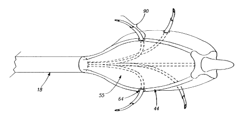
electrodes 90 are attached to arms 44 and have an
insulating layer 92, covering an insulated segment 94
except for an exposed segment 95. For purposes of this
disclosure, an insulator or insulation layer is a barrier
to either thermal, RF or electrical energy flow.
Insulated segment 94 is of sufficient length to extend
into sphincter wall 26 and minimize the transmission of
RF energy to a protected site 97 near or adjacent to
insulated segment 94 (see FIG.13). Typical lengths for
insulated segment 94 include, but are limited to, 1-4
mms. Suitable materials for needle electrodes 90
include, but are not limited to, 304 stainless steel and
other stainless steels known to those skilled in the art .
Suitable materials for insulating layer 92 include, but
are not limited to, polyimides and polyamides.
During introduction of sphincter treatment
apparatus 10, basket assembly 50 is in a contracted
state. Once sphincter treatment apparatus 10 is properly
positioned at the treatment site 12, needle electrodes 90
are deployed by expansion of basket assembly 50,
resulting in the protrusion of needle electrodes 90 into
the smooth muscle tissue of sphincter wall 26 (refer to
FIG. 14). The depth of needle penetration is selectable
from a range of 0.5 to 5 mms and is accomplished by
indexing movable fitting 82 so as to change the camber 54
CA 02391301 2002-O1-24
WO 01/06942 PCT/US00/17734
- 19 -
of arm 44 in fixed increments that can be selectable in
a range from 0 . 1 to 4 inms . Needle electrodes 90 are
coupled to power source 24 via insulated wire 60.
In another embodiment of sphincter treatment
apparatus 10 shown in FIG. 15, needle electrodes 90 are
advance out of apertures 64 in basket arms 44 into the
smooth muscle of the esophageal wall or other sphincter
16. In this case, needle electrodes 90 are coupled to RF
power source 24 by electrode delivery member 60. In this
embodiment, the depth of needle penetration is selectable
via means of stepped sections 66 or tapered sections 68
located in apertures 64. Referring to FIG. 16, apertures
64 and needle electrodes 90 are configured such that the
penetration angle 96 (also called an emergence angle 96)
of needle electrode 90 into sphincter wall 26 remains
sufficiently constant during the time needle electrode 90
is being inserted into sphincter wall 26, such that there
is not tearing or unnecessary trauma to sphincter wall
tissue. This is facilitated by the selection of the
following parameters and criteria: i) the emergence
angle 96 of apertures 64 which can vary from 1 to 90°,
ii) the arc radius 98 of the curved section 100 of
aperture 64 which can vary from 0.001 to 2 inch, iii) the
amount of clearance between the aperture inner diameter
102 and the needle electrode outside diameter 104 which
can vary between 0.001"; and, iv) use of a lubricous
coating on electrode delivery member 60 such as a Teflon
or other coating s well known to those skilled in the
art. Also in this embodiment, insulated segment 94 can
be in the form of a sleeve that may be adjustably
positioned at the exterior of electrode 90.
In another alternative embodiment shown in FIG.
17, electrode delivery member 60 with attached needle
electrodes 90, can exit from lumen 36 at distal shaft end
32 and be positioned into contact with sphincter wall 26.
CA 02391301 2002-O1-24
WO 01/06942 PCT/LTS00/17734
- 20 -
This process may be facilitated by use of a hollow
guiding member 101, known to those skilled in the art as
a guiding catheter, through which electrode delivery
member 60 is advanced. Guiding catheter 101 may also
include stepped sections 66 or tapered sections 68 at
it's distal end to control the depth of penetration of
needle electrode 90 into sphincter wall 26.
RF energy flowing through tissue causes heating
of the tissue due to absorption of the RF energy by the
tissue and ohmic heating due to electrical resistance of
the tissue. This heating can cause injury to the
affected cells and can be substantial enough to cause
cell death, a phenomenon also known as cell necrosis.
For ease of discussion for the remainder of this
application, cell injury will include all cellular
effects resulting from the delivery of energy from
electrode 88 up to, and including, cell necrosis. Cell
injury can be accomplished as a relatively simple medical
procedure with local anesthesia. In one embodiment, cell
injury proceeds to a depth of approximately 1-4 mm. from
the surface of the mucosal layer of sphincter 16 or that
of an adjoining anatomical structure.
Referring now to FIGS. 18A, 18B and 18C,
electrodes 88 and/or apertures 64 may be distributed in
a variety of patterns along expansion device 20 or basket
assembly 50 in order to produce a desired placement and
pattern of lesions 14. Typical electrode and aperture
distribution patterns include, but are not limited to, a
radial distribution 105 (refer to FIG. 18A) or a
longitudinal 106 (refer to FIG. 18B). It will be
appreciated that other patterns and geometries for
electrode and aperture placement, such as a spiral
distribution 108 (refer to FIG. 18C) may also be suitable.
These electrodes may be cooled as described hereafter.
FIG.19 is a flow chart illustrating one
CA 02391301 2002-O1-24
WO 01/06942 PCT/US00/17734
- 21 -
embodiment of the procedure for using sphincter treatment
apparatus 10. In this embodiment, sphincter treatment
apparatus 10 is first introduced into the esophagus under
local anesthesia. Sphincter treatment apparatus 10 can
be introduced into the esophagus by itself or through a
lumen in an endoscope (not shown), such as disclosed in
U.S. Patents Nos. 5,448,990 and 5,275,608, incorporated
herein by reference, or similar esophageal access device
known to those skilled in the art. Basket assembly 50 is
expanded as described herein. This serves to temporarily
dilate the LES or sufficiently to efface a portion of or
all of the folds of the LES. In an alternative
embodiment, esophageal dilation and subsequent LES fold
effacement can be accomplished by insufflation of the
esophagus (a known technique) using gas introduced into
the esophagus through shaft lumen 36, or an endoscope or
similar esophageal access device as described above.
Once treatment is completed, basket assembly 50 is
returned to its predeployed or contracted state and
sphincter treatment apparatus 10 is withdrawn from the
esophagus. This results in the LES returning to
approximately its pretreatment state and diameter. It
will be appreciated that the above procedure is
applicable in whole or part to the treatment of other
sphincters in the body.
The diagnostic phase of the procedure can be
performed using a variety of diagnostic methods,
including, but not limited to, the following: (i)
visualization of the interior surface of the esophagus
via an endoscope or other viewing apparatus inserted into
the esophagus, (ii) visualization of the interior
morphology of the esophageal wall using ultrasonography
to establish a baseline for the tissue to be treated,
(iii) impedance measurement to determine the electrical
conductivity between the esophageal mucosal layers and
CA 02391301 2002-O1-24
WO 01/06942 PCT/LTS00/17734
- 22 -
sphincter treatment apparatus 10 and (iv) measurement and
surface mapping of the electropotential of the LES during
varying time periods which may include such events as
depolarization, contraction and repolarization of LES
smooth muscle tissue. This latter technique is done to
determine target treatment sites 12 in the LES or
adjoining anatomical structures that are acting as foci
107 or pathways 109 for abnormal or inappropriate
polarization and relaxation of the smooth muscle of the
LES (Refer to FIG.20).
In the treatment phase of the procedure, the
delivery of energy to treatment site 12 can be conducted
under feedback control, manually or by a combination of
both. Feedback control (described herein) enables
sphincter treatment apparatus 10 to be positioned and
retained in the esophagus during treatment with minimal
attention by the physician. Electrodes 88 can be
muliplexed in order to treat the entire targeted
treatment site 12 or only a portion thereof. Feedback
can be included and is achieved by the use of one or more
of the following methods: (i) visualization, (ii)
impedance measurement, {iii) ultrasonography, (iv)
temperature measurement; and, (v) sphincter contractile
force measurement via manometry. The feedback mechanism
permits the selected on-off switching of different
electrodes 88 in a desired pattern, which can be
sequential from one electrode 88 to an adjacent electrode
88, or can jump around between non-adjacent electrodes
88. Individual electrodes 88 are multiplexed and
volumetrically controlled by a controller.
The area and magnitude of cell injury in the
LES or sphincter 16 can vary. However, it is desirable
to deliver sufficient energy to the targeted treatment
site 12 to be able to achieve tissue temperatures in the
range of 55-95°C and produce lesions 14 at depths ranging
CA 02391301 2002-O1-24
WO 01/06942 PCT/LTS00/17734
- 23 -
from 1-4 mms from the interior surface of the LES or
sphincter wall 26. Typical energies delivered to the
esophageal wall include, but are not limited to, a range
between 100 and 50,000 joules per electrode 88. It is
also desirable to deliver sufficient energy such that the
resulting lesions 14 have a sufficient magnitude and area
of cell injury to cause an infiltration of lesion 14 by
fibroblasts 110, myofibroblasts 112, macrophages 114 and
other cells involved in the tissue healing process (refer
to FIG.21). As shown in FIG.22, these cells cause a
contraction of tissue around lesion, 14, decreasing its
volume and, or altering the biomechanical properties at
lesion 14 so as to result in a tightening of LES or
sphincter 16. These changes are reflected in transformed
lesion 14~ shown in FIG.19B. The diameter of lesions 14
can vary between 0.1 to 4 mms. It is preferable that
lesions 14 are less than 4 mms in diameter in order to
reduce the risk of thermal damage to the mucosal layer.
In one embodiment, a 2 mm diameter lesion 14 centered in
the wall of the smooth muscle provides a 1 mm buffer zone
to prevent damage to the mucosa, submucosa and
adventitia, while still allowing for cell infiltration
and subsequent sphincter tightening on approximately 500
of the thickness of the wall of the smooth muscle (refer
to FIG.23).
From a diagnostic standpoint, it is desirable
to image the interior surface and wall of the LES or
other sphincter 16, including the size and position of
created lesions 14. It is desirable to create a map of
these structures which can input to a controller and used
to direct the delivery of energy to the treatment site.
Referring to FIG.24, this can be accomplished through the
use of ultrasonography (a known procedure) which involves
the use of an ultrasound power source 116 coupled to one
or more ultrasound transducers 118 that are positioned on
CA 02391301 2002-O1-24
WO 01/06942 PCT/US00/17734
- 24 -
expansion device 20 or basket assembly 50. An output is
associated with ultrasound power source 116.
Each ultrasound transducer 118 can include a
piezoelectric crystal 120 mounted on a backing material
122 that is in turn, attached to expansion device 20 or
basket assembly 50. An ultrasound lens 124, fabricated
on an electrically insulating material 126, is mounted
over piezoelectric crystal 120. Piezoelectric crystal
120 is connected by electrical leads 128 to ultrasound
power source 116. Each ultrasound transducer 118
transmits ultrasound energy into adjacent tissue.
Ultrasound transducers 118 can be in the from of an
imaging probe such as Model 21362, manufactured and sold
by Hewlett Packard Company, Palo Alto, California. In
one embodiment, two ultrasound transducers 118 are
positioned on opposite sides of expansion device 20 or
basket assembly 50 to create an image depicting the size
and position of lesion 14 in selected sphincter 16.
It is desirable that lesions 14 are
predominantly located in the smooth muscle layer of
selected sphincter 16 at the depths ranging from 1 to 4
mms from the interior surface of sphincter wall 26.
However, lesions 14 can vary both in number and position
within sphincter wall 26. It may be desirable to produce
a pattern of multiple lesions 14 within the sphincter
smooth muscle tissue in order to obtain a selected degree
of tightening of the LES or other sphincter 16. Typical
lesions patterns shown in FIGS. 25A-D include, but are
not limited to, (i) a concentric circle of lesions 14 all
at fixed depth in the smooth muscle layer evenly spaced
along the radial axis of sphincter 16, (ii) a wavy or
folded circle of lesions 14 at varying depths in the
smooth muscle layer evenly spaced along the radial axis
of sphincter 16, (iii) lesions 14 randomly distributed at
varying depths in the smooth muscle, but evenly spaced in
CA 02391301 2002-O1-24
WO 01/06942 PCT/US00/17734
- 25 -
a radial direction; and, (iv) an eccentric pattern of
lesions 14 in one or more radial locations in the smooth
muscle wall. Accordingly, the depth of RF and the
thermal energy penetration sphincter 16 is controlled and
selectable. The selective application of energy to
sphincter 16 may be the even penetration of RF energy to
the entire targeted treatment site 12, a portion of it,
or applying different amounts of RF energy to different
sites depending on the condition of sphincter 16. If
desired, the area of cell injury can be substantially the
same for every treatment event.
Referring to FIG.26, it may be desirable to
cool all or a portion of the area near the
electrode-tissue interface 130 before, during or after
the delivery of energy in order to reduce the degree and
area of cell injury. Specifically, the use of cooling
preserves the muscosal layers of sphincter wall 26 and
protects, or otherwise reduces the degree of cell damage
to cooled zone 132 in the vicinity of lesion 14.
Referring now to Fig. 27, this can be accomplished
through the use of cooling solution 70 that is delivered
by apertures 64 which is in fluid communication with
shaft lumen 36 that is, in turn, in fluid communication
with fluid reservoir 134 and a control unit 136, whose
operation is described herein, that controls the delivery
of the fluid.
Similarly, it may also be desirable to cool all
or a portion of the electrode 88. The rapid delivery of
heat through electrode 88, may result in the build up of
charred biological matter on electrode 88 (from contact
with tissue and fluids e.g., blood) that impedes the flow
of both thermal and electrical energy from electrode 88
to adjacent tissue and causes an electrical impedance
rise beyond a cutoff value set on RF power source 24. A
similar situation may result from the desiccation of
CA 02391301 2002-O1-24
WO 01/06942 PCT/US00/17734
- 26 -
tissue adjacent to electrode 88. Cooling of the
electrode 88 can be accomplished by cooling solution 70
that is delivered by apertures 64 as described
previously. Referring now to FIG. 28, electrode 88 may
also be cooled via a fluid channel 138 in electrode 88
that is in fluid communication with fluid reservoir 134
and control unit 136.
As shown in FIG. 29, one or more sensors 140
may be positioned adjacent to or on electrode 88 for
sensing the temperature of sphincter tissue at treatment
site 12. More specifically, sensors 140 permit accurate
determination of the surface temperature of sphincter
wall 26 at electrode-tissue interface 130. This
information can be used to regulate both delivery of
energy and cooling solution 70 to the interior surface of
sphincter wall 26. In various embodiments, sensors 140
can be positioned at any position on expansion device 20
or basket assembly 50. Suitable sensors that may be used
for sensor 140 include: thermocouples, fiber optics,
resistive wires, thermocouple IR detectors, an the like.
Suitable thermocouples for sensor 140 include: T type
with copper constantene, J type, E type and K types as
are well known to those skilled in the art.
Temperature data from sensors 140 are fed back
to control unit 136 and through an algorithm which is
stored within a microprocessor memory of control unit
136. Instructions are sent to an electronically
controlled micropump (not shown) to deliver fluid through
the fluid lines at the appropriate flow rate and duration
to provide control temperature at the electrode-tissue
interface 130 (refer to FIG. 27).
The reservoir of control unit 136 may have the
ability to control the temperature of the cooling
solution 70 by either cooling the fluid or heating the
fluid. Alternatively, a fluid reservoir 134 of
CA 02391301 2002-O1-24
WO 01/06942 PCT/US00/17734
- 27 -
sufficient size may be used in which the cooling solution
70 is introduced at a temperature at or near that of the
normal body temperature. Using a thermally insulated
reservoir 142, adequate control of the tissue temperature
may be accomplished without need or refrigeration or
hating of the cooling solution 70. Cooling solution 70
flow is controlled by control unit 136 or another
feedback control system (described herein) to provide
temperature control at the electrode-tissue interface
130.
A second diagnostic phase may be included after
the treatment is completed. This provides an indication
of LES tightening treatment success, and whether or not
a second phase of treatment, to all or only a portion of
the esophagus, now or at some later time, should be
conducted. The second diagnostic phase is accomplished
through one or more of the following methods: (i)
visualization, (ii) measuring impedance, (iii)
ultrasonography, (iv) temperature measurement, or (v)
measurement of LES tension and contractile force via
manometry.
In one embodiment, sphincter treatment
apparatus 10 is coupled to an open or closed loop
feedback system. Referring now to FIG. 30, an open or
closed loop feedback system couples sensor 346 to energy
source 392. In this embodiment, electrode 314 is one or
more RF electrodes 314.
The temperature of the tissue, or of RF
electrode 314 is monitored, and the output power of
energy source 392 adjusted accordingly. The physician
can, if desired, override the closed or open loop system.
A microprocessor 394 can be included and incorporated in
the closed or open loop system to switch power on and off
as well as modulate the power. The closed loop system
utilizes microprocessor 394 to serve as a controller,
CA 02391301 2002-O1-24
WO 01/06942 PCT/US00/17734
- 28 -
monitor the temperature, adjust the RF power, analyze the
result, refeed the result, and then modulate the power.
With the use of sensor 346 and the feedback
control system a tissue adjacent to RF electrode 314 can
be maintained at a desired temperature for a selected
period of time without causing a shut down of the power
circuit to electrode 314 due to the development of
excessive electrical impedance at electrode 314 or
adjacent tissue as is discussed herein. Each RF
electrode 314 is connected to resources which generate an
independent output. The output maintains a selected
energy at RF electrode 314 for a selected length of time.
Current delivered through RF electrode 314 is
measured by current sensor 396. Voltage is measured by
voltage sensor 398. Impedance and power are then
calculated at power and impedance calculation device 400.
These values can then be displayed at user interface and
display 402. Signals representative of power and
impedance values are received by a controller 404.
A control signal is generated by controller 404
that is proportional to the difference between an actual
measured value, and a desired value. The control signal
is used by power circuits 406 to adjust the power output
in an appropriate amount in order to maintain the desired
power delivered at respective RF electrodes 314.
In a similar manner, temperatures detected at
sensor 346 provide feedback for maintaining a selected
power. Temperature at sensor 346 is used as a safety
means to interrupt the delivery of energy when maximum
pre-set temperatures are exceeded. The actual
temperatures are measured at temperature measurement
device 408, and the temperatures are displayed at user
interface and display 402. A control signal is generated
by controller 404 that is proportional to the difference
between an actual measured temperature and a desired
CA 02391301 2002-O1-24
WO 01/06942 PCT/US00/17734
- 29 -
temperature. The control signal is used by power
circuits 406 to adjust the power output in an appropriate
amount in order to maintain the desired temperature
delivered at the sensor 346. A multiplexer can be
included to measure current, voltage and temperature, at
the sensor 346, and energy can be delivered to RF
electrode 374 in monopolar or bipolar fashion.
Controller 404 can be a digital or analog
controller, or a computer with software. When controller
404 is a computer it can include a CPU coupled through a
system bus. This system can include a keyboard, a disk
drive, or other non-volatile memory systems, a display,
and other peripherals, as are known in the art. Also
coupled to the bus is a program memory and a data memory.
User interface and display 402 includes
operator controls and a display. Controller 404 can be
coupled to imaging systems including, but not limited to,
ultrasound, CT scanner, X-ray, MRI, mammographic X-ray
and the like. Further, direct visualization and tactile
imaging can be utilized.
The output of current sensor 396 and voltage
sensor 398 are used by controller 404 to maintain a
selected power level at RF electrode 314. The amount of
RF energy delivered controls the amount of power. A
profile of the power delivered to electrode 314 can be
incorporated in controller 404 and a preset amount of
energy to be delivered may also be profiled.
Circuitry, software and feedback to controller
404 result in process control, the maintenance of the
selected power setting which is independent of changes in
voltage or current, and is used to change the following
process variables: (i) the selected power setting, (ii)
the duty cycle (e.g., on-off time), (iii) bipolar or
monopolar energy delivery; and (iv) fluid delivery,
including flow rate and pressure. These process
CA 02391301 2002-O1-24
WO 01/06942 PCT/US00/17734
- 30 -
variables are controlled and varied, while maintaining
the desired delivery of power independent of changes in
voltage or current, based on temperatures monitored at
sensor 346.
Referring now to FIG. 31, current sensor 396
and voltage sensor 398 are connected to the input of an
analog amplifier 410. Analog amplifier 410 can be a
conventional differential amplifier circuit for use with
sensor 346. The output of analog amplifier 410 is
sequentially connected by an analog multiplexer 412 to
the input of A/D converter 414. The output of analog
amplifier 410 is a voltage which represents the
respective sensed temperatures. Digitized amplifier
output voltages are supplied by A/D converter 414 to
microprocessor 394. Microprocessor 394 may be a type
68HCII available from Motorola. However, it will be
appreciated that any suitable microprocessor or general
purpose digital or analog computer can be used to
calculate impedance or temperature.
Microprocessor 394 sequentially receives and
stores digital representations of impedance and
temperature. Each digital value received by
microprocessor 394 corresponds to different temperatures
and impedances.
Calculated power and impedance values can be
indicated on. user interface and display 402.
Alternatively, or in addition to the numerical indication
of power or impedance, calculated impedance and power
values can be compared by microprocessor 394 to power and
impedance limits. When the values exceed predetermined
power or impedance values, a warning can be given on user
interface and display 402, and additionally, the delivery
of RF energy can be reduced, modified or interrupted. A
control signal from microprocessor 394 can modify the
power level supplied by energy source 392.
CA 02391301 2002-O1-24
WO 01/06942 PCT/US00/17734
- 31 -
FIG. 32 illustrates a block diagram of a
temperature and impedance feedback system that can be
used to control the delivery of energy to tissue site 416
by energy source 392 and the delivery of cooling solution
70 to electrode 314 and/or tissue site 416 by flow
regulator 418. Energy is delivered to RF electrode 314
by energy source 392, and applied to tissue site 416. A
monitor 420 ascertains tissue impedance, based on the
energy delivered to tissue, and compares the measured
impedance value to a set value. If the measured
impedance exceeds the set value, a disabling signal 422
is transmitted to energy source 392, ceasing further
delivery of energy to RF electrode 314. If measured
impedance is within acceptable limits, energy continues
to be applied to the tissue.
The control of cooling solution 70 to electrode
314 and/or tissue site 416 is done in the following
manner. During the application of energy, temperature
measurement device 408 measures the temperature of tissue
site 416 and/or RF electrode 314. A comparator 424
receives a signal representative of the measured
temperature and compares this value to a pre-set signal
representative of the desired temperature. If the tissue
temperature is too high, comparator 424 sends a signal to
a flow regulator 418 (connected to an electronically
controlled micropump, no shown) representing a need for
an increased cooling solution flow rate. If the measured
temperature has not exceeded the desired temperature,
comparator 424 sends a signal to flow regulator 418 to
maintain the cooling solution flow rate at its existing
level.
The foregoing description of a preferred
embodiment of the invention has been presented for
purposes of illustration and description. It is not
intended to be exhaustive or to limit the invention to
CA 02391301 2002-O1-24
WO 01/06942 PCT/LTS00/17734
- 32 -
the precise forms disclosed. Obviously, many
modifications and variations will be apparent to
practitioners skilled in the art. It is intended that
the scope of the invention be defined by the following
claims and their equivalents.