Note: Descriptions are shown in the official language in which they were submitted.
WO 01/45564 CA 02392205 2002-05-16 PCT/SE00/02629
TITLE
DEVICE FOR COMPRESSION OF THE LOWER ENTREMITIES FOR
MEDICAL IMAGING PURPOSES
DESCRIPTION
Technical field
The present invention relates to a device for compression of the lower
extremities for
medical imaging purposes, in particular for diagnostic purposes at
complementary
examination using computed tomography (CT) or magnetic resonance tomography
(MRT).
The object of the present invention is to obtain a device the use of which
provides an aid
for an adequate and reproducible examination of the lower extremities under
load in
connection with computed tomography and/or magnetic resonance tomography, in
particular for the diagnosis of development of arthrosis, changes of cartilage
in beginning
arthrosis, luxation, sacroiliac joint changes with regard to the hip joint;
diagnosis of
cruciate ligaments and meniscus damages, development of cartilage, patellar
luxation,
subluxations, preconditions for meniscus transplantations in the knee;
diagnosis of
cartilage damages, changes of tendons, luxations, and osteochondritis in the
ankle.
Background of the invention
Diagnosis of the lower extremities with regard to development of arthrosis,
changes of
cartilage in beginning arthrosis, luxation, sacroiliac joint changes with
regard to the hip
joint; diagnosis of cruciate ligaments and meniscus damages, development of
cartilage,
patellar luxation, subluxations, preconditions for meniscus transplantations
in the knee;
diagnosis of cartilage damages, changes of tendons, luxations, and
osteochondritis in the
ankle are hard to carry out under well-defined conditions and the market does
not recognise
any such diagnostic tools or equipment for said purpose.
EP 95920357.1 discloses a device for compression of the lumbar spine for
medical imaging
purposes, and then in particular for the diagnosis of the spinal cord canal
and nerve
structures (spinal stenosis) present.
WO 01/45564 CA 02392205 2002-05-16 PCT/SE00/02629
2
Further, it is previously knov~m from US-A-3,629,581 a device for positioning
a patients
shoulders in connection with an X-ray examination of the spine of a patient,
whereby the
upper spinal column is pressed downwards towards the examination table and
makes it
possible to obtain good X-ray pictures of the upper vertebras. Hereby a
pressure is applied
over the spinal column of the lying patient via two flexible strings provided
with handles
for the patient which strings are arranged around a foot plate.
US-A-4,202,355 discloses an X-ray grid orthometer used for measuring the leg
length of
individuals that may have an anatomical leg length imbalance, where the device
comprises
a frame means mountable on a support for adjusting to the length of the legs
of a patient,
whereby the frame comprises two foot plates onto which the patient's feet are
placed.
Further the X-ray grid orthometer comprises a pair of pulleys and a flexible
elongated
member arranged as to be engageable at the end portion by a patient for
compressing the
lumbar spine and legs the patient pulling with both hands on the flexible
member.
There is thus a problem to be solved, viz. to obtain an apparatus or device
for the diagnosis
of development of arthrosis, changes of cartilage in beginning arthrosis,
luxation, sacroiliac
joint changes with regard to the hip joint; diagnosis of cruciate ligaments
and meniscus
damages, development of cartilage, patellar luxation, subluxations,
preconditions for
meniscus transplantations in the knee; diagnosis of cartilage damages, changes
of tendons,
luxations, and osteochondritis in the ankle under well-defined conditions.
Description of the present invention
It has now surprisingly been shown possible to be able to solve this problem
by the present
invention which is characterized in that the device comprises
a waist arrangement to be placed around and in firm contact with the hip
region of a
patient,
a knee arrangement to be placed in the vicinity of and in firm contact with
the knee of a
patient,
a foot plate comprising pressure sensors and being arranged to accommodate the
feet of a
patient, whereby said sensors are arranged to monitor the pressure of the
respective foot on
said foot plate,
WO 01/45564 CA 02392205 2002-05-16 PCT/SE00/02629
3
strings connecting said waist arrangement and said foot plate arrangement,
which strings
can be stretched independently of each other and comprise each a tension meter
to monitor
the force by which the strings are strained.
By means of the present invention a controlled load of the lower extremities
can be
obtained when the waist arrangement applied around a patient's hip region and
the knee
arrangement is placed in the vicinity of the knee and the strings are
stretched to a certain
load between the waist arrangement and the foot plate. Further, the load can
be applied
asymmetric as well, whereby a single knee can be examined and diagnosed in any
bent and
luxated position. Thus a knee can be examined during load at different degrees
of flexion
and rotation or both, examined when bent at different angles, optionally at
fixation of the
knee at three dimensions. The diagnosis and examination is further facilitated
as the foot
plate in one particular embodiment is made rotatable with regard to foot blade
and heel, or
both.
The invention will now be described more in detail with reference to the
attached drawing,
showing a preferred device of the invention, however, without being restricted
thereto,
wherein
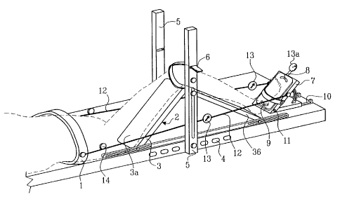
FIG. 1 shows a perspective view of one embodiment of a device according to the
present
invention.
1 denotes a waist arrangement which is arranged to be placed around the waist
and imme-
diately in connection with the hip region of a patient and thereby resting on
the hipbones
(innominate bones), whereby it is fastened conveniently using cords of the
burdock type.
The waist arrangement is applied firmly but still comfortably, but in such a
manner that it
does not slide over the body/hipbones (innominate bones).
A knee/leg arrangement 2 is provided with means to have it arranged firmly and
non-slid-
able to a knee. The knee/leg arrangement 2 is comprises a resting plate 3 for
the thigh and
the lower part of the leg of a patient, which resting plate 3 can be raised to
an upright
inclined position, whereby it is preferably adjustable as to its length to fit
any thigh length.
WO 01/45564 CA 02392205 2002-05-16 PCT/SE00/02629
4
The resting plate 3 is divided in the position to be placed under a knee (at
the hollow of the
knee) to bend the resting plate 3 in such a way that a bent leg at the knee
joint will still rest
with all its parts on the resting plate 3. Thereby the resting plate 3,
preferably consisting of
two subsections 3a, 3b is construed in such a way that it does not damage or
harm the
hollow of the knee by e.g., having a foldable, flexible sheet placed over the
joint of the two
subsections. The resting plate 3 is raised either manually or by using a motor
operating on
a screw means raising said subsections. On either side of the resting plate 3
a number of
holes 4 are arranged in parallel with the extension of the resting plate 3.
This holes 4 are
arranged to receive a pole 5 to which a straining band 6 can be attached. By
applying the
straining band to a knee, the knee and leg can be flexed sidewise and be held
in such a
position during CT or MRT attaching the band to the said pole.
A foot plate 7 is part of the device according to invention as well. The foot
plate 7
comprises a support for a foot, whereby the support is supported in two
different points 8,9.
Hereby the upper part of the support is provided with a journal running in a
track or groove
and being arranged to be locked in a central position. The lower part of the
support is in the
same way provided with a journal running in a track or groove and likewise
arranged to be
locked in a central position. Both journals are arranged to be either locked
simultaneously,
or one be locked and the other free in its track or groove. The tracks or
grooves allow the
support to be turned sidewise up to 30° from a vertical line in the
centre, i.e., in total 60°
from side to side. This is due for both the upper as well as the lower parts.
This means that
a foot placed upon the support can be rotated sidewise to the left or the
right from a
forward directed position either around the heel or around the matrix. The
foot support is
hereby preferably provided with a stop to prevent the foot from going back
into a normal
position.
Further the foot plate 7 is arranged to slide along a track 10 in the resting
plate 3 in order to
have the foot plate 7 follow the foot when, and if, the knee is raised to a
bent position.
Thereby the foot plate 7 is arranged to be locked in a position allowing a
normal resting of
the foot onto the foot plate 7 when the knee has been raised.
WO 01/45564 CA 02392205 2002-05-16 PCT/SE00/02629
The adjustment of the position of the foot plate 7 can be made either manually
or by means
of a motor operating to move the foot plate 7 to and fro. Care should be taken
not to
interfere with the CT or MRT if a motor should be installed.
Further, the foot plate 7 is arranged to adopt the raising of the knee in such
a way that the
foot angle visavi the leg remains in a normal position, i.e., about 90°
between leg and foot.
In case, however, the ligament controlling the bending forward of a foot
should be
examined, there is a further possibility to turn the foot plate 7 around a
horizontal axis 11
as well. To this effect there is a further pivot attached horizontally at the
middle of the foot
plate 7 around which the foot plate 7 can be turned to effect such an
adjustment.
Between the foot plate 7 and the waist arrangement there are at least two
pulling cords 12
or strings arranged, whereby each string individually can be
disconnected/connected to the
foot plate 7/waist arrangements. Each string comprises a tension meter 13 and
a straining
means 14 arranged in such a way that each string can be stretched
individually, whereby
the tension in each string can be monitored. The straining means can, in a
simple
embodiment, be a mechanical rolling device, manually or motor driven. The
tension meters
are preferably of an electronic type so that they can be attached to a
computer for collecting
data necessary to determine and document the conditions used.
The patient provided with the device of the invention is placed upon a resting
surface of a
patient table being suitable for being introduced in a device for computed
tomography or
magnetic resonance tomography. Computed tomography and magnetic resonance
tomography are known units and are subject of the present invention.
By having at least four cords or strings the different bending conditions can
be achieved.
During normal examination the tension in the cords will be about 25 to 50 kpm,
whereby
under certain extreme conditions it may be as high as 50 to 100 kpm or even
higher, e.g.
when studying a damage, where the ankle, knee and hip joints have been subject
to very
high forces and loads. The number of cords can be increased if more specific
bending
conditions will be achieved. In such cases four, six or even eight cords or
strings with their
respective tension meters can be attached.
WO 01/45564 CA 02392205 2002-05-16 PCT/SE00/02629
6
The tension meters 13 may also be connected to a motor which affects the
straining of the
cords v~~hereby a predetermined, adjusted value can be maintained as the
tension meters
control the motor.
At an examination a patient is placed on his back on the resting surface of
the patient table
of a tomography apparatus the waist arrangement is arranged around the hip
region and the
knee and foot plate 7 arrangements are arranged to the of the patient,
v~~hereby the knee
bend determines the position of the foot plate 7 along the resting plate 3,
whereafter the
cords or strings 12 with their respective tension meters 13 are attached
between the two
arrangements (waist and foot plate 7). The cords or strings are strained to a
predetermined
value, and the patient is brought into the tomography apparatus to obtain the
pictures
wanted. To determine conditions using a magnetic resonance tomograph certain
special
accessories (such as flex coils) can be needed to obtain the right signals due
to a distance
between the signal receiving parts and the patient. However, this is common
knowledge to
the operator of such tomographs, and is not part of the invention.
An alternative to the tension meters 13 is a meter 13a sensing the force by
which the foot
presses upon the foot plate 7.