Note: Descriptions are shown in the official language in which they were submitted.
CA 02392243 2007-12-27
1
LOW PROFILE VALVE
Field of the Invention
The present invention is related generally to medical devices. More
specifically, the present invention is directed to occlusion catlieters.
Catheters of the
present invention incorporate devices and methods allowing a balloon or other
occlusion device to be inflated or expanded and remain inflated or expanded
while a
second catheter is advanced over the proxinial end of the occlusion catheter.
Background of the Invention
Body vessels and conduits, for example coronary arteries, the carotid artery,
and lumens of the biliary tree, are frequently treated from within using
catheters
having means for treating conditions or affected areas at locations within the
vessels.
Treatment device examples include angioplasty balloons, stents and associated
stent
delivery catheters, drug delivery catheters, atherectomy devices, and devices
for
crushing or dissolving blockages in the biliary tree. When using these and
other
devices, it may be desirable to position and expand an occlusion device such
as an
inflatable distal occlusion balloon in proximity to the device. In coronary
artery
applications, the occlusion device can be disposed distally and downstream of
the
more proximal treatment apparatus such as a rotatable atherectomy burr or an
angioplasty balloon. In this application, the occlusion device is a distal
occlusion
device. A distal occlusion device may also be placed downstream of a stent and
associated stent delivery catheter while the stent is being expanded against
the vessel
wall.
CA 02392243 2002-05-17
WO 01/62329 PCT/US01/00638
Distal occlusion devices can be used to prevent byproducts of treatment from
leaving the treatment area. For example, small particles of plaque may be
freed by an
atherectomy process. Distal occlusion devices may also be used to provide a
quiescent region of a body vessel where treatment can occur. In one example, a
coronary artery region may be blocked off from blood flow to allow treating a
stenosed region vessel wall with an agent to inhibit restenosis. In another
example, a
stone may be isolated between a distal and a proximal occlusion balloon, with
the
space being filled with a chemical to dissolve the stone. In many of these
applications, the vessel region proximal of the distal occlusion device is
aspirated
through a catheter lumen to remove byproducts prior to deflating or removing
the
distal occlusion device.
An alternative application of an occlusion device is disclosed by Parodi et
al.
in published PCT Application WO 99/45835. The Parodi et al. disclosure is
directed to an occlusion device to guard against embolization during carotid
angioplasty. The occlusion device is placed within the vessel lumen proximal
to the
treatment site, and the device is expandable against the vascular duct to
occlude the
anterograde blood flow while a vacuum suction device is used to reverse blood
flow
distal of the occlusion device. The occlusion device includes a mouth for
drainage of
the retrograde blood flow containing any emboli therein. In this way, the
protective
device allows the temporary reversal of the flow of blood to prevent emboli
from
reaching the brain and allows for the drainage of emboli to the outside of the
patient's
body. During treatment with an angioplasty balloon distal of the occlusion
device, the
occlusion device in conjunction with vacuum suction and monitoring of the
patient's
blood flow allows controlled reversal of the blood flow.
Inflating an occlusion device is often accomplished in a manner similar to
inflating an angioplasty balloon. Proximal manifolds and adapters such as Luer
fittings can provide a secure channel between a pressurized fluid supply
outside the
body and the distal occlusion device such as a balloon. Luer fittings are
often bulky
and significantly larger than the tubes to which they are attached. Because it
may be
required to advance a second catheter over the occlusion catheter while the
occlusion
catheter remains in place, it is generally not possible to advance a second
catheter
over the occlusion catheter while the conventional fitting is attached. If the
-2-
CA 02392243 2002-05-17
WO 01/62329 PCT/US01/00638
conventional fitting were removed from the occlusion device catheter shaft,
the distal
occlusion device shaft proximal end would require sealing to avoid loss of
inflation
pressure. The seal itself would have to be small enough to allow the second
catheter
to pass over the seal while the seal maintained the pressure within the
occlusion
device and balloon.
Examples of a low profile occlusion device are described by Zadno-Azizi et
al. in published PCT Application No. WO 99/26692 and by Teitelbaum in U.S.
Patent
No. 5,807,330. Both the Zadno-Azizi et al. and Teitelbaum devices never become
completely sealed systems during operation. Both devices have proximal ports
that
must be opened and closed when inflating or deflating the occlusion balloon.
What would be advantageous is an occlusion catheter having a proximal end
profile sufficiently small so as to allow a second catheter to be advanced
over the
proximal end of the occlusion balloon catheter shaft, while maintaining the
occlusion
balloon in an inflated state. A device allowing inflation and rapid deflation
while a
catheter is inserted over the distal occlusion catheter would be desirable as
well. A
device that does not require opening and closing a part to operate the balloon
would
also be desirable.
Summary of the Invention
The present invention provides occlusion devices for occluding body conduits
and vessels. The devices include expandable distal portions and an elongate
tubular
shaft. The occlusion devices allow other devices to be advanced over and
retracted
from the occlusion device shafts while the occlusion devices occlude the
conduit or
vessel. One device includes an elongate tubular shaft having an inflatable
occlusion
device disposed near the distal end and a lumen extending within the shaft
walls. An
elongate fluid displacement rod is disposed within the shaft. The fluid
displacement
rod is preferably at least half the length of the tubular shaft length. The
tubular shaft
can have a distal fluid preparation portion near the distal balloon for
infusing inflation
fluid into the shaft prior to use.
In use, the elongate fluid displacement rod can be advanced distally, wherein
the volume of the rod within the lumen forces an equal volume of fluid into
the
distally disposed balloon. The fluid displacement rod can provide precise
linear
control of the amount of fluid forced into the balloon and a linear
relationship
-3-
CA 02392243 2002-05-17
WO 01/62329 PCT/USO1/00638
between the linear displacement of the rod and the fluid in the balloon. The
placement of the rod also provides control of pressure within the balloon. The
rod can
also provide for rapid inflation of the balloon and rapid deflation of the
balloon.
Rapid deflation can be advantageous where it is desirable for the occlusion to
be
ended or reduced rapidly in order to restore fluid flow. One example of this
advantage may be found in rapidly deflating a distal occlusion balloon where
the
balloon is occluding a coronary vessel and patient condition indicates that
rapid
balloon deflation may be called for.
Another aspect of the invention includes alignment devices for aligning
hypodermic needles for insertion into the proximal end of occlusion devices.
The
hypodermic needle aligmnent devices are particularly suitable for use with
distal
occlusion devices having proximally disposed sealable or self-sealing seals.
Brief Description of the Drawings
Figure 1 is a schematic plan view of a representative occlusion device having
a proximal seal, suitable for use with one aspect of the present invention;
Figure 2 is a longitudinal cross-sectional view of the proximal region of the
occlusion device of Figure 1, illustrating injection of inflation fluid
through a self-
sealing valve;
Figure 3 is a longitudinal cross-sectional view of a jawed alignment device
for
aligning the needle for penetration through the device seal of Figure 2;
Figure 4 is an enlarged schematic view of detents on the alignment device of
Figure 3;
Figure 5 is a perspective view of an alignment device for aligning a side
entry
hypodermic needle with the shaft of an occlusion device such as the device of
Figure
1;
Figure 6 is a perspective view of an aligmnent device for aligning a top
entry,
curved hypodermic needle, with the shaft of an occlusion device such as the
device of
Figure 1;
Figure 7 is a fragmentary, longitudinal cross-sectional view of an occlusion
device having a fluid displacement rod disposed within the inflation lumen;
-4-
CA 02392243 2002-05-17
WO 01/62329 PCT/US01/00638
Figure 8 is a fragmentary, longitudinal cross-sectional view of the occlusion
device of Figure 7, having a second catheter disposed over the occlusion
device shaft;
and
Figure 9 is a fragmentary, longitudinal cross-sectional view of the device of
Figure 8, incorporating features to incrementally inflate the occlusion
balloon.
Detailed Description of the Invention
Figure 1 illustrates an occlusion device 20 having a distal region 22, a
proximal region 24, a distal end 26, a proximal end 28, an elongate tubular
shaft 30, a
distally disposed occlusion balloon 32, and a proximally disposed seal 34.
Occlusion
device 20 illustrates one type of occlusion device suitable for use with a
hypodermic
needle alignment device, described later. Balloon 32 can be formed of a non-
compliant polymeric material such as polypropylene, polyethylene and nylon or
compliant polymeric materials such as polyvinyl chloride, olefin copolymers
and
ionomer resins, in a manner well known to those skilled in the art. The
elongate
tubular shaft 30 is preferably made of a material such as stainless steel
hypotubing or
other materials well known to those skilled in the art such as a relatively
stiff polymer
or a nickel titanium alloy.
Figure 2 illustrates part of the elongate shaft proximal portion 24 of Figure
1
in greater detail. Seal 34 can be formed of a sealable or self-sealing
material such as
medical grade silicone rubber or other suitable polymeric material, which is
illustrated
as fonning a proximal plug region 36. Seal 34 can also include a proximalmost
layer
38 formed of a material such as polycarbonate. Proximal seal or plug portion
36 can
be formed by injecting a polymeric material between walls 40 of elongate shaft
30 to
fill the lumen therein. Proximalmost film or barrier 38 can be a formed by
affixing
polymeric material over proximal end 28. A hypodermic needle 42 having a sharp
end 44 can be inserted through seal material 36 and into a lumen 46 disposed
between
walls 40. In use, liypodermic needle 42 or other suitable injection device may
be used
to inject inflation fluid into lumen 46 to inflate balloon 32. Proximal end 38
has an
outside diameter as indicated at D1. At a more distal portion within proximal
region
24, elongate shaft 30 has an outside diameter D2. In one embodiment, Dl and D2
are
substantially equal, elongate shaft 30 having a substantially uniform outer
diameter
over much of its length. In one embodiment, Dl is equal to D2. In another
-5-
CA 02392243 2002-05-17
WO 01/62329 PCT/US01/00638
embodiment, D1 is only slightly larger than D2. In a preferred embodiment, D1
is not
substantially larger than D2. Having the outside diameter of elongate shaft
proximal
end 28 substantially equal to the outside diameter of the shaft provides a
small profile
for advancing other devices over elongate shaft 30. In particular, in a
preferred
embodiment, there is no proximal seal having an outer diameter substantially
larger
than the outer diameter of the shaft, for example at the shaft midpoint well
distal of
the proximal region seal.
Having a distal occlusion device with a proximal end outside diameter
approximately the same as the shaft outside diameter at its midpoint
longitudinally
can provide an elongate shaft which can be used for advancing a second medical
device over the elongate shaft. Elongate shaft 30 can thus be used in ways
similar to a
guide wire. In one use, elongate shaft 30 can be used to guide a therapeutic
device
such as an atherectomy catheter, an angioplasty catheter, or a stent delivery
catheter
over the shaft. In another use, elongate shaft 30 can be used to guide a
diagnostic
device such as an angiography catheter over its length. "Over the wire"
catheters can
be guided to a target site, having shaft 30 disposed within most of their
length. Single
operator exchange catlieters can be guided to a target site, having elongate
shaft 30
disposed primarily within a distal region of the device. For such uses, it is
preferred
that the shaft have an outside diameter of about 0.010 inches to about 0.018
inches.
It may be possible for hypodermic needle 40 to be hand guided into proximal
seal 34. Given the small dimensions of the distal occlusion device catheter
shaft,
however, guiding a hypodermic needle into the proximal seal can be difficult.
Referring now to Figure 3, an alignment device 50 is illustrated. Aligmnent
device 50
can be used to guide a hypodermic needle into the proximal end of a distal
occlusion
device. In one embodiment, alignment device 50 includes two opposing jaws 52
disposed about elongate shaft 30. Another embodiment has three jaws,
preferably
spaced equidistantly about shaft 30. In other embodiments, multiple jaws,
fingers, or
a single cylindrical mouth may be disposed about elongate shaft 30. The
alignment
device illustrated includes a hypodermic needle 54 disposed within a lumen 56
extending through the central longitudinal axis of the device 50. Needle 54
has a
lumen which is in fluid communication with a proximal fitting 58 which can be
used
for attachment to an inflation fluid source such as a syringe. In one
embodiment,
-6-
CA 02392243 2002-05-17
WO 01/62329 PCT/US01/00638
proximal fitting 58 includes a series of internal threads 60 for attachment of
a syringe.
In the distal end near the jaws, one embodiment includes a pair of alignment
pads 62
for grasping shaft 30. Alignment pads 62 can be formed of elastomeric gripping
material for grasping shaft 30. One embodiment also includes a pair of stops
64 for
positioning the occlusion device proximal end. In the embodiment illustrated,
a sharp
distal end 66 of needle 56 is shown protruding toward seal 34.
In one embodiment, a clamp is included for forcing together jaws 52 about
catheter shaft 30. The clamping device can be used to securely fix alignment
device
50 to distal occlusion device 20 prior to inserting the needle. In one
embodiment, the
clamping device includes a collar or sleeve 68 disposed about alignment device
50 at
a mid portion 70. Collar 68 can be disposed between a proximal stop 72 and a
distal
stop 74. In one embodiment, proximal stop 72 and distal stop 74 are formed as
annular rings about the device. In another einbodiment, discrete protruding
regions or
bumps form the proximal and distal stops. The alignment device can also
include
detents 76 securely engaging corresponding structures on the clamping collar
68.
Figure 4 illustrates in greater detail one embodiment of detents 76 and
corresponding
teeth 78 on the clamping collar 68. Collar 68 can also be threadably secured
between
stops 72 and 74. This eliminates the need for detents. As the collar is
advanced
across the mid portion, a wider jaw section, the jaws are forced inward to
clamp on
catheter shaft 30.
In use, jaws 52 can be disposed about seal 34 which is guided between
alignment pads 62. With shaft 30 somewhat aligned, clamping ring 68 can be
slid
proximally toward jaws 52. Shaft 30 can be slid further into jaws 50 across
alignment
pads 62 until stops 64 is reached. In one embodiment, the distal end of needle
56
extends distally past stops 64 such that when seal 34 is finally in contact
with stops
64, the distal end of needle 56 extends sufficiently far into catheter shaft
30 so as to be
in fluid communication with distal occlusion device lumen 46. At this point,
rings 68
can be slid distally to engage detents 76, and in some embodiments, to abut
distal
stops 74. With alignment device 50 securely affixed to shaft 38, fluid can be
injected
through needle 56, into shaft 30, into an occlusion balloon.
Referring now to Figure 5, anotller alignment device 80 is illustrated.
Alignment device 80 includes a first or top surface 82 and an opposing second
or
-7-
CA 02392243 2002-05-17
WO 01/62329 PCT/US01/00638
bottom surface 84. Aligrunent device 80 includes a longitudinal channel 88
disposed
on second surface 84 and a grasping pad 90 is disposed on first surface 82.
Other
embodiments have channels or partial channels in both the first and second
surfaces
and can include other means for grasping. Catheter shaft 30 is illustrated
disposed
longitudinally and within longitudinal channel 88. In the device illustrated,
a pair of
pads 92 are disposed on either side of longitudinal channel 88, with both pads
92 and
90 being formed of an elastomeric material. In one embodiment, longitudinal
channel
88 allows longitudinal, but not lateral, movement of shaft 30. In another
embodiment,
the geometry of longitudinal channel 88 and pads 92 are such that botli
longitudinal
and lateral movement of shaft 30 is precluded after first and second surfaces
82 and
84 are fully brought together. A second longitudinal channel 94 is
illustrated, also
along the longitudinal axis of shaft 30. Again, channel 94 can be formed as
either a
full or partial channel in both the first and second surfaces 82 and 84. In
one
embodiment, chaimel 88 and corresponding pads 92 preclude lateral and
longitudinal
movement of shaft 30 once enclosed, and second channel 94 allows longitudinal,
but
not lateral, movement of an inserted hypodermic needle.
In use, a device such as aligmnent device 80 can, in the open position,
receive
an inserted catheter shaft such as shaft 30 within longitudinal channel 80.
With
proximal end 28 in position, first surface 82 and second surface 84 can be
closed
about hinge 86, laterally and longitudinally immobilizing shaft 30. A
hypodermic
needle can be inserted into longitudinal channel 94, bringing the sharp tip of
the
hypodermic needle into shaft proximal end 28. With shaft 30 firmly held in
place,
inflation fluid can be injected from the hypodermic needle into shaft 30.
Referring now to Figure 6, another alignment device 100 is illustrated, being
similar in many respects to alignm.ent device 80 illustrated in Figure 5.
Alignment
device 100 includes a first surface 102 and a second opposing surface 104,
attached to
each other about a hinge 106. Pads 90 and 92 can be as illustrated in Figure
5, and as
previously discussed. In the embodiment illustrated, device 100 includes
longitudinal
channel 88 and has a carrier 108 disposed within a second longitudinal chamlel
110
shown on second surface 104. Carrier 108 is preferably slidably mounted within
second channel 110, providing for longitudinal movement toward and away from
shaft proximal end 28. A curved or bent hypodermic needle 112 is mounted on
-8-
CA 02392243 2007-12-27
carrier 108 and can be received within shaft proximal end 28. Hypodermic
needle
112 is illustrated having a proximal port 114 which can protrude through a
substantially longitudinal slot 116 in first surface 102.
In use, shaft 30 can be disposed between pads 90 and 92 within longitudinal
channel 88. First surface 102 can be brought into close proximity to second
surface
104, allowing hypodermic needle proximal port 114 to protrude through
longitudinal
slot 116. With the first and second surfaces brought together, hypodermic
needle
proximal port 114 can protrude through the top of device 100. A syringe or
other
fluid source can be attached to hypodermic needle port 114, preferably after
the first
and second surfaces are brought together. Before attachment of the fluid
source such
as a syringe, carrier 108 can be longitudinally slid toward shaft proximal end
28,
causing hypodermic needle 112 to protrude sufficiently far into shaft 30.
Inflation
fluid can then be injected through hypodermic needle 112 and into shaft 30,
inflating
a distal occlusion device. Alignment device 100 has the advantage of allowing
the
syringe and hypodermic to be inserted into the proximal port 114 after the
first and
second opposing services are closed. After inflation, the fluid source can be
detached
from port 114, and the opposing services opened. Slidable hypodermic needle
112
can be retracted out of shaft seal region 28.
Referring now to Figure 7, an inflatable occlusion device 130 is illustrated,
extending from a proximal region 150 to a distal region 152. Occlusion device
130
can terminate distally as illustrated, in an atraumatic tip such as a spring
coil tip 154.
Occlusion device 130 includes a pushable elongate rod or displacement rod 132
inserted through the device, which can be used to displace inflation fluid
from the
proximal portion of the shaft lumen into an occlusion balloon 134. Occlusion
device
130 functions similar to the inflatable device described in U.S. Patent No.
5,785,685
entitled BALLOON CATHETER WITH IMPROVED PRESSURE SOURCE.
Displacement rod 132 has an outer
diameter D1 indicated near a proximal end 136 of rod 132. Rod 132 includes a
distalmost end 138 illustrated as disposed distally well into device 130.
Displacement rod 132 is illustrated as directly disposed within an elongate
tubular member 140 having a shaft distal region 155 proximal of balloon 134
and a
-9-
CA 02392243 2007-12-27
lumen 142 within. Lumen 142 can serve as a means for inflating device 130,
containing inflation fluid which can be displaced by rod 132 which forces
fluid into
distally disposed occlusion balloon 134. Inflation fluid can be retained
within lumen
142 by a proximal seal 144 disposed between rod 132 and tube 140. In preferred
embodiments, the displacement rod 132 is preferably pre-loaded into lumen 142
during manufacturing, with fluid filling the shaft lumen. Alternatively, the
catheter
could be prepped on-site. In either application, proper function of the
displacenient
rod requires venting substantially all compressible gas from the lumen and
balloon
interior. One such niethod and device is disclosed in U.S. Patent No.
5,785,685,
wherein a one-way valve is provided to force gas from the distal portion of
the
catheter out the proximal end by injection of fluid through the one-way valve.
In use, occlusion device 130 can also be prepared by injecting inflation fluid
into lumen 142 sufficient to largely fill the length of the lumen. After the
initial
filling with inflation fluid, displacement rod distal end 132 can be displaced
near a
proximal end 146 of outer tube 140. After device 130 is inserted well into the
body,
displacement rod 132 can be advanced distally, thereby forcing inflation fluid
from
lumen 142 into balloon 134, thereby inflating balloon 134. As can be seen in
Figure
7, displacement rod 132 provides a small proximal profile for device 130,
which can
allow a second catheter to be inserted over outer tube proximal end 146,
thereby using
outer tube 140 as a guide wire to guide a second catheter into position.
Other methods and devices can also be used to prepare the occlusion device
130 for use. Inflation fluid can be injected into tube 140 after pulling a
vacuum on
tube 140 and balloon 134, using methods well known to those skilled in the
art.
Inflation fluid can also be initially injected into outer tube 140 using
features and
procedures described in U.S. Patent Application Serial No. 09/208,145, filed
December 9, 1998, entitled CATHETER WITH DISTAL MANIFOLD PREP
VALVE/MANIFOLD.
As described in the aforementioned application, inflation fluid can be
injected into
tube 140 tlirough an additional valve disposed near catheter shaft distal
region 144.
Injecting inflation fluid from a distal location has the advantage of forcing
any air
proximally out of the shaft.
-10-
CA 02392243 2002-05-17
WO 01/62329 PCT/US01/00638
Distal occlusion device 130 can be both rapidly inflated and deflated,
relative
to a syringe inflated catheter of similar dimensions. Using a fluid
displacement rod as
the inflation fluid pressure source can also provide control over balloon
inflation
through control over linear position of the fluid displacement rod. In
particular, the
ability to rapidly deflate the balloon can be advantageous in coronary artery
applications, where patient indications may require rapid deflation of the
balloon.
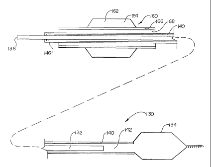
Referring now to Figure 8, distal occlusion device 130 is further illustrated
having a second catheter 160 disposed over outer tube 140. Second catheter 160
can
be a therapeutic or diagnostic catheter. In the example illustrated, second
catheter 160
is a highly diagraminatically illustrated angioplasty balloon catheter, having
only the
distal region illustrated. Second catheter 160 includes a distal balloon 162
having
interior 164 which is disposed about an elongate tube 166 having a lumen 168
for
receiving a guide wire and/or outer tube 140 of the first or occlusion
catheter 130.
Figure 8 illustrates how a second catheter can be inserted over the distal
occlusion
catheter where the distal occlusion catheter proximal profile is sufficiently
small so as
to fit within the lumen of the second catheter. In some embodiments, not
requiring
illustration, after displacement rod 132 is moved distally further into
occlusion device
lumen 142, the displacement rod proximal end can be clamped in a desired
position to
maintain inflation of distal occlusion balloon 134 while enclosed by outer
tube 146.
Referring now also to Figure 9, an altern.ative embodiment of the catheter of
Figure 8 is depicted. The embodiment of Figure 9 includes the additional
feature of
radial expansion of balloon 134 being incrementally controlled through a
ratcheting or
detent mechanism 170 that interacts with corresponding indentations 172 on
displacement rod 132. As shown in Figure 9, the detents include projections
extending radially inward. One or more of such detents can be incorporated in
combination with one or more indentations on the displacement rod. The
combination of detents and indentations can also act to enhance the desired
seal 144,
necessary for use. Thus, in alternative embodiments, the combination of
detents and
indentations could replace the seal or work in combination with the seal. The
distance
d' between detents 170 are preferably set to correspond to certain degrees of
radial
expansion of balloon 134. For example, each distance d' rod 132 is moved in a
proximal to distal direction could correspond to a 0.5 mm increase in diameter
of
-11-
CA 02392243 2002-05-17
WO 01/62329 PCT/US01/00638
balloon 134. Correspondingly, each distance d' displacement rod 132 is moved
in the
distal to proximal direction would result in a 0.5 mm decrease. Other
mechanisms for
controlling incremental changes in radial expansion of balloon 134 include
markings
on the side of displacement rod 132. A threaded design is also possible.
In use, distal occlusion catheter 130 preparation can include first filling
the
catheter with inflation fluid while maintaining balloon 134 in an uninflated
state.
Fluid displacement rod 132 can be inserted into a proximal portion of outer
tube 140.
The distal occlusion device with rod partially inserted can be advanced past a
target
site in a body conduit such as a coronary artery. Second catheter 160 can be
advanced
over first catheter outer tube 140, receiving outer tube 140 within lumen 168.
Second
catheter 160 can be advanced to a treatment site, and distal occlusion device
130 can
be inflated by advancing rod 132 distally within tube 140. With the vessel
occluded,
catheter 160 can be used to treat the target site. In some applications, for
example,
this may include either angioplasty or atherectomy.
Numerous advantages of the invention covered by this document have been
set forth in the foregoing description. It will be understood, however, that
this
disclosure is, in many respects, only illustrative. Changes may be made in
details,
particularly in matters of shape, size, and arrangeinent of parts without
exceeding the
scope of the invention. The invention's scope is, of course, defined in the
language in
which the appended claims are expressed.
-12-