Note: Descriptions are shown in the official language in which they were submitted.
CA 02392659 2002-05-24
WO 01/49211 PCT/USOO/01712
1
VASCULAR GRAFTS AND METHODS FOR BRIDGING
A VESSEL SIDE BRANCH
Field of the Invention
The present invention relates to prosthetic vascular grafts and, more
particularly, to a vascular graft for a primary vessel adapted to bridge a
side
branch, especially for providing a support tube for a primary graft located in
the
primary vessel on one side of the side branch.
Background of the Invention
An aneurysm is a ballooning of the wall of an artery resulting from
weakening due to disease or other condition. Left untreated, the aneurysm may
rupture, resulting in severe loss of blood and potentially death. An aneurysm
in
the abdominal aorta is the most comrnon form of arterial aneurysm. The
abdominal aorta connects the ascending aorta at the heart to the circulatory
system of the trunk and lower body. The abdominal aorta extends downward
from the heart in front of and parallel to the spine, through the thorax and
abdomen, and branches off in a plurality of side vessels. Among other
branching vessels, the abdominal aorta supplies the two kidneys via oppositely-
directed renal arteries. Below the renal arteries, the abdominal aorta
continues
to about the level of the fourth lumbar vertebrae and divides at a Y-junction
into
the left and right iliac arteries, which supply blood to the lower
extremities.
A common location for an aortic aneurysm is in the section of aorta
between the renal and iliac arteries. Without rapid surgical intervention, a
rupture of the abdominal aorta is commonly fatal because of the high volume of
blood flow within the aorta. Conventional surgical intervention involves
penetrating the abdominal wall to the location of the aneurysm to reinforce or
replace the diseased section of the aorta. Typically, a prosthetic tube graft
replaces the area of, or proximal and distal zones abutting, a potential
rupture
CA 02392659 2002-05-24
WO 01/49211 PCT/USOO/01712
2
portion of the aorta. Unfortunately, conventional surgical intervention has
resulted in substantial morbidity rates, and at the very least a protracted
recovery period. Likewise, cost and other constraints militate for a
longstanding need for endovascular intervention.
In recent years, methods and devices have been developed to treat an
aortic aneurysm without opening up the abdominal wall. These new techniques
typically involve a catheter-carried tubular graft delivered upward from the
femoral artery through the iliac artery and into the region of the aneurysm.
The
graft normally includes a tubular graft body supported by an expandable stent,
either self-expanding or balloon-expanding. The balloon-expanding type of
stent naturally requires an expansion balloon, while the self-expanding type
is
simply deployed from the end of a tubular sheath. Implacement issues impact
upon both known techniques.
If the aneurysm affects the Y-junction between the abdominal aorta and
the iliac arteries, a bifurcated graft is typically used. A trunk portion of
the
bifurcated graft is secured to a healthy section of the abdominal aorta just
below
the renal arteries, and branched legs of the graft are secured within each of
the
iliac arteries, sometimes via a tubular extension graft. This procedure does
not
involve cardiopuhnonary bypass, and thus blood continues to flow downward
through the abdominal aorta. Certain complications arise in anchoring the
graft
to the inner wall of the vessel, because of the high blood flow both during
the
procedure and afterward. Indeed, the risk of grafts migrating within a vessel
is
a problem in many locations, not just in the abdominal aorta. In addition, the
abdominal aorta may be aneurysmic very close to the renal arteries, which
results in a fairly poor substrate within which to secure a repair graft. - In
fact,
surgeons require various minimum lengths of healthy aortic wall below the
renal arteries before an endovascular graft repair is indicated, or else a
conventional invasive technique must be used. Moreover, the same
consideration of a minimum healthy portion of the host vessel applies in other
CA 02392659 2002-05-24
WO 01/49211 PCT/USOO/01712
3
areas, especially with regard to the portion of the aorta adjacent the
branching
subclavian or carotid arteries.
A number of techniques have been proposed for anchoring grafts to
vessel walls, most notably the use of barbs or hooks extending outward from
S graft that embed themselves into the vessel wall. Although these devices
secure
the graft, they may damage the vessel wall and cause complications.
Alternatively, portions of the stent may extend beyond the upstream end of the
graft body and be bent outward into contact with the vessel wall, either from
a
pre- or shape memory-bias, or from expansion of a balloon in this region.
In the context of repairing an aneurysm in the abdominal aorta, some
manufacturers have provided a stent at the upper end of a bifurcated graft
that
extends across the renal arteries. For example, the TALENT brand of
Endovascular Stent-Graft System available from World Medical of Sunrise,
Florida, includes an undulating wire support frame extending above the graft
body intended for supra-renal fixation. Likewise, the ZENITH AAA brand of
Endovascular Graft from Cook, Inc. of Bloomington, Indiana, utilizes an
undulating wire support having barbs for supra-renal fixation of the graft.
However, because these wires extend across the opening of the branching renal
arteries they present a certain impediment to blood flow therethrough.
Moreover, any structure placed in the path of blood flow may tend to initiate
the
blood clotting cascade, which in turn, may generate free-floating emboli that
would adversely impact the kidneys, or other organ that is perfused through
the
affected side branch. Because the kidneys are highly susceptible to injury
from
incursion of such emboli, it is highly desirable to avoid even the possibility
of
blood clotting at the mouth of the renal arteries.
Despite much work in this highly competitive field, there is still a need
for a more secure means of anchoring a bifurcated graft in the abdominal
aorta.
More generally, there is a need for a more secure means of anchoring a tubular
graft in a primary vessel in the vicinity of a vessel side branch.
CA 02392659 2002-05-24
WO 01/49211 PCT/USOO/01712
4
Summary of the Invention
The present invention comprises a vascular graft adapted for placement
in a primary blood vessel and suited to bridge a vessel side branch. The graft
comprises
a tubular structure defining an outer surface, a first portion of the outer
surface
being sized to contact and support the blood vessel on one side of the side
branch, and a second portion of the outer surface being sized to contact and
support the blood vessel on the other side of the side branch. The tubular
structure defines an aperture for alignment with the side branch so as to
permit
blood flow between the blood vessel and the side branch.
The first and second portions may be separated across a gap and the graft
further may include at least one bridging member traversing the gap and
connecting the first and second portions so as to prevent relative axial
separation of the two portions after implantation, the aperture being defined
between the bridging member and the first and second portions. There are
desirably at least two bridging members and two apertures, and potentially
four
bridging members and four apertures. Further, the bridging member may be a
relatively rigid strut.
In another aspect, the invention provides a vascular graft adapted for
placement in a primary blood vessel and suited to bridge a vessel side branch,
comprising:
a first tubular structare sized to contact and support the blood
vessel on one side of the side branch;
a second tubular structure sized to contact and support the blood
vessel on the other side of the side branch; and
at least one bridging member connecting the first and second
tubular structures so as to define an aperture in the vascular graft sized
for blood to flow through between the blood vessel and the side branch.
CA 02392659 2006-08-25
At least one of the first and second tubular structures desirably
comprises a flexible graft body and a support stent, wherein the
strut is directly connected to the graft body. More preferably, the
flexible graft body is only provided in one of the first or second
tubular structures, the other tubular structure being defined solely
by the stent.
In a further aspect, the invention provides a vascular graft system
adapted for placement in a primary blood vessel and adjacent a
vessel side branch. The system includes a tubular support graft
including a first tubular structure sized to contact and support the
blood vessel on one side of the side branch, and a second tubular
structure spaced from and connected to the first tubular structure
and sized to contact and support the blood vessel on the other side
of the side branch. The system further includes a tubular primary
graft sized to co-axially couple with the first tubular structure.
At least one bridging member may connect the first and second
tubular structures so as to prevent relative axial separation of the
two tubular structures after implantation, an aperture being defined
between the bridging member and the first and second tubular
structures of a sufficient size to permit blood flow though the
vessel side branch. In one application of the system, the primary
vessel is the abdominal aorta, the vessel side branch comprises the
renal arteries, and the tubular primary graft is a portion of a
bifurcated graft. In addition, at least one of the first and second
tubular structures preferably comprises a flexible graft body and a
support stent, and more preferably the flexible graft body is only
provided in the tubular structure that is disposed infra-renally,
the other tubular structure disposed supra-renally being defined
solely by the stent. The stent may be self-expandable or balloon-
expandable.
The present invention in another aspect provides a vascular graft,
said vascular graft being adapted for placement in a primary blood
vessel and being suited to bridge a vessel side branch,
said vascular graft comprising:
CA 02392659 2008-05-29
5a
a tubular structure comprising a first tubular element configured to contact
and support a
primary blood vessel on one side of a vessel side branch, and a second tubular
element
configured to contact and support the primary blood vessel on the other side
of the vessel
side branch,
said vascular graft further including an intermediate element disposed between
said first
and second tubular elements,
said intermediate element comprising a bridging component and a side branch
aperture
component,
said bridging component connecting the first and second tubular elements so as
to
prevent relative axial separation of the first and second tubular elements
after
implantation, and
said side branch aperture component consisting of a single unobstructed side
branch
aperture, said side branch aperture being alignable with the vessel side
branch for
permitting blood flow between the primary blood vessel and the vessel side
branch and
whereby said blood flow is unobstructed by the bridging component.
The present invention in accordance with an additional aspect provides a
vascular graft, said
vascular graft being adapted for placement in a primary blood vessel and being
suited to
bridge two vessel side branches,
said vascular graft comprising:
a tubular structure comprising a first tubular element configured to contact
and support a
primary blood vessel on one side of two vessel side branches, and a second
tubular
element configured to contact and support the primary blood vessel on the
other side of
the two vessel side branches,
said vascular graft further including an intermediate element disposed between
said first
and second tubular elements,
said intermediate element comprising a bridging component and a side branch
aperture
component,
CA 02392659 2008-05-29
5b
said bridging component connecting the first and second tubular elements so as
to
prevent relative axial separation of the first and second tubular elements
after
implantation, and
said side branch aperture component consisting of two unobstructed side branch
apertures,
said side branch apertures each being alignable with a respective vessel side
branch for
permitting blood flow between the primary blood vessel and the respective
vessel side
branch and whereby said blood flow is unobstructed by the bridging component.
In accordance with the present invention a vascular graft is provided wherein
the bridging
component may comprise a relatively rigid strut component.
Methods of supporting a tubular primary graft in a primary blood vessel
adjacent a vessel
side branch is also provide by the present invention. One method includes,
providing a tubular support graft including a first tubular section and a
second tubular section
connected to the first tubular section;
CA 02392659 2002-05-24
WO 01/49211 PCT/USOO/01712
6
delivering the tubular support graft into an implant position;
deploying the tubular support graft so that the first tubular
section contacts and supports the blood vessel on one side of the side
branch and the second tubular section contacts and supports the blood
vessel on the other side of the side branch;
providing a tubular primary graft having a first end;
delivering the first end of the primary graft within the support
graft second tubular section; and
radially expanding the first end of the primary graft against the
inner surface of the second tubular section.
Another method includes the steps of:
providing a tubular primary graft having a first end;
delivering the first end of the primary graft into an implant
position; and
radially expanding the first end of the primary graft against the
inner surface of the blood vessel on one side of the side branch.
providing a tubular support graft including a first tubular section
and a second tubular section connected to the first tubular section;
delivering the tubular support graft so that the second tubular
section is within the primary graft first end; and
radially expanding the tubular support graft so that the first
tubular section contacts and supports the blood vessel on one side of the
side branch and the second tubular section contacts and supports the
inner surface of the primary graft first end.
Either method is preferably accomplished by endoluminally delivering
both the tubular support graft and the tubular primary graft.
CA 02392659 2002-05-24
WO 01/49211 PCT/USOO/01712
7
A further understanding of the nature and advantages of the invention
will become apparent by reference to the remaining portions of the
specification
and drawings.
Brief Description of the Drawings
Figure 1 is a sectional view through an abdominal aorta showing the
branching renal and iliac arteries, and illustrating one embodiment of a graft
of
the present invention for supporting a trunk portion of a bifurcated graft,
shown
in phantom;
Figure 2 is a perspective view of the graft of Figure 1;
Figure 3 is a perspective view of an alternative graft in accordance with
the present invention having two planar bridging members;
Figure 4 is a perspective view of a further graft of the present invention
having two wire-like bridging members;
Figure 5 is a perspective view of a further graft of the present invention
having four bridging members;
Figure 6 is a sectional view of the abdominal aorta in the region of the
renal arteries illustrating a still further embodiment of a graft of present
invention used to support the trunk portion of a bifurcated graft, shown in
phantom;
Figure 7 is a perspective view of the graft of Figure 6;
Figure 8 is an elevational view of the graft of Figure 6 showing certain
axial dimensions; and
Figure 9 is an axial sectional view of an abdominal aorta in the region of
the renal arteries showing certain anatomical dimensions.
Description of the Preferred Embodiments
Figure 1 illustrates a graft 20 of the present invention deployed within a
primary vessel, in this case the abdominal aorta. 22. A pair of side branches
24
is shown intersecting the primary vessel 22 at approximately the same axial
CA 02392659 2002-05-24
WO 01/49211 PCT/USOO/01712
8
location across the vessel. In the context of an abdominal aorta. 22, two
important side branches are the renal arteries 24, as shown. The abdominal
aorta. 22 continues downward from the renal arteries 24 and bifurcates at a Y-
junction 26 into the left and right iliac arteries 28.
The present invention provides a tubular graft within a primary vessel
for supporting another tubular graft in the primary vessel in proximity to a
side
vessel. It should therefore be understood that although the drawings and
description involve a graft in the abdominal aorta for supporting another
graft in
the region of the renal arteries, the same principles apply whichever primary
vessel or side vessel is involved. For example, as illustrated in Figure 1,
the
graft 20 could be used in the vicinity of a side branch 30 in the iliac
arteries 28.
Representative conditions suitable for repair with the grafts of the present
invention include the abdominal aortic aneurysm (AAA) described herein, a
thoracic aortic aneurysm (TAA), and an aortic uni-iliac (AUI) aneurysm. For
purpose of explanation, however, the term "side branch" will be used
interchangeably herein with "renal artery," and the term "primary vessel" will
be used interchangeably with "abdominal aorta."
As illustrated in Figure 1, the graft 20 helps anchor a trunk portion 34 of
a bifurcated graft 36, shown in phantom. The bifurcated graft 36 typically
comprises the trunk portion 34 that diverges at a septum 38 into a pair of
legs
40. One or both of the legs 40 may extend a sufficient distance to form a seal
within the iliac arteries 28, or tubular extensions 42 may be provided for
this
purpose. The end result is that the bifurcated graft 36 (and optional tubular
extensions 42) extends from a healthy portion 44 of the abdominal aorta. 22 to
both of the iliac arteries 28, spanning an aneurysmic region 46. Once the
bifurcated graft 36 is in place, blood flows therethrough and blood pressure
is
reduced between the aneurysm 46 and the exterior of the graft. Ultimately, the
aneurysm 46 collapses inward around the graft, which remains in place.
With reference to Figures 1 and 2, the graft 20 of the present invention
comprises a first tubular section 50 and a second tubular section 52 connected
CA 02392659 2002-05-24
WO 01/49211 PCT/USOO/01712
9
via at least one bridging member 54. The first tubular section 50 is spaced
from
the second tubular section 52 across a gap that, in conjunction with the
bridging
member 54, defines an aperture 56 for blood flow. If the first and second
tubular sections 50, 52 are co-linear, then the bridging member 54 is
generally
axially disposed. Alternatively, if the graft 20 is intended for implantation
in a
curvilinear vessel, the first and second tubular sections 50, 52 may be
aligned
along a curvilinear axis, in which case the bridging member 54 will also be
generally disposed along the same curve. Still further, the graft 20 may be
multi-curvate, for example S-shaped, in which case the first and second
tubular
sections 50, 52 and bridging member 54 will follow the multiple curves.
As illustrated in Figure 1, the aperture 56 is aligned with at least one of
the side branches 24. In a preferred application, the graft 20 is used to
support a
bifurcated graft 36 in proximity with the renal arteries 24, and thus defines
two
apertures 56, each aligned with one of the renal arteries. In this context,
the first
tubular section 50 is secured in contact with a supra-renal portion of the
abdominal aorta 22, while the second tubular section 52 is secured in contact
with an infra-renal portion. The apertures 56 are sized large enough so that
no
portion of the graft 20 resides in the blood flow path of the renal arteries
24, and
also so that renal arteries that are slightly axially offset from one another
can be
accommodated.
With specific reference to Figure 2, the graft 20 comprises a tabular
graft body 60 internally supported by a stent 62. The tubular graft body 60
may
be formed of one or more pieces, typically of a biocompatible fabric such as
polyester (e.g., polyterepthalate). Alternatively, the graft body 60 may be an
extruded PTFE tube. In a particular preferred embodiment, the graft body 60 is
one piece, with the apertures 56 formed by diametrically-opposed, generally
oval-shaped windows 64 cut in the body and extending circumferentially around
the body into proximity with one another. Two bridge segments 66 of the graft
body 60 extend between the first and second tubular sections 50, 52 of the
graft
and separate the windows 64. Preferably, the bridge segments 66 extend
CA 02392659 2002-05-24
WO 01/49211 PCT/USOO/01712
circumferentially around the graft body 60 a small arc in relation to the
adjacent
windows 64 so as to maximize the size of the blood flow apertures 56. In one
embodiment, the bridge segments 66 each circumferentially extends between
about 1-90 around the graft body 60, and more preferably each extends about
5 5-10 .
The blood flow apertures 56 are sized to enable alignment with side
branches of varying sizes. Of course, the particular size is defined by the
axial
dimension and the circumferential arc of the windows 64, which depends on the
overall graft diameter and length. For instance, a graft that is designed for
small
10 arteries and small side branches will have a reduced diameter and reduced
window size. Additionally, if the graft is intended to bridge only one side
branch then only one window is required. In a preferred embodiment, for use in
the abdominal aorta 22 to bridge the renal arteries 24, the graft 20 has a
diameter of between about 19 and 30 mm, and a length of between about 22 and
46 mm. The opposed windows 64 have an axial length of between about 6 and
mtn, and extend circumferentially around the graft body 60 between about
90 and 189 . The renal arteries 24 typically have a diameter of between
about
8-10 mm, and thus the windows 64 are desirably oversized to ensure open blood
flow through the renals, and to accommodate offset or otherwise misaligned
20 pairs of renals.
The stent 62 actually comprises a first stent portion within the first
tubular section 50, and a second stent portion within the second tubular
section
52. The first and second stent portions may be substantially similar in
construction, or may be configured differently, as desired. Those of skill in
the
art will understand that a variety of different types of stents may be used to
internally support a tubular graft body.
In a preferred embodiment, the stent 62 comprises a plurality of
separate, spaced-apart wireforms 70, each formed in an undulating, or
sinusoidal pattern. Each of the wireforms 70 includes alternating peaks and
valleys, with either the peaks or valleys being woven through the graft body.
CA 02392659 2002-05-24
WO 01/49211 PCT/USOO/01712
11
More specifically, as seen in Figure 2, there are three axially-spaced rows of
wireforms 70 in the first tubular section 50, and four axially-spaced rows of
wireforms in the second tubular section 52. Either the peaks or valleys of
these
rows of undulating wireforms are woven through slits 72 formed in the graft
body 60. In this manner, the wireforms 70 are prevented from migrating axially
within the graft body 60 with respect to one another, and thus provide a
fairly
uniform inner support structure for the flexible graft body. As mentioned,
each
wireform is either radially self-expandable to the configuration shown, or is
capable of plastic deformation when balloon-expanded. In either case, the
stent
62 (comprising the array of wireforms 70) compresses the graft body 62 against
the inner wall of a tubular blood vessel to form a fluid seal therebetween.
Moreover, certain materials and/or sleeve-like structures are available to
enhance the seal between the exterior of the graft 20 and the vessel wall, and
may be combined with the present invention.
A plurality of crimps 74 is visible on the exterior of the graft body 62.
The crimps 74 join free ends of each wireform 70, which comprise one or more
wire segments bent into the undulating pattern, and into the annular shape
required. Though the crimps 74 are not sharp, they provide an irregular
surface
structure on the exterior of the graft 20, and thus help secure the graft in
position within the vessel.
The bridging member 54 seen in Figure 1 and 2 comprises a reinforcing
strut 80 and the aforementioned bridge segments 66 of the graft body 60. The
reinforcing strut 80 is a relatively rigid elongate member extending between
the
first and second tubular sections 50, 52 of the graft 20. In a preferred
embodiment, the reinforcing strut 80 is a biocompatible metal (e.g., stainless-
steel) strip or rod secured at each end to either the graft body 60 or the
stent 62.
If the ends of the reinforcing strut 80 are secured to the graft body 60 as
shown,
sutures are typically used to sew an eyelet, hook or other such feature (not
shown) provided on each end of the reinforcing strut. If the ends of the
reinforcing strut 80 are secured to the stent 62, crimps are preferably used
CA 02392659 2002-05-24
WO 01/49211 PCT/USOO/01712
12
between juxtaposed ends of the closest wireforms and the reinforcing strut. As
shown, the reinforcing struts 80 are desirably located to the outside of the
bridge
segments 66, although the reverse configuration is contemplated as well.
The bridging members 54 serve to anchor one of the first and second
tubular sections 50, 52 of the graft 20 with respect to the other, and
desirably
maintain the spacing between the tubular sections, while at the same time
present very little in the way of structure that might occlude or otherwise
interfere with the blood flow between the primary vessel 22 and the affected
side branch 24. The bridging members 54 must have tensile strength to
withstand migratory forces that may tend to separate the first and second
tubular
sections 50, 52. In an exemplary configuration, the upstream section 50 or 52
serves to anchor the downstream section by virtue of their connection with the
bridging members 54. In addition, the bridging members 54 may be relatively
rigid in the sense that they have column strength sufficient to prevent the
tubular sections 50, 52 from migrating toward each other after implantation.
The bridging members 54 have a radial dimension that is approximately
the same as the rest of the graft 20; that is, they do not project radially
into or
out from the side wall of the graft. The circumferential width of each
bridging
member 54 depends on the intended use for the graft 20. That is, if the graft
20
is to be used in the abdominal aorta 22 to bridge the renal arteries 24 as
shown
in the drawings, then there are two bridging members 54 diametrically spaced
apart of relatively narrow circumferential width. In this way, the bridging
members 54 each axially extend along the wall of the abdominal aorta 22 at 90
orientations from the openings to the renal arteries 24, and there is no
chance of
occluding blood flow between the abdominal aorta 22 and renal arteries 24.
Alternatively, if there is only one side branch then there need only be one
bridging member of relatively greater circumferential width than as shown.
That is, the bridging member might extend 180 or more around the graft, with
the corresponding window opening up the remaining portion. In general, as
CA 02392659 2002-05-24
WO 01/49211 PCT/USOO/01712
13
long as care is taken to orient the window(s) in registration with the side
branch
or branches, then the bridging member(s) will not occlude blood flow.
The embodiment of Figures 1 and 2 shows relatively rigid bridging
members 54 that are constructed of, for example, wires. Alternatively, the
bridging members 54 may be strips of biocompatible fabric or even sutures that
provide tensile strength to prevent the downstream tubular section 52 from
migrating with respect to the upstream section 50. In the illustrated example,
the upstream section 50 anchors the graft 20, and in particular the downstream
tubular section 52, with respect to the renal arteries 24. In this context,
one or
the other of the tubular sections 50, 52 may be designed to better anchor the
graft 20 in the primary artery 22, and the other may perform another function,
such as supplementing a damaged section of the artery so that another graft
may
be secured adjacent the side branch 24. Of course, however, both tubular
sections 50, 52 can be constructed to have identical anchoring and vessel
supporting characteristics if the graft 20 is used to repair a damaged length
of
the vessel that extends upstream and downstream of the side branch.
Figure 3 illustrates an alternative graft 90 of the present invention
having a first tubular section 92 separated from a second tubular section 94
across a gap 96 and connected across the gap by two bridging members 98.
Again, the graft 90 comprises a graft body 100 and an internal stent 102. The
graft body 100 may be a tubular biocompatible fabric, and in the illustrated
embodiment is separated across the gap 96 into two tubular portions in the
respective first and second tubular sections 92, 94. Because the facing edges
of
the two tubular portions of the graft body 100 are circular, the gap 96 is
tubular.
The stent 102 again coniprises a plurality of spaced-apart annular wireforms,
although it should be noted that the first tubular section 92 only has a
single
wireform 104.
The bridging members 98 are elongated planar bars or strips of
relatively rigid material, such as stainless-steel or a suitable polymer
connected
directly to the stent 102 or to the graft body 100 in the first and second
tubular
CA 02392659 2002-05-24
WO 01/49211 PCT/USOO/01712
14
sections 92, 94. Again, the bridging members 98 must have must have tensile
strength to withstand migratory forces that may tend to separate the first and
second tubular sections 92, 94 after implantation, while at the same time must
not occlude or otherwise interfere with the blood flow between the primary
vessel and the affected side branch or branches. Therefore, instead of being
relatively rigid, the bridging members 98 may be strips of fabric, such as
polyester, or sutures for that matter.
Figure 4 illustrates an alternative graft 110 of the present invention
having a first tubular section 112 separated from a second tubular section 114
across a gap 116 and connected across the gap by two bridging members 118.
Again, the graft 110 comprises a graft body 120 and an internal stent 122. The
graft body 120 may be a tubular biocompatible fabric, and in the illustrated
embodiment is separated across the gap 116 into two tubular portions in the
respective first and second tubular sections 112, 114. In this case the facing
edges of the two tubular portions of the graft body 120 are uneven by virtue
of a
plurality of notches 123, and thus the gap 116 is uneven as well. The stent
122
again comprises a plurality of spaced-apart annular wireforms, with the first
tabular section 112 having two wireforms and the second tubular section 114
having three.
The bridging members 118 each comprises lengths of wire either
separate from the stent 122 or defined by extensions of one or the wireforms.
If
the bridging members 118 are separate from the stent 122, they are connected
directly to the stent using a crimp 124, for example, or are connected
indirectly
via stitching 126 to the graft body 120. In an exemplary embodiment as
illustrated, the bridging members 118 are connected via crimps 124 to free
ends
of the lowest wireform in the first tubular section 112 and sewn to the graft
body 120 in the second tubular section 114.
Figure 5 illustrates a still further exemplary graft 130 of the present
invention having a first tubular section 132 separated from a second tubular
section 134 across a gap 136 and connected across the gap by four (4) bridging
CA 02392659 2002-05-24
WO 01/49211 PCT/USOO/01712
members 138. Again, the graft 130 comprises a graft body 140 and a stent 142.
The graft body 140 is desirably a tubular biocompatible fabric. The stent 142
again comprises a plurality of spaced-apart annular wireforms, with the first
tubular section 132 having a single wireform 144 disposed on the exterior of
the
5 graft body 140. The external wireform 144 can either be woven through slits
in
the graft body 140 as described above, or may be secured thereto with the use
of
suture thread.
The four bridging members 138 are distributed generally equidistantly
around the circumference of the graft 130 and each comprises a narrow strip of
10 fabric 146 and a reinforcement strut 148. Again, the reinforcement struts
148
may be connected directly to the stent 142 using a crimp, for example, or are
connected indirectly via stitching 149 to the graft body 140. The use of four
bridging members 138 may be desirable for stability when smaller branching
vessels are involved so that the windows defmed between the bridging members
15 need not be as large as the previous embodiments.
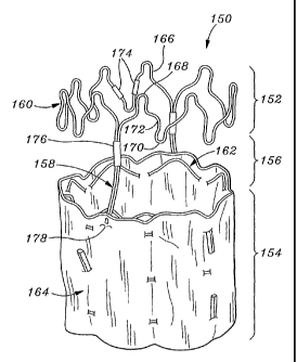
Figure 6 and 7 illustrate a still further embodiment of a graft 150 of the
present invention that defines a tubular structure having a first portion 152
and a
second portion 154 separated from the first portion across a gap 156. Two
bridging members 158 extend generally axially between and couple the first and
second portions 152, 154 to prevent their relative movement before during and
after implantation. In this embodiment, the first portion 152 of the tubular
structure is defined solely by a stent 160, while the second portion 154 is
defined by a stent 162 internally supporting a tubular graft body 164.
The upper stent 160 comprises an annular wireform 166 having
alternating peaks 168 and valleys 170 and contoured curvilinear segments 172
extending therebetween. The curvilinear segments 172 are shaped so as to nest
together when the graft 150 is in a radially constricted state, so as to
enable
smaller compaction of the graft. The wireform 166 includes one or more
segments connected into the annular shape by one or more crimps 174. The
CA 02392659 2002-05-24
WO 01/49211 PCT/USOO/01712
16
lower stent 162 includes a plurality of axially-spaced undulating wireforms
woven through the graft body 164, as previously described.
The bridging members 158 each comprise lengths of wire either separate
from the stents 160, 162 or defined by extensions of one or the wireforms. If
the bridging members 158 are separate from the stents 160, 162, they are
connected directly to the upper stent 160 using a crimp 176, and are connected
directly to the lower stent 162 using a crimp or indirectly via stitching 178
to
the graft body 164. In an exemplary embodiment as illustrated, the bridging
members 158 are connected via crimps 176 to free ends of the wireform 166 in
the first portion 152 and sewn to the graft body 164 in the second portion
154.
Figure 6 shows the graft 150 in place within a primary vessel 180 (e.g.,
the abdominal aorta) and bridging two oppositely-directed vessel side branches
182 (e.g., the renal arteries). The first portion 152 is located to contact
and
support the primary vessel 180 on one side of the side branches 182, while the
second portion 154 is located to contact and support the primary vessel on the
other side of the side branches. The gap 156 is positioned to permit blood
flow
between the primary vessel 180 and side branches 182, as indicated by the flow
arrows 184. The bridging members 158 extend axially across the gap 156
against the wall of the primary vessel 180 at approximately 90 orientations
from the side branches 182. Another graft 186 (e.g., the trunk of a bifurcated
graft) is seen positioned within the second portion 154. In this way, the
graft
186 is secured within the uniform and tubular second portion 154, which in
turn
is anchored within the primary vessel 180 from its own contact with the vessel
wall, and by virtue of its connection to the first portion 152 via the
bridging
members 158. This system of supporting one graft with another permits graft
positioning very close to the vessel side branches 182, and is especially
effective when the primary vessel is distended even very close to the side
branches.
The axial dimensions of the various grafts disclosed herein may be
selected to match the particular anatomical dimensions surrounding the
affected
CA 02392659 2002-05-24
WO 01/49211 PCT/USOO/01712
17
side branch. That is, the grafts, including two tubular sections with an
aperture
or gap therebetween and bridging members connecting the sections, are sized so
as to permit blood flow through the affected side branch and any adjacent side
branches. For example, the graft 150 seen in Figures 6 and 7 is positioned so
that the first portion 152 is above the renal arteries 182 and the second
portion
154 is below the renals.
A more detailed depiction of the relative axial dimensions for the graft
150 and region of the abdominal artery 180 near the renals 182 is seen in
Figures 8 and 9. In addition to the renal arteries 182, the openings for the
superior mezzanteric artery 190 and the ciliac artery 192 are shown in Figure
9.
These arteries typically project in the posterior direction, in contrast to
the
laterally-directed renals 182, and are located close to but upstream of the
renals.
The distance from the lowest of the arteries 190 or 192 and the highest of the
renals 182 is given as A, the distance from the upstream side of the highest
of
the renals 182 to the downstream side of the lowest of the renals is given as
B,
and the distance between the downstream side of the lowest of the renals to
the
end of the perceived healthy portion of the abdominal aorta 180 is given as C.
In addition, the diameter of one of the renal arteries 182 is given as D. The
axial dimensions of the graft 150 are given in Figure 8 as: L for the overall
for
the tubular structure, Ll for the first portion 152, L2 for the gap 156, and
L3 for
the second portion 154.
In a preferred embodiment, L2 > D, and if the renal arteries 182 are
offset, L2 > B. In addition, Ll is preferably smaller than or equal to A, so
that
the first portion 152 does not occlude either of the arteries 190 or 192.
Finally,
the lengtli L3 of the second portion 154 is desirably less than the length C
of the
healthy portion of the abdominal aorta.180, but may be greater than C.
In a specific embodiment, for use in the abdominal aorta 180 to bridge
the renal arteries 182, the graft 150 has a diameter of between about 19 and
30
mm, and a length L of between about 22 and 46 mm. The renal arteries 182
typically have a diameter of between about 5-10 mm, and may be offset center-
CA 02392659 2002-05-24
WO 01/49211 PCT/USOO/01712
18
to-center up to 10 cm. Thus the gap 156 has an axial length L2 of between
about 6 and 20 mm, and is desirably oversized to ensure open blood flow
through the renals and to accommodate offset or otherwise misaligned pairs of
renals. The length Li for the first portion 152 is desirably about 6 mm, but
may
vary depending on need. The length C of the healthy portion of the abdominal
aorta 180 should be at least 5 mm to enable the proper seal of the second
portion
154 with the aorta, which is smaller than an endovascular repair would
currently
be indicated. The length L3 of the second portion 154 is preferably at least 6
mm, more preferably about 10-20 mm. Of course, if the graft 20 is used to
repair a longer section of vessel as a primary graft, the length L3 of the
second
portion 154 can be longer than 20 mm, up to the currently accepted maximum
length of straight tube vascular graft.
To ensure the proper size/configuration of graft, the surgeon first
determines the anatomical landscape through the use of angioscopy; that is, by
injecting a contrast media and visualizing flow through the affected vessels
with
an X-ray device. The dimensions noted in Figure 9 can thus be obtained. A
range of different sized grafts are preferably available, and the surgeon then
selects the graft to match the anatomy in conformance with the above preferred
guidelines.
During implantation, the surgeon can ensure proper placement and
orientation of the grafts of the present invention with the use of radiopaque
markers on the graft. For example, the stent structure, or portions thereof,
could
be radiopaque, or markers can be attached to the stent or graft body. In
Figure
7, for instance, the wireform 160 and the upper wireform in the stent,162 are
desirably radiopaque so as to enable the surgeon to monitor the approximate
axial borders of the gap 156. Furthermore, the bridging members 158 or crimps
176 may be radiopaque to enable rotational orientation with respect to the
respective side branch or branches.
A method of supporting a tubular primary graft in a primary blood
vessel adjacent a vessel side branch, in accordance with the present invention
CA 02392659 2002-05-24
WO 01/49211 PCT/USOO/01712
19
can be illustrated with reference to the embodiment of Figure 6. First, the
tubular graft 150 is implanted in the primary vessel 180 such that the first
portion 160 contacts and supports the primary vessel on one side of a side
branch 182, in this case the two renal arteries, and the second portion 154
contacts and supports the primary vessel on the other side of the side branch.
Implantation of the tubular graft 150 can be accomplished by releasing a self-
expandable version of the graft from within a catheter sheath in the proper
location, or positioning a balloon-expandable version of the graft and
inflating a
balloon within the interior of the graft. A primary graft 186 is then
delivered in
a radially constricted state to a position overlapping the end of the second
portion 154 and radially expanded into contact therewith. Again, the primary
graft 186 may be either self-expanding or balloon-expanding.
An alternative method comprises implanting the tubular graft 150 after
the implantation of the primary graft 186. That is, the second portion 154 of
the
tubular graft 150 is self- or balloon- expanded outward into contact with the
primary graft 186. Indeed, the primary graft 186 may be implanted for a
significant period of time before the need for the supporting function of the
tubular graft 150 is recognized.
As mentioned above, one tubular portion of the graft-may perform an
anchoring function to maintain the position of the other portion that may or
may
not have the same anchoring characteristics. For instance, the graft portion
upstream of the side branch may anchor the downstream portion, which in turn
reinforces, supplements or seals with the primary vessel so as to enable
placement of another graft in that location. The present invention has been
described so far in terms of self- or balloon-expandable stents for anchoring,
but
those of skill in the art will recognize that there are other ways to anchor.
For
instance, staples, bent or corkscrew, are becoming more sophisticated and
effective, and may be used for anchoring. For that matter, any means for
anchoring one portion of the graft can be used.
CA 02392659 2002-05-24
WO 01/49211 PCT/USOO/01712
While the foregoing is a complete description of the preferred
embodiments of the invention, various alternatives, modifications, and
equivalents may be used. Moreover, it will be obvious that certain other
modifications may be practiced within the scope of the appended claims.
5