Note: Descriptions are shown in the official language in which they were submitted.
CA 02393104 2003-04-23
PERCUTANEOUS ELECTRICAL THERAPY SYSTEM AND ELECTRODE
TECHNICAL FIELD
This invention relates generally to percutaneous electrical
therapy systems for medical use.
s BACKGROUND OF THE INVENTION
Electrical therapy has long been used in medicine to treat pain
and other conditions. For example, transcutaneous electrical nerve
stimulation (TENS) systems deliver electrical energy through electrode
patches placed on the surface of a patient's skin to treat pain in tissue
io beneath and around the location of the patches. The efficacy of TENS
systems in alleviating pain is questionable at best; however.
More recently, a technique in which electrodes are placed
through the patient's skin into the target tissue has been proposed.
Percutaneous Neuromodulation Therapy ("PNT") (also sometimes called
15 Percutaneous Electrical Nerve Stimulation or "PENS") using percutaneously
placed electrodes achieves significantly better pain relief results than TENS
treatments using skin surface electrodes. This therapy is described in
Ghoname et al., "Percutaneous Electrical Nerve Stimulation for Low Back
Pain," JAMA 281:818-23 (1999); Ghoname et al., "The Effect of Stimulus
Zo Frequency on the Analgesic Response to Percutaneous Electrical Nerve
Stimulation iii Patients with Chronic Low Back Pain," Anesth. Analg.
88:841-6 (1999); Ahmed et al., "Percutaneous Electrical Nerve Stimulation
(PENS): A Complementary Therapy for the Management of Pain Secondary
to Bony Metastasis," Clinical Journal of Pain 14:320-3 ( 1998); and Ahmed
2s et al., "Percutaneous Electrical Nerve Stimulation: An Alternative to
Antiviral Drugs for Herpes Zoster," Anesth. Ar~alg. 87:911 -4 (1998).
'Thus far, PNT practitioners have used percutaneously placed
acupuncture needles attached to waveform generators via cables and
3o alligator clips to deliver the therapy to the patient. This arrangement and
design of electrodes and generator is far fiom optimal. For example, the
prior art has not addressed the issue of how to control the entry angle of
percutaneous electrodes used in PNT and other electrical therapies, or how
to prevent percutaneous electrodes from buckling when they are inserted into
_1_
CA 02393104 2002-05-29
WO 01/39829 PCT/US00/32559
the skin. Another drawback with conventional arrangements is that they
may not control the depth to which the percutaneous electrodes are inserted,
and may not prevent the electrodes from being inadvertently withdrawn from
the skin. Conversely, conventional electrodes may also be difficult or
s awkward to deliberately withdraw from the pu~ients. Still another drawback
is that the electrical connection to the electrode may be unreliable and
difficult to use. The patient may also experience discomfort when the
electrode is inserted into the skin. Still further, some conventional systems
may permit the patient's caregiver and/or a bystander to inadvertently
to contact the sharp end of the electrode, for example, when inserting or
withdrawing the electrode.
SUMMARY OF THE INVENTION
The present invention is directed to apparatuses and methods
for administering percutaneous electrical therapy. In one aspect of the
is invention, the apparatus can include an electrode electrically connectable
to
a control unit to deliver electrical therapy to a patient during operation.
The
electrode can have a first end and a second end opposite the first end with
the first end having a sharp point configured to be inserted into tissue of
the
patient. The apparatus can further include an electrode housing operatively
2o coupled to the electrode and positioned to support the electrode during
insertion of the electrode into the tissue. The housing can be positioned
relative to the electrode to control the motion of and/or the access to the
electrode.
The housing can include a channel disposed annularly about
2s the electrode to engage and guide at least a portion of the electrode
during
operation as the electrode moves relative to the housing into the tissue. The
housing can include a pressure element positioned adjacent to the electrode
to provide pressure against the tissue adjacent to an electrode insertion
point
through which the electrode enters the tissue. The apparatus can fiuther
so include an electrode actuator attached to the electrode and movable with
the
electrode relative to the housing, and an actuator tool removably attached to
the actuator to move the actuator and the electrode relative to the tissue.
The
housing can include a limit stop positioned to engage the electrode actuator
for stopping the motion of the electrode actuator when the electrode reaches
3s a selected depth in the tissue.
-2-
CA 02393104 2002-05-29
WO 01/39829 PCT/iJS00/32559
In another aspect of the invention, the housing forms an
interface with the skin of the patient when the housing is engaged with the
skin so that the housing and the skin completely surround the sharp point of
the electrode as the electrode moves into the tissue. The apparatus can
s further include n housing alignment member disposed on the tissue and
adapted to mechanically interact with the housing to align the housing
relative to the tissue. In a further aspect of this embodiment, the alignment
member can include a patch adhesively attached to the patient's skin.
The invention is also directed to a percutaneous electrode
io remover that includes a housing configured to be held in a human hand. The
housing can have an aperture at a distal end and an actuator configured to be
engaged by the human hand. In one aspect of the invention, the actuator is
movable relative to the housing between a first position with the actuator
coupled to an electrode while the electrode is inserted in a patient, and a
~s second position with the actuator coupled to the electrode and the
electrode
withdrawn through the aperture and completely into the housing.
The invention is also directed to a method for administering
percutaneous electrical therapy to a patient. The method can include
aligning an electrode housing with tissue of the patient, moving at least one
20 of the electrode and the housing relative to the other to insert a sharp
point
of the electrode into the tissue, and controlling a motion of and/or access to
the electrode with the housing. The method can further include applying an
electrical current to the electrode while the electrode is inserted in the
tissue.
In one aspect of the invention, the method can include guiding
2s the electrode in an axial direction by engaging at least a portion of the
electrode with walls of a channel disposed annularly about the electrode as
the electrode moves relative to the housing. The method can also include
halting movement of the electrode when the sharp point of the electrode
reaches a selected depth in the tissue. The method can further include
3o applying pressure to a skin of the patient adjacent to an electrode
insertion
point as the electrode is passed into the patient at the electrode insertion
point.
In another embodiment, the method can include grasping a
housing of a percutaneous electrode remover, engaging the housing with
3s tissue proximate to the percutaneous electrode while the percutaneous
electrode is inserted in the tissue, and manipulating an actuator of the
percutaneous electrode remover to couple the actuator to the percutaneous
-3-
CA 02393104 2002-05-29
WO 01/39829 PCT/US00/32559
electrode. The method can further include activating the actuator to
withdraw the percutaneous electrode from the tissue and into the housing.
BRIEF DESCRIPTION OF THE DRAWINGS
Figures lA-G are schematic renderings of a percutaneous
s electrical therapy system according to one embodiment of this invention.
Figure lA shows electrode and angle of insertion assemblies
wherein the electrode is in an undeployed and uninserted state.
Figure 1B shows the electrode and angle of insertion
assemblies of Figure lA during deployment but prior to insertion of the
to electrode into a patient's tissue.
Figure 1C shows the electrode and angle of insertion
assemblies of Figure lA during deployment and insertion of the electrode
into the patient's tissue.
Figure 1D shows the electrode of Figure lA inserted into the
is patient's tissue.
Figure lE shows the electrode of Figure lA attached to a
control unit to provide percutaneous electrical therapy.
Figure 1F shows the electrode and angle of insertion
assemblies of Figure lA during undeployment but prior to removing the
2o electrode from the patient's tissue.
Figure 1G shows the electrode and sharp point protection
assemblies of Figure lA during undeployment and after removing the
electrode from the patient's tissue.
Figures 2A-E are schematic renderings of a percutaneous
2s electrical therapy system according to another embodiment of this
invention.
Figure 2A shows a percutaneous electrical therapy system with
electrode and angle of insertion assemblies wherein the electrode is in an
undeployed and uninserted state.
Figure 2B shows the percutaneous electrical therapy system of
3o Figure 2A during deployment, but prior to insertion, of the electrode.
Figure 2C shows the percutaneous electrical therapy system of
Figure ZA with the electrode in a deployed and inserted state.
Figure 2D shows the percutaneous electrical therapy system of
Figure 2A during undeployment of the electrode.
-4-
CA 02393104 2002-05-29
WO 01/39829 PCT/US00/32559
Figure 2E shows the percutaneous electrical therapy system of
Figure 2A after the electrode has been undeployed.
Figure 3 shows an electrode montage for use in percutaneous
neuromodulation therapy to treat low back pain.
s Figure 4 is an exploded sectio:.vview of an electrode and
angle of insertion assembly according to yet another embodiment of this
invention.
Figure 5 is a partially exploded elevational W ew of the
embodiment of Figure 4.
to Figure 6 is an elevational view of the embodiment of Figure 4
showing the electrode and angle of insertion assemblies and an actuator tool.
Figure 7 is a sectional view of the embodiment of Figure 4
showing the electrode and angle of insertion assemblies and an actuator tool.
Figure 8 is a sectional view of the embodiment of Figure 4
is showing the actuator tool in engagement with the electrode and angle of
insertion assemblies prior to insertion of the electrode into a patient's
tissue.
Figure 9 is a sectional view of the embodiment of Figure 4
with the electrode in its deployed and inserted state.
Figure 10 shows a montage for using the embodiment of
2o Figure 4 to treat low back pain with the electrodes in a partially deployed
but
uninserted state.
Figure 11 shows the electrode montage of Figure 10 at the
beginning of the electrode insertion step.
Figure 12 shows the electrode montage of Figure 10 with the
2s electrodes deployed, inserted and attached to a control unit to provide
electrical therapy to the patient.
Figure 13 is an exploded view of an electrode introducer and
angle of insertion assembly of yet another embodiment of this invention.
Figure 14 is a partial sectional view of the introducer and angle
30 of insertion assembly of Figure 13.
Figure 15 is a sectional view of the introducer and angle of
insertion assembly of Figure 13.
Figure 16 is an elevational view of gear assemblies of the
introducer and angle of insertion assembly of Figure 13.
3s Figure 17 shows part of the electrode assembly of the
embodiment of Figures 13-16 in a montage used for treating low back pain
using PNT.
-5-
CA 02393104 2002-05-29
WO 01/39829 PCT/US00/32559
Figure 18 is an elevational view showing the introduces of
Figure 13 in the process of deploying an electrode.
Figure 19 is a sectional view showing the introduces of Figure
13 in the process of deploying an electrode, prior to insertion of the
s electrode.
Figure 20 is a sectional view showing the introduces of Figure
13 in the process of deploying an electrode, during insertion of the
electrode.
Figure 21 is a sectional view showing the introduces of Figure
13 in the process of deploying an electrode, also during insertion of the
to electrode.
Figure 22 is a sectional view of an inserted electrode assembly
of the embodiment of Figures 13-16.
Figure 23 is a partial sectional view of an electrode remover
and angle of insertion assembly according to yet another embodiment of the
is invention prior to removal of an electrode.
Figure 24 is a partial sectional view of the electrode remover
and angle of insertion assembly of Figure 23 partially actuated but prior to
removal of an electrode.
Figure 25 is a partial sectional view of the electrode remover
2o and angle of insertion assembly of Figure 23 partially actuated but prior
to
removal of an electrode.
Figure 26 is a partial sectional view of the electrode remover
and angle of insertion assembly of Figure 23 partially actuated and engaged
with an electrode but prior to removal of the electrode.
2s Figure 27 is a partial sectional view of the electrode remover
and angle of insertion assembly of Figure 23 during removal of an electrode.
Figure 28 is a partial sectional view of the electrode remover
and angle of insertion assembly of Figure 23 after removal of an electrode.
DETAILED DESCRIPTION OF THE PREFERRED
so EMBODIMENTS
Percutaneous electrical therapy systems, such as PNT systems,
deliver electric current to a region of a patient's tissue through electrodes
that pierce the skin covering the tissue. The electric current is generated by
a control unit external to the patient and typically has particular waveform
3s characteristics such as frequency, amplitude and pulse width. Depending on
-6-
CA 02393104 2002-05-29
WO 01/39829 PCT/US00/32559
the treatment or therapy being delivered, there may be one electrode
containing both a cathode and an anode or a plurality of electrodes with at
least one serving as a cathode and at least one serving as an anode.
The electrode has a sharp point not only to facilitate insertion
s through the patient's skin but also to enhance local current density
durir_g;
treatment. The placement and location of the electrode's point is therefore
an important aspect of the therapy. The angle at which the electrode enters
the patient's tissue helps determine where the electrode's point will end up.
One aspect of the invention therefore provides an electrode angle of entry
to assembly for use with a percutaneous electrical therapy system.
Insertion of percutaneous electrodes can be painful. The
thinner the electrodes, however, the less pain on insertion. One drawback of
conventional thin percutaneous electrodes is they may bend or buckle if they
are inserted into the patient improperly. In addition to potentially causing
is pain to the patient, the sharp point of a bent or buckled electrode will
not
likely be positioned at the target location for providing the therapy. Since
the sharp point of the electrode enhances local current density during
treatment, a displaced point could adversely affect the efficacy of the
treatment. Another aspect of this invention therefore provides an axial
2o electrode insertion supporter for a percutaneous electrical therapy system.
Furthermore, patient apprehension of imagined or impending pain can cause
discomfort. Therefore, yet another aspect of this invention provides an
electrode insertion pain reducer for use with a percutaneous electrical
therapy system and provides other features for minimizing patient
is discomfort.
Once the electrode is placed in the patient, it is important that
the electrode remain stationary so it does not move back out or become
completely dislodged. Accordingly, another aspect of this invention
provides an inserted electrode holding mechanism for use with a
so percutaneous electrical therapy system. Furthermore, once the electrode is
inserted into the skin, the sharp point may become exposed to pathogens,
microbes, toxins, etc. in the patient's tissue and/or blood. After removal of
the electrode from the patient's tissue, a caregiver or other bystander may be
stuck accidentally with the sharp point of the electrode, thereby exposing the
3s caregiver to any pathogens that may be on the used electrode. Another
aspect of this invention therefore provides an electrode assembly and/or
CA 02393104 2002-05-29
WO 01/39829 PCT/US00/32559
remover for a percutaneous electrical therapy system that provides sharp
point protection and is easy to use.
Figures lA-G are block diagrams showing deployment and use
of one embodiment of this percutaneous electrical therapy system and
s electrode assembly invention. As shown in Fig,~~s lA and 1B, the system
includes an electrode 1 having a sharp point 2 at its distal end and a housing
4 surrounding at least the electrode's sharp point 2 when the electrode is in
its undeployed and uninserted states. The undeployed and uninserted states
include pre-deployment and post-deployment states of the electrode.
to Housing 4 has an aperture 5 at its distal end. An actuator 6 interacts with
a
handle 11 at the proximal end of electrode 2 as shown.
Deployment of the electrode assembly includes the steps taken
to place the electrode assembly in proper position and condition for use in
electrical therapy. Figure lA shows the electrode assembly in an
is undeployed (pre-deployed) state. During deployment, the distal face 7 of
housing 4 is placed against a patient's skin 22, as shown in Figure 1B. This
action supports housing 4 with respect to the patient's skin, thereby
controlling the angle between the housing and the patient's skin. Electrode 2
is then inserted through aperture 5 into the tissue underlying the patient's
2o skin by moving actuator 6 distally, as shown in Figure 1C. As it moves,
actuator 6-and therefore electrode 2-is supported by housing 4 to control
the electrode's angle of entry into the patient's tissue.
Actuator 6 may have a limit stop 9 element cooperating with a
limit stop area 8 of housing 4 to limit distal motion of actuator 6 and to
2s control the depth of insertion of sharp point 2 of electrode 1. In a
preferred
embodiment of the invention, for example, where the electrical therapy
system is used to provide percutaneous neuromodulation therapy, the
predetermined electrode depth is 3 cm. Other electrode depths may be used,
of course, depending on the intended application and therapy.
After insertion, housing 4 and actuator 6 (which have
heretofore acted as an electrode introducer) are preferably removed, as
shown in Figure 1D. Electrode 1 is connected to a control unit 10 via a
conductor or cable 16. For use with PNT, control unit 10 preferably
supplies a current-regulated and current-balanced waveform with an
3s amplitude of up to approximately 20 mA, frequency between approximately
4 Hz and 50 Hz, and pulse width of between approximately 50 .sec and 1
msec. Other electrical waveforms having other parameters may be used, of
-g_
CA 02393104 2002-05-29
WO 01/39829 PCT/US00/32559
course, depending on the therapy to be provided. Also, while Figure lE
shows only one electrode connected to the control unit, it should be
understood that a plurality of electrodes may be connected to a single control
unit, as called for by the desired electrical stimulation treatment.
After ~w~npletion of the electrical therapy, the electrode
assembly is undeployed. The patient, therefore, does not have an
opportunity to view the length or amount of the electrode that had been
inserted into his or her tissue. One embodiment of this invention therefore
minimizes any discomfort the patient may experience due to fear or
to apprehension regarding percutaneous electrodes. In this embodiment, as
shown in Figure 1F, the aperture 5 of housing 4 is placed over the handle
portion 11 of electrode 1. Housing 4 may be the same used to deploy and
insert the electrode (i. e., the electrode introducer), or it may be an
entirely
different assembly (e.g., an electrode remover). The sharp point 2 of
~s electrode 1 is then drawn into housing 4 of sharp point protection assembly
3
by moving actuator 6 proximally, as shown in Figure 1G. Thus, sharp point
protection assembly 3 of Figures lA-G helps prevent all unintended contact
between the electrode's sharp point and a caregiver or other bystander
before, during and after deployment of the electrode.
2o Figures 2A-E are block diagrams showing another embodiment
of our invention. A control unit 10 is connected to an electrode 12 within an
electrode assembly 13 via a conductor 16. As above, for use with PNT,
control unit 10 preferably supplies a current-regulated and current-balanced
waveform with an amplitude of up to approximately 20 mA, frequency
2s between approximately 4 Hz and 50 Hz, and pulse width of between
approximately 50 sec and 1 msec. Other electrical waveforms having other
parameters may be used, of course, depending on the therapy to be provided.
Also, while Figures 1 A-E show only one electrode connected to the control
unit, it should be understood that a plurality of electrodes may be connected
3o to a single control unit, as called for by the desired electrical
stimulation
treatment.
As shown in its undeployed state in Figure 2A and in its
uninserted state in Figure 2B, the system includes a housing 18 surrounding
the sharp point 20 of electrode 12 when the electrode point 20 has not yet
3s been inserted through the patient's skin 22. To begin deployment, distal
face
21 of housing 18 is placed against the patient's skin 22, as shown in Figure
2B. As with the previous embodiment, this action supports housing 18 with
-9-
CA 02393104 2002-05-29
WO 01/39829 PCT/US00/32559
respect to the patient's skin, thereby controlling the angle between the
housing and the patient's skin. In one aspect of this embodiment, the
housing is held in place with an adhesive. The system includes an electrode
actuator 19 that enables deployment and insertion of the sharp point 20 of
s electrode 12 through the patient's skin 22 into the underlying tissue
through
an aperture 24 in housing 18, as shown in Figure 1C. Actuator 19 has an
interference fit with housing 18. Since the housing 18 is fixed on the
patient's skin, the interference fit between the actuator 19 and the housing
18
requires a minimum force to move actuator 19 with respect to housing 18.
~o This interference fit will keep actuator 19-and therefore electrode 12-in
place after electrode point 20 has been placed at the desired location. The
combination of the housing's attachment to the patient and the actuator's
fixed position with respect to the housing constitutes the electrode holding
mechanism of this embodiment.
is Actuator 19 may be part of the electrode assembly 13 or a
separate component of the system. Actuator 19 may also have a limit stop
element 23 that cooperates with a limit stop area 17 of housing 18 to limit
distal movement of actuator 19, thereby controlling depth of insertion of
electrode 12. In one embodiment of the invention, for example, where the
2o electrical stimulation system is used to provide percutaneous
neuromodulation therapy, the predetermined electrode depth is
approximately 3 cm., although other electrode depths may be used
depending on the application. The control unit 10 may then provide the
appropriate therapy to the patient through electrode 12 and any other
2s electrodes connected to it.
During undeployment, actuator 19 is used to draw electrode 12
back proximally into housing 18. After removal of the electrode from the
patient's skin, housing 18 of sharp point protection assembly 14 once again
surrounds the sharp point 20 of the now uninserted electrode 12, as shown in
3o Figures 2D and 2E. Actuator 19 helps enable this operation to occur without
ever exposing the sharp point of the electrode when the sharp point is no
longer in the patient. In fact, the operator of the electrode assembly never
sees the sharp point of the electrode. Thus, sharp point protection assembly
14 shields the potentially contaminated portion of the undeployed electrode
3s and protects the patient's caregiver or other bystander from unintended
contact with the sharp point of the electrode before, during and after
electrical therapy.
-10-
CA 02393104 2002-05-29
WO 01/39829 PCT/US00/32559
While Figures 2A-E show the electrode connected to the
control unit prior to deployment and insertion of the electrode into the
patient's skin, the connection between the control unit and the electrode
could be made during deployment or after insertion. Also, while Figures
2A-E show only one electrode connected to the wo:ntrol unit, it should be
understood that a plurality of electrodes may be connected to a single control
unit, as called for by the desired electrical stimulation treatment.
To use the percutaneous electrical therapy systems of Figures
lA-G and Figures 2A-E to treat a patient, one or more electrodes are inserted
to through the patient's skin into the underlying tissue. As an example, to
treat
low back pain using PNT with unipolar electrodes, an array or montage such
as that shown in Figure 3 may be used. The "T 12" D "S 1" designations refer
to the patient's vertebrae. The control unit or generator supplies current
pulses between pairs of electrodes for durations of a few minutes to several
~s hours, preferably delivering the current-regulated waveform described
above. Thirty-minute treatments are recommended in the Ghoname et al.
low back pain treatment articles.
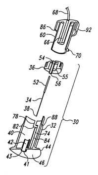
Figures 4-12 show another embodiment of this invention. An
electrode assembly 30 includes a base 32, an electrode 34, and a plunger or
2o actuator 36. Base 32 has a flange or flared end 44 that is adapted to make
contact with a patient's skin. Base 32 may be formed from any suitable
polymer or metal, such as a high-density polyethylene (HDPE). Base 32 is
preferably opaque so that the electrode cannot be seen by a needle-shy
patient.
2s Actuator 36 fits within a housing portion 40 of base 32 in a
slidable arrangement. A locking assembly is operable to prevent relative
movement between actuator 36 and housing 40 of base 32. In this
embodiment, the locking assembly of actuator 36 has integrally-formed
resilient detents 48 on its exterior cylindrical surface. In the undeployed
3o state of electrode assembly 30, detents 48 mate with a corresponding
openings 50 in base 32 to hold actuator 36 and base 32 in place with respect
to each other to prevent electrode 34 from moving outside of the protective
housing 40 of base 32 and thereby providing sharp point protection.
Mechanisms other than the detent and opening arrangement shown here may
3s be used to hold the actuator and base in place may be used without
departing
from the invention.
-11-
CA 02393104 2002-05-29
WO 01/39829 PCT/US00/32559
In this embodiment, electrode 34 is preferably a 3-cm long 32
gauge stainless steel needle. Other sizes and materials may be used for
electrode 34, of course, without departing from the scope of the invention.
Actuator 36 is preferably formed from HDPE as well, although other
s suitable materials may b,r ~~sed.
Electrode 34 has a larger-diameter handle 52 at its proximal
end. Handle 52 fits within a channel 54 formed within actuator 36. Channel
54 has a narrow opening 56 at its distal end whose diameter is slightly larger
than the diameter of electrode 34 but narrower than the diameter of handle
l0 52 to hold electrode 34 in place within actuator 36 after initial
manufacture
and assembly. As shown in Figure 7, in an undeployed state the sharp point
38 of electrode 34 is disposed within housing portion 40 of base 32,
specifically, within a narrow channel 42 of the housing 40.
To deploy one or more electrode assemblies on a patient in
is order to provide electrical stimulation therapy (such as PNT), the distal
surface 46 of flange portion 44 of base 32 is mounted on the desired site on
the patient's skin, preferably with a compressible adhesive pad (not shown)
surrounding a ring 43 extending downward from surface 46 around an
aperture 41 formed at the distal end of channel 42, although other means of
2o attaching base 32 to the patient may be used as appropriate. This action
aligns base 32 with respect to the patient's skin. Flange portion 44 of base
32 provides extra stability for the electrode assembly during electrode
insertion and use.
An electrical connector and actuator tool 60 is used to insert
2s the electrode and connect the electrode electrically with a control unit
62.
Actuator tool 60 and electrode assembly 30 also interact to provide the sharp
point protection assembly of this embodiment. When the distal end of
actuator tool 60 is placed against the proximal ends of base 32 and actuator
36, the exposed proximal end 64 of electrode handle 52 makes electrical
3o contact with a contact surface 66 within actuator tool 60. Contact surface
66, in turn, is electrically connected to the control unit 62 via a cable or
other conductor 68.
Actuator tool 60 has two oppositely disposed pegs 70
extending outward from the distal portion of its cylindrically surface. Pegs
3s 70 mate with two corresponding slots 72 in actuator 36 and with two
corresponding grooves 74 in base 32. (The second slot 72 and second
groove 74 are each opposite the slot 72 and groove 74, respectively, shown
-12-
CA 02393104 2002-05-29
WO 01/39829 PCT/US00/32559
in Figures 4 and 5.) When connecting actuator tool 60 to electrode assembly
30, pegs 70 move along longitudinal portions 76 of slots 72 and along
longitudinal portions 78 of grooves 74. Concurrently, exposed distal end 64
of electrode handle 52 begins to make sliding contact with contact surface 66
of actuator tool 60 to create the electrical connection between actuator tool
60 and electrode 32.
Clockwise rotation (looking down on the assembly) of actuator
tool 60 after pegs 70 reach the end of longitudinal portions 76 and 78 moves
pegs 70 into short circumferential portions 80 and 82, respectively, of slots
io 72 and grooves 74. The length of circumferential portions 80 of slots 72 is
less than the length of circumferential portions 82 of grooves 74. Continued
movement of pegs 70 along circumferential portions 82 will therefore move
pegs 70 against the ends 81 of circumferential slots 80. Further clockwise
rotation of actuator tool 60 will cause actuator 36 to rotate clockwise as
well,
~s thereby moving detents 48 out of openings 50 and allowing the electrode 34
and actuator 36 to move with respect to base 32.
Second longitudinal portions 84 of grooves 74 are formed in
base 32 at the end of circumferential portions 82. Movement of pegs 70
distally along longitudinal portions 84 pushes pegs 70 against the distal
2o edges of circumferential slot portions 80, thereby moving actuator 36 and
electrode 34 in a controlled fashion distally toward the patient's skin 22.
As it moves, electrode 34 passes through channel 42, and the
sharp point of electrode 34 moves out through aperture 41. Channel 42 and
actuator 36 provide axial support to electrode 34 during this forward
2s movement and also, along with the support provided by flange 44, provide
entry angle guidance to the electrode. In addition, downward pressure on
the patient's skin during electrode deployment and/or movement of the
actuator tool and actuator compresses the compressible adhesive pad and
presses ring 43 against the patient's skin 22, which helps ease electrode
3o entry through the skin and also lessens the insertion pain experienced by
the
patient.
The alignment of base 32 with respect to the patient's skin and
the controlled movement of actuator 36 and electrode 34 within base 32
controls the electrode's angle of entry into the tissue underlying the
patient's
3s skin. Distal movement of the electrode and its actuator within base 32
continues until the distal surface 86 of a cylindrical cap portion 92 of
actuator tool 60 meets an annular surface 88 of housing 40. At this point,
-13-
CA 02393104 2002-05-29
WO 01/39829 PCT/US00/32559
sharp point 38 of electrode 34 has extended a predetermined depth into the
tissue underlying the patient's skin. In the preferred embodiment, this
predeternlined depth is approximately 3 cm., although other electrode depths
may be desired depending on the treatment to be performed. In one aspect
of this embodiment, an interference fit between the Ln,der surface of channel
42 and the outer surface 5~ of channel 54 performs this function.
The interaction of the actuator tool with the actuator and
electrode enables the electrode to be inserted into the patient and connected
electrically with the control unit in a single motion. From a time and motion
to standpoint, this design provides increased efficiency through the
elimination
of a motion (e.g., separately connecting the electrode to the control unit
after
inserting the electrode in the patient). This efficiency can save the
caregiver
a great deal of time when multiplied by, e.g., ten electrodes per patient and
five patients per hour.
is Electrical stimulation treatment may begin once the electrodes
have been deployed and inserted. Control unit 62 supplies stimulation
current to the electrodes, e.g., in the manner described in the Ghoname et al.
articles. The electrical waveform provided by the control unit depends on
the application. For example, in an embodiment of a system providing
2o percutaneous neuromodulation therapy, control unit 62 would preferably
provide a current-regulated and current-balanced waveform with an
amplitude of up to approximately 20 mA, frequency between approximately
4 Hz and 50 Hz, and pulse width of between approximately 50 .sec and 1
msec.
2s The interaction of actuator tool 60 and base 32 provides
stability to electrode 34 and its electrical connection to the control unit
during treatment by holding the electrode in place, by providing strain relief
for tugging forces on cable 68, and by providing a robust mechanical
connection. It should also be noted that the sharp point of the electrode is
so not exposed to the operator or to any other bystander at any point during
deployment and use of the electrode assembly.
After treatment has been completed, the electrode may be
removed from the patient. To do so, actuator tool 60 is moved proximally
away from the patient. As pegs 70 move proximally along longitudinal
3s portions 84 of grooves 74, pegs 70 push against proximal edges of the
actuator's circumferential slot portions 80, thereby moving actuator 36 and
electrode 34 proximally as well. When pegs reach the proximal end of
-14-
CA 02393104 2002-05-29
WO 01/39829 PCT/US00/32559
longitudinal groove portions 84, the sharp end 38 of electrode 34 is out of
the patient and safely inside housing 40 of base 32. Counterclockwise
movement of actuator tool 60 moves pegs along circumferential portions 80
and 82 of slot 72 and groove 74, respectively. Since, as discussed above,
s circumferential portion a is shorter than circumferential portion 82, this
counterclockwise movement will turn actuator 36 counterclockwise.
At the limit of the counterclockwise movement, detents 48
move back into openings 50 to prevent further movement of the electrode
and actuator with respect to base 32. Further distal movement of actuator
io tool 60 moves pegs 70 distally along longitudinal portions 76 and 78 of
slot
72 and groove 74, respectively, to disconnect actuator tool 60 from electrode
assembly 30. Base 32 can then be removed from the patient. It should be
noted that the patient never sees the length or amount of the electrode that
had been inserted into his or her tissue. One embodiment of this invention
is can therefore minimize any discomfort the patient may experience due to
fear or apprehension regarding percutaneous electrodes.
Once again, the interaction of the actuator tool with the
actuator and electrode enables the electrode to be removed from the patient
and disconnected electrically from the control unit in a single motion. From
2o a time and motion standpoint, this design provides increased efficiency
through the elimination of a motion, particularly when multiplied by many
electrodes and many patients. Also, at no time during the electrode
deployment, use or removal processes is the sharp point of the electrode
exposed to the operator or bystanders.
2s Figures 10-12 show the use of the electrode and sharp point
protection assemblies of Figures 4-9 to treat low back pain using PNT. As
shown in Figure 10, ten electrode assemblies 30a j are arranged in a montage
on the patient's back and attached with adhesive. Next, ten actuator tools
60a j are attached to the ten electrode assemblies 30a j. In this example,
3o prior to deployment the actuator tools are mounted on an actuator tool tray
61 that provides electrical communication to a control unit 62 via cable 69.
The actuator tools electrically connect with tool tray 61, and thereby to
cable
69 and control unit 62, via individual cables 68a-j. It should be understood
that the tool tray 61 and its electrical connection scheme play no part in
this
3s invention. Figure 11 shows the beginning of the electrode insertion
process.
Once each electrode assembly has been actuated by its
respective actuator tool to insert an electrode into the patient's tissue (as
-ls-
CA 02393104 2002-05-29
WO 01/39829 PCT/US00/32559
shown in Figure 12), control unit 62 provides electrical signals to treat the
patient. Preferably, half the electrodes (e.g., assemblies 30b, 30d, 30g, 30h
and 30i) are treated as anodes, and the other half as cathodes. In the
preferred embodiment, control unit 62 would provide a current-regulated and
s .,~.urent-balanced waveform with an amplitude of up to approximately 20
mA, frequency between approximately 4 Hz and 50 Hz, and pulse width of
between approximately 50 sec and 1 msec. to treat the patient's low back
pain using PNT.
Another embodiment of the invention is shown in Figures
l0 13-28. In this embodiment, an electrode introducer and an alignment
member mounted on the patient's skin provide an electrode angle of
insertion assembly controlling the electrode's angle of entry into the
patient's tissue. The electrode introducer and an electrode remover can
cooperate to connect and disconnect an electrode and an electrode holding
is mechanism, and can provide sharp point protection. In a preferred
embodiment of an electrode introducer 100 shown in Figures 13-16 and
19-21, introducer 100 is designed to insert multiple electrodes. It should be
understood that the principles of this invention could be applied to an
introducer designed to hold and insert any number of electrodes.
2o Twelve electrodes 102 are disposed within a magazine 103
rotatably mounted within a housing 104. In this embodiment, housing 104 is
a two-part injection molded polystyrene assembly. Housing 104 is
preferably opaque so that the patient cannot see the length of the electrodes.
As seen best in Figure 14, magazine 103 rotates about a hub 105 mounted on
2s supports formed in housing 104. A leaf spring 106 mates with one of twelve
radial grooves 108 formed in magazine 103 to form a twelve-position ratchet
mechanism for rotatable magazine 103 in housing 104.
Magazine 103 has twelve electrode chambers 115 arranged
radially about hub 105. When introducer 100 is completely full, each
so chamber 115 contains one electrode 102. The diameter of upper portion 118
of chamber 115 is sized to form an interference fit with the wider portions
112 and 114 of electrode handle portion 107 of electrode 102. Lower wide
portion 114 of electrode 102 is formed from a compressible material. The
diameter of lower portion 119 of chamber 115 is slightly larger so that there
ss is no interference fit between chamber portion 119 and electrode handle
107,
for reasons explained below. Each time leaf spring 106 is within a groove
-16-
CA 02393104 2002-05-29
WO 01/39829 PCT/US00/32559
108, the opening 106 of a magazine chamber 115 is lined up with the
aperture 117 of introduces 100, as shown in Figures 14 and 15.
A slide member 109 is disposed on a rail 110 formed in
housing 104. Extending longitudinally downward from slide member 109 is
s a drive rod 11 l, and extending longitudinally upward fr~m slide member 109
is a gear rack 120. The teeth of gear rack 120 cooperate with teeth on a
rotational gear 122 mounted about a shaft 124 extending into a shaft mount
126 formed in housing 104. A second set of teeth are mounted on a smaller
diameter rotational gear 128 (shown more clearly in Figure 16) which is also
to mounted about shaft 124. Gears 122 and 128 rotate together about shaft
124.
The teeth of smaller diameter gear 128 mesh with the teeth of a
second gear rack 130 extending from a longitudinally movable actuator 132.
A spring 134 mounted between actuator 132 and a spring platform 136
is biases actuator 132 away from housing 104. Actuator 132, gears 122 and
128, gear racks 120 and 130, slide member 109 and drive rod 111 form the
introducer's transmission assembly.
To deploy the electrode assembly of this embodiment, a
flexible and compressible annular patch 140 is placed on the patient's skin at
2o the desired site, preferably with adhesive (not shown). For example, to
treat
low back pain using PNT, the arrangement or montage shown in Figure 17
may be used. In this montage, five electrodes serve as cathodes and five
serve as anodes.
As shown in Figures 19 and 20, patch 140 has an annular rigid
2s member 141 disposed in its center and extending upwardly from it. Rigid
member 141 has a smaller diameter opening 142 leading to a larger diameter
opening 144. The diameter of opening 142 is slightly smaller than the lower
wide portion 114 of the handle portion 107 of electrode 102 and slightly
larger than the diameter of the central portion 113 of handle portion 107 of
3o electrode 102.
After the patch 140 is in place, the distal end of introduces 100
is placed against patch 140 so that introduces aperture 117 surrounds the
upwardly extending portion of rigid patch member 141, as shown in Figure
18. This interaction aligns the opening 116 of one of the introducer's
3s magazine chambers 115 with the opening 142 of rigid member 141 and helps
control the electrode's angle of entry, as shown in Figure 19. The line-of
sight action of the introduces (i. e., the electrode moves along, or parallel
to,
-17-
CA 02393104 2002-05-29
WO 01/39829 PCT/US00/32559
the introducer's longitudinal axis) helps in the accurate placement of the
electrodes.
Downward pressure on introducer 100 compresses patch 140,
thereby causing the upper surface of rigid member 141 to engage a lower
s surface of magazine 103 a.cl pressing rigid member 141 downward into the
patient's skin 22. This pressure on the patient's skin around the insertion
site minimizes the pain of insertion of the electrode.
Depressing actuator 132 moves gear rack 130 distally, which
causes gears 128 and 122 to rotate. Because of the relative diameters and
to relative tooth counts of gears 128 and 122, gear rack 120 moves
longitudinally a much greater distance than the corresponding longitudinal
movement of gear rack 130. This feature enables the electrode to be inserted
its required distance into the patient's skin using only a comparatively small
movement of the operator's thumb and (along with the opaque introducer
is housing) helps minimize discomfort caused by patient fear and apprehension
regarding the length of the electrode being inserted into his or her tissue.
Distal movement of gear rack 120 is guided by the movement of slide
member 109 along rail 110. As slide member 109 moves distally, drive rod
111 moves into a magazine chamber 115 until the distal end of drive rod 111
2o engages the top surface of the electrode's handle portion 107. As shown in
Figure 20, further distal movement of drive rod 111 pushes electrode 102
downward so that sharp point 108 of electrode 102 leaves the introducer
housing and enters the patient's skin 22 and the tissue beneath the skin.
Chamber 115 provides axial stability to the electrode 102 during insertion.
2s When the top portion 112 of electrode handle portion 107
leaves the smaller diameter portion 118 of magazine chamber 115, it enters
the larger diameter portion 119 of chamber 115. At this point (shown in
Figure 21), because the diameter of chamber portion 119 is wider than the
diameter of the electrode handle 107, the electrode is no longer attached to
3o introducer 100.
Continued downward movement of actuator 132 and drive rod
111 pushes the lower larger diameter portion 114 of electrode handle 107
through the smaller diameter portion 142 of rigid member 141 by
compressing handle portion 114. Further downward movement pushes
3s handle portion 114 into the larger diameter portion 144 of rigid member 141
so that the rigid member's smaller diameter portion lies between the larger
diameter portions 112 and 114 of the electrode handle 107. This interaction
-18-
CA 02393104 2002-05-29
WO 01/39829 PCT/US00/32559
holds the electrode in place in the patient's tissue and helps provide depth
control for electrode insertion. In this embodiment, the preferred depth of
the electrode's sharp point 108 is approximately 3 cm., although other
electrode depths may be desired depending on the treatment to be performed.
s ~'_ider member 109 also acts as a limit stop at this point when it engages
the
limit stop area 145 of housing 104, thereby also controlling electrode
insertion depth.
In one embodiment, actuator 132 and electrode 102 move in
the same direction during insertion: along, or parallel to, the longitudinal
to axis of the introduces. This common directional movement, along with the
ergonomic design of the introduces allowing it to be held and operated by
one hand, helps control electrode insertion speed and pressure on the patient.
Magazine 103 is rotated to a new insertion position and placed
against an empty patch 140 after insertion of each electrode until all
is electrodes have been deployed and inserted. A suitable electrical connector
148 such as an alligator clip is electrically connected to electrode 102
through an aperture (not shown) formed in the upper larger diameter portion
112 of electrode handle 107 to provide electrical communication between a
control unit 150 and electrode 102 via a cable or other conductor 149, as
2o shown in Figure 22. Patch 140 provides strain relief for electrode 102 by
preventing tugging forces on cable 149 from dislodging the electrode from
the patient, thereby helping keep the electrode in place.
Control unit 150 supplies stimulation current to the electrodes,
e.g., in the manner described in the Ghoname et al. articles. Once again, the
2s electrical waveform provided by the control unit depends on the
application.
For example, in an embodiment of a system providing percutaneous
neuromodulation therapy, control unit 150 would preferably provide a
current-regulated and current-balanced wavefoim with an amplitude of up to
approximately 20 mA, frequency between approximately 4 Hz and 50 Hz,
3o and pulse width of between approximately 50 sec and 1 msec.
It should be noted that in one embodiment, at no time during
the electrode deployment, insertion and electrical therapy treatment
processes was the sharp point of the electrode exposed to the operator or
bystanders.
3s In an alternative embodiment, the lower wide portion of the
electrode handle is formed from a rigid material and has rounded caroming
edges. The central annulus of patch 140 in this alternative embodiment is
-19-
CA 02393104 2002-05-29
WO 01/39829 PCT/US00/32559
either compressible or has a resilient caroming opening under the caroming
action of the electrode handle.
Figures 23-28 show a sharps-safe electrode remover according
to one embodiment of this invention. Remover 200 is designed to work with
s the electrode and electrode patch assembly described with u:spect to Figures
13-22 above. It should be understood that the principles of sharps-safe
remover 200 may apply to other electrode designs as well.
Remover 200 has a housing 202 with an aperture 204 at its
distal end. A number of previously undeployed electrodes 102 are stored
to within housing 202. Housing 202 can be opaque so that the patient cannot
see the length of the electrodes being removed. This feature helps minimize
discomfort caused by patient fear and apprehension regarding the length of
inserted electrodes. A pair of rails 214 and 216 hold the electrodes 102 in
alignment via the electrode handles 107, as shown. While this embodiment
is of the remover is designed to provide sharps-safe removal and storage of a
plurality of electrodes, the invention applies to removers designed to remove
and store one or any number of electrodes.
As described above, electrodes for percutaneous electrical
therapy are inserted through a patient's skin into underlying tissue with
2o handle portions exposed above the skin. The first step in undeploying and
removing an inserted electrode is to line up the exposed handle 107 of an
electrode with the remover's aperture 204, as shown in Figure 23, by placing
the distal face 205 of remover 200 against the patient's skin or against any
portion of the electrode assembly (such as an adhesive patch) surrounding
2s the electrode. While not shown in Figures 23-28, aperture 204 is sized to
surround an annular member (such as annular member 141 discussed above)
holding an electrode handle of an electrode assembly (such as that shown in
Figures 13-22 above), the sharp point of which has been inserted through a
patient's skin.
3o An electrode engagement fork 206 is pivotably attached to a
longitudinally movable actuator 208 via an arm 209 and a hinged pivot 210.
A coil spring 212 biases actuator 208 upward towards the actuator and fork
position shown in Figure 28. A leaf spring 218 extends from arm 209. A
cross-bar 220 at the end of leaf spring 218 slides in groove 222 and a
3s corresponding groove (not shown) on the other side of housing 202. Leaf
spring 218 is in its relaxed state in the position shown in Figure 23. In this
position, a cross-bar 224 extending from the distal end of arm 209 adjacent
-20-
CA 02393104 2002-05-29
WO 01/39829 PCT/US00/32559
fork 206 lies at the top of a caroming member 226 and a corresponding
caroming member (not shown) on the other side of housing 202.
Downward movement of actuator 208 (in response, e.g., to
pressure from a user's thumb) against the upward force of spring 212 moves
s cross-bar 224 against a first c~ ~.:ming surface 228 of catnming member 226,
as shown in Figure 24. Caroming surface 228 pushes crossbar 224 of arm
209 against the action of leaf spring 218 as actuator 208, arm 209 and fork
206 move downward.
Figure 25 shows the limit of the downward movement of fork
l0 206. At this point, crossbar 224 clears the caroming member 226, and leaf
spring 218 rotates fork 206 and arm 209 about pivot 210 to engage fork 206
with electrode handle 107, as shown in Figure 26. The tine spacing of fork
206 is shorter than the diameter of the upper wide portion 112 of electrode
handle 107 but wider than the diameter of the narrow middle portion 113 of
is electrode handle 107.
Release of actuator 208 by the user permits spring 212 to move
actuator 208, arm 209 and fork 206 proximally. The engagement between
fork 206 and electrode handle 107 causes the electrode to begin to move
proximally with the fork out of the patient and into the remover housing, as
2o shown in Figure 27. At this point, crossbar 224 is now engaged with a
second caroming surface 230 of caroming member 226. Caroming surface
230 pushes cross-bar 224 against the action of leaf spring 218 in the other
direction (to the left in the view shown in Figure 27) as the electrode, fork
and arm rise under the action of coil spring 212.
2s The electrode and fork continue to rise until they reach the
upward limit of their permitted motion, as shown in Figure 28. At this point,
electrode handle 107 has engaged rails 214 and 216 and the most recent
electrode previously stored in remover 200. Electrode handle 107 pushes
against the electrode handle of the previously stored electrode handle, which
3o in turn pushes against any electrode handles stored above it in the stack.
In
this manner, the latest electrode removed by remover 200 goes into the
bottom of the stack of used electrodes stored in remover 200. Now that the
sharp point 108 of electrode 102 is safely inside housing 202, remover 200
can be withdrawn from the site on the patient's skin through which the
3s electrode had been inserted. Once cross-bar 224 clears the top of caroming
member 226, and leaf spring 218 moves arm 209 back to the center position
shown in Figure 23.
-21-
CA 02393104 2002-05-29
WO 01/39829 PCT/US00/32559
It should be noted that the remover 200 can provide sharp
point protection for the entire electrode undeployment and removal process.
Once all electrodes have been removed, the used electrodes can be safely
transported in the sharps-safe container provided by the housing 202 of
s re~~.ver 200.
Modifications of the above embodiments of the invention will
be apparent to those skilled in the art. For example, while the invention was
described in the context of percutaneous electrical therapy in which
electrodes are used to deliver electricity to a patient, the entry angle
control
io features may be used with electrodes designed for medical monitoring and/or
diagnosis. In addition, the entry angle control features of this invention may
be used with acupuncture needles or other needles not used for conducting
electricity to or from a patient.
-22-