Note: Descriptions are shown in the official language in which they were submitted.
CA 02394194 2008-10-22
. ` = WO 01/44783 PCT/US00/33760
R.APID TTSSUE PROCESSOR
BACKGROUND OF TIffi INVE'NTION
1. Field of the Invention
The present invention relates to the rapid, continuous flow, processing of
tissue for
histology, from fixation to impregnation. In particular, it relates to an
automated tissue
processing system that can be operated with continuous throughput and uses a
sequential
series of different non-aqueous chemical solutions to harden a tissue specimen
and to
produce a wax-impregnated tissue specimen suitable for embedding and
sectioning.
2. Description of the Related Art
Conventional methods prepare tissues for histology by incubation in separate
solu-
tions of phosphate-buffered 10% formaldehyde for fixation, a series of
increasing concen-
trations of ethanol for dehydration, and xylene for clearing tissue of
dehydration agent,
prior to impregnation. Because of the time required for this process, usually
8 hours or
longer, it is customary to complete these separate steps - fixation,
dehydration, clearing,
and impregnation - oveinight in automated mechanical instruments designed for
those
tasks (see, for example, U.S. Patent Nos. 3,892,197; 4,141,312; and
5,049,510).
Automated tissue processors implementing such conventional processes are manu-
factured and sold by, for example, Shandon (HYPERCENTERTM and PATHCENTERTM
models), Miles-Sakura (TISSUE-TEKTM models), and Mopec-MediteTM (TPC 15
model).
A disadvantage of the prior art is that such automated systems have not been
capable of continuous throughput. Given the time required to complete tissue
processing,
cassettes containing tissues are loaded into the system during the day and
tissue process-
ing is completed in an overnight cycle. Thus, operation of the prior art
systems did not
allow tissue-containing cassettes to be processed to completion during the
work day.
I
CA 02394194 2002-06-12
WO 01/44783 PCT/US00/33760
For example, the TISSUE-TEK vacuum infiltration processor (VIP) series
requires
more than eight hours for completion of processing. Baskets holding the
cassettes are
placed in a retort in which tissue is processed. In addition, 14 stations
supply solutions of
various compositions to the retort. User-programmable software controls this
automated
process. A rotary valve regulates the movement of solutions between the retort
and the
various stations; applying pressure or vacuum to the retort when the valve is
open causes
solution to be pumped out of or pumped into the retort, respectively. Upon
completion of
a processing run, the instrument automatically prompts the use for a cleaning
cycle; this
requirement can be overridden only if no paraffin is used. Typically, tissue
specimens are
batch processed according to the following program:
Solution Concen- Set Set P/V Agitation Volume
tration Time Tempe- ** of
(min) rature Solution
1 Buffered 10% 50 40 C On On 2.2-3.2L
formalin
2 Buffered 10% 50 40 C On On 2.2-3.2L
formalin
3 Alcohol* 80% 50 40 C On On 2.2-3.2L
4 Alcohol 95% 50 40 C On On 2.2-3.2L
5 Alcohol 95% 50 40 C On On 2.2-3.2L
6 Alcohol 100% 50 40 C On On 2.2-3.2L
7 Alcohol 100% 50 40 C On On 2.2-3.2L
8 Alcohol 100% 50 40 C On On 2.2-3.2L
9 Xylene 100% 50 40 C On On 2.2-3.2L
10 Xylene 100% 50 40 C On On 2.2-3.2L
11 Paraffin 50 60 C On On 4L
12 Paraffin 50 60 C On On 4L
13 Paraffin 50 60 C On On 4L
14 Paraffin F-450 60 C On On 4L
** - P/V (Pressure/Vacuum): agitation is provided by alternating the
application of
pressure and vacuum to the retort when "P/V" is On. Otherwise, when
"Agitation" is On,
agitation can also be provided by pumping in and then pumping out the same
solution
every 20 minutes.
* - The alcohol used in most laboratories is a mixture of 90% ethyl, 5%
methyl, and 5%
isopropyl alcohol.
Typically such conventional methodology demands sending tissue specimens from
2
CA 02394194 2002-06-12
WO 01/44783 PCT/US00/33760
the operating room, medical office or other sites, to a pathology laboratory
sometime
during the working day; overnight batch processing of the specimens, so that a
tissue
specimen suitable for blocking and sectioning is only available on the moming
of the next
day; and rendering a diagnosis by a pathologist based on microscopic
examination of
sections prepared from a blocked and sectioned specimen later on that next day
(Figure
1). This requires almost 24 hours between receipt of the specimen and delivery
of the
pathologist's report. Although a shortened version of the conventional method
is presently
practiced, it is feasible only for small biopsies. These biopsies need to be
fixed for at least
about 30 minutes before initiating the processing cycle. The instrument
processing cycle
can be programmed to last a minimum of 70 minutes, but is preferably 2 to 2'/z
hours.
In addition to the minimum one-day delay in giving a surgeon the benefit of a
report from the pathologist, there are also problems associated with impeded
work flow in
the pathology laboratory necessitated by the requisite batch processing of
specimens, the
safety concerns that attend having instruments operating overnight, the risk
of possible
instrument failures and the need to monitor the instruments, and the waste of
using large
volumes of reagents for such processing when automated. Moreover, expensive
measures
are required to prevent exposure of laboratory personnel to noxious fumes and
toxic sub-
stances associated with the reagents used in this process. Also, the large
volumes of sol-
vent waste and paraffm debris produced by the conventional methodology will
pollute the
environment if not properly disposed.
Conventional fixation and processing also cause irreversible damage (e.g.,
hydro-
lysis of a phosphodiester bond and/or deamidation) to the structure of nucleic
acids (e.g.,
DNA, and especially RNA) that limits the application of genetic techniques for
diagnosis
and research. Consequently, most DNA and certainly RNA analysis require
special pre-
cautions with handling of material, such as immediate freezing of fresh
tissues to prevent
degradation, because retrospective genetic analysis is impaired by the
conventional meth-
odology.
Histological diagnosis of a frozen section suffers from multiple disadvantages
in
comparison to sections prepared from paraffm blocks. U.S. Patent No. 3,961,097
cautions
that the slide prepared from a frozen section "does not possess ... uniformity
of quality;"
"it is technically more difficult for serial sections of the same specimen to
be examined;"
3
CA 02394194 2002-06-12
WO 01/44783 PCT/US00/33760
"extreme caution must be exercised in cutting the specimen in order to ensure
a suffi-
ciently thin section and to avoid the possibility of damaging details of the
specimen;" and
all the slides must be prepared "while the tissue is in the initial frozen
state" because "[i]f
the tissue is thawed and refrozen for sectioning, it is severely damaged."
There is an ever present interest in expediting tissue processing and analysis
for
diagnostic purposes. Furthermore, recent healthcare focus has been directed to
lessening
the cost of various procedures including tissue processing. The costs of
tissue processing
are related to the time for processing and analysis of the specimens, the
space required for
the personnel and equipment in the laboratory, the volume of reagents (both
the purchase
price of the pure chemicals and the charges for discarding waste), and the
number of per-
sonnel required. More importantly, patients and their physicians depend on
evaluation and
diagnosis by the pathologist to guide treatment. Reducing the amount of time
needed to
complete tissue processing would lessen the anxiety experienced during the
period
between obtaining the specimen and delivering the pathologist's report to the
surgeon.
Others have recognized the need to shorten the time required for tissue
processing,
but they have made only modest improvements in the conventional methods. To
accele-
rate tissue processing, U.S. Patent Nos. 4,656,047, 4,839,194, and 5,244,787
use micro-
wave energy; U.S. Patent Nos. 3,961,097 and 5,089,288 use ultrasonic energy;
and U.S.
Patent No. 5,023,187 uses infrared energy. U.S. Patent No. 5,104,640 disclosed
a non-
aqueous composition of a fixative, a stabilizing agent, and a solubilizing
agent that
adheres a blood smear to a slide. But the aforementioned patents do not teach
or suggest
that the entire process of preparing diagnostic tissue slides could be
accomplished in less
than two hours, starting from fixation and ending with impregnation, with
continuous
processing of specimens.
Microwave ovens similar in design to those used in home cooking have been used
to accelerate the time required for tissue processing. U.S. Patent No.
4,656,047 claims a
method of tissue processing in which at least one of the dehydrating,
clearing, or impreg-
nating steps utilizes microwave energy. Fixation may be accomplished by
immersing the
tissue specimen in chemical fixative and then exposing the specimen to
microwave
energy for a time sufficient to chemically fix the specimen. U.S. Patent No.
4,839,194
claims a method of fixing a tissue specimen at a temperature not to exceed 40
C in which
4
CA 02394194 2002-06-12
WO 01/44783 PCT/US00/33760
the non-thermal effects of microwave energy are used. U.S. Patent Nos.
4,839,194 and
5,244,787 claim a method of staining tissue specimens utilizing microwave
energy.
In such conventional methods of tissue processing, the distribution of
microwave
energy is not uniform because of reflection and interference effects within
the chamber in
which the microwaves resonate and the waveguide that conducts the microwave
energy
from the source to the chamber. U.S. Patent No. 4,835,354 proposes a
mechanical solu-
tion utilizing a rotating platform to ensure uniform contact with the
microwaves, and
mixers and isolaters that disperse and absorb microwaves. U.S. Patent No.
5,289,140
proposes a solution that utilizes a combination of microwaves of different
wavelengths
and/or intensities, or sources emitting microwaves of different frequencies.
U.S. Patent
No. 5,796,080 discloses adjustable moderating means between the waveguide and
a
plurality of resonance chambers to individually control the chemical reaction
in each
chamber, such that the propagated mode of the microwaves in the wave-guide is
not
substantially changed.
We now describe a microwave unit that provides gentle uniform heating during
tissue processing in a manner distinct from that disclosed in the
aforementioned patents.
Such operation causes minimal damage to the processed tissue, and results in a
superior
specimen for subsequent histologic studies by a pathologist or cell biologist.
In contrast
to the solutions disclosed in the patent discussed above, the microwave unit
of the present
invention does not use a resonance chamber which would be sensitive to the
contents of
the chamber. This is an important consideration when heating a region that is
larger in all
dimensions than about 10%-20% of the wavelength of the microwave used and the
chem-
ical compositions in the chamber change in different steps of the process. In
the invention,
microwave energy is distributed into the solution and tissue in such a way as
to minimize
interference effects. By distributing the energy, it is absorbed by the
solution and tissue in
one pass through the materials.
Some improvements that result from the invention are summarized here, but
other
improvements are described below. Convective heat losses from the reaction
chamber and
the evaporation rate of liquid in the reaction chamber are reduced, volatile
substances are
prevented from contacting electronic components and vented to protect the
laboratory
personnel in the vicinity of the unit, errors committed during processing by a
human ope-
5
CA 02394194 2002-06-12
WO 01/44783 PCT/US00/33760
rator are eliminated, the power required by the unit to maintain the liquid
temperature in
the reaction chamber is reduced, and labor and reagent costs are reduced with
this system
as compared to manual operation. More subjectively, consistency in the quality
of tissue
specimens processed by the disclosed process is improved. Although one
microwave unit
may be used advantageously, multiple units may be operationally and physically
linked to
accelerate chemical reactions performed in batch and/or continuous mode.
SUMMARY OF THE INVENTION
It is an object of the invention to provide a microwave unit and a system for
tissue
processing that reduces the time required for processing and analysis, and
reduces the cost
thereof. The tissue processing system is capable of automation and,
preferably, accepts
specimens in a continuous manner. This allows conversion of existing practice
to rapid
response surgical pathology for the patient undergoing an operation, and may
even allow
point-of-care diagnosis by the pathologist in the vicinity of the operating
room.
In particular,.the microwave unit can provide gentle heating of tissue
specimens
and prevents over cooking. Uniform heating in the reaction chamber ensures
specimens at
different locations in the chamber are maintained at about the same
temperature. Thus,
both the temperature throughout the chamber and during steps of the process
are kept
substantially the same. A preferred configuration for the chamber is built in
whispering
gallery mode. Disadvantages of conventional microwave ovens (e.g., hot spots
that over
cook tissue and do not maintain a solution at substantially the same
temperature within
the chamber) are avoided by the invention.
The system for tissue processing may utilize the microwave unit as at least
one
module of the system. Such system may be manually operated or automated.
Tissue speci-
mens may be loaded into the system and processed either continuously and/or
batchwise.
Throughput may also be increased by using a plurality of individual systems
arranged in
parallel. Continuous processing is accessing an individual series of modules
with a tissue
specimen or batches thereof prior to the completion of processing without
otherwise inter-
rupting the system. The system may be adapted for use in the processes
described herein
and in previously filed applications; or in other histochemical reactions.
6
CA 02394194 2008-10-22
WO 01/44783 PCT/US00/33760
A microwave unit of the invention is comprised of (a) a source for the
microwave
energy, (b) a waveguide that transmits the microwave energy from the source to
a reaction
chamber, and (c) a reaction chamber that receives the tiansmitted microwave
energy and
processes a tissue specimen by at least initiating hardening (e.g., fixation,
dehydration, or
a combination thereof). The reaction chamber may contain a plurality of
different tissue
specimens; for example, the reaction chamber may be configured to contain a
carrier or
basket loaded with tissue specimens. Preferably, the interior geometry of the
reaction
chamber is configured to achieve uniform distribution of microwave energy and
heating
of its contents. Similarly, the source and the waveguide may be configured to
achieve
minimal energy loss during traasmission of the microwave radiation. Power
delivered by
the microwave source, and thus the heating of the reaction chamber's contents,
may be
regulated by a variable cmrent source to allow continuous variation of the
power.
The microwave unit may be further comprised of any combination with or without
a removable container adapted to fit within the reaction chamber; at least one
temperature
and/or pressure probe to monitor conditions in the reaction chamber, a closure
adapted to
fit the reaction chamber and to isolate the reaction chamber from the
operator's surroun-
dings (e.g., a lid attached or removable from the reaction chamber); thermal
insulation to
retain heat in the reaction chamber, a seal to isolate electronic components
from chemi-
cals in the reaction ehamber; and control circuitry to receive input from at
least one probe
and/or timer, and to regulate the microwave energy emanating from the source.
30
7
CA 02394194 2008-10-22
The invention further provides a microwave unit for processing a tissue
specimen
of less than about three millimeters for histology, the microwave unit
comprising:
(a) a source which generates microwave radiation as a form of energy,
(b) a waveguide which transmits the microwave radiation, and
(c) a first reaction chamber which receives the microwave radiation, wherein
at least a
first chemical composition and the tissue specimen in contact therewith are
surrounded within walls of the first reaction chamber;
wherein the microwave radiation is transmitted from the source to the first
reaction
chamber by the waveguide, the first reaction chamber comprises a whispering
gallery mode
which provides a substantially uniform distribution of temperature therewithin
due to the
energy of the microwave radiation, the first chemical composition is brought
from a first
storage chamber to the first reaction chamber, and the tissue specimen is at
least initially
hardened by the first chemical composition, the microwave radiation, or both.
The invention further provides a system for processing a tissue specimen of
less
than about three millimeters for histology, the system comprising a plurality
of modules
each comprised of a reaction chamber and a chemical composition contained
therein,
wherein the tissue specimen is processed by being brought into contact with
each chemical
composition in the reaction chamber of each module:
(a) at least one first module comprising a microwave unit mentioned above,
wherein at
least a second chemical composition is brought from a second storage chamber
to
the first reaction chamber and the tissue specimen is thereby at least
initially
impregnated;
(b) at least one second module comprising a second reaction chamber, wherein
impregnation of the tissue specimen is substantially completed under less than
atmospheric pressure within walls of the second reaction chamber; and
(c) a conveyance which transfers the tissue specimen between a said first
module and a
said second module.
The invention further provides a microwave unit for tissue processing
comprising:
(a) a source generating microwave radiation,
(b) a reaction chamber comprising a whispering gallery mode to provide a
substantially
uniform distribution of microwave radiation transmitted therein,
7a
CA 02394194 2008-10-22
(c) a waveguide transmitting the microwave radiation from the source to the
reaction
chamber,
(d) a first storage chamber in fluid communication with the reaction chamber,
(e) a second storage chamber in fluid communication with the reaction chamber,
and
(f) a third storage chamber in fluid communication with the reaction chamber.
In contrast to the invention, batch processing is required by the prior art
because that
conventional methodology may take eight hours or longer. In the prior art,
specimens are
loaded into an automated instrument and cannot be loaded with additional
specimens until
the entire instrument cycle is completed. All the tissue specimens loaded into
the prior art
instrument are at the same stage of processing during the entire instrument
cycle.
Further advantages of and improvements due to the invention are described
below.
BRIEF DESCRIPTION OF THE DRAWINGS
FIGURE 1 is a flow chart showing that almost 24 hours elapse between the time
a
tissue specimen is obtained by a surgeon and the time a diagnosis by a
pathologist can be
prepared from microscopic examination of sections of the tissue.
7b
CA 02394194 2002-06-12
WO 01/44783 PCT/US00/33760
FIGURE 2 is a flow chart showing that diagnosis by a pathologist in accordance
with the invention can be made available to the surgeon who provided the
tissue specimen
in about two hours or less.
FIGURE 3 is a schematic illustration of a tissue processing system that may be
manually operated in batch and/or continuous mode.
FIGURE 4 shows a shaker bath, which does not provide either microwave heating
or vacuum, for use in a manually-operated tissue processing system.
FIGURE 5 shows a conventional microwave oven provided for use in a manually-
operated tissue processing system.
FIGURE 6 shows a paraffin bath provided for use in a manually-operated tissue
processing system.
FIGURE 7 is a schematic illustration of a tissue processing system that is
auto-
mated, and may be operated in batch and/or continuous mode.
FIGURE 8 is shows a microwave unit of the invention. Figure 8A is a cutaway
top
view and Figure 8B is a cutaway side view. Figure 8C is a more detailed side
view of the
reaction chamber of the microwave unit.
FIGURE 9 shows electrical components of a microwave unit of the invention.
FIGURE 10 is a block diagram of the control features of a microwave unit of
the
invention.
FIGURE 11 shows an impregnator unit of the invention.
FIGURE 12 is a schematic illustration of an alternative tissue processing
system of
the invention.
FIGURE 13 is a schematic illustration of an alternative tissue processing
system of
the invention (i.e., two series of modules arranged for parallel processing).
DETAILED DESCRIPTION OF THE INVENTION
The microwave unit disclosed herein may be used to advantage in conventional
tissue processing, but it has been developed in the context of and may be
adapted for use
in the process described herein and in U.S. Appln. Nos. 60/056,102 and
09/136,292.
Over 150,000 tissue specimens have been processed by the invention (see Figure
2
for an illustrative example). This represents about 30,000 cases per year, and
an average
8
CA 02394194 2002-06-12
WO 01/44783 PCT/US00/33760
of three specimens processed per case. The steps of fixation, dehydration, and
impregna-
tion can be performed in less than about two hours. This allows a pathologist
to evaluate
specimens shortly after receipt; perhaps while the patient is still in the
operating or reco-
very room. Patient anxiety can be advantageously reduced by shortening the
time required
for pathological diagnosis. Rapid and continuous flow processing is
accomplished by
decreasing the thickness of tissue specimens, use of non-aqueous solutions
composed of
admixtures, solution exchange at elevated temperature and with agitation,
uniform heating
of tissues and solutions with microwave radiation (e.g., less than about 3 C
or 1 C vari-
ation throughout), impregnation under vacuum pressure, or combinations
thereof.
With regard to the processing and analysis of solid tissue, a tissue slice
must be on
the order of 3 to 6 microns to be examined under a microscope, whereas the
thinnest slice
of fresh tissue that can be obtained by cutting is about 1 mm with the typical
slice being
on the order of about 2-3 mm. In order to produce a sufficiently thin slice
for micro-
scopic examination, it is necessary to harden the tissue so that a finer slice
can be obtained
(e.g., by sectioning with a microtome). The present invention greatly
accelerates the tissue
hardening process and thereby turns the conventional overnight processing into
a process
which totals on the order of about 65 minutes.
We have developed a simple, safe, low cost, expeditious, and reliable process
that
permits preparation of impregnated tissue blocks suitable for microtome
sectioning in less
than two hours from the moment tissue is received in the pathology laboratory.
The inven-
tion allows continuous throughput and flow of specimens, is adaptable to
automation, pre-
cludes the need for formalin and xylene with their noxious fumes, allows
standardization
of tissue processing, and requires considerably smaller volumes of reagents
than conven-
tional methods. Either fresh or previously fixed tissues can be processed.
In addition to the reduction in time required for tissue processing, the rapid
prepa-
ration of tissue by the present process is capable of preserving tissue
structures and mor-
phology that were lost with conventional methodology. Glycogen is almost
always lost
using the conventional methodology. Lymphatic vessels, particularly of the
myometrium,
collapse during conventional processing while they remain widely patent when
the present
invention is used.
9
CA 02394194 2002-06-12
WO 01/44783 PCT/US00/33760
Moreover, studies with tissues processed in accordance with the invention
indicate
better preservation of DNA and RNA extraction as compared to conventional
processing
methods. Tissues obtained in hospitals and other surgical settings can be
processed for
both histologic and genetic studies soon after delivery to the laboratory, and
archival
material may be made available for future research and other applications.
Improvements
may be expected in the yield of genetic material, the stability of the genetic
material in
archival form, the size and integrity of the genetic material, and reducing
chemical modi-
fication of the genetic material in comparison to the prior art.
In the context of the invention, a "tissue specimen" is a piece of tissue that
may be
processed by the methods disclosed herein. It may also refer to single cells
from any bio-
logical fluid (e.g., ascites, blood, pleural exudate), or cell suspensions
obtained from aspi-
ration of solid organs or lavage of body cavities. Single cells may be
pelleted by sedimen-
tation or buoyant centrifugation prior to processing. As shown in the
examples, solid
pieces (i.e., tissue slices) are commonly processed for histology and
pathology.
By "continuous" processing, we mean accessing the system of the invention with
additional tissue specimens at intervals determined by the time required to
complete an
individual step of the process (i.e., a few minutes) instead of the time
required to complete
the process (i.e., an hour to several hours). At any given time with the
invention, there can
be tissue specimens at different stages of processing. In other words, there
can be conti-
nuous throughput and flow of specimens along the various stages of tissue
processing
with the invention. Continuous processing may be accomplished manually or by
an auto-
mated instrument.
In one aspect of the process, a tissue specimen is fixed, dehydrated, and fat
is
removed (i.e., defatted). A suitable admixture for use is a non-aqueous
solution comprised
of fixative and dehydrating agents, preferably a ketone and an alcohol; the
volume ratio of
alcohol to ketone may be between about 1:10 to about 10:1 (although such
extremes may
change the processing time or results may be less reliable), greater than
about 1:6 or about
1:3, less than about 3:1 or about 6:1, about 1:1, or any intermediate range
thereof (e.g.,
between about 1:1 to 6:1). The tissue specimen may be incubated for a time of
about 25
minutes or less, about 15 minutes or less, or about 5 minutes or less. The
temperature of
incubation may be between about 30 C and about 80 C, greater than about 40 C
or about
CA 02394194 2002-06-12
WO 01/44783 PCT/US00/33760
50 C, less than about 70 C or about 75 C, or any intermediate range thereof
(e.g., between
about 40 C and 75 C).
Another aspect of the process is fixation, dehydration, defatting, and
clearing of a
tissue specimen. A preferred solution is alcohol and a clearant. This step of
the process
may be accomplished in about 5 minutes or less.
In yet another aspect of the process, a tissue specimen is cleared and
impregnated
in a single solution comprised of a clearant and an impregnating agent. This
step of the
process may be accomplished in about 5 minutes or less. Prior to sectioning,
the impreg-
nated tissue specimen may be embedded in the impregnating agent.
A tissue specimen which has been fixed, dehydrated, and defatted may then be
impregnated in a wax solution. Consistent with dehydration of the tissue
specimen, the
wax solution is preferably as low as possible in water content. Thus, the wax
solution may
be prepared prior to impregnation by heating the wax to evaporate any
dissolved water
and by degassing under reduced pressure. Impregnation of the tissue specimen
may take
place under less than atmospheric pressure and at elevated temperature to
remove any
solvents from the tissue specimen and to draw the wax solution into the tissue
specimen.
Vacuum decreases impregnation time by accelerating diffusion and reducing the
evapora-
tion temperature of any solvents that may be present in the specimen. The wax
solution
may comprise degassed paraffin and/or mineral oil. Impregnation of the tissue
specimen
may be completed in about 25 minutes or less, 20 minutes or less, or about 15
minutes or
less. The wax solution may be solid at room temperature and molten above about
65 C or
about 70 C (e.g., impregnation at a temperature between about 45 C and about
75 C).
Prior to sectioning, the impregnated tissue specimen may be embedded in the
impregna-
ting agent to form a tissue block.
Another embodiment of the invention is processing a tissue specimen from
fixation to impregnation in a series of solutions, at least some of which are
admixtures
performing more than one task at the same time: fixation, dehydration, removal
of fat, and
impregna-tion. The admixture may include fixative, dehydrating agent, and fat
solvent
(e.g., ketone and alcohol). Another solution may include fixative, dehydrating
agent, fat
solvent, and clearant (e.g., alcohol and xylene). Yet another solution may
include a
clearant and an impregnating agent (e.g., xylene and paraffin). The tissue
specimen may
11
CA 02394194 2008-10-22
WO 01/44783 PCTIUSOO/33760
be impregnated in a wax solution comprised of a mixture of different chain
lengths (e.g.,
at room tempera-ture, mineral oil which is Iiquid and paraffin which is
solid). Although
many chemicals are multifunctional, preferred admixtures contain more than one
chemical. Preferably, an admixture contains at least two or three different
chemicals (e.g.,
alcohol and ketone; alcohol, ketone, and mineral oil or wax).
Processing time may be reduced by a non-aqueous admixture (e.g., fixative-dehy-
drating agent-fat solvent, fixative-dehydrating agent-fat solvent-clearant,
clearant-impreg-
nating agent), microwave energy as a source to achieve uniform heating within
the tissue
specimen, and reducing the pressure by using a vacuum source. Diffusion of the
solution
'into the tissue specimen and chemical exchange may be promoted by mechanical
agita-
tion, heat, reduced pressure, or a combination thereof.
The above steps may be accelerated by adding an enhancer, a surfactant, or
both to
the solutions used in the process. The enhancer may be polyethylene glycol
(PEG), mono-
and dimethyleneglycol, propylene glycol, polyvinyl pyrrolidone, or the like;
the polymer
used may be between about 100 and about 500 average molecular weight, or about
300
molecular weight. The surfactant may be dimethyl sulfoxide (DMSO),
polyoxyethylene
sorbitan esters (e.g., TWEENTM 80), sodium dimethyl sdfosuccinate, mild
household
detergents, or the like.
Fixation initiates hardening of the tissue specimen, and may preserve cell
morpho-
logy by stabilizing proteins and halting cellular degradation. Without
chemical fixation,
endogenous enzymes will catabolize and lyse the cell, and the tissue
microanatomy will
be altered. The fixative may be a ketone (e.g., acetone, methyl ethyl ketone);
aldehyde
(e.g., acetylaldehyde, formaldehyde, glutaraldehyde, glyoxal); alcohol (e.g.,
methanol,
ethanol, isopropanol); acetic acid; heavy metals (e.g., lead acetates and
citrate, mercuric
salts, chromic acid and its salts, picric acid, osmium tetroxide); or the
tike. Indications
that fixation was inadequate can include: disassociation of tissue structures,
bubbles in
tissue sections, poor and irregular staining, shrunken cells, clumping of
cytoplasm,
condensation and less distinct nuclear chromatin, and autolysis/hemolysis of
erythrocytes.
Generally, fixation with acetone is accomplished on a time scale of minutes
instead of
hours because long exposure turns tissue britde and causes extreme shrinkage.
Further
contrasting with conventional fixation using formalin, use of ketones and
alcohols is
12
CA 02394194 2002-06-12
WO 01/44783 PCT/US00/33760
believed to act as fixatives by physically stabilizing proteins (e.g.,
precipitation) without
chemically combining with them.
Dehydration removes water from the tissue specimen to promote hardening.
Replacement of water in the tissue specimen with a dehydrating agent also
facilitates
subsequent replacement of the dehydrating agent with material used for
impregnation.
This solution exchange is enhanced by using a volatile solvent for
dehydration. The dehy-
drating agent may be low molecular weight alcohols (e.g., methanol,
isopropanol, ethanol,
propanol, butanol, isobutanol, ethyl butanol, amyl alcohol), ketones, dioxane,
alkylene
glycols, ethylene glycol, or polyalkylene glycols. Failure to dehydrate the
specimen can
lead to inadequate impregnation, poor ribbon formation during sectioning,
clefts in tissue
sections, dissociation of structures, water crystals in tissue sections, and
poor staining.
Microwave radiation may also assist hardening in a physical rather than
chemical
manner. As noted in the discussion of chemical admixtures, the combination of
physical
and chemical processes may decrease processing time and/or increase specimen
quality.
The effect of a particular physical or chemical treatment can be determined by
noting the
effect on tissue processing of omitting the treatment.
Fat in the tissue specimen is removed with a solvent because fat impairs
clearing
and impregnation. Inadequate fat removal can result in spreading artifacts of
tissue sec-
tions, wrinkling of tissue sections, and poor staining. Fat may be removed
from the tissue
specimen with an organic solvent such as, for example, acetone, chloroform or
xylene.
Optionally, the tissue specimen is cleared. The clearant extracts solvents
used for
dehydrating and/or defatting from the tissue specimen if they are not miscible
with the
impregnating agent. The tissue may become "clear" and its opacity may be
reduced due to
this extraction. Examples of clearants include xylene, limonene, benzene,
toluene, chloro-
form, petroleum ether, carbon bisulfide, carbon tetrachloride, dioxane, clove
oil, or cedar
oil.
Finally, once the tissue specimen is suitably fixed and dehydrated, it is
hardened
by impregnation with and/or embedded in an agent such as paraffin, mineral
oil, non-
water-soluble waxes, celloidin, polyalkylene glycols, polyethylene glycols,
polyvinyl
alcohols, agar, gelatin, nitrocelluloses, methacrylate resins, epoxy resins,
or other plastics.
Hardening of the tissue specimen with adequate preservation of cellular
morphology is
13
CA 02394194 2002-06-12
WO 01/44783 PCT/US00/33760
required prior to placing the impregnated specimen in a block and obtaining
ten micron or
thinner sections with a microtome knife. Preferred impregnation materials are
commercial
wax formulae, mixtures of waxes of different melting points (e.g., liquid
mineral oil and
solid paraffin), paraplast, bioloid, embedol, plastics, and the like. Paraffin
has been
chosen for use in the examples herein because it is inexpensive, easy to
handle, and
ribbon sectioning is facilitated by the coherence of structures provided by
this material.
This methodology is specially suitable for tissue specimens in which cell-cell
contact, tissue organization, organ structure, or a combination thereof must
be preserved.
With the present invention (e.g., Example 3), such a specimen is a tissue
slice less than
about 3 mm in its smallest dimension, about 2 mm or less, about 1.5 mm or
less, or about
1 mm or less.
The tissue specimen may be fresh, partially fixed (e.g., fixation in 10%
formalin
for 2-3 hours), or fixed (e.g., overnight fixation in 10% formalin or any
other fixative).
The above process allows processing of a tissue specimen from fixation to
impregnation
in less than about two hours, less than about 90 minutes, less than about one
hour, or less
than about 45 minutes or about 30 minutes. The time required for solution in
each step to
reach the appropriate temperature is insignificant compared to incubation time
for each
step, and may be disregarded to calculate the total time for processing. In
particular, small
biopsies and tissues less than about 1.5 mm thick, as well as those containing
little or no
fat, could be processed quickly. Tissue may be transported from the operating
room to the
pathology laboratory in a non-aqueous solution; such a transport solution may
consist of
equal volumes of PEG and the non-aqueous admixture described herein.
Following impregnation, the tissue specimen can be embedded to produce a
block.
The agent used to embed the tissue specimen is preferably the same as the
material used
for impregnation, but a different impregnating agent may also be used. The
blocked tissue
specimen can be mounted on a microtome to produce tissue sections of between
about 1
micron and about 50 microns, or between about 2 microns and about 10 microns.
The
tissue sections may be further processed for histochemical staining, antibody
binding, in
situ nucleic acid hybridization/amplification, or a combination thereof. The
tissue- speci-
3.0 mens are then typically examined by microscopy, but other techniques for
detecting cellu-
14
CA 02394194 2002-06-12
WO 01/44783 PCT/US00/33760
lar properties may be used to examine the processed tissue specimen (e.g.,
automated
cytometry, autoradiography, electrophoresis of nucleic acid).
Fixation, dehydration, and removal of fat are required for the preparation of
tissue
prior to impregnation. These steps are facilitated by trimming the tissue to a
suitable size
prior to processing, and using cassettes which hold such tissue blocks and
allow their easy
transfer between solutions for fixation, dehydration, removing fat, and
impregnation.
If processing of the tissue specimen is incomplete, the sections cut by the
micro-
tome knife will appear cracked or "exploded". Tissue processing is deemed a
failure when
one or more of the following problems is encountered: embedded tissue blocks
are too
soft or too hard, sections fall out or show an amount of compression different
from the
embedding agent, sections appear mushy, tissue ribbons fail to form or are
crooked,
sections crumble or tear, erythrocytes are lysed, clumping of cytoplasm,
condensation of
chromatin, basophilic staining of nucleoli, shrunken cells, spreading
artifacts, and moth-
eaten effect. Another indication of incomplete processing is the odor of
organic solvent
coming from the block and/or shrinkage of embedded tissue after storage.
For wax-impregnated sections on glass slides made by the present invention,
the
wax may be melted and removed prior to staining or immunohistochemistry. The
tissue
section is rehydrated and then analyzed as described below with stains or
antibodies. After
staining is completed or the histochemical reaction is developed, the slide
may be cover-
slipped and viewed under a microscope. Alternatively, the stained or antibody-
decorated
specimen may be studied with an instrument for cytometry. The tissue blocks
may be
stored for archival purposes or retrospective studies.
The present invention is compatible with preparation of nucleic acids, DNA or
RNA, from processed tissues. Thus, genetic study is possible for specimens
collected
2 5 routinely in the clinical pathology laboratory. The combined power of
these technologies
will be great. Histological observations may be correlated with genetics by
analyzing one
section by staining or immunohistochemistry, and preparing nucleic acids from
an adja-
cent section for genetic analysis. For example, diseased and normal regions of
the same
section may be compared to detect genetic differences (e.g., mutations, levels
of trans-
cription), disease progression may be characterized by comparing genetics
differences in
CA 02394194 2002-06-12
WO 01/44783 PCT/US00/33760
samples taken at several time points, and tumor evolution may be assessed by
following
the accumulation of genetic differences from primary cancer to metastasis.
Mutations may be germline and used to trace genetic predisposition of disease,
or
mutations may be somatic and used to determine genetic alterations in disease
pathoge-
nesis. The disease may be a metabolic or neurologic disorder, malignancy,
developmental
defect, or caused by an infectious agent. The present invention preserves
material for
genetic analysis by a simple procedure and room temperature storage.
It is envisioned that the present invention will preserve tissue that yields
greater
amounts of nucleic acid with ahigher average molecular weight than tissues
processed by
conventional processes.
Many features distinguish the invention over conventional methods for tissue
pro-
cessing: (a) thin slicing of the tissues prior to processing; (b) continuous
input of tissue
specimens and continuous flow through the system; (c) elimination of water
from solu-
tions (i.e., non-aqueous solutions); (d) fixation, dehydration, fat removal,
clearing, and
impregnation of tissue performed with uniform heating (e.g., microwave
energy); (e) ad-
mixture solutions to fix-dehydrate-remove fat, fix-dehydrate-remove fat-clear,
and clear-
impregnate; (f) impregnation of tissue under reduced pressure with degassed
impregnating
agent; (g) elimination of toxic chemicals (e.g., formaldehyde and xylenes);
and (h) reduc-
tion of the volume of reagents. These features make the present process
simple, practical,
easily implemented for continuous throughput and flow, and amenable to
automation.
One or more such features may be used for tissue processing in accordance with
the
invention to obtain the benefits thereof.
Hematoxylin-eosin staining is commonly used for histological study and may be
considered a standard for comparison by pathologists. In addition, the present
process has
been found to be compatible with other stains including trichrome, reticulin,
mucicar-
mine, and elastic stains as described in general references such as Thompson
(Selected
Histochemical and Histopathological Methods, C.C. Thomas, Springfield,
Illinois, 1966),
Sheehan and Hrapchak (Theory and Practice of Histotechnology, C.V. Mosby, St.
Louis,
Missouri, 1973), and Bancroft and Stevens (Theory and Practice of Histological
Tech-
niques, Churchill Livingstone, New York, New York, 1982). Such staining
procedures
would take between 30 minutes and several hours to complete, although rapid
staining
16
CA 02394194 2002-06-12
WO 01/44783 PCT/US00/33760
procedures are available from Fisher Scientific that require only five minutes
to accom-
plish.
Tissue may be obtained from an autopsy, a biopsy (e.g., endoscopic biopsy), or
from surgery. For cancer surgery, the ability to provide a pathological
diagnosis from a
stained tissue section will provide the surgeon with information that may be
used prior to
the patient's departure from the operating room. For example, an indication
from the
pathologist that the cancer is confined to the resected tissue may allow the
surgeon to be
conservative in treatment and to preserve neighboring healthy tissue.
Alternatively, a
finding by the pathologist that cancer is not confined to a resected organ
would permit
more aggressive surgical treatment while the patient was still in the
operating room.
Over 150,000 samples of tissue have been successfully processed by the
invention,
including: brain, breast, carcinoma (e.g., bowel, nasopharynx, breast, lung,
stomach),
cartilage, heart, kidney, liver, lymphoma, meningioma, placenta, prostate,
thymus, tonsil,
umbilical cord, and uterus. Mineralized tissue (e.g., bone, teeth) would
require decalcifi-
cation prior to processing by the present process. For example, tissue may be
decalcified
with a hydrochloric acid/ethylenediaminetetraacetic acid (EDTA) solution from
Stephens
Scientific (Allegiance Healthcare Supply, catalog no. 1209-1A) according to
the manufac-
turer's instructions. Decalcification of large bone fragments may require
several hours or
even days, but bone marrow biopsies may be decalcified in about 30 minutes to
about one
hour. Tissue samples from almost every organ of the human body and a large
number of
different diseased tissues have been successfully processed.
Tissue sections processed by the present process may also be used in immunohis-
tochemistry. The present process provides tissue specimens in which antigen is
recovered
and preserved, the choice of fixative may be optimized for recovery and
preservation of
particular antigens. Non-specific binding sites are blocked, antigen is bound
by specific
antibody (i.e., the primary antibody), and non-bound antibody is removed. If
labeled with
a probe or signal generating moiety, the primary antibody may be detected
directly but it
is preferred to attach the probe to a protein (e.g., a secondary antibody)
that specifically
binds the primary antibody. Secondary antibody may be raised against the heavy
or light
chain constant region of the primary antibody. This amplifies the signal
generated by an
antigen-antibody conjugate because each primary antibody will bind many
secondary anti-
17
CA 02394194 2002-06-12
WO 01/44783 PCT/US00/33760
bodies. Alternatively, amplification may occur through other specific
interactions such as
biotin-streptavidin. Antibody binding is performed in a small volume to reduce
usage of
expensive reagents and maintain a high binding rate; evaporation of this small
volume is
reduced by incubation in a humidity chamber. The signal generating moiety is
preferably
an enzyme which is not otherwise present in the tissue. For example, alkaline
phosphatase
and horseradish peroxidase may be attached to the secondary antibody or
conjugated to
streptavidin. Substrates are available for these enzymes that generate a
chromogenic, fluo-
rescent, or luminescent product that can be detected visually.
The staining pattern for antigen may be used to localize expression of the
antigen
in the context of cellular structures revealed by counterstaining. Antigen
expression can
identify cell or tissue type, developmental stage, tumor prognostic markers,
degenerative
metabolic processes, or infection by a pathogen.
Antigen-antibody binding may also be visualized with fluorescent, radioactive,
or
colloidal metal probes by epifluorescence, autoradiography, or electron
microscopy. Simi-
lar probes may be used to detect nucleic acid in the tissue section by in situ
hybridization
to identify genetic mutations or transcripts; alternatively, the nucleic acid
(DNA or RNA)
may be extracted from tissue sections and analyzed directly by blotting, or
amplified prior
to further genetic analysis.
In accordance with an exemplary system for tissue processing provided in accor-
dance with the present invention, a series of tissue processing stations may
be provided,
e.g., in a single tissue processing unit or area. By way of non-limiting
example, a suitable
tissue processing facility is illustrated in Figure 3. Such a facility is
suitable for manual
operation of the tissue processing system, in either batch and/or continuous
mode.
The first step in the process, which may be carried out at the surgical
theater, path-
ology laboratory, or elsewhere, is to prepare a suitable tissue specimen for
hardening and
ultimate examination. Typically, a slice of the tissue of interest is
prepared. A fine slice
may be obtained for processing: about 1 mm to about 3 mm thick, about 1 mm to
about
2.5 mm thick, or about 1.5 mm to about 2 mm thick. The tissue slice is placed
in a tissue
cassette or other holder in which the tissue is contained during subsequent
processing
until the hardened specimen is ready for sectioning. For ease of handling many
cassettes,
18
CA 02394194 2008-10-22
WO 01/44783 PCTIUSOO/33760
the cassettes may be placed in a carrier or basket. The cassette or holder is
next placed in
a Srst solution provided in accordance with the invention.
By way of example, the cassette or holder 10 may be placed in a PyrexTM beaker
12,
having the first solution 14 therein, by itself or in batches together with a
limited number
of other, similar tissue cassettes or holders. Processing may be manually
processed in a
substantially continuous manner. With reference to Figure 4, the beaker 12 is
then placed
in a shaker bath 16 for gently agitating and heating the same. We have used a
LAB-LdNF./
DUBNOFF'm incubator-shaker bath (B in Figure 3) for this purpose. Rather than
water, as it
is our goal to minimize moisture to which the tissue specimens are exposed
and, in fact,
ultimately to dehydrate the same, we have provided glycerin as the temperature
conduc-
ting fluid 18 in the shaker bath 16. Glycerin has the advantage that it is an
effective
conductor of thermal energy, but it does not evaporate. Evaporation would
undesirably
increase the moisture of the environment in which the tissue is processed, and
would
require periodic replenishment. Because the glycerin neither needs replacement
nor adds
moisture to the environment, it is most preferred. For this stage of the
process, the tissue
specimen (in cassette or holder 10) is disposed in the first solution, in the
shaker bath 18
for about 3 minutes to about 15 minutes.
Supplemental agitation is desirably also provided during the shaker bath step.
An
extern,al pump (A in Figure 3) is provided with a tube (not shown) therefrom
inserted into
the solution beaker 12 or other receptacle for bubbling and thus agitating its
contents. An
aeration diffusion nozzle or plate may be provided to provide for more uniform
solution
agitation as deemed necessary or desirable.
To ensure that the tissue cassette or holder 10 and first solution containing
beakers
12 remain upright and in a desired disposition, we have modified the
conventional shaker-
=
bath to provide transverse wires or stays 20, e.g., four wires, defining,
e.g., five longitu
dinal channels in which tissue cassette- or holder-containing beakers 12 may
be disposed.
Thus, for example, specimen-containing beakers 12 may be regularly added to
the shaker
bath 18 and sufficiently processed tissue specimens removed in turn therefrom
for further
processing as described below, by adding new specimens on the left end of the
shaker
3 0 bath and removing sufficiently processed specimens from the right end
thereof.
A tissue cassette or holder 10 is exposed to a series of fluids while
simultaneously
19
CA 02394194 2002-06-12
WO 01/44783 PCT/US00/33760
being agitated and subjected to microwave radiation. In one embodiment, three
micro-
wave units are provided, as shown at (C, D and E in Figure 3), each having a
different
solution in which the tissue cassette or holder is submerged for a prescribed
period. In the
alternative, a single source of microwave energy could be provided if a
waveguide trans-
mits the microwave energy to the different solutions or there is a sequential
transfer of the
different solutions. A waveguide would require more complex manufacture and,
while for
a single tissue specimen such solution placement and replacement would not
significantly
increase the duration of the tissue processing cycle, it can be appreciated
that the use of a
single microwave unit that receives multiple solutions may hinder the
continuity of the
process with respect to subsequent specimens. Indeed, where a series of
microwave units
are provided, as a given tissue specimen is moved from one microwave to the
next having
the next solution, a subsequent tissue specimen can then be received in the
first micro-
wave unit. Thus, providing a microwave unit for each of the respective
solutions means
that a subsequent tissue specimen need not be held while all microwave
processing steps
of the proceeding specimen have been completed. It is to be understood,
however, that
with the noted hindrance of continuity, the three microwave units illustrated
could be
reduced to two or even one. Likewise, other steps in the process may be
combined or sub-
combined as deemed necessary or desirable from a balance of process continuity
versus a
potential reduction in manpower, equipment, space requirements, etc. An
exemplary such
more compact unit is discussed in greater detail below, with reference to
Figure 7.
An exemplary microwave unit 22 for tissue processing is illustrated in Figure
5.
For applying microwave radiation, we have used laboratory microwave ovens
obtained
from Energy Beam Sciences, Inc. We have used two microwave processor models, H-
2800 and H-2500, either model or another similar such system could be used. By
way of
example, a Pyrex beaker or other microwave transparent fluid receptacle 24 is
utilized to
hold respectively second, third and fourth solutions provided in accordance
with the
invention in each of the three microwave units (Figure 3). A temperature probe
26 is
placed in the solution to ensure that the temperature of the respective bath
is within the
desired range. Moreover, to provide for agitation which accelerates tissue
processing,
aeration is provided. The microwave units we have used include a tube 28 for
aeration. A
single tube may be inserted into the bath, but for more uniform and complete
agitation, a
CA 02394194 2002-06-12
WO 01/44783 PCT/US00/33760
diffusion plate or nozzle head (not shown) in cooperation with the gas tube 28
for diffu-
sing the agitating bubbles, e.g., across a substantial portion of the diameter
of the solution
receptacle for uniform agitation of the entire volume of solution. Such
diffusion plates
and nozzles are well known and can be provided, e.g., at the base of the
solution recep-
tacle. Agitation may also be provided by pumping solution into and out of the
receptacle
(e.g., circulating the solution through the receptacle) or using P/V cycles
(e.g., 10 to 30
seconds spent under pressure, reduced to a partial vacuum, and under pressure
again).
Conventionally, paraffin is degassed as a part of the tissue processing
procedure.
Degassing removes organic solvents from the paraffin. To enhance this process
and to
reuse the paraffm in the system, we propose continuous degassing. This is
accomplished
by maintaining the vacuum within the covered Pyrex dessicator jar 32 at 640 mm
Hg.
Following the three sequential steps employing microwave radiation, the tissue
cassette(s) or holder(s) are placed in a paraffin bath (J in Figure 3). With
reference to
Figure 6, we provide a paraffm bath comprising three paraffm bath stations
(beakers) 30
provided within a covered jar 32. For the purpose of temperature control, the
jar 32 is
placed in, e.g., a Poly Science brand water bath 34 at (G in Figure 3). By
applying grease
or the like to the internal edges of the flanges on both the lid and jar, an
airtight coupling
can be provided between the lid and jar and thus a vacuum can be pulled
through a tooled
hose connector 36 provided in the lid. Suitable such covered jars are
available from Fisher
Scientific (model 01-092-25). To create a vacuum within the covered jar 32, a
conven-
tional pressure/vacuum pump 38 (F in Figure 3) is coupled to a tube 40 that is
in turn
coupled to connector 36. A suitable such power operated pump is available from
Fisher
Scientific and a pressure of about 100 psi max.
Next the tissue specimen must be embedded. For that purpose, we use a conven-
tional TISSUE-TEK embedding console system (I in Figure 3) available from
Miles-
Sakura, e.g., Model No. 4708.
The embedded tissue specimen is then cut in a conventional manner with a micro-
tome (L in Figure 3) and floated (M in Figure 3) for placement, we use the
Leitz 1512
microtome, and the Lipshaw electric tissue float Model 375. A hot plate is
provided (N in
Figure 3).
21
CA 02394194 2002-06-12
WO 01/44783 PCT/US00/33760
After the slice is placed on the slide, the slide is heated to melt the
paraffm and
adhere the sections to the glass. We have used the Isotemp Oven 300 series
available from
Fisher (K in Figure 3).
Next the slides are stained. To accelerate the staining process, we propose to
use
an automated stainer (0 in Figure 3) to reduce the number of personnel and
time required.
A non-continuous process could use the Miles-Sakura diversified stainer DRS-
601 which
stains slides in batches; alternatively, a continuous process could use a
Leica autostainer
XL which contains a dewaxing stage so that a separate incubation step in an
oven may be
omitted. An immunohistochemical (IHC) stainer (P in Figure 3) and IHC controls
(Q in
Figure 3) are also shown. The fixed and stained tissue specimen is then
covered, e.g., with
the TISSUE-TEK coverslipper, Model No. 4764 (R in Figure 3).
As described above, the system for carrying out the dehydration and
impregnation
in accordance with the present process can be a series of discrete units. In
the alternative,
as also noted above, one or more steps can be carried out in a single
processing compo-
nent or unit. The number of units provided and the steps carried out by each
unit affects
the continuity of the tissue processing system and the time required for
completion. Thus,
in low volume environments, a single unit for carrying out a plurality of the
tissue proces-
sing steps may be advantageous and will not significantly affect continuity of
processing.
In higher volume environments, a tissue processing system may be comprised of
two or
more units or a parallel series of units.
An exemplary tissue processing system 42 is illustrated in Figure 7. The
system 42
includes two subunits; a microwave unit 44 and an impregnator unit 46. The
microwave
unit 44 is provided for sequentially submerging the tissue being processed in
solution A,
solution B, and solution C, in each instance agitating the solution and
exposing the tissue
to microwave energy. Thus, in the illustrated embodiment, a vessel 48 is
provided for
receiving, e.g., one or more tray carriers 50 on which one or more tissue
cassettes 10 may
be placed. The vessel 48 is fluidly coupled to a source of each of the
solutions for tissue
dehydration. Thus, once the tissue cassette(s) are placed on the respective
tray carrier(s)
50, solution A is conducted to the vessel 48 and microwave energy is applied
thereto
simultaneous with agitation via, for example, an aeration tube (not shown in
Figure 7).
Agitation may be provided with a mechanical device that causes bubbling in,
shaking or
22
CA 02394194 2002-06-12
WO 01/44783 PCT/US00/33760
vibration of, or transfer of ultrasound energy into the solution.
Alternatively, agitation
may be provided by P/V cycles or circulation using a pump. After a sufficient
time of
exposure has passed, solution A is drained and the tissue cassettes are
preferably flushed
either with solution B or with a combination of solution A and solution B so
as to sub-
stantially eliminate residual solution A. Solution B is then fed to the vessel
48, whereupon
microwave energy and agitation are again applied for a prescribed period. At
the conclu-
sion of administration of solution B, solution B is returned to a storage
vessel therefor and
the tissue specimens are flushed either with solution C or a combination of
solution B and
solution C. Thereafter, solution C is fed to the vessel 48, agitation and
microwave energy
are applied, and ultimately solution C is drained. Transfer of different
solutions into the
vessel 48 can be facilitated by using the same number of dedicated storage
vessels, a
pump to move fluid in a tube or pipe between storage and reaction vessels, and
a multi-
position rotary valve to connect the different storage vessels to the reaction
vessel using a
common tube or pipe. The tissue specimens are then ready for impregnation.
In the illustrated embodiment, impregnation is carried out in a second
impregnator
unit 46 of the tissue processing system. This allows impregnation to be
carried out while a
subsequent tissue specimen(s) are subject to microwave energy application. If
a single
unit is provided, then the vessel used for microwave processing can be used
for impregna-
tion however the microwave energy would not be applied thereto during the
impregnation
steps.
In accordance with the proposed impregnation process, a series of paraffin
solu-
tions, e.g., three or four, are applied to the tissue cassettes disposed,
e.g., or_ suitable tray
carriers 52 in a vessel 54, to provide sequential paraffin baths to effect the
impregnation
of the tissue specimen as a final step in the tissue preparation process. In
the impregnator
unit 46, the tissue specimens are placed under a vacuum at a controlled
elevated tempera-
ture. Agitation may be provided during this step with a mechanical device that
causes
bubbling in, shaking or vibration= of, or transfer of ultrasound energy into
the solution.
Alternatively, a pump may be used for agitation using P/V cycles or
circulating the
solution.
Here, the tray carrier may be transferred between vessels manually, or by an
arma-
ture or track conveyance (not shown in Figure 7). Movement of the tissue
specimens can
23
CA 02394194 2002-06-12
WO 01/44783 PCT/US00/33760
be minimized by filling the vessel containing a stationary carrier with
different solutions
and then draining the vessel between changes of solution. The remaining
embedding, etc.
steps of slide preparation are carried as outlined above with reference to
Figure 3.
The microwave unit provides gentle heating of a tissue specimen while
preventing
its over cooking, and uniform heating in the reaction chamber which to ensure
specimens
at different locations in the chamber are maintained at about the same
temperature. Over
cooking is defined as a change in the histologic structure of the tissue
specimen because
the microwave field is too intense. Microwaves can heat the tissue specimen
better than
the surrounding solution; this effect is minimized by allowing sufficient time
for the heat
to be dissipated from the tissue specimen into the surroundings.
A microwave unit of the invention is comprised of (a) a source of the
microwave
energy (e.g., magnetron, klystron, traveling wave tube), (b) a waveguide that
transmits the
microwave energy from the source to a reaction chamber, its dimensions and
shape being
adapted for this purpose, and (c) a reaction chamber that receives the
transmitted micro-
wave energy and is adapted to process a tissue specimen by at least chemical
fixation,
dehydration, and defatting. The reaction chamber may contain a plurality of
different
tissue specimens. Preferably, the interior geometry of the reaction chamber is
configured
to achieve uniform distribution of microwave energy and heating of its
contents. Unifor-
mity is achieved primarily by consideration of two factors.
First, the circumference of the reaction chamber is made to be an integral
number
of half wavelengths of the microwave radiation in the chamber. With proper
arrangement
of the waveguide entrance into the reaction chamber, a mode will be excited
that will
propagate around the exterior wall. This type of mode is characterized by the
microwave
field being predominantly near the exterior wall. A similar phenomenon occurs
in acous-
tics where sound waves travel very efficiently next to solid walls. These
types of modes
are referred to as whispering gallery modes.
A second consideration is the radial distance between the boundary of solution
in
the reaction chamber and its wall. The optimum spacing is determined
empirically by
changing that spacing. If the spacing is too narrow, the microwave radiation
is absorbed
primarily near the waveguide entrance to the reaction chamber. If the spacing
is too wide,
the reaction chamber becomes a resonant cavity and is sensitive to the amount
of non-
24
CA 02394194 2002-06-12
WO 01/44783 PCT/US00/33760
aqueous solution and solids (e.g., tissue specimens, cassettes, and basket)
therein. With
the proper spacing, efficient heating of the solution and solids is achieved
over an
extensive range of heights of the contents as measured by a level sensor
outside the
reaction chamber (i.e., volumes therein). As little as 10% of the full height
(i.e., total
volume) still provides efficient heating of the contents.
Similarly, the source and the waveguide are configured to achieve minimal
energy
loss during transmission of the microwave radiation. The microwave unit is
configured
with a waveguide to have no more than about 2% energy loss from the source to
the reac-
tion chamber. A higher energy loss would require the use of expensive
shielding and other
protection devices for the source of the microwave energy.
Heating may be controlled by cycling power on-off in cycles of about 10
seconds
to about 25 seconds because a minimum time is required by the heating
characteristics of
the cathode of the microwave source. But this may burn the tissue, so heating
may be
controlled through a variable current source to allow continuous variation in
the power
delivered by the microwave source to the reaction chamber. Such burning or
over cooking
is typified by homogeneous staining of tissue structures without
distinguishing cellular
features.
The microwave unit may be further comprised of any combination of a removable
container adapted to fit in the reaction chamber and to receive at least one
tissue specimen
(e.g., a basket); at least one temperature and/or pressure probe to monitor
conditions in the
reaction chamber; one or more energy probes to monitor microwave energy being
sent by
the source, transmitted through the waveguide, and/or received by the.
reaction chamber; a
closure adapted to fit the reaction chamber and to isolate the reaction
chamber from the
operator's surroundings; thermal insulation to retain heat in the reaction
chamber; shiel-
ding to isolate electronic components from chemicals in the reaction chamber;
and control
circuitry to receive input from at least one probe or timer and thereby
regulate at least one
of the microwave energy from the source, transmitted through the waveguide,
and/or
received in the reaction chamber.
Characteristics of the materials used for the seal are the ability to
hermetically iso-
late the reaction chamber from the environment, substantial transparency to
microwave
radiation, malleability to ensure a tight fit which conforms to the closure,
and chemical
CA 02394194 2002-06-12
WO 01/44783 PCT/US00/33760
resistance to solutions of the process. Modifying the reaction chamber with
(a) a closure
and a hermetic seal to reduce evaporation and (b) thermal insulation can
reduce the power
required to operate the microwave unit by two- or three-fold.
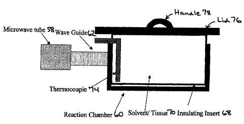
Figure 8A shows a cutaway top view and Figure 8B shows a cutaway side view of
an exemplary microwave unit. Microwave energy is transferred from the
microwave tube
58 to the reaction chamber 60 by the waveguide 62. Interlocks 64 ensure that
the micro-
wave unit will not operate while open and alignment pins 66 ensure that the
unit is closed.
An insulating insert 68 surrounds the contents 70 of the reaction chamber 60
to reduce
heat loss. An agitator 72 and a thermocouple 74 is shown projecting into the
reaction
chamber 60. The lid 76 must be removed (e.g., by a robot arm lifting the
handle 78) prior
to grabbing a basket containing tissue specimens (not shown) and placing it
into or taking
it out of the reaction chamber 60.
A more detailed view of the reaction chamber 60 of the exemplary microwave
unit
is shown in Figure 8C. The microwave unit is alternatively called a MW retort
80 because
the reaction chamber 60 is isolated from the environment, but a vacuum is not
required
for hardening the tissue specimen. Reagent ports 82 may be used to transfer
solutions into
and out of the reaction chamber 60, or may be used to as an air port 84. A
welded '/4-inch
socket provides a seal between the insulating insert 68 and the MW retort 80.
The solution
level can be visualized through an external sight tube 86 connected to the
interior of the
2 0- reaction chamber 60. A proximity switch 88 serves as a level sensor.
Electrical components of the exemplary microwave unit are shown in Figure 9.
Control of the temperature of the contents of the reaction chamber 60 is shown
in Figure
10. The temperature controller 90 is programmed with the desired temperature.
A control
signal 92 is sent to the microwave unit to apply power 94 to the microwave
source 58,
which microwave energy is transmitted by the waveguide 62 to the reaction
chamber 60.
The thermocouple 74 senses the temperature of the contents of the reaction
chamber 60
and is fed back to the temperature controller 90. An algorithm or other
program in the
temperature controller 90 then adjusts the control signal 92 to make the
sensed tempera-
ture approximate'ly equal to the desired temperature.
The system for tissue processing may be comprised of a physically linked
series of
modules (e.g., reaction chambers with or without an operably linked microwave
unit) to
26
CA 02394194 2002-06-12
WO 01/44783 PCT/US00/33760
accomplish a combination of fixation, dehydration, defatting, clearing, and/or
impregna-
tion of a tissue specimen. The system may be comprised of one module or a
plurality of
them. Each module would constitute a part of the entire processing cycle, but
an indivi-
dual module may accomplish more than one of the steps of tissue processing
(i.e., fixa-
tion, dehydration, defatting, clearing, and impregnation) because of the
chemical compo-
sition contained therein. A recorder may be included to receive measurements
of reaction
conditions in at least one module and other performance characteristics of the
system
(e.g., amount of chemical in a module, time spent by a tissue specimen within
a module or
in contact with a chemical), and to store the measurements for retrieval by
the operator.
The modules may occupy the same space and/or the tissue specimen may remain
stationary. Microwave or thermal energy may be regulated and transmitted into
the same
space, or onto the stationary tissue specimen at different times in the
process. Chemical
solutions and/or vapors may be moved into or out of the same space, or brought
into or
out of contact with the stationary tissue specimen. Preferred is minimizing
space require-
ments for the system by using one or two reaction chambers, and transporting
the different
chemical compositions into a reaction chamber by tubing or piping from
separate storage
and/or waste chambers. A controller can receive input from the reaction
chamber and/or
from timing that part of the processing cycle, and thereby regulate the
transport of the
different chemical compositions.
Alternatively, a plurality of modules containing at least four, five, or six
different
chemical compositions and to have at least one armature or track conveyance to
move the
tissue specimens among the modules may be provided. Thus, the system may
comprise at
least one, two, three, or four microwave units. In some embodiments, if a
tissue specimen
is transferred from one chemical composition to another with the same chemical
composi-
tion, it may be possible to combine parts of the processing cycle into the
same module
with an exchange of the chemical composition therein. Thus, certain parts of
the proces-
sing cycle may be combined and the number of different modules that are
required could
be reduced. Plumbing for fluid transfer may be simplified as compared to other
embodi-
ments previously described because the chemical composition may remain in the
reaction
chamber during the entire processing cycle and be moved into the reaction
chamber only
at the initiation of the cycle in a filling step, or out of the reaction
chamber at the termina-
27
CA 02394194 2008-10-22
WO 01/44783 PCT/US00/33760
tion of the cycle in an emptying step. The number of modules may also be
decreased to
one or two (e.g., only one microwave unit and one impregnation unit) by using
storage
chambers in fluid communication with the reaction chamber and that are
comprised of the
different chemical compositions, and moving them into and out of the
appropriate reac-
tion chamber as needed. For example, pumps and multi-position rotary valves
may be
used to control fluid movement between reservoir and reaction chamber by
pumping.
Controller circuitry may also be simplified if movement between modules occurs
in an
integral multiple of a common block of time. Movement of the tissue specimen
may be
controlled by a program. stored in memory such that the carrier or basket
loaded with
tissue specimens encounters modules in a particular order for set incubation
times. The
number of different modules, some of which may contain the same chemical
composition,
may be at least any integer from four to ten. Two, three, or four lines of
modules -may be
arranged for parallel processing.
An exemplary impregnator unit is illustrated in Figure 11 with a hot fluid
(e.g.,
water) passed through the heating coil 112 to maintain the impregnating agent
114 as a
liquid. Radiant heat may be provided by such coil within the interior (e.g.,
heating coil
112) of the reaction chamber 120 or an electrical wire wrapping its exterior
(not shown).
A lid 100 and gasket 102 covers the reaction chamber 120 and is displaced
using the
handle 104 before a basket (not shown) containing tissue specimens is grabbed.
Here, a
2 o hinge 106 shows how the lid 100 is attached to the reaction chamber 120.
Stainless steel
may be used as the exterior surface of the impregnator unit. An alignment ring
108 over
an insulating insert 110 ensures proper placement of the basket into the
reaction chamber
120. The insulating insert 110 (e.g., DELRINTM or other plastic material)
reduces heat loss
during impregnation of the tissue specimen. Reducing the pressure within the
reaction =
chamber 120 using the vacuum port 116 hastens impregnation. The lid 100 and
gasket
102 maintains the vacuum within the reaction chamber 120 after its evacuation.
During
transfer, the temperature of the contents of the reaction chamber 120 of the
impregnator
unit is maintained within about 2 C.
Either transferring different solutions into and out of the reaction chamber
or
transferring the basket among reaction chambers containing different solutions
may effect
changes in reaction steps. Holding the basket above the interior of the
reaction chamber
28
CA 02394194 2002-06-12
WO 01/44783 PCT/US00/33760
for about 10 seconds allows excess solution to drain back through one or more
openings
in the bottom and/or sides before the basket is transferred. Thus, the
sequence in which
the basket is transferred among reaction chambers, each containing a
particular composi-
tion of tissue processing chemicals, and the time the basket is incubated in
each reaction
chamber will dictate the series of chemical reactions necessary to accomplish
the process
according to the invention.
The lid can be removed; the gasket can be attached to the lid and moved with
it.
This process of removing the lid and gasket is performed for both the reaction
chamber
which initially contains the tissue specimens and the next reaction chamber
into which the
tissue specimens will be subsequently transferred. The basket is then removed,
allows
solution to drain from the basket and any cassettes which may be contained
therein back
into the reaction chamber for about 10 seconds, and transfers the basket to
the reaction
chamber containing the next chemical solution in the process. Finally, the
lids and gaskets
are replaced. The total time for such a transfer is about one minute.
For tissue processing, a plurality of modules containing at least four, five,
or six
different chemical compositions and to have at least one armature or track
conveyance to
move the tissue specimens among the modules may be used. The system will may
be
comprised of at least one, two, or three microwave units. In preferred
embodiments of the
invention, if a tissue specimen is transferred from one chemical composition
to another
with the same chemical composition, it may be possible to combine these parts
of the
processing cycle into the same module with an exchange of the chemical
composition
therein. Thus, certain parts of the processinb cycle may be combined and the
number of
different modules that are required could be reduced. Plumbing may be
simplified as
because, in many of the envisioned embodiments, the chemical composition may
remain
in the.reaction chamber during the entire processing cycle and be moved into
the reaction
chamber only at the initiation of the cycle in a filling step, or out of the
reaction chamber
at the termination of the cycle in a emptying step. Controller circuitry may
also be simpli-
fied if movement between modules occurs in an integral multiple of a common
block of
time. The number of different modules, some of which may contain identical
chemical
compositions, may be at least any integer from four to ten.
29
CA 02394194 2002-06-12
WO 01/44783 PCT/US00/33760
In accordance with the invention, variations on the above embodiments are envi-
sioned. Various configurations of the tissue processing system are possible,
and optional
modules may be connected to form a portion of the system. The specific
configuration
chosen may be dictated by the average number of specimens that will be
processed on a
daily basis by the clinical laboratory, and/or the speed with which histology
or pathology
reports must be prepared.
The system may incorporate a conventional microwave oven, the improved micro-
wave unit of the invention, or any combination thereof.
The system may be manually operated or automated. Manual operation is particu-
larly suited for research and development because variations in the process or
apparatus
may be quickly assessed. For automated instruments, tissue specimens may be
transported
by armature or track conveyance and/or chemical compositions may be
transferred by
corrosion-resistant plumbing. Thus, tissue processing may be automated by
moving tissue
specimens between stationary modules in a particular sequence for set times,
filling and
emptying modules of different chemicals such that stationary tissue specimens
are
incubated in a particular sequence for set times, or any combination thereof.
The armature conveyance may, for example, grab the specimen with a pincer-like
mechanism or catch the specimen with a hook-like device. The arm may be
articulated to
perform human-like motion; or may be mounted in a fixed coordinate rack with
linear or
two dimensional movement, and optionally another dimension of movement
provided by
varying the height of the arm over the system. The track conveyance may be
made from
resilient or tacky material to fix the specimen on the track by friction, or
there may be a
regular series of bumps or walls to trap the specimen therebetween. The track
may be
formed as a continuous belt or may be a series of belts that convey the tissue
specimen,
with the belt put into motion with a roller or sprocket mechanism. The
cassette or holder
may be adapted for conveyance by having a stem (with or without a knob) to be
grabbed
or a loop to be caught by the arm, or by fitting within a groove or
indentation in the track.
Similarly, the cassette or holder may be organized in a carrier or basket for
processing a
large number of specimens, the carrier or basket being adapted for transport
by the arma-
3 0 ture or track conveyance.
CA 02394194 2008-10-22
WO 01/41783 PCT/US00/33760
Electric motors and controllers may be used to transport a tissue specimen by
the
operator's real-time command or selection of a stored program. A simple
mechanism of
controlling the time spent by the tissue specimen in each module would be to
move the
tissue sample or holder thereof at a constant speed and to adjust the length
of the path
through each module to accommodate the intended incubation time.
The piping or flexible tubing, as well as other components of the plumbing,
should
resist corrosion by the chemicals used in tissue processing (e.g.,
polyethylene, polyvinyl
chloride, Teflon, stainless steel). Mechanical or electric pamps/valves and
controllers may
be used to move chemical compositions in any combination from storage chamber
to
reaction chamber, from reaction chamber to storage chamber if the composition
can be
reused, from reaction chamber to waste chamber if the composition is to be
flushed from
the system, to fill the storage chamber, and to flush the waste chamber by the
operator's
real-time conunand or selection of a stored program. Heating a combination of
plumbing
components may be necessary to maintain the chemical composition at reaction
temper-
ature or to ensure that the chemical composition (e.g., paraffin-containing)
is kept in a
transportable fluid state. In contrast, vapor seals and/or cooling may be
necessary to
isolate corrosive vapors from the mechanical and electrical components of the
system.
Specimens may be processed continuously and/or batchwise.
Safety considerations and precautions for an automated instrument (e.g., alarm
monitor, proximity sensor) can be incorporated into the system.
Furthermore the accessories, disposable parts (e.g., cassettes, mesh bags),
and rea-
gents that have been adapted for use in the system may also be considered as a
part of the
system. These specially designed instruments and apparatuses have also been
described in
. U.S. Patent No. 6,207,408. =
The present invention will have many advantages over conventional methods in
the areas of the practice of pathology, patient care, biomedical research, and
education.
The availability of microscopic diagnosis of tissue specimens within about one
to
six hours after receipt will allow rapid, or even real-time, clinical
interaction between
surgical intervention and pathological evaluation. For example, if 65 minutes
is taken to
process tissue, a stat diagnosis may be given in about two hours. This may
bring about
31
CA 02394194 2008-10-22
WO 01/44783 PCT/USOO/33760
significant improvements in patient care by eliminating or reducing to a
minimum patient
anxiety during the wait for diagnosis of disease, prognosis, and planning for
treatment
Consequently, there will be a drastic reordering of the workflow in pathology
lab-
oratories. Clinical laboratory space, pathological expertise, and clerical and
technical per-
sonnel will be utiiized more efficiently. Continuous workflow will improve
accessibility
and re.sponsiveness of pathologists who process and evaluate specimens, reduce
the
number of pathologists needed to process and evaluate specimens, and may also
improve
medical education, particularly the accessibility and responsiveness of
residency
Programs=
The smaller volume of reagents will also result in cost savings. Elimination
of for-
maldehyde and xylene, and the diminished requirement for other bazardous
chemicals,
will provide benefits to the environment and increased safety in the
laboratory. The costs
involved in handling and disposal of hazardous chemicals will be reduced.
Standardization of tissue fixation and processing procedures will ease
comparison
of specimens from different laboratories. Artifacts in histology due to the
use of formal-
dehyde and/or prolonged processing will be eliminated; thus, allowing more
precise eval-
uation of microscopic morphology of normal and diseased tissues. Similarly,
antigen
retrieval and staining will be improved. For genetic analysis, formaldehyde-
induced DNA
mutations will be eliminated and extraction of nucleic acid from archival
material may be
enhanced. The feasibility of RNA studies from stored, fixed paraffin-embedded
tissue
opens unliauted avenues for diagnostic and research applications.
The following examples are meant to be illustrative of the present invention,
but =
the practice of the invention is not limited or restricted in any way by them.
N.B. Energy
Beam Sciences' tissue microwave processors are examples of conventional
microwave
ovens that are available for commercial use.
32
CA 02394194 2002-06-12
WO 01/44783 PCT/US00/33760
EXAMPLES
EXAMPLE 1
Thick tissues were sliced to a maximum of 2 mm, preferably 1.5 mm or less. Two
mm thick or thinner slices, or small biopsies of fresh or previously fixed
tissue were held
in tissue cassettes and placed in a non-aqueous first solution of:
40% isopropyl alcohol,
40% acetone,
20% polyethylene glycol (average molecular weight 300), and
1% dimethyl sulfoxide (DMSO) (i.e., 10 ml per liter of the above mixture).
Tissues specimens were incubated for 15 minutes at a glycerin bath temperature
between 45 C and 50 C. The 400 ml solution for fixation was placed in a 500 ml
beaker
in a water bath shaker (linear displacement of 5 cm/sec). Additional agitation
of the fixa-
tion solution was provided by bubbling with an air pump.
Fixation, dehydration, fat removal, clearing, and impregnation are
accomplished
by sequential exposure of the tissue specimen to three different solutions
(i.e., the second,
third, and fourth solutions described below), one in each of three microwave
ovens from
Energy Beam Sciences. A one liter solution of 70% isopropyl alcohol and 30%
polyethy-
lene glycol (average molecular weight 300) is placed in the first oven (model
H2800) in a
1500 ml beaker, the solution in the second oven (model H2800) consists of one
liter of
70% isopropyl alcohol and 30% xylene in a 1500 ml beaker, and the third oven
(model
H2500) contains a solution of 1000 ml of xylene and 300 gm of paraffin in a
1500 ml
beaker. Ten ml of DMSO per liter are added to these three solutions. Heating
at 60 C by
microwave radiation is effected for 15 minutes in the first oven, and 5
minutes each in the
second and third ovens (75% power setting with a cycle of 2 seconds).
To continue paraffin impregnation after completion of the microwave radiation
steps, tissue sections were incubated in four 500 ml baths of molten paraffm
placed within
a large dessicator filled with paraffin, and resting in a glycerin bath at 75
C. Tissue sec-
tions were transferred from one paraffm bath to the next at 3 minute
intervals, for a total
impregnation time of 12 minutes. Each 3 minute interval was measured from the
time that
the pressure reading is about 640 mm of Hg..No agitation was used in this
step.
33
CA 02394194 2002-06-12
WO 01/44783 PCT/US00/33760
EXAMPLE 2
Fixation, dehydration, fat removal, and paraffin impregnation of fresh or
fixed
tissue sections, approximately 1 mm thick, was accomplished in 40 minutes by
exposing
these tissue sections to four successive steps as follows.
Step 1.
In this example, the first solution consisted of:
60% isopropyl alcohol,
10% acetone,
30% polyethylene glycol (average molecular weight 300), and
dimethyl sulfoxide (DMSO) added at an approximate concentration of 1% of the
total volume. One liter of this solution suffices to fix 60 specimens of
tissue held in tissue
cassettes. The specimens were incubated at 55 C in a commercial tissue
microwave
processor (H2500 or H2800, Energy Beam Sciences) for 5 min each in a series of
three
baths containing the first solution (15 min total incubation); agitation of
the solution was
obtained by bubbling to accelerate solution exchange.
Step 2.
The specimens were incubated in a solution of 70% isopropyl alcohol, 30% ace-
tone, and DMSO added at an approximate concentration of 1% at 60 C. Specimens
were
heated in a commercial tissue microwave processor (H2800, Energy Beam
Sciences) for 5
min each in two beakers containing the solution (10 min total incubation),
which were
agitated by bubbling.
Step 3.
Following microwave irradiation, impregnation was initiated by incubation in a
wax solution of 25% mineral oil and 75% molten paraffin placed in a large
dessicator
resting in a 60 C or 70 C glycerin bath, under a vacuum of about 200 mm of Hg,
for 5
min. Paraffin was degassed prior to use as described in Example 1.
Step 4.
Impregnation was completed by incubation in four baths of molten paraffin
placed
within a large dessicator resting in a glycerin bath at 75 C. Tissue sections
were transfer-
red from one paraffin bath to the next at 3 min intervals, for a total
impregnation time of
34
CA 02394194 2002-06-12
WO 01/44783 PCT/US00/33760
12 min. Each 3 min interval was measured for the time that the pressure
reading is about
640 mm of Hg.
In this example, 6 ml of a color indicator stock solution (10 gm methylene
blue in
1000 ml of isopropyl alcohol) was added to each of the solutions of isopropyl
alcohol and
acetone. Tissue specimens acquire a blue tint that facilitates their handling
during impreg-
nation and handling; penetration of the tissue specimen may also be monitored
by obser-
vation of an even blue color throughout the tissue specimen.
EXAMPLE 3
Fixation, dehydration, fat removal, and paraffin impregnation of fresh or
fixed
tissue sections, up to about 1 to 2 mm thick, were accomplished in about 65
minutes as
follows. Sections of 1.5 mm or less are preferred for consistency.
Step 1.
In this example, the first solution consists of:
40% isopropyl alcohol,
40% acetone,
20% polyethylene glycol (average molecular weight 300),
glacial acetic acid added at an approximate concentration of 0.5% of the total
volume, and
dimethyl sulfoxide (DMSO) added at an approximate concentration of 1% of the
total volume. One liter of this solution suffices to fix 60 specimens of
tissue held in tissue
cassettes. The specimens are incubated at 65 C in a commercial tissue
microwave proces-
sor (H2500 or H2800, Energy Beam Sciences) for 15 min in a 1500 ml beaker
containing
the first solution; agitation of the solution is obtained by bubbling to
accelerate solution
exchange.
Step 2.
The specimens are incubated in a solution of 55% isopropyl alcohol, 25%
acetone,
10% polyethylene glycol (average molecular weight 300), 10% low viscosity
mineral oil,
glacial acetic acid added at an approximate concentration of 0.5% of the total
volume, and
DMSO added at an approximate concentration of 1%. Specimens are heated at 65 C
in a
CA 02394194 2002-06-12
WO 01/44783 PCT/US00/33760
commercial tissue microwave processor (H2800, Energy Beam Sciences) for 15
minutes
in a 1500 ml beaker containing the solution, which is agitated by bubbling.
Step 3.
The specimens are incubated in a solution of 55% isopropyl alcohol, 25%
acetone,
20% low viscosity mineral oil, glacial acetic acid added at an approximate
concentration
of 0.5% of the total volume, and DMSO added at an approximate concentration of
1% of
the total volume. Specimens are heated at 65 C in a commercial tissue
microwave proces-
sor (H2800, Energy Beam Sciences ) for 5 minutes in a 1500 ml beaker
containing the
solution, which is agitated by bubbling.
1 o Step 4.
Following microwave irradiation, impregnation is initiated by incubation in
two
baths of a wax solution of 30% low viscosity mineral oil and 70% molten
paraffin placed
in a large dessicator resting in a 60 C glycerin bath, under a vacuum of about
640 mm of
Hg, for 5 minutes in each bath.
Step 5.
Impregnation is completed by incubation in four baths of molten paraffin
placed
within a large dessicator resting in a glycerin bath at about 75 C to 80 C and
a reduced
pressure of about 640 mm of Hg, for 5 minutes each. Tissue specimens were
transferred
from one paraffm bath to the next at 5 minute intervals, for a total
impregnation time of
2 0 20 minutes. Each 5 minute interval was measured for the time that the
pressure reading is
about 640 mm of Hg.
EXAMPLE 4
Tissue processing may be performed in the following manner using the system
illustrated in Figure 12. Fluid levels in the reservoirs are checked, retorts
are cleaned, and
plumbing is flushed prior to operation. Vacuum is drawn and air pressure is
raised to
transfer solutions and, if needed, provide agitation of solutions within the
retort by P/V
cycling. Only impregnation in the vacuum retort requires a reduction in the
pressure
because tissue processing in the microwave retort (e.g., hardening and initial
impregna-
tion) is done at atmospheric pressure. Solutions and retorts are warmed to
appropriate
operating temperatures.
36
CA 02394194 2002-06-12
WO 01/44783 PCT/US00/33760
A basket containing samples in cassettes is loaded. If the system illustrated
in
Figure 13 is being used, then tissue processing can be performed in parallel
because each
series of retorts can be accessed independently. An arm or track moves the
loaded basket
from the loading station to the microwave retort, and then to the vacuum
retort. Retorts
may resemble the reaction chamber shown in Figures 8 and 11 for hardening and
impreg-
nation, respectively: no beaker insert is used and the lid is attached to the
rest of the reac-
tion chamber by a hinge. The lid can be moved aside (e.g., by grabbing a
handle on the
top of the lid) to open the reaction chamber. This allows access to the
interior without
having to rest the lid at a holding station. Finally, when tissue impregnation
is completed,
the loaded basket is moved from the vacuum retort to an unloading station
containing
molten paraffm. The time required to transfer the basket between stations is
less than
about 10 seconds. The tissue cassettes can then be unloaded from the basket.
The reaction chamber containing an impregnation agent (e.g., mineral oil, wax)
may be heated using a common heating source. Alternatively, -a heater
maintains the tem-
perature of water circulating in tubing in contact with the impregnation agent
to keep it in
a molten state. The hot water can be circulated to each station for which it
is needed; each
reaction chamber can be attached to a supply and return manifold. For example,
a coil of
tubing can be located inside the reaction chamber; this heating coil would
then transfer
heat to the contents. Preferably the heating coil is eliminated by wrapping
the outside wall
of the reaction chamber with electrical wire that conducts heat through the
walls into the
contents of the reaction chamber.
The process described in Example 3 may be used in this system. Each different
solution is stored in one of three reservoirs and can be transferred to or
from the retort.
For example, a three-position rotary valve can select the appropriate
reservoir for that step
and pumping at nominal pressures of 250 mm Hg can transfer solution into the
retort
while 0.35 Kg/cm2 can transfer solution out of the retort; agitation can be
performed by
P/V cycles of nominal pressure 0.35 Kg/cm2 and 500 mm Hg vacuum. The
connections
(e.g., flexible tubing) between reservoirs and retorts, and the port where the
connection
joins each reservoir or retort, are not shown. Other conditions (e.g., times
or temperatures
for each step) are as described in Example 3.
37
CA 02394194 2002-06-12
WO 01/44783 PCT/US00/33760
EXAMPLE 5: Detection of Antigen in Tissue Sections
Paraffin sections are cut on a microtome to a thickness of 3 microns, placed
in a
water bath, and floated onto a glass slide. Paraffin was melted by placing
slides in either a
58 C oven for 30 minutes, or preferably in a 37 C oven for approximately 18
hours or
overnight, and then dewaxed in a xylene bath for 10 minutes. Slides were
rehydrated in
decreasing ethanol solutions for 1 min each (two baths of absolute, two baths
of 95%, and
one bath of 90%) and rinsed by submerging in tap water for 2 minutes.
Endogenous peroxidase was blocked with a solution of 6% hydrogen peroxide
(H202) and methanol, or 35 ml of 6% H202 with 140 ml of methanol, incubated
for 15
minutes. Slides were rinsed by submerging in tap water for 2 minutes and PBS
for 2
minutes, then dried.
Slides were transferred to a humidity chamber and normal horse serum (NHS) was
added to block for 10 minutes. Excess normal horse serum was decanted from
slides, and
specific primary antibody was incubated for 30 minutes on the tissue section
in a humidity
chamber at room temperature. Slides were flushed with PBS with back and forth
motion
using a squeeze bottle, submerged in a PBS bath for 2 minutes, and excess PBS
was dried
off each slide. Linking solution (also known as secondary antibody or
biotinylated anti-
rabbit or anti-mouse) was added to each tissue section and incubated for 25
minutes in a
humidity chamber at room temperature. Such rabbit, rat, and mouse secondary
antibodies
(e.g., anti-IgM, anti-IgG) were obtained from Dako (Carpinteria, CA) and used
at a dilu-
tion of about 1:600. Slides were flushed with PBS using a squeeze bottle,
submerged in a
PBS bath for 2 minutes, and excess PBS was dried off each slide.
Signal was developed according to the manufacturer's instructions (Vector Labo-
ratories). Avidin-biotin complex (ABC) solution was added to the tissue
section and incu-
bated for 25 minutes in humidity chamber. Slides were flushed with PBS in a
squeeze
bottle and submerged in a rack in a PBS bath for 2 minutes. The rack was
submerged in a
bath of diaminobenzidine (DAB) chromogen for 6 minutes, then submerged under
run-
ning water to wash gently for 4 minutes. Tissue sections were counterstained
with hema-
toxylin (staining time will depend on the age of the hematoxylin) from about
15 seconds
to 90 seconds at room temperature. Slides were washed under running water for
3 minutes
38
CA 02394194 2002-06-12
WO 01/44783 PCT/US00/33760
to remove excess counterstain, dehydrated in alcohol baths (about 10 seconds
in each)
from 85% to 100%, cleaned in xylene, and coverslipped.
Better antigen reactivity has been shown for progesterone receptor, factor
VIII-
related antigen, CD-3 1, CD-68, cytokeratin-7, chromogranin, and smooth muscle
antigen,
probably because of better preservation of antigen (e.g., greater signal-to-
noise ratio).
Table 1.
Reagents Catalog # Source
Microscope slides - snow coat X-TRA 00206 Surgipath
Elite ABC Kit (standard) PK-6100 Vector Labs.
Biotinylated anti-mouse IgG (H&L) BA-2000 Vector Labs.
Biotinylated anti-mouse IgM (H&L) BA-2020 Vector Labs.
Biotinylated anti-mouse/anti-rabbit IgG (H&L) BA-6000 Vector Labs.
Normal horse serum (NHS) S-2000 Vector Labs.
Diaminobenzidine tetrahydrochloride K3466 DAKO Corp.
Potassium phosphate (monobasic) 7100-500 NY Baxter Scientific
Sodium phosphate (dibasic) 7917-2.5 NY Baxter Scientific
Sodium chloride (AR Crystals) 7581-2.5 NY Baxter Scientific
30% Hydrogen peroxide 5240-500 NY Baxter Scientific
Xylene 8644-20 NY Baxter Scientific
Harris hematoxylin S-7735-3 Baxter Scientific
Methyl alcohol 3016-20 NY Baxter Scientific
95% Alcohol Florida Distillers
Absolute Ethyl Alcohol Florida Distillers
39
CA 02394194 2002-06-12
WO 01/44783 PCT/US00/33760
Table 2. Antibodies, Dilutions and Incubation Times
Rabbit (R) Microwave (M) 30' Incubation
Mouse (MIgG) Trypsin (T) 45' Incubation
Mouse (MIgM) Protease (P) 90' Incubation
Goat (G) Fast Green (FG)
Table 3. Antibodies, Dilutions and Incubation Times
Abbrev. Antibody Special Incub. Linking
Procedure Time Sol.
(ACTH) Adrenocorticotropin Hormone 1:2000 30' R
(AACT) Alpha-1 Antichymotrypsin 1:50000 30' R
(AAT) Alpha-1 Antitrypsin 1:2000 30' R
(ADENO) Adenoviurs 1:1000 30' MIgG
(AFP) Alpha Fetoprotein 1:2500 30' R
(AEI/3) Cytokeratin 1:200(M) 45' MIgG
(ALA) Alpha Lactalbumin 1:600 30' R
(ACTIN) Actin Muscle 1:200 30' MIgG
(APP-A4) Anti-Alzheimer Precursor Protein 1:500(M) 45' MIgG
A4
(ASPE) Aspergillus 1:500 30' R
(AR) Androgen Receptor 1:20(M) 45' MIgG
(FG)
(BCA) B-Cell 1:200 30' MIgG
(bcl-2) Anti-Human Oncoprotein 1:100(M) 45' MIgG
(BerEp4) Human Epithelial Antigen 1:25 30' MIgG
(B72.3) TAG72 Tumor-Associated 1:100 30' MIgG
Glycoprotein 72
(BLA36) B Lymphocyte Antigen 1:100 30' MIgG
(CMV) Cytomegalovirus 1:50(P) 30' MIgG
(CHRG) Chromogranin 1:50 30' MIgG
(CALC) Calcitonin 1:2000 30' R
(CEA) Carcinoembryonic Antigen 1:6000 30' R
(CERb'B2) c-erbB-2 Oncogene Mabl 1:1500 90' R
CA 02394194 2002-06-12
WO 01/44783 PCT/US00/33760
Table 3 (continued). Antibodies, Dilutions and Incubation Times
Abbrev. Antibody Special Incub. Linking
Procedure Time Sol.
(CATH) Cathepsin D 1:2000(M) 45' R
(CAM 5.2) Cytokeratin 1:500(M) 45' R
(CK 7) Cytokeratin 1:200(M) 45' MIgG
(CK 20) Cytokeratin 1:25(M) 45' MIgG
(COLL IV) Collagen IV 1:25(P) 30' MIgG
(CA 125) Anti-Human CA 125 (MII) 1:20(M) 45' MIgG
(CD 30) Anti-Human Ki-1 Antigen 1:200(M) 45' MIgG
(BER-H2)
(ER) Estrogen Receptor 1:50(M)(FG) 45' MIgM
(FVIII) Von Willebrand Factor 1:50(P) 30' MIgM
(FSH) Follicle Stimulating Hormone 1:3000 30' R
(5 HT) Serotonin 1:50 30' MIgM
(FXIII) Anti-coagulation Factor 1:1200 30' R
(GAST) Gastrin 1:2000 30' MIgM
(GFAP) Glial Fibrillary Acidic Protein 1:1500 30' R
(GLUC) Glucagon 1:10000 30' R
(GH) Growth Hormone 1:5000 30' R
(GCDFP) Gross Cystic Disease Fluid 1:250 30' MIgM
Protein
(GRP) Gastrin-Releasing Peptide 1:1000 30' R
(HMWK) High Molecular Weight 1:10 45' MIgM
Keratin (34DE12)
(Hbcore) Hepatitis B Core Antigen 1:5000 30' R
(HBsAg) Hepatitis B Surface Antigen 1:100 30' MIgM
(HSV I) Herpes Simplex Type I 1:10 30' R
(HSV II) Herpes Simplex Type II 1:10 30' R
(HCG) Human Chorionic 1:50000 30' R
Gonadotropin
(HPL) Human Placental Lactogen 1:100000 30' R
41
CA 02394194 2002-06-12
WO 01/44783 PCT/US00/33760
Table 3 (continued). Antibodies, Dilutions and Incubation Times
Abbrev. Antibody Special Incub. Linking
Procedure Time Sol.
(HIST) Histoplasma 1:1000 30' R
(H.Pyl) Heliobacter pylori 1:500(M) 45' R
(P-HCG) (3-Human Chorionic 1:10000 30' R
Gonadotropin
(IgA) Alpha Heavy Chain 1:400 30' R
(IgG) Gamma Heavy Chain 1:1000 30' R
(IgAs) Secretory Piece of IgA 1:200 30' R
(IgM) Mu Heavy Chain IgM 1:1000 30' R
(INS) Insulin 1:100 30' R
(Ki-67) Nuclear Antigen MIB-1 1:50(M)(FG) 45' MIgG
(K) Kappa Light Chain 1:200(M) 45' MIgG
(KERATIN) AE1/3 CAM 1:50/1:500(M) 45' MIgG
(LCA) Leucocyte Common Antigen 1:50 30' MIgG
(Leu M1) Leu M1 Antigen 1:200(M) 45' MIgM
(Leu 7) Leu 7 Antigen 1:50(M) 45' MIgM
(Lectin) Lectin 1:4000 USE INSTEAD
OF NHS
(Anti-Lectin) Anti-Lectin Antigen 1:10000 30' G
(LEA 135) Anti-Human Luminal 1:50 30' MIgG
Epithelial Antigen
(LH) Luteinizing Hormone 1:3000 30' R
(L) Lambda Light Chain 1:6000(M) 45' MIgG
(LMK-8) Low Molecular Weight 1:25(M) 45' MIgG
Keratin
(LIP-AS 105) Lipase 1:400 30' MIgG
(MCA) Myeloid Histiocyte Antigen 1:400(M) 45' MIgG
(MAC 387)
(MUR) Muramidase 1:2000 30' R
(MYOGL) Myoglobin 1:5000 30' R
(MAPH) Macrophage 1:50 30' MIgG
42
CA 02394194 2002-06-12
WO 01/44783 PCT/US00/33760
Table 3 (continued). Antibodies, Dilutions and Incubation Times
Abbrev. Antibody Special Incub. Linking
Procedure Time Sol.
(MTLT) Metallothionein 1:50 30' MIgG
(MEL) Melanoma HMB 45 1:50 30' MIgG
(MAK 6) Anti-Cytokeratin 1:50(T) 90' MIgG
(MBP) Myelin 1:500 30' R
(MESO) Mesothelial Antigen 1:500 30' MIgM
(MAST-C) Mast Cell 1:2000(T) 30' MIgG
(MPO) Myeloperoxidase 1:5000 30' R
(MGN) Myogenin 1:15 45' MIgG
(NB) Neuroblastoma 1:200 90' MIgG
(N-FIL) N-Filament (2F11) 1:250 30' MIgG
(NSE) Neuron Specific Enolase 1:4000(M) 45' MIgG
(PAMYL) Pancreatic Amylase 1:20 30' MIgG
(PCP) Pneumocystis carinii 1:25 30' MIgM
(PLAP) Placental Alkaline 1:800 30' R
Phosphatase
(PPP) Pancreatic Polypeptide 1:3000 30' R
(PTH) Parathyroid Hormone 1:250(M) 45' (RAT)
(PROL) Prolactin 1:500 30' R
(PAPH) Prostatic Acid Phosphatase 1:4000 30' R
(PML)(SV40) Progressive Multifocal 1:10000 30' R
Leucoencephalopathy
(PR) Progesterone Receptor 1:100(M) 45' R
(PR 1A6) Progesterone Receptor 1:50(M) 45' MIgG
(PSA) Prostate Specific Antigen 1:750 30' R
(PCNA) Proliferating Cell Nuclear 1:1 oo(M)(FG) 45' MIgG
(PS2) PS2 Protein 1:1000 45' R
(P53) p53 Antigen 1:50(M)(FG) 45' MIgG
43
CA 02394194 2008-10-22
WO 01/44783 PCT/USOO/33760
Table 3 (continued). Antibodies, Dilutions and Incubation Times
Abbrt,w. Antibody Special Incub. Linking
Procedure Time Sol.
(S 100 A) S 100 A Protein 1:3000 30' R
(S 100) S 100 Protein 1:2000 30' R
(SOMAT) Somatostatin 1:3000 30' R
(SYNAP) Synaptopbysin 1:800(M) 45' R
(SMA) Smooth Muscle Actin 1:100 30' MIgG
(ocSR-1) Sarcomeric Actin 1:100 30' MIgG
(TESTOS) Testosterone 1:250 30' R
(TGB) Thyroglobulin 120000 30' R
(TP-103) Treponema 1:50(T) 30' MIgG
(TM) Thrombomodulin 1:50 30' MIgG
(TSH) Thyroid Stimulating Hormone 1:2000 30' R
(TCA) T-Cell Antigen 1:800(M) 45' MIgG
(TOXO) Toxoplasma 1:1000 30' R
(UBT) Ubiquitin 1:250 30' R
(VIP) Vasoactive intestinal peptide 1:1500 30' R
(VIM) Vimentin 1:800(M) 45' 11NIIgG
(VZV) Variecella-Zoster Virus 1:100 30' MIgG
(WSKER) W-ide Spectrum Keratin 1:500 30' R
F.XAIvIPLE 6: DNA Extraction from Processed Tissue Sections
5- Two six micron tissue sections were placed in a 1.5 ml microfuge tube, 800
l
xylene was added and mixed by vortexing, 400 l absolute ethanol was added and
mixed
by vortexing, the tube was centrifuged for 5 minutes in a high speed
microfuge, and the
supernatant was decanted. To the pellet, 800 0 absolute ethanol was added and
mixed by
vortexing.
The supernatant was decanted after centrifugation as above, and 100 l of a
deter-
gent/proteinase K solution (1% NP40 or Triton X-100TM, 2.4 I of 2.5 mg/ml
proteinase K)
was added to the pellet and incubated at 55 C for one hour. Proteinase K was
inactivated
44
CA 02394194 2008-10-22
WO 01/11783 PCT/US00/33760
by incubation at 95 C for 10 minutes. Save the supernatant containing DNA
after centrifu-
gation in the microfuge for 5 minutes. This material is ready for PCR. It
should be preci-
pitated and/or extracted further if Southern blotting is planned. More
sections would be
required to obtain enough DNA for restriction analysis.
.5
EXAMPLE 7: RNA Extraction from Processed Tissue Sections
Ten sections (7 m each) of a paraffin block were cut using disposable blades.
The
blocks were prepared aceording to the present invention and by the
conventional proce-
dure. They were placcd in 50 mi Falcon tubes, depataffiniud with 20 ml of
xylene, and
the remaining tissue was then washed twice with absolute alcohol for 30
minutes. The
tissue was suspended at 0.5 gm/ml in a solution containing 4M guanidinium
thiocyanate,
25 mM Na citrate pH 7.0, 0.5% 1V laurylsarcosine, and 0.1 M of 2-
mercaptoethanol. The
solution was mixed by vortexing and DNA was sheared by passage through an 18
to 22
gauge syringe needle.
The RNA-containing solution was carefully layered on 2.8 ml of 5.7 M CsCI in
several 5 ml centrifuge tubes (SorvallTM), and RNA was sedimented by
centrifugation in an
SW55Ti rotor at 35,000 rpm -and 18 C for 14 hours in a Beckman L8-53
ultracentrifuge.
The top fraction was carefully removed to leave an RNA pellet at the bottom of
the tube.
The pellet was resuspended with n'bonuclease-free water, and the Eppendorf
tube was
spun at 14,000 rpm for 10 minutes. The supernatant containing RNA was saved
and the
ultraviolet (UV) absorbance was measured: an extinction coefficient of 1
OD2so%m is
estimated to be the equivalent of about 40 911111 RNA and the OD26/OI>no
ratio should
be between about 1.8 and about 2Ø A total of 45 pg RNA was extracted from
tissue
specimens prepared according to the present invention whereas no RNA was
detectable
from tissue specimens processed conventionally.
While the invention has been described in connection with what is presently
consi-
dered to be practical and preferred embodiments, it is understood that the
present inven-
tion is not to be limited to the disclosed embodiments but, on the contrary,
is intended to
cover various modifications and equivalent arrangements included within the
spirit and
scope of the appended claims. Thus, it is to be understood that variations in
the described
invention will be obvious to those skilled in the art without departing from
the novel
CA 02394194 2002-06-12
WO 01/44783 PCT/US00/33760
aspects of the present invention and such variations are intended to come
within the scope
of the claims below.
Furthermore, it should be understood that an element contained in this specifi-
cation should not be construed as a limitation of the claimed invention unless
explicitly
recited in the claims. Thus, the claims are the basis for determining the
scope of legal
protection granted instead of using the specification to import a limitation
which is not
explicitly recited in the claims.
46