Note: Descriptions are shown in the official language in which they were submitted.
CA 02395203 2002-07-02
WO 01/49181 PCT/CA01/00008
OPHTHALMOLOGICAL ULTRASONOGRAPHY
SCANNING APPARATUS
FIELD OF THE INVENTION
The invention is in the field of medical ultrasound apparatus, particularly
apparatus for
use in ultrasonography of the eye.
BACKGROUND OF THE INVENTION
Ultrasound may be used in a variety of medical applications, including
diagnostic
ultrasonography of the eye. Diagnostic information is typically provided by an
ultrasound
pulse from a piezoelectric transducer, which is directed into a tissue.
Reflected acoustic energy
is detected (as 'echoes'), so that the amplitude of the received energy may be
correlated with
the time delay in receipt of the echo. The amplitude of the echo signal is
proportional to the
scattering strength of the refractors in the tissue, and the time delay is
proportional to the range
of the refractors from the transducer. A variety of hand-held ultrasound
instruments for
measuring corneal thickness (called pachymeters) have been developed (for
example see U.S.
Patent Nos. 4,564,018; 4,817,432; 4,930,512). Many prior art ultrasonic
pachymeters provide
A-scan output, in the form of waveforms displayed on a cathode ray tube,
representing
acoustic reflections in a single dimensional 'column' of tissue.
In B-scan ultrasonography, a two-dimensional image is formed, in which pixel
brightness reflects the amplitude of the reflected acoustic signal. A B-scan
image therefore
represents a cross-sectional slice of the imaged tissue. The cross-sectional
information is
typically provided by correlating information from a series of adjoining
columnar scans (each
of which may be used to produce A-scan output). For the purpose of producing B-
scans,
adjoining columnar scans may be produced by a number of methods: rectilinear
translocation
of a transducer over the tissue of interest; pivoting angular displacement of
a single transducer
over a fan-shaped area; or through the use of a linear array of transducers.
In some applications, three dimensional images may be reconstructed from a
series of
B-scans. U.S. Patent No. 4,932,414 to Coleman et al. for example describes a
system in which
the transducer is electronically swept or physically rotated to produce a
series of sectored (fan-
-1-
CA 02395203 2002-07-02
WO 01/49181 PCT/CA01/00008
shaped) scan planes which are separated by a known angular distance, to
produce a 3-
dimensional display. In a similar fashion, U.S. Patent No. 5,487,388 to Rello
et al. discloses
an ultrasonic scanning system in which sequential fan-shaped B-scan image
planes are
obtained by movement of the transducer probe in an arc, a movement which
allows the apex
of the scanned 3-dimensional volume to be located below the probe to
facilitate imaging
between closely-spaced surface obstructions.
The structure of the eye, particularly the cornea, presents special problems
for optimal
ultrasonographic B-scan imaging. The human cornea is an asphere, flattening
concentrically,
typically approximately 11 mm across with an average central radius of
curvature of 7.8mm
which increases towards the periphery. The high resolution required for
ultrasonic imaging of
some corneal structures is optimally achieved if ultrasound data is collected
from the focal
point of the transducer, and the ultrasound beam is normal to the suxface of
the cornea. As a
result, rectilinear scanning of the cornea provides optimal imaging
information only from
I 5 relatively small segments of the cornea which are normal to the transducer
beam and in the
plane of beam focus. Similarly, volumetric 3-dimensional scanning by
reconstruction of a
series of fan-shaped B-scan planes, as for example described in U.S. Patent
Nos. 4,932,414
and 5,487,388, is not a system adapted to provide the degree of resolution
required for
biometry of the corneal surface.
High frequency ultrasound has been used in ophthalmological ultrasonography to
obtain biometric B-scan images of the human cornea, by arcuate translocation
of a single
element focused transducer. Silverman et al., 1997, J. Ultrasound Med. I6: I
I7-124, describe a
system for sonographic imaging and biometry of the cornea in which a
sophisticated
programmable motion system permits ultrasonographic arc scanning. In the
Silverman et al.
system, the ultrasonic transducer is translated through an axc matched to the
approximate
radius of curvature of the cornea using fve servo motors and a controller.
Similarly, U.S.
Patent No. 5,331,962 discloses an ultrasound system for corneal arc scanning,
in which a
transducer is translocated along a curved track that approximates the surface
curvature of the
cornea.
-2-
CA 02395203 2002-07-02
WO 01/49181 PCT/CA01/00008
SUMMARY OF THE INVENTION
In one aspect of the invention, an apparatus for ultrasound scanning of the
eye is
provided comprising a virtual center translocation mechanism that facilitates
precise arcuate
motion of an ultrasonic transducer to maintain focal distance from the eye and
to maintain
normality of the ultrasound beam with surfaces of the eye. The arcuate
movement of the
transducer focal path may closely approximate the surface of the cornea. Some
embodiments
of the invention may include a radius adjust mechanism for changing the radius
of ultrasound
scanning, to accommodate different eye sizes and to facilitate positioning of
the ultrasound
transducer focal point on selected surfaces of the eye, such as the cornea.
Centration optics
may also be provided, for aligning the translocation path of the ultrasound
transducer with an
axis such as, but not limited to, the Purkinje axis of a patient's eye.
In one embodiment, the invention pxovides an ultrasound transducer support
comprising a transducer mount adapted to accommodate an ultrasound transducer
having a
focal point. The support may be provided with a virtual centre mechanism
attached to the
transducer mount, for moving the ultrasound transducer along an arcuate
translation path. The
arcuate translation path of the transducer may be offset from a virtual centre
of translocation
by a radius of transducer translocation, so that the focal point of the
ultrasound transducer
traverses an arcuate focal path about the virtual centre of translocation. A
radius adjust
mechanism may be provided for adjusting the position of the transducer mount
to change the
radius of transducer translocation.
In an alternative embodiment, the invention provides a method of
ophthamological
ultrasonography comprising centring an ultrasound transducer having a focal
point in
alignment with the Purkinje or other optical or geometric axis of a patient's
eye using
centration optics, and moving the ultrasound transducer along an arcuate
translation path
intersecting the Purkinj a or other optical or geometric axis of the patient's
eye. The arcuate
translation path of the transducer may be offset from a virtual centre of
translocation by a
radius of transducer translocation, so that the focal point of the ultrasound
transducer traverses
an arcuate focal path about the virtual centre of translocation.
-3-
CA 02395203 2002-07-02
WO 01/49181 PCT/CA01/00008
BRIEF DESCRIPTION OF THE DRAWINGS
Figure 1A is a side elevational view of an ultrasound transducer support of
the
invention, showing a cam-actuated radius adjust mechanism.
Figure 1B is an isometric view of an alternative embodiment of the ultrasound
transducer support of the invention, showing shaped arm linkages, as are also
shown in Figure
4.
Figure 1 C is a schematic diagram showing a linking element connecting the
front and
rear swinging linkages which may form part of the transducer part of the
invention.
Figure 2 is a schematic diagram showing the motion of the transducer support
of the
invention.
Figure 3A and 3B are elevations views of the embodiment of the invention shown
in
Figure 1, showing the cams that are part of the radius adjust system in
different positions.
Figure 4A is a side elevational view showing the ultrasound transducer support
of the
invention with accessory apparatus for sealing a fluid-filled chamber against
the patient's eye.
Figure 4B is a schematic illustration showing alternative optics which may be
used in
conjunction with methods of centering the transducer using the apparatus of
the invention.
Figure S is a schematic illustration of a series of meridional ultrasound
scanning paths
which intersect at a point near the apex of the cornea.
Figure 6 is an isometric view of a stage for the scanning apparatus of the
invention,
providing for rotational movement of the scanning apparatus, as well as
movement in X, Y
and Z axes.
-4-
CA 02395203 2002-07-02
WO 01/49181 PCT/CA01/00008
Figures 7 and 7A are cross-sectional side views showing a membrane which may
be
used in some embodiments to isolate a volume of fluid around a patient's eye.
Figure 8 is an elevational view showing a mechanical safety stop mechaiusm.
DETAILED DESCRIPTION OF THE INVENTION
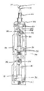
In one aspect, the invention provides an ultrasound transducer support
comprising a
transducer mount adapted to accommodate an ultrasound transducer, and a
virtual centre
mechanism. Figure 1A illustrates an embodiment of a virtual center mechanism.
First and
second arm linkages 65A and 65B are each connected via three pivots to moving
parts of the
mechanism. Rear swinging pivots 96 connect first and second arm linkages 65A
and 65B to
rear radius adjust slider 71, and rear radius adjust slider 71 is attached to
rear swinging linkage
81. Similarly, front swinging pivots 95 connect arm linkages 65A, 65B to front
radius adjust
slider 70, and front radius adjust slider 70 is attached to front swinging
linkage 80. The front
ends of the arm linkages 65A, 65B are connected by transducer pivots 60 to
transducer mount
55, and transducer mount 55 is adapted to accommodate ultrasonic transducer
50. Front pivot
85 and rear pivot 86 are stationary relative to the swinging motion of front
swinging linkage
80 and rear swinging linkage 81.
The virtual centre mechanism is attached to transducer mount 55 for moving the
ultrasound transducer 50 along an arcuate translation path 56 offset from a
virtual centre of
translocation 52 by a radius of transducer translocation, so that the focal
point 51 of the
ultrasound transducer 50 traverses an arcuate focal path 59 about virtual
centre of
translocation 52. As shown in Figure 2, when rear swinging linkage 81 rotates
about rear pivot
86, rear swinging pivots 96 describe arcuate paths about rear pivot 86. Arm
linlcages 65A, 65B
move front swinging pivots 95, so that front swinging pivots 95 describe
identical paths about
front pivot 85. Similar triangles 53 show that this swinging motion causes
ultrasonic
transducer 50 to move in an arc such that its axis pivots about virtual center
52. In addition,
transducer focus point 51 traverses an arc 59 about virtual center 52 at image
radius 54. The
pivoting motion of the apparatus may be driven by scanning driver 82, which
may for example
be a servo motor. It will be seen that focal point 51 may also lie behind
virtual center 52, for
example to scan the back of the eye.
-5-
CA 02395203 2002-07-02
WO 01/49181 PCT/CA01/00008
The mounting of transducer 50 in transducer mount 55 may be adapted so that
the
position of transducer 50 is adjustable relative to transducer mount 55. Such
an adjustment
may be difficult to accomplish during operation, due to the configuration of
the assembled
apparatus, as shown in Figure 4. A radius adjust mechanism for adjusting the
radius of
transducer translocation may be provided, for example by radius adjust sliders
70, 71 which
are movable relative to the respective pivot points 85, 86. In operation, the
effect of movement
of radius adjust sliders 70, 71 is to elongate similar triangles 53. The
elongation of triangles 53
reflects simultaneous changes to three radii: a'first' radius of rotation of
front swinging pivots
95; a'second' radius of rotation of rear swinging pivots 96, and the radius of
transducer
translocation circumscribed by transducer pivots 60. In addition, image radius
54 is changed
(the distance between virtual centre 52 and the arcuate focal path 59
traversed by the focal
point 51 of transducer 50). The radius adjustment may be driven by rotating
radius adjust cams
75, 76 relative to swinging linkages 80, 81. Radius adjust cams 75, 76 may be
linked by a
rotation linking mechanism, such as anti-backlash belt 90, which operates so
that adjusting
one cam automatically adjusts the other cam by the same amount. Alternatively,
a single cam
75 or 76 could be used on either slider 70 or 71, in which case the other
slider would follow.
Mechanisms other than cams, such as motors, gears, or other mechanical
linkages may be used
to actuate sliding movement of radius adjust sliders 70, 71.
To provide extra rigidity to the mechanism supplementary linking such as that
shown
in figure 1 C may be used. Linking element 203 may for example be a steel band
or a belt or a
chain or a cable and may engage sheaves 201 and 202. Alternatively the linking
may be
supplied many other ways including driving both swinging linkages 80 and 81
directly with
wormgears or flexures.
Ultrasonic transducers for use in accordance with various aspects of the
invention may
be high frequency transducers, operating for example at frequencies between 50
and 100 MHz.
A saline bath may be used to acoustically couple ultrasound transducer 50 to
patient's eye 105.
Figure 4A shows the general arrangement of an embodiment of the ultrasound
transducer
support of the invention with accessory apparatus including a saline bath
adapted for
diagnostic use. In the illustrated embodiment, a patient may be scanned in a
seated position by
-6-
CA 02395203 2002-07-02
WO 01/49181 PCT/CA01/00008
placing the patient's orbit against eye seal 15. The patient's head may be
supported by head
support 170 which may be adapted to immobilize the patient's head during
ultrasound
scanning. The overall axis of the apparatus, shown as line 25 in Figure 4, may
be at an angle
of about 45 degrees to horizontal. Alternative angles from horizontal to
vertical may also be
used. In some embodiments, a patient's mandible may be supported with an
upward force
which encourages the teeth into mechanical contact to stabilize the patient's
head. Arranging
the apparatus at an overall axis of 45 degrees may help to reduce the
accumulation of bubbles
in the vacinity of the patient's orbit, particularly when saline fluid fills
reservoir 20 and eye
seal 15.
Coarse alignment of the eye on axis 25 may be done visually, for example using
video
camera 140, which preferably has a very high sensitivity. The seal may be
tested by slowly
filling the saline chamber with saline and watching for leaks. The position of
the patient's head
may be adjusted, or the eye seal changed, in order to achieve a good seal.
Once an acceptable
position has been found, the patient's head may be locked into position by
immobilizing the
head support. With the head stationary the scanning mechanism 10 can be moved
relative to
saline chamber 15 to make scan axis 25 coincident with the Purkinjie (or other
optical or
geometric) axis of the patient's eye.
In accordance with one aspect of the invention, corneal scanning may be
undertaken
along a series of meridional paths which intersect at a point near the apex of
the cornea, as
shown in Figure 5. In some embodiments, this intersection point may be the
Purkinje (or other
optical or geometric) axis of the eye, which may be used as an approximation
of the optical
axis of the eye (defined by the Iine between the object of regard and the
fovea of the retina).
The Purkinje axis may be located by shining a focused beam of light into the
patient's eye, and
examining the Purkinje reflections from four optical surfaces of the eye: the
front and rear
surfaces of the cornea, and the front and rear surfaces of the lens. The
Purkinje reflections are
observable along the axis of the Iight beam. The Purkinje axis is located when
the reflections
from these four surfaces are coincident. A light beam used to locate the
Purkinje axis may also
conveniently serve as a view target for the patient. Other axes may be used as
an intersection
point for meridional scanning such as the vertex-fixation axis. When a light
is shone axially
toward the eye onto the corneal surface, two reflected images can be seen -
the specular
CA 02395203 2002-07-02
WO 01/49181 PCT/CA01/00008
(Normal to incident light) reflection and the diffuse reflection (not
necessarily Normal
reflection). When the position of the light source is adjusted such that the
specular and diffuse
reflections from the corneal surface are coincident, the light source will now
be perpendicular
to the vertix of the cornea. The vertex fixation axis is obtained when the
patient's eye is
looking directly at a fixation target, while observing coincidence of the
diffuse and specular
corneal surface reflections.
Figure 4A shows an embodiment that includes accessory centration optics for
centering
the transducer in alignment with the Purkinje axis of the patient's eye.
Centration light source
120 may be refined using aperture 126 and focused using centration optics 125.
Centration
light source 120 may for example be a laser, laser diode, light emitting diode
or incandescent
source. The centration light beam may be aligned with machine axis 25 using
reflector 130,
such as a prism or mirror, and beam splitter 135. The centration light beam
then passes
through fluid-sealed camera window 136 and through the fluid (saline) in
cavity 175 before
reaching the patient's eye 105. As shown in figure 4B in order to address
potential back
reflection problems from window 136, both camera 140 and window 136 may be
tipped
relative to machine axis 25 in such a way that the centration beam still
travels along the
machine axis 25 within the saline chamber 175. The centration light beam
thereby intersects
the arcuate translation path of transducer 50. The Purkinje reflections then
return back through
beam splitter 135 and may be recorded by camera 140 through lens 145. As shown
in Figure 4,
in order for the light to reach the patient's eye 105, transducer 50 must be
swung over to the
side as shown in Figure 2. During an ultrasound scan, because the centration
light beam
intersects the arcuate translation path of transducer 50, the patient using
the centration light as
a view target will see the light disappear momentarily as the light is blocked
by the passing
transducer. This flashing behavior may be helpful in facilitating alignment of
the eye, since the
photoreceptors in the retina would otherwise saturate after a few seconds of
staring at a f red
target light which may cause the eye to shift slightly to compensate.
Figure 4A also illustrates focus point illuminator 155, which shines through
focus
point optics 160 and aperture 161 to produce a focus point spot on eye 105.
The angle of focus
point illuminator 155 is set so that when the focus point spot is
appropriately positioned on the
eye, the transducer apparatus is in a selected vertical position at a known
distance from eye
_g_
CA 02395203 2002-07-02
WO 01/49181 PCT/CA01/00008
105. The centration optics may for example be used to determined when the
focus point spot
joins the Purkinje (or other axis) reflections from the centration light 120.
In some
embodiments, this positioning of the focus point spot may be used to identify
the point at
which the apparatus of the invention is positioned at the correct distance
from the eye to have
the cornea within the focal point of transducer 50.
For extra illumination to improve the eye image on camera 140, an infra-red
light may
be shone through either of windows 136, 150, in which case the camera will be
adapted to be
sensitive to the wavelength selected.
In addition to the scanning motion shown in Figure 5, several other motions
may be
produced by the mechanism of the invention to scan an eye. In order to produce
various
meridian angles theta as shown on Figure 5, the scan mechanism I O may rotate
about the
machine axis 25 (shown in Figure 4). Rotational motion of the scanning
apparatus may be
accomplished using rotary table 210. Motion in the Z axis, which shifts the
mechanism toward
or away from the eye, may be used to compensate for the degree of insetting of
a patient's eye.
Motion in the Z axis may be accomplished using a Z-slide 215, which may be
motorized or
manually controllable. Motion along the X and Y axes, perpendicular to the
machine axis 25,
may be used to adjust the position of the ultrasound scanning apparatus once a
patient has
been positioned in front of the machine. These motions may be produced by X
slide 220 and Y
slide 225. In some embodiments, the X and Y slides may be motorized to
facilitate X and Y
motion of the scanning apparatus in planar scans of eye structures, such as
the iris plane.
These axes may of course be arranged differently than shown in figure 6 while
retaining the
same essential operation.
In order to provide a mechanical means of preventing the transducer from
approaching
an eye too closely, a safety stop as shown in figure ~ may be used. The
transducer may be
shifted closer to the eye by either a radius adjustment or Z axis adjustment.
A curved stop bar
212 may be fixed to the body of the Z axis stage 215. Stop pads 210 and 211
are fixed to
radius adjust slider 205 so that an excess motion of either the radius or Z
axes causes one of
the pads to touch the stop bar. These stop pads 210, 211 may be supplemented
with sensors
for operator feedback.
_9_
CA 02395203 2002-07-02
WO 01/49181 PCT/CA01/00008
In some embodiments, it may be desirable to provide a barrier to inhibit the
passage of
an infection from one patient to another. In some embodiments, it will be
necessary for the
centration light beam and the Purkinje (or other axis) reflections to pass
through such a barrier
without significant shifting or distortion. In one embodiment, membrane 180 as
shown in
Figure 7 may be used, which has saline fluid on both sides of it and is
selected to have a
similar index of refraction to saline so that light rays passing through
membrane 180 will be
affected very little by its presence. A filling and draining system may be
provided, as shown
by tube 181 in Figure 7. The outer edges of the membrane 180 may be draped
over the eye seal
and provide the sealing surface fox the face. Near its center membrane 180 may
be attached by
clamp 190 to transducer 50. Clamp 190 may be adapted to accommodate rotation
of
transducer 50 relative to the eye seal 15 during a scan, for example by
permitting rotational
movement of transducer 50 within clamp 190. Alternatively, membrane 180 may be
continuous, and adapted to permit transmission of ultrasonic vibrations
through the membrane
itself as shown in Figure 7A. In some embodiments, bellows seal 173 may be
provided over
ultrasound transducer 50 and linkage anus 65A, 65B.
Although various embodiments of the invention are disclosed herein, many
adaptations
and modifications may be made within the scope of the invention in accordance
with the
common general knowledge of those skilled in this art. Such modifications
include the
substitution of known equivalents for any aspect of the invention in order to
achieve the same
result in substantially the same way. Numeric ranges are inclusive of the
numbers defining the
range. In the claims, the word "comprising" is used as an open-ended term,
substantially
equivalent to the phrase "including, but not limited to".
-10-